Thrombotic Microangiopathies


Thrombotic Microangiopathies


Zoltan G. Laszik

Neeraja Kambham

Fred G. Silva


Thrombotic microangiopathy (TMA) is the pathologic term for a condition characterized by microvascular changes including thrombosis in association with laboratory abnormalities of microangiopathic hemolytic anemia (MAHA) and thrombocytopenia. In addition to the two flagship diseases of TMA, hemolytic-uremic syndrome (HUS) and thrombotic thrombocytopenic purpura (TTP), there is a broad spectrum of other disorders and conditions with diverse etiology and overlapping clinical manifestations that can also present with TMA. Such diseases and conditions include systemic infections, autoimmune diseases, exposure to certain drugs, pregnancy/postpartum states, eclampsia-preeclampsia, HELLP (hemolytic anemia, elevated liver enzymes, and low platelets) syndrome, solid organ and hematopoietic stem cell transplantation (HSCT), various glomerulopathies, malignancies, malignant hypertension, scleroderma renal crisis, and radiation nephropathy. This chapter discusses all major forms of TMA including those of systemic sclerosis (SSc) with the exception of eclampsia-preeclampsia that are covered in Chapter 19.


HISTORICAL BACKGROUND AND NOMENCLATURE

In 1925, Moschcowitz (1) published a case report of a 16-year-old girl who had a sudden onset of fever, anemia, and central nervous system involvement. After an episode of pulmonary edema, she lapsed into coma and died. She did not have renal failure, and clinical findings were limited to traces of albumin, hyaline casts, and granular casts in the urine. At autopsy, hyaline thrombi were present in the capillaries and terminal arterioles of the heart, liver, and kidneys. This report was followed by the observations that thrombocytopenia was an additional feature in Moschcowitz disease and that the thrombi were composed predominantly of platelets (2). Singer et al. (3) were the first to use the term thrombotic thrombocytopenic purpura. The term thrombotic microangiopathy was introduced in 1952 by Symmers (4) for the vascular lesions of TTP.

In 1955, Gasser et al. (5) coined the term hemolytic uremic syndrome to describe a syndrome encountered in five children that consisted of hemolytic anemia, thrombocytopenia, and acute renal failure. The patients had cortical necrosis of the kidneys, and cerebral symptoms were also present. HUS was considered a new syndrome distinct from TTP for the following reasons: (a) HUS affected mostly infants, whereas TTP was primarily recognized as a disease of adults; (b) all patients with HUS presented with acute renal failure, whereas renal involvement was not considered a characteristic feature of TTP; and (c) the dominating pathologic finding in HUS (renal cortical necrosis) was different from findings observed previously in TTP. However, renal cortical necrosis is present only in some patients with HUS, and the characteristic glomerular and arteriolar changes of HUS were subsequently described by Habib et al. in 1958 (6). Shortly thereafter, Habib’s group proposed the term thrombotic microangiopathy, which they borrowed from Symmers (4), to consolidate the vascular lesions of HUS and TTP. Since the original description of HUS, it has become apparent that HUS may also be seen in adults and that the severity of renal involvement may vary from patient to patient. In addition to TMA, Symmers (4) also used the term thrombotic microangiopathic hemolytic anemia for the cases he described. The term has now been shortened to microangiopathic hemolytic anemia and is used to describe the special form of anemia in both TTP and HUS.

The first comprehensive description of TTP with a review of all published cases until 1966 was published by Amorosi and Ultmann (7). This paper establishes the diagnostic criteria for TTP consisting of a “pentad” of characteristic clinical and laboratory features, including fever, MAHA, thrombocytopenia, neurologic abnormalities, and renal failure, which occurred in 88% to 98% of the 271 patients. The next major milestone was the documentation that plasma exchange is a highly effective treatment for TTP improving mortality from 90% to 22% (7,8). Since the disease can be rapidly fatal, there is an urgency to initiate treatment, which led to the implementation of the current, less stringent clinical diagnostic criteria for TTP that includes only MAHA and thrombocytopenia with no alternative etiology (9,10). In contrast, HUS is currently defined by the diagnostic “triad” of MAHA, thrombocytopenia, and renal impairment in the absence of associated diseases and TTP (11).

The microscopic features of HUS and TTP are similar to those seen in other conditions featuring TMA such as scleroderma renal crisis, some additional forms of autoimmune diseases, malignant hypertension, eclampsia or preeclampsia, postpartum renal failure, HSCT, antibody-mediated transplant rejection, HIV infection, and exposure to drugs, among others. Therefore, thrombotic microangiopathy is an appropriate term to describe the pathologic findings in HUS, TTP, and related conditions and is in widespread use.


CLASSIFICATION

The classification of TMAs is quite complex mostly because there are multiple terms in use to describe overlapping clinical syndromes with diverse etiologies and major differences in
prognosis and therapy. Current clinical classification is based primarily on the patient’s age, clinical presentation, and anticipated treatment rather than etiology. This is because there is an urgency to initiate treatment usually well before etiologic diagnosis is typically available. Once etiology is determined, treatment can be adjusted, if needed. However, the lack of etiologic information at the time of presentation is not the only barrier for the implementation of a purely etiologic classification. Etiologic diagnoses based on some of the key laboratory parameters, such as ADAMTS13 activity, may not be appropriate for the critical initial decision needed for treatment. For example, abnormally low ADAMTS13 activity does not identify all patients who will respond to plasma therapy (9). There is no uniformly accepted classification of TMAs in use. The clinical classification discussed below has four major diagnostic categories (Table 18.1), that is, classic HUS, atypical HUS (aHUS), TTP, and “other” TMAs. Because of historical as well as practical reasons, this classification also includes the secondary forms of aHUS (i.e., those other than familial and idiopathic forms) and the secondary forms of TTP (i.e., those other than congenital and idiopathic forms), under the aHUS and TTP categories, respectively. It should be emphasized that clinical recognition of various subsets, including the secondary forms, within the major categories at the onset of the disease is often difficult or impossible. Therefore, the rule of thumb is that at the time of presentation, patients are presumptively assigned into one of the four major diagnostic categories to guide initial treatment. MAHA, thrombocytopenia, and renal impairment with a diarrhea prodrome caused by Shiga toxin (Stx)-producing Escherichia coli (E. coli) are the diagnostic hallmarks of classic HUS. Most of these patients are children under the age of 5. However, adults with a history suggesting Stx-producing E. coli (STEC) infection are also treated with plasma exchange because the etiology cannot be certain when decision about plasma exchange needs to be made (12). Diagnosis of aHUS relies on the presence of MAHA, thrombocytopenia, and renal impairment, and exclusion of associated disease(s), Stx-associated HUS, and TTP (11). In most instances, however, aHUS cannot be distinguished from TTP at the time of presentation. Until recently, this had only limited therapeutic ramifications since the first line of treatment for aHUS has been plasmapheresis, similar to that of TTP. If additional clinical and laboratory data do not confirm the presumptive clinical diagnosis, the diagnosis is revised and treatment is readjusted, as needed. Those patients who fulfill the diagnostic criteria of TTP (MAHA, thrombocytopenia, with or without renal or neurologic abnormalities, and exclusion of systemic infections and other causes of TMA) are diagnosed as such and treated accordingly with plasmapheresis (9). The majority of patients diagnosed with TTP are adults; however, children without renal impairment can also be diagnosed with TTP. Those cases that do not fit into any of the aforementioned categories will be classified as “other TMAs” with a designation of associated disease(s) or condition(s). The current definition of aHUS limits the subgroups within the aHUS category to those familial and idiopathic forms (13), and all secondary forms are delegated to the “other TMA” category and designated as such (Tables 18.2 and 18.3). Some investigators further narrow the idiopathic group of aHUS to only those cases with complement abnormalities (11) and refer to them, along with the familial forms with complement abnormalities, as “primary” HUS or “complement”-HUS. HUS caused by Streptococcus pneumoniae is classified as a form of aHUS by some and, as a separate category, often designated as S. pneumoniae-associated HUS by others (14,15,16). The term TTP is applied to only those cases in the congenital and idiopathic groups, while all other cases formerly considered as various subgroups of TTP (“secondary” TTPs) are now designated as “TMAs” or “other TMAs.” In this modified classification, the clinical diagnoses are better aligned with etiology in the HUS and TTP groups; however, the “other TMA” group is still quite heterogeneous. To avoid potential confusion due to terminology, some experts prefer the all-inclusive morphologic term TMA instead of HUS and TTP, while others use the hybrid term HUS/TTP.


TABLE 18.1 Clinical classification of TMA


  1. Classic HUS


  2. Atypical HUS


    1. Familial


    2. Sporadic (idiopathic)


    3. Streptococcus pneumoniae associated


    4. Cobalamin C (cblC) disorder


    5. Secondary forms


      1. Infections


      2. Autoimmune disorders


      3. Drugs


      4. Pregnancy/postpartum


      5. HELLP syndrome


      6. Other


  3. TTP


    1. Congenital


    2. Idiopathic


    3. Secondary forms


      1. Infections


      2. Autoimmune disorders


      3. Drugs


      4. Pregnancy/postpartum


      5. HELLP syndrome


      6. Other


  4. Other TMAs


    1. Glomerulopathies


    2. Malignant hypertension


    3. Malignancies


    4. Solid organ transplantation


    5. Scleroderma renal crisis


    6. Radiation nephropathy


    7. HSCT


Classic HUS, Also Known as Diarrhea-Positive (D+) or Epidemic HUS

Classic or typical HUS is associated with prodromal bloody diarrhea. This form occurs mainly in young children, accounts for most cases (90% to 95%) seen in North
America and Europe, and develops in isolated cases or as outbreaks occurring mostly in the summer (17). Although the designation “epidemic” HUS is also used for the classic form of HUS, the majority of cases are indeed sporadic. Furthermore, gastroenteritis is a common trigger for the atypical form of the disease, and therefore the term D+ HUS can be misleading. In North America and Western Europe, most of the classic forms are associated with O157:H7 serotype of Shiga toxin-producing E. coli (STEC) infection (18,19,20,21). However, many other serotypes of STEC have also been linked to classic HUS (20,22,23,24,25,26). Infection with Shiga toxin-producing Shigella dysenteriae serotype 1 has been a common cause of classic HUS in developing countries in Asia (27) and Africa (11,28,29,30), but not in industrialized countries (31). The annual incidence of classic HUS is estimated to be 21 per 1 million with a peak incidence in children under the age of 5 years (61 per 1 million) and the lowest rate in adults in the age group of 50 to 59 years (5 per 1 million) (32). TMA in the classic form due to STEC infection is most often confined to the glomeruli, with a consequently good prognosis.


TABLE 18.2 Classification of HUS and TTPa


Forms


Subgroups


Etiology


Age at onset


Classic HUS


Shiga and verocytotoxin (Shiga-like toxin)-producing bacteria


Mostly in children under the age of 5


Atypical HUSb


Familial


Genetic disorders of complement regulation


In both children and adults


Sporadic (former idiopathic subset)


Genetic and acquired disorders of complement regulation


Streptococcus pneumoniae-associated HUSc


Streptococcus pneumoniae infection


Mostly in children


Defective cobalamin metabolism-associated HUSc


Cobalamin C (cblC) disease (hereditary defect of cobalamin metabolism)


Mostly in infants


TTP


Congenital (Upshaw-Schulman syndrome)


Inherited via ADAMTS13 mutations


Neonatal onset


Idiopathic


Acquired, secondary to anti-ADAMTS13 autoantibodies, present in ˜50% of cases


Mostly in adults


aTMAs in these forms have no associated underlying diseases or conditions, and the etiology is well defined in the majority of cases.

b The term “primary” HUS is also used for a subset of these cases with disorders of complement dysregulation.

c In some publications, these forms are classified under the atypical category of HUS.


TABLE 18.3 Thrombotic microangiopathies other than classic and aHUS and TTPa


Infections


Systemic infections, human immunodeficiency virus infection, H1N1 influenza, Salmonella typhi, others


Autoimmune diseases


SLE, APS, others


Drugs


Quinine, ticlopidine, clopidogrel, anti-VEGF agents, oral contraceptives, mitomycin C, interferon, gemcitabine, CNIs, sirolimus, and others


Pregnancy/postpartum


HELPP syndrome


Glomerulopathies


Malignant hypertension


Malignancies


Solid organ transplantation


Scleroderma renal crisis


Radiation nephropathy


HSCT


aThese are TMAs associated with underlying diseases and conditions. The precise etiology for many of these forms remains uncertain.


Atypical HUS

Since prodromal bloody diarrhea, characteristic of classic (D+) HUS, is typically absent in aHUS, this type has also been designated as nonenteropathic or diarrhea-negative (i.e., postdiarrhea negative) (D-) HUS. It should, however, be pointed out that gastroenteritis is a common trigger for at least some forms of aHUS (33,34), and therefore the term diarrhea-negative HUS is inaccurate. The atypical form affects both children and adults and in various reports accounts for approximately 5% to 12% of all cases of HUS (11,35). The onset is usually insidious, and marked proteinuria and hypertension are characteristic features. The majority of cases are sporadic, and approximately 20% to 30% are familial (34,36). Those classified as “idiopathic” (same as “sporadic”) are the nonfamilial cases with no apparent association with underlying diseases. Genetic or acquired abnormalities of the complement regulatory proteins have been identified as the most common etiology in the aHUS group (11,37,38,39,40,41,42). Although the precise incidence of aHUS with complement abnormalities is not known, the best available data indicate that genomic abnormalities account for 17% to 60% (34,36,43) and autoantibodies for 6% to 10% of cases (44,45,46). However, in a significant proportion of patients, the etiology is still unknown.


Historically, the term aHUS also encompasses the “secondary” forms of HUS related to a wide variety of triggers and etiologic agents, such as infections other than STEC, including S. pneumoniae, human immunodeficiency virus, and H1N1 influenza A, autoimmune diseases, drugs, pregnancy, HELLP syndrome, hematopoietic stem cell or solid organ transplantation, various glomerulopathies, malignant hypertension, malignancies, ionizing radiation, and, in children, methylmalonic aciduria with homocystinuria and cblC type, a rare hereditary defect of cobalamin metabolism (47,48,49,50,51,52,53,54,55,56,57,58,59). Complement regulatory abnormalities, both genetic and acquired, are also seen in a variable but usually a small proportion of patients within various subgroups of “secondary” aHUS (34,36), currently classified in the “other TMA” category. In general, cases with “secondary” aHUS have no well-characterized specific etiology identified except those developing in association with S. pneumoniae infection and perhaps those rare cases with concurrent complement regulatory abnormalities.

Although most of the recent publications still include at least some “secondary” forms of HUS under the umbrella of aHUS (34,35,36,43), some limit the term of aHUS specifically to those cases related to complement regulatory abnormalities (11,60). However, sometimes it might be difficult to define the secondary forms and separate them from the idiopathic (sporadic) forms of aHUS. For example, a significant number of aHUS cases are associated with pregnancy; however, complement regulatory abnormalities are very common in these patients (61). Therefore, the cause is likely complement dysregulation, and pregnancy serves only as a trigger. The European Pediatric Research Group for HUS proposed a new simplified classification for HUS and TTP with only two major categories: one with well-defined underlying etiology and the other with clinical associations but unknown etiology (47). The evolving changes in the classification are the reflection of our better understanding of these complex diseases with the pendulum shifting from purely clinical toward clinical-etiologic classifications.

The prognosis of aHUS is poor; the mortality rate is 10% to 15% during the acute phase (35), and up to 50% of patients develop end-stage renal disease (ESRD) (11,34). Although the renal morphologic findings of TMA are similar to those seen in the classic form, vascular (i.e., arterial and arteriolar) involvement is more common in the atypical forms.

Based on the clinical presentation, etiologic factors, and underlying diseases, the following subgroups of aHUS are distinguished:


Familial Forms

Familial occurrence of HUS was recognized nearly four decades ago by Kaplan (62). These forms represent approximately 20% to 30% of cases with aHUS (34). They occur in more than one member of the same family, may occur at any age, and may follow a recurrent pattern. The transmission is either autosomal recessive or autosomal dominant (34). Abnormalities in the complement regulatory proteins with underlying hereditary genetic disorder(s) have been identified in approximately 70% of the affected patients and also in some asymptomatic family members (34,63). Changes are common in the renal arteries, hypertension may be present, and the disease is usually severe.


Sporadic (Noninfectious) Forms

Genetic or acquired deficiencies of some of the complement regulatory proteins have been identified in 41% to 80% of patients with these forms of aHUS (11,34). The morphologic changes are similar to those seen in the familial form, and the disease is, just like in the familial form, usually severe (11,34,36).


Streptococcus pneumoniae Infection-Associated Form (Pneumococcal HUS)

This form affects mostly children under the age of 2 (15,35). Pneumococcal HUS is a relatively rare complication of invasive S. pneumoniae infection. Of the 435 children with culture-confirmed invasive pneumococcal disease in Utah from 1997 through 2008, only 7 patients (1.7%) developed HUS (64). The incidence of pneumococcal HUS was 0.015/100,000 child years averaged over the time period of the study, accounting for 5.6% of total HUS cases in Utah children (64). The relatively low incidence of pneumococcal HUS from 1997 to 2008 still represents a significant rise in the number of cases compared to those from 1971 to 1996 when no pneumococcal HUS cases were identified (64). A similar rise in the rate of incidence for pneumococcal HUS was also reported from other countries, including the United Kingdom, during the same time period (65). The rise in the incidence of pneumococcal HUS might be related to the introduction of heptavalent pneumococcal conjugate vaccine (PCV-7) in 2000 (64), which changed considerably the epidemiology of invasive pneumococcal infections (66). Marked reductions in invasive pneumococcal disease because of vaccine serotypes and the emergence of nonvaccine serotypes might have influenced the epidemiology of pneumococcal HUS as well (64,66). In the most recent and largest series of pneumococcal HUS reported from North America, of the 37 cases between 1997 and 2009, 76% of patients had completed their heptavalent pneumococcal conjugate vaccination (PCV7) series (15). Among 24 serotyped isolates in this series, 96% were non-PCV7 serotypes, most commonly 19A (50%) (15). In another study also from the United States, the pneumococcal HUS accounted for approximately 38% of all nonenteropathic cases of HUS (i.e., of all nonclassic forms) and 4.7% of all HUS cases (35). One study reported pneumococcal HUS as the most common form of pediatric HUS in Taiwan (16). This form of HUS carries an increased risk of mortality and renal morbidity compared with classic HUS according to some studies (15,67,68,69) but good long-term prognosis according to others (35).


Forms Associated With Cobalamin C (cblC) Disorder

This form of aHUS occurs in association with a rare genetic abnormality of cobalamin metabolism (cblC) with autosomal recessive inheritance. Most of the patients present during early infancy; however, late-onset disease up until teenage years has also been documented. Only a small number of cases have been described in the literature with mostly poor prognosis and high mortality rate (56,70). However, a few cases with early treatment and renal functional recovery have also been reported recently (56,70).


Thrombotic Thrombocytopenic Purpura

The great majority of patients diagnosed with TTP are adults featuring MAHA and thrombocytopenia, with or without renal or neurologic abnormalities (9). Children can also be affected, although rarely (71). In the rare congenital form (Upshaw-Schulman syndrome), the onset is during the neonatal period in about 75% of cases (71). The clinical diagnosis of TTP requires exclusion of other etiologies, such as systemic infection or another cause of TMA. TTP defined as such, that is, without an alternate cause that mimics TTP, is also referred to as idiopathic TTP. Severe deficiency of the activity of ADAMTS13 (A disintegrin-like and metalloprotease with a thrombospondin type 1 motif 1), a protease that cleaves von Willebrand factor (vWF), has been detected in 48% and 69% of idiopathic cases in two large registries from the United States and Japan, respectively (9,72). Since deficiency of ADAMTS13 results in elevated plasma levels of ultralarge vWF multimers (ULvWF), which are prone to induce platelet aggregation, significantly deficient ADAMTS13 activity is believed to play a key role in the development of at least some forms of TTP.

Similar to that of aHUS, historically the term TTP also incorporated the secondary forms of TTP, such as those associated with hematopoietic stem cell and solid organ transplantation, pregnancy/postpartum period, exposure to drugs, autoimmune diseases, infections, malignant hypertension, malignancy, and multiorgan failure. Severe ADAMTS13 deficiency is less common in the secondary forms than those with the idiopathic or congenital forms of the disease. In the Oklahoma Registry, the incidence of severe ADAMTS13 deficiency among the secondary forms of TTP varied from 0% to 21% (9).

Since renal involvement in TTP is typically mild, the renal prognosis is good. However, the mortality rates, although significantly improved since the introduction of plasma therapy, are still in the range of approximately 8.5% to 30% (73,74).


Other TMAs (Formerly Secondary HUS and Secondary TTP)

These forms represent the most diverse group of all TMAs (see Table 18.3). Patients in this category develop morphologic features of TMA with or without the characteristic clinical and laboratory manifestations in the background of other diseases and conditions, such as infections other than STEC, autoimmune diseases, exposure to drugs, pregnancy/postpartum states, HELLP syndrome, solid organ and HSCT, various glomerulopathies, malignant hypertension, malignancies, scleroderma renal crisis, and ionizing radiation. The etiology of TMA in most of these cases is unknown; however, ADAMTS13 deficiency or abnormality in complement regulation is suspected in some cases with such abnormalities present. These, along with other factors with potential pathogenetic significance, are discussed under pathogenesis.


Relationship Between Hemolytic-Uremic Syndrome and Thrombotic Thrombocytopenic Purpura

Although the morphologic lesions of these two conditions are thought by many to be virtually identical, certain clinical differences have been enumerated. Chief among them are that TTP (a) occurs among adults, (b) affects the central nervous system more commonly, (c) exhibits less frequent and less severe involvement of the kidney, and (d) involves multiple organs and has a poorer prognosis. However, these differences are not absolute, and some differences are less consistent than others.

First, with regard to age, TTP is not restricted to adults, and appreciable numbers of cases are seen in infants and children (71). Similarly, HUS is not confined to childhood, as was originally thought. Although the classic form of HUS is most common in infancy and childhood, many cases are reported in adults (17,75). The onset of the atypical form of HUS is more common during childhood (34,76); however, a significant proportion of cases with aHUS manifest in adults and the majority of adult patients with HUS have the atypical form of the disease (11).

Second, severe central nervous system involvement can also be encountered in HUS. In a recent major outbreak of classic HUS in Europe caused by E. coli O104:H4 during the summer of 2011, severe neurologic involvement developed in 26% of children (77), an incidence comparable to that reported by some prior series (18,78). In a subset of adult patients with HUS during the same outbreak, of 63 patients admitted to intensive care units, 12 patients (19%) developed severe neurologic symptoms (79). One study from the United States and one from Norway reported an even higher incidence of neurologic involvement in outbreaks caused by E. coli O111 (80) and O103:H25, respectively (80,81). However, the French national survey that included all classic HUS cases in France between 1993 and 2007 found a significantly lower prevalence of severe neurologic complications affecting approximately 3% of patients (82). Differences in the inclusion criteria for neurologic involvement and differences in the type of bacterial infections causing HUS may account, at least in part, for the differences in the reported incidence for neurologic involvement in various studies. In one of the largest reported series of 52 patients with classic HUS and severe neurologic complications, 17% of the patients died due to neurologic complications, 23% survived with severe sequelae, and 50% had full neurologic recovery (82). In the Oklahoma Registry, the incidence of major neurologic abnormalities was 37% among idiopathic TTP cases with no difference in the incidence between those with and without severe ADAMTS13 deficiency (9).

Third, clinical and morphologic renal abnormalities can be found in TTP and may occasionally be severe (9). Interestingly, the severity of renal involvement seems to cluster with ADAMTS13 activity. In a large series of 107 patients with idiopathic TTP, acute renal failure was present only in 6 out of 51 (12%) patients with severe ADAMTS13 deficiency (9). In contrast, acute renal failure was observed in 50 out of 56 (89%) patients with normal ADAMTS13 activity (9).

Fourth, extrarenal involvement can be seen in HUS, as in TTP, including the colon, liver, pancreas, heart, and brain, as shown in two autopsy series (83,84).

In addition, a historical review of a series of patients with TTP showed that the classic pentad of symptoms, as defined by Amorosi and Ultmann (7) and consisting of fever, thrombocytopenic purpura, MAHA, neurologic manifestations, and renal dysfunction, was present in only 40% of patients during the course of their illness (85). The frequent clinical presentation of TTP lacking the features of the classic pentad and the urgency to initiate treatment early in the disease led to the
implementation of MAHA and thrombocytopenia as the new diagnostic criteria for TTP in a proper clinical setting (86). In a recent publication relevant to the current clinical practice, the diagnostic pentad of TTP was only present in 3 out of 69 patients (5%) with severe ADAMTS13 deficiency at the time of presentation (9). This change in the diagnostic criteria of TTP over time has further narrowed the differences between HUS and TTP. However, distinction between TTP and HUS has gained ground with the recognition that a significant proportion of patients with TTP have very low ADAMTS13 activity, while many of the patients with the atypical form of HUS have complement regulatory factor abnormalities. Although these findings proved to be very helpful to our understanding of the pathogenesis, abnormal ADAMTS13 activity is not diagnostic of TTP since patients with other conditions, such as systemic infections, might also have significantly diminished ADAMTS13 activity. Furthermore, the activity is normal in approximately 50% of cases with idiopathic TTP, and abnormal ADAMTS13 activity does not identify all patients who will benefit from plasmapheresis (9,72). In regard to complement regulatory factor abnormalities, since penetrance is only 50% in those family members who carry the mutations, the mutations seem to be risk factors rather than unique causes of the disease. In addition, mutations are only identified in approximately 50% of patients with idiopathic aHUS, further limiting the diagnostic and prognostic utility of the test. Finally, since the prevalence of the complement regulatory factor mutations in the general population is unknown, the finding of a mutation in an asymptomatic patient is difficult to interpret.

Therefore, to many investigators and clinicians, the distinction between HUS and TTP appears to be less clear than has been generally claimed. Indeed, with the exception of classic and S. pneumoniae-associated HUS, there are no specific diagnostic criteria to distinguish TTP from HUS. Yet, demonstration of severely diminished ADAMTS13 activity or complement dysregulation in proper clinical context is strongly supportive of the diagnosis of TTP and aHUS, respectively.


Microangiopathic Hemolytic Anemia and Thrombocytopenia

MAHA and thrombocytopenia are the two principal laboratory abnormalities of TMA required for the clinical diagnosis. MAHA is a nonimmune hemolytic anemia (hemoglobin less than 10 g/dL) characterized by deformed and fragmented erythrocytes (helmet cells, burr cells, schistocytes) in the peripheral circulation; the Coombs test is usually negative. Thrombocytopenia (platelets less than 150,000/mm3) is secondary to platelet consumption; small blood vessels, particularly the renal arterioles and glomerular capillaries, show fibrin deposition in their walls and lumina. The microthrombotic lesions are “platelet rich” in TTP and “fibrin rich” in HUS. Investigators have suggested that the bizarre morphologic features of the red cells and the hemolytic anemia are due to mechanical red-cell fragmentation by shearing by fibrin strands at the site of vascular lesions. However, it has also been pointed out that in the early, and sometimes later, stages of HUS, thrombi in the glomeruli and arterioles are either absent or too scanty to be responsible for the mechanical destruction of red blood cells (87,88). Neuraminidase, secreted by bacteria or viruses, has been invoked as a possible cause of the damage to the red blood cells by its action on the cell membrane (89). There are also a number of well-documented cases of TMA in the literature with no evidence of MAHA and/or thrombocytopenia (11,90).


HEMOLYTIC-UREMIC SYNDROME


Epidemiology


Classic HUS

The reported mean annual incidence of classic HUS was 7.1 cases per 1 million children less than 15 years of age and 18.7 cases per 1 million children less than 5 years of age in a recent French study for years 1996-2006 (91). A similar incidence rate of 6.7 cases per 1 million children was reported from California for 1994-1999 for children of 17 years of age or less (92). In New Mexico, the incidence rate was 17 cases per 1 million population in 2007 (93). Standardized incidence rates of HUS in 3 geographic areas from the 1990s were also comparable 2.7 per million person-years in the United States, 2.1 per million person-years in the United Kingdom, and 2.1 per million person-years in Saskatchewan (Canada) (94). However, the annual incidence rate of classic HUS in 2008 was substantially higher (170 cases per 1 million children less than 5 years of age) in Argentina, a country where classic HUS is considered to be endemic (95). At least some data indicate that the incidence is higher in females than in males (77,78,94,96). The prognosis is relatively good with most patients fully recovering, and the reported mortality rates are less than 5% for most studies (17,77,91,96,97).


Atypical HUS

The precise incidence of aHUS is not known. In the United States, it is estimated to be 2 per million, derived from the incidence of aHUS in children, including those with pneumococcal HUS (11,35). However, according to the most recent data more than 1000 cases of aHUS have been reported mostly from Europe and also from the United States (11,33,34,43,46,76,98,99,100,101,102). In general, the outcome is significantly worse, and the mortality is higher than in classic HUS (34). However, the outcome is closely related to the specific etiology of aHUS and is discussed below.


Clinical Presentation


Classic HUS

Although classic HUS is considered to be most prevalent in infants and young children, it can occur at any age (75,77,103,104). In a systematic review of the literature on classic (diarrhea-associated) HUS that included 49 studies conducted between 1950 and 2001 with 3476 patients enrolled, the mean age of patients was 2.4 years (range 0.1 to 18 years at recruitment into studies) (105). The age of patients with classic HUS was comparable in more recent studies from France, Germany, Austria, Great Britain, and the United States with an average age of less than 5 years (18,78,106,107). Data from two large E. coli O157:H7 outbreaks in Japan in 1990 and in 1996 revealed slightly higher median age for patients who developed HUS (3.7 and 7 years, respectively) (108,109). In sharp contrast to these historical data, of the 845 patients with classic HUS in the most recent and largest ever E. coli outbreak during the summer of 2011 in Europe, 88% of patients were adults (i.e., older than 17 years of age) (75). The median age
of all patients with HUS during this outbreak was 44 years, and among those 101 pediatric patients (i.e., 17 years of age or younger), the median age was 11 years (75). It remains unclear whether the atypical age distribution in this most recent outbreak reflects the pattern of sprout consumption (the most likely vehicle for the infection) or is attributable to the specific properties of the serotype (O104:H4) of the causative E. coli strain (75).

The classic form of HUS usually begins with watery diarrhea and may progress rapidly to bloody diarrhea with hemorrhagic colitis. Clinically, the colitis may mimic appendicitis, diverticulitis, ulcerative colitis, regional enteritis, or intussusception. However, concurrent appendicitis, intussusception, and volvulus may occur as a complication (95,109). In severe cases, pseudomembranous enterocolitis, necrosis, and perforation of the colon with subsequent peritonitis can also be seen. Some evidence suggests that the colonic changes are part of the TMA, with ischemia as the underlying mechanism. Vascular microthrombi have been described in surgically removed segments of colon, as well as in autopsy material (83,110,111). However, microthrombotic lesions are seen only in a few small vessels even when hemorrhagic colitis is extensive; therefore, Shiga toxin-producing E. coli (STEC) possibly cause direct endothelial damage in the colon leading to mucosal hemorrhage. It is also possible that the thrombi are secondary to tissue ischemia and/or necrosis. The natural history of the colitis is usually favorable; it can, however, be fatal (95).

The second phase of classic HUS caused by STEC infection occurs abruptly within 3 to 4 days of the onset of gastrointestinal symptoms and is characterized by various combinations of acute renal failure, proteinuria, hematuria, anemia, bleeding abnormalities, central nervous system disorders (e.g., headache, altered consciousness, paresis, aphasia, syncope, seizures, visual changes, and dysarthria), and cardiovascular changes (e.g., hypertension and congestive heart failure). However, during a recent outbreak in Germany caused by a new hybrid strain of E. coli O104:H4, the incubation period from the onset of diarrhea to the development of HUS was longer than usual at 8 days (75).

The characteristic renal manifestations include oliguria or anuria, hematuria, hemoglobinuria, proteinuria, various types of casts in the urine, hyperkalemia, and elevated blood urea nitrogen (BUN) and serum creatinine level. Approximately one half of the pediatric patients with classic HUS require dialysis; however, some series reported significantly higher rates of dialysis requirement mostly in association with non-E. coli O157 infections (77,78,80,97). Hematuria is usually microscopic, hemoglobinuria is present in a few cases, and proteinuria may be severe.

Manifestations of both anemia and a tendency to bleed include intense pallor, weakness, melena, hematemesis, hematuria, petechiae, and ecchymoses in the skin. Hemoglobin levels are low—sometimes as low as 2 to 3 g/dL—with increased plasma bilirubin levels. The morphologic features of the red blood cells are abnormal, with the presence of helmet cells, burr cells, and fragmented cells (schistocytes) in the peripheral blood; reticulocytes are increased. The Coombs test is almost invariably negative. Leukocytosis is common in the early stages. Platelets are decreased to varying degrees, but megakaryocytes in the bone marrow are usually present in normal numbers. Additional laboratory findings include marked elevation in the serum lactic dehydrogenase level and concomitant reduction in the serum haptoglobin level, and in some cases, especially those with delay in diagnosis, hyperkalemia (≥6 mmol/L), acidosis (serum bicarbonate less than 15 mmol/L), and hyponatremia (less than 125 mmol/L) may be observed. The results of coagulation studies in HUS are not strikingly abnormal; a slight prolongation in prothrombin time occurs in approximately 50% of patients. Prolongation of the partial thromboplastin time is less common; plasma fibrinogen levels are variable, and accelerated fibrinolysis with fibrin split products in the circulation has been recorded. Various tests also indicate functional platelet impairment (112) and destruction (113).

Central nervous system disturbances such as irritability, restlessness, tremors, and ataxia occur. In more severe cases, coma, stupor, and decerebrate rigidity can also be seen. In most series, significant neurologic complications are reported in less than one third of the patients (18,77,78,80,81,97). Cardiovascular disturbances include hypertension and congestive heart failure. Hypertension is present in many patients, but clinically apparent cardiac involvement is infrequent (114). Pancreatic involvement with the clinical features of acute pancreatitis is relatively frequent and has been recorded in 26% of cases in one series from Japan (109). Diabetes mellitus can also be seen (114).

In addition to the classic clinical presentation, incomplete forms of classic HUS have also been described in children (115). These patients present either with bloody diarrhea, hemolytic anemia, and thrombocytopenia without renal failure or with bloody diarrhea, anemia, hematuria, or proteinuria but without azotemia or thrombocytopenia.


VEROTOXIN-PRODUCING ESCHERICHIA COLI INFECTION

In the mid-1980s, verotoxin-producing E. coli (VTEC) clearly emerged as the major etiologic factor in the pathogenesis of classic HUS. The term VTEC refers to various strains of E. coli that produce one or two distinct bacteriophagemediated protein exotoxins, VT1 and VT2. Both VT1 and VT2 are composed of a single A subunit of 32-kDa and five 7.7-kDa B subunits (116). These toxins are closely related to Shiga toxin (Stx), the exotoxin produced by S. dysenteriae (117), and therefore the terms VT and Stx are used interchangeably. VTEC bacteria are also referred to as STEC. Cattle and other farm and wild animals are the main reservoirs of many STEC serotypes, and cattle are considered to be the most important source of human infections (118). Human infection typically occurs through acquisition of the bacteria via consumption of contaminated food, water, or by person-to-person transmission (17). Most but not all STEC bacteria causing HUS have the capacity to colonize the intestinal mucosa with an attachingand-effacing (A/E) mechanism (119). The A/E lesion is mediated by proteins encoded within a large pathogenicity island called the locus of enterocyte effacement or LEE, and this subset of STEC bacteria is referred to as enterohemorrhagic E. coli (EHEC) (120). The bacteria also possess mobile genetic elements carrying additional virulence genes such as plasmids, phages, and pathogenicity islands (e.g., O-I 122) (119). Unlike all other STEC strains linked to HUS, humans are the only known natural reservoir for the enteroaggregative hemorrhagic E. coli (EAHEC) O104:H4 responsible for a major HUS outbreak in Germany in 2011 (25). EAHEC is a highly
pathogenic hybrid organism possessing features common to both enteroaggregative E. coli (EAEC) and STEC (121,122,123). EAHEC strains have evolved from EAEC that cause watery diarrhea in children and travelers’ diarrhea, by acquiring genes for Stx2a and antibiotic resistance (121,122). Except for Stx2a, no other EHEC-specific virulence markers including the locus of enterocyte effacement (LEE) are present in EAHEC strains. EAHEC O104:H4 colonizes the bowel through aggregative adherence fimbrial pili encoded by the EAEC plasmid. The aggregative adherence fimbrial colonization mechanism substitutes for the LEE functions for bacterial adherence and delivery of Stx2a into the colonic mucosa, ultimately resulting in HUS (25,121,122).

The clinical significance of STEC was first recognized by Riley et al. (124) in 2 outbreaks of hemorrhagic colitis and subsequently by Karmali et al. (125,126) in 11 of 15 cases of sporadic HUS. It is now well established that STEC, especially the O157:H7 serotype, cause enteric illness that results in a spectrum of outcomes including asymptomatic infection, uncomplicated diarrhea, hemorrhagic colitis, and HUS. Data from Western Europe and North America indicate that about 90% of children with HUS have some evidence of STEC infection, and the O157 serogroup is the most commonly involved, seen in up to 83% of the cases (18,114,127). Among various serotypes, the O157:H7 is the most common; however, geographic differences in the occurrence of various serotypes are apparent. In the United States, 63% of 558 patients with diarrhea-associated HUS in 1997-2009 tested for evidence of STEC were positive with the great majority belonging to the O157 serogroup (96). However, an emerging role of non-O157 serogroups has been reported from Europe (20,75,78) and non-O157:H7 STEC strains predominate in Australia (97). In the United States, serogroup STEC O111 was the second most common cause of classic HUS after STEC O157:H7 in 1983-2002 (128). A large outbreak in the United States in 2008 caused by STEC O111 infection affected mostly adults (80). The largest ever outbreak in Europe during the summer of 2011 was caused by the unusual STEC O104:H4 strain and also affected mostly adults (75). Worldwide, the serogroups O26, O103, O111, O118, O121, O145, and O157 are responsible for the majority of HUS cases (24). It should, however, be emphasized that compared with STEC O157 infections, identification of non-O157 STEC infections is more complex. Currently, there are limited public health surveillance data on the occurrence of such infections, and many of the non-O157 STEC infections may go undiagnosed or unreported (http://www.cdc.gov/ecoli/2012/O145-06-12/index.html).

The number of outbreaks of E. coli 0157 infections reported to the United States Centers for Disease Control and Prevention has increased dramatically, from 4 in 1992 to 46 in 2002 (17). Out of the total of 350 outbreaks between 1982 and 2002, there were 8598 cases of E. coli 0157 infection and 354 (4.1%) patients developed HUS (17). The largest reported outbreak of STEC 0157 in North America affected 501 patients with 45 cases of HUS (approximately 9%) and 3 deaths (129). This outbreak was traced to undercooked hamburgers from a fast food restaurant chain in Washington State. One of the largest ever outbreaks of STEC O157 infection occurred during the summer of 1996 in Japan and affected a total of 12,680 patients (109). The probable source of the infection was lunch food supplied to elementary school children; 121 patients developed HUS (0.9%) and 3 children died. A survey report from the United Kingdom showed that 15% of 1275 patients with STEC 0157 infection developed HUS (130). The largest ever outbreak in Europe during the summer of 2011 caused by the enteroaggregative STEC O104:H4 strain affected 3816 patients of which an unusually high proportion (22%) developed HUS and 54 patients (1.4%) died (75). Contaminated sprouted fenugreek seeds were the suspected primary vehicle of transmission (25,131).

Higher initial leukocyte count and antibiotic use are reported as risk factors for the development of oliguric HUS among those children with STEC O157:H7 infection (132). Antibiotics may potentiate the synthesis and release of Stxs from EHEC (133).

The type of Stx produced by bacteria may also play a significant role in the pathogenicity. Bacteria can possess one or more different stx alleles, and the different stx subtypes are associated with different clinical outcomes of infections (134). Certain Stxs, such as Stx2, Stx2c, and Stx2dactivatable, are associated with severe disease such as HUS and bloody diarrhea, while other Stx2 variants have been mostly identified from patients with uncomplicated diarrhea or from asymptomatic shedders (134). However, in spite of the significant progress made in our understanding the biology of STEC infection, the virulence and the evolution of the different STEC serotypes have only been partially unraveled (119).


TRANSMISSION

The main reservoir of the STEC is the intestinal tract of healthy cattle. Most outbreaks in the United States have resulted from transmission of the organism through the consumption of undercooked ground beef or dairy products including raw milk (17). Various other sources and modes of transmission have also been reported, including swimming in infected water, drinking water, consumption of lettuce, apple cider and apple juice, coleslaw, spinach, raw milk, melons, cookie doughs, and grapes (17,104,135,136,137,138). Outbreaks due to contaminated drinking water tended to be much larger than all other outbreaks (17). Outbreaks due to secondary transmission of the organisms from person to person by the fecal-oral route occurred at child day care centers, homes, and communities (17,136,139,140). Outbreaks due to animal contact are one of the more recently recognized transmission routes; they occurred at various settings including farms, county fairs, and petting zoos (17).


SHIGELLA DYSENTERIAE INFECTION

In addition to STEC, infection with S. dysenteriae type 1 has also been identified as an etiologic factor for classic (D+) HUS. In 1978, Koster et al. (141) reported a series of HUS cases from Bangladesh in association with shigellosis, severe colitis, and endotoxemia. HUS associated with S. dysenteriae type 1 infection is clinically and morphologically similar, but not identical, to the classic (i.e., VTEC-associated) form of HUS (142). Morphologically, renal necropsy specimens from eight of nine patients with Shigella-associated HUS showed renal cortical necrosis, extensive glomerular thrombosis, or arterial thrombosis (142). Subsequently, several other reports confirmed the association of S. dysenteriae infection with HUS in both children and adults (28,30,143,144).


Atypical HUS

The clinical manifestations of aHUS are similar to those of classic HUS except that they typically develop without prior diarrhea and/or hemorrhagic colitis. The onset is usually sudden with general distress, fatigue, vomiting, and drowsiness. In most patients, the diagnostic triad of MAHA, thrombocytopenia, and renal impairment are present. Arterial hypertension is common sometimes accompanied by cardiac failure or neurologic complications. Extrarenal manifestations are observed in approximately 20% of patients (33,34) with central nervous system involvement (10% of patients) being the most frequent. Less frequent complications include myocardial infarction in approximately 3% of patients, distal ischemic gangrene, and multiorgan failure secondary to localized or diffuse microvascular thrombosis (33,34,145,146). Sudden death due to cardiac involvement has also been reported (34,145).

In approximately one fifth of the patients with aHUS, the clinical onset is characterized by subclinical anemia and fluctuating thrombocytopenia for weeks or months with preserved renal function at diagnosis (33). The course may alternate between remissions and acute relapses with hypertension and renal impairment developing over several weeks or months. Since anemia and thrombocytopenia are not uniformly present, arterial hypertension, proteinuria, or progressive deterioration of renal function may be the only manifestations of renal TMA in these incomplete forms. Among patients with aHUS, approximately half of the children and the majority of adults need dialysis at admission (11).


HEREDITARY ASPECTS—FAMILIAL FORMS

Familial occurrence of HUS has been well documented, both in siblings and in related family members of different generations (34,62,147). Familial forms represent a subset accounting for approximately 20% to 30% of cases of aHUS in various reports (33,34,36). In a series of 82 patients with the familial forms of aHUS, gene mutations, mostly of complement regulatory proteins, were identified in 74% of patients (34). When compared with the sporadic forms of aHUS, the familial forms had a worse prognosis (34). In contrast with the 74% of patients in the familial group, only 49% of patients in the sporadic group died or developed ESRD at 3 years (34). Both autosomal dominant and recessive patterns of inheritance have been reported (11,62,148).

Patients with the autosomal recessive pattern of inheritance have gradual clinical onset of disease, frequent relapses, and renal disease characterized by vascular lesions; ESRD and death are common (149). The clinical and morphologic features of patients with autosomal dominant pattern of inheritance are similar to those of the patients with autosomal recessive inheritance, except the onset is typically in adults, and death occurs even more frequently in patients with the autosomal dominant pattern of inheritance (greater than 90%) (149).


RECURRENCES

Recurrent attacks of HUS in the same patient occur mostly in those with the atypical form of the disease. They have been described in patients with various forms of genetic abnormalities of the alternative complement regulatory proteins, thrombomodulin and C3 (34), in association with anti-factor H autoantibodies (34,44). Typical HUS may recur following reinfection with STEC (150). In addition, rare cases of atypical (D-) recurrences in patients with an initial episode of classic HUS have been reported (151). In one recent study, recurrences were more frequent in patients with membrane cofactor protein (MCP) mutations than in patients either with CFI mutations or without mutations (34). However, despite the more frequent recurrences, patients with MCP mutations had a better outcome than those in the other groups (34).


TRIGGERING EVENTS

In a significant proportion of patients with aHUS, the onset of HUS is preceded by a triggering event or disease. Infections, mainly upper respiratory tract infections or diarrhea/gastroenteritis, are the most common, reported in 50% to 80% of patients in various cohorts (33,34,152). Since diarrhea and gastroenteritis are present in up to 28% of patients preceding aHUS (33,34), the presence or absence of diarrhea cannot be used to distinguish the classic form of HUS from aHUS. Other infections, including H1N1 influenza, varicella, and interestingly STEC, have also been reported in association with aHUS (51,153,154,155). The possibility of H1N1 influenza as a cause of aHUS rather than a trigger has also been entertained (51). Pregnancy is also a well-established risk factor for triggering aHUS (156). In approximately 20% of women with aHUS, the disease is linked to pregnancy, with the onset being most often during the third trimester or postpartum period (61,156). The presence of complement abnormalities in a significant proportion of these patients is a strong argument for the pregnancy being a trigger rather than a specific cause of the disease.


THROMBOTIC THROMBOCYTOPENIC PURPURA

TTP is a rare disease with a reported incidence of 6 cases per million in the United Kingdom (73). The age-sex-race standardized annual incidence rate of idiopathic TTP with ADAMTS13 activity of less than 5% in the Oklahoma TTP-HUS registry was 1.7 cases per million (86). The relative incidence of acquired TTP is significantly higher among females than in males and also higher among African Americans and obese patients. The reported age-sex-race standardized incidence rate ratio for African Americans to non-African Americans was 9.3 and that for women to men was 2.7 (86,157), similar to the demographics of systemic lupus erythematosus (SLE) (158). The clinical features of TTP are similar to those of aHUS, and in most instances, TTP cannot be distinguished from aHUS at the time of presentation.

The congenital form of the disease (159,161) is due to an inherited deficiency of ADAMTS13, while the more common acquired (idiopathic) TTP (71,163) is due to the reduction of ADAMTS13 activity by anti-ADAMTS13 autoantibodies. Although common, severe deficiency in ADAMTS13 activity is not a universal feature of the acquired (idiopathic) form of the disease. The reported incidence of severe ADAMTS13 deficiency among patients with idiopathic TTP varies from 48% to 69% in three large registries, from the United States, South East England, and Japan (9,72,73). Importantly, measurements of ADAMTS13 levels are not required for initial management decision to begin or not begin plasma exchange (9). Furthermore, since severe ADAMTS13 deficiency can also be seen in various conditions other than TTP, the presence of severe ADAMTS13 deficiency may not conclusively confirm the diagnosis of TTP (9).


With the current clinical practice and revised diagnostic criteria that require only thrombocytopenia and MAHA to consider the diagnosis of TTP (10), the classic pentad (fever, MAHA, thrombocytopenia, neurologic abnormalities, and renal failure) is rarely seen and was present only in 5% of patients in the Oklahoma TTP-HUS registry (9). Importantly, major neurologic abnormalities, once considered to be one of the cornerstones of the diagnosis of TTP, may only be present in a minority of patients (164).

Mild renal manifestations with elevation of serum creatinine, proteinuria, and hematuria are not uncommon in TTP (73,164,165); however, acute renal failure is present only in a small proportion of patients (73,164,166). Additional clinical signs and symptoms at presentation include pallor, jaundice, fatigue, arthralgia or myalgia, chest pain, heart failure, hypotension, and abdominal pain (165).

Some patients respond quickly to plasma exchange therapy, while others may have a prolonged course with multiple exacerbations and complications. Relapses occur in up to 50% of patients who survive the first episode of TTP (167,168). However, recurrences are generally milder than the first episodes with fewer neurologic symptoms, lower mortality rates, and higher platelet count and hemoglobin levels (168). Although triggering events in TTP are considered to be relatively uncommon, underlying conditions, such as infections (169), acute pancreatitis (170), or pregnancy (156), may promote the onset of the disease. Some data indicate that pregnancy might be the initiating event in as many as 25% of cases with TTP including late-onset congenital forms and the idiopathic forms (73,171). Although historical data show an approximately 90% mortality rate for TTP (7), the current mortality rates of 8.5% to 30% are much lower due to the efficiency of plasma exchange therapy (73,74).


THROMBOTIC MICROANGIOPATHY IN ASSOCIATION WITH OTHER RENAL OR SYSTEMIC DISEASES

Historically, most forms of TMA developing in the background of other diseases or conditions were classified as secondary HUS or TTP. Such secondary forms of HUS and TTP may be seen in association with a wide array of renal and systemic diseases and conditions, such as infections other than STEC, autoimmune diseases, exposure to drugs, pregnancy/postpartum states, HELLP syndrome, solid organ and HSCT, various glomerulopathies, malignancies, malignant hypertension, scleroderma renal crisis, and ionizing radiation. In the current schema of TMA classification, most of these secondary forms of HUS and TTP are listed under the category of “other TMAs” (see Tables 18.2 and 18.3). However, it should be acknowledged that in some of these forms, such as those associated with pregnancy, distinction between “primary” and “secondary” may be difficult. It should also be emphasized that some forms of TMA, such as those associated with HSCT, malignant hypertension, scleroderma renal crisis, radiation nephropathy, antibody-mediated transplant rejection, as well as malignancies should always be designated as TMAs rather than secondary HUS or TTP. This is because names (i.e., diagnoses) have therapeutic ramifications and the diagnosis of HUS or TTP (even secondary forms) may prompt the implementation of unnecessary and potentially harmful therapies.

SSc, scleroderma renal crisis, radiation nephropathy, and HSCT are discussed in this chapter on pages 775-786. Hypertension and malignant hypertension are discussed in further detail in Chapter 20.


Systemic Infections

Severe systemic infections can mimic the clinical features of TTP or HUS. Out of the 451 patients in the Oklahoma TTP-HUS Registry, 31 (7%) had severe infections, 16 of which (52%) presented with the classic pentad of TTP, and 4 of them had severe ADAMTS13 deficiency with demonstrable inhibitor activity in 2 of them (172). A review of the literature conducted in conjunction with the analysis of the Oklahoma TTP-HUS Registry revealed a wide array of systemic infections in association with the clinical presentation of TTP. Although some of these infections, such as brucellosis, streptococcal infection with acute glomerulonephritis, angioinvasive fungal infections, cytomegalovirus (CMV), HIV, ehrlichiosis, and Rocky Mountain spotted fever, may cause microvascular injury, in the majority of the cases, the etiology of the MAHA is uncertain. The possibility that some of these cases may have severe systemic infections with concurrent TTP or HUS, triggered by infection, has also been entertained (173). Well-documented association of certain infections and also inflammatory conditions with TTP supports the role of severe infections as potential triggers for TTP (169,174). Since TTP can never be excluded with certainty in patients who are acutely ill and present with MAHA and thrombocytopenia, these cases represent a difficult differential diagnostic and therapeutic challenge.


Human Immunodeficiency Virus

The reported frequency of HIV infection-associated TMA varies greatly, from 0% to 83% in various series (175,176,177,178,179,180,181,182,183,184,185,186,187). Some of the very high frequencies of TMA in the background of HIV infection may reflect the high regional prevalence of HIV infection. Some studies seem to indicate that the frequency of HIV-associated TMA decreased since the advent of HAART (highly active antiretroviral) era (179,180). The clinical manifestations are those of TTP, or less frequently HUS. It can occur in patients with full-blown AIDS, as well as in patients with asymptomatic HIV infection.

The prognosis also varies significantly with reported mortality as high as 100% in some series (180), and as low as 4%, in others (187). A study from France identified two distinct subsets of HIV-associated TMA on the basis of ADAMTS13 activity (186). Those with severe ADAMTS13 deficiency were associated with less profound immune deficiency and a good prognosis. The possibility that a detectable ADAMTS13 may be associated with a less favorable prognosis was raised. In a cohort of HIV-associated TTP patients from South East England, the mortality rate was only 4%, significantly better than in those with idiopathic TTP (187). The majority of the patients in this study had severely diminished ADAMTS13 activity. However, the clinical laboratory manifestations of advanced HIV infection can be similar to that of TTP, including those of MAHA, thrombocytopenia, abnormal renal function, and sometimes even severely diminished ADAMTS13 activity (181,185,188,189). Therefore, the clinical diagnosis of TTP is often uncertain in these patients. It has been suggested that in some patients, HIV infection can trigger TTP, similar to that seen in association with other infections, inflammatory
disorders, and pregnancy (185). In some, and perhaps a significant proportion of patients, TTP can be mimicked by AIDS-related conditions, comorbidities, and multiple drugs in use. Also, the possibility of a specific HIV-associated form of TMA (other than HUS or TTP) has been hypothesized (190). Findings that may support the existence of such form of TMA include the presence of endothelial dysfunction in association with HIV infection (191), and also human herpesvirus 8 infection involving endothelial cells, common in HIV-infected patients (192). A number of other factors have also been implicated as possible triggers or predisposing factors for TMA in HIV-infected patients. These include CMV infection, cryptosporidiosis, and AIDS-related malignancies (180,181,193).


Systemic Lupus Erythematosus

TMA may develop in patients with connective tissue diseases, among which SLE is the most common. TMA in patients with SLE can be seen in association with a variety of clinical-laboratory manifestations, including those of lupus nephritis, antiphospholipid antibody nephropathy/lupus anticoagulant syndrome, HUS, or TTP. TMA can develop in any class of lupus nephritis and can be the sole finding in a kidney biopsy. The morphologic changes are those of classic TMA with glomerular and arteriolar and less frequently arterial involvement. Those forms associated with antiphospholipid antibodies are discussed in a separate paragraph and further discussed in Chapter 14.

Some data indicate that severely diminished ADAMTS13 activity may correlate with the clinical features of TMA in patients with SLE. Severe ADAMTS13 deficiency was reported in 3 patients with SLE who had clinical evidence of TMA (194,195); however, none of the 36 patients with SLE but without clinical signs of TMA had severely diminished ADAMTS13 activity (196). Unfortunately, no morphologic data were available in either of these studies. In contrast, another study documented laboratory features of TMA with normal ADAMTS13 activity in 3 patients with biopsy-proven severe proliferative lupus nephritis, but there was no morphologic evidence of TMA (197). It is unclear whether the lack of TMA in these biopsies was due to sampling error, timing of the biopsy, or perhaps some other factors. However, good clinical response to mycophenolate mofetil treatment and the lack of abnormal ADAMTS13 activity was interpreted as suggestive of lupus nephritis being the direct cause of clinically diagnosed TMA.

It is also important to emphasize that clinical distinction of TTP or HUS from SLE may be difficult since both disorders can present with similar clinical and laboratory manifestation (158).


Renal Transplantation

TMA with renal involvement is a well-known complication of solid organ transplantation including the kidney, lung, heart, liver, pancreas, and intestines. It is most commonly observed in renal transplants during the early posttransplant period (i.e., within the first 3 to 6 months); however, it may develop later. In renal transplants, it may occur either as a recurrent or as a de novo disease. De novo TMA in renal transplants has been linked to treatment with various immunosuppressant drugs, antibody-mediated rejection (AMR), viral infections, ischemia-reperfusion injury, and anticardiolipin antibodies. Recurrent posttransplant HUS is a complication of the posttransplant course of patients whose original renal disease was HUS, mostly the atypical form. Among immunosuppressant drugs, the ones that are most commonly associated with TMA in kidney transplant patients are CNIs and mTOR inhibitors. Similarly, in patients with transplanted organs other than the kidney who develop TMA in their native kidneys, association with CNI and mTOR inhibitors is the most frequent.

In a historical cohort study of 15,870 renal transplant recipients from the United States Renal Data System (USRDS) between January 1, 1998, and July 31, 2000, the incidence of de novo TMA was 0.8% with a 1.26-year mean follow-up (229). The risk of TMA was highest for the first 3 months after transplant. Risk factors for de novo TMA included younger recipient age, older donor age, female recipient, and initial use of sirolimus. Patient survival rate was approximately 50% at 3 years. In two more recent single-institution studies, the prevalence of de novo TMA among renal transplant recipients was 6.1% and 3.4%, respectively (230,231).


Calcineurin Inhibitors

Cyclosporine-associated TMA was first recognized in bone marrow transplant patients, followed by descriptions in patients with solid organ transplants, including the kidneys (232,233,234,235). The precise incidence of cyclosporine-associated TMA is not well established. The frequency varies between 3% and 14% in various reports (236,237). The difference might, in part, be attributed to clinical variables, differences in biopsy practices, as well as differences in biopsy interpretation. Tacrolimus (FK506, Prograf), another CNI and mainstream immunosuppressant in transplant patients, has also been associated with de novo TMA in renal transplants (237,238,239). According to some studies, the incidence of tacrolimus-associated TMA is somewhat lower than that associated with cyclosporine. However, other studies showed no differences between the rates of TMA with the use of cyclosporine versus tacrolimus (229). De novo TMA has also been linked to mTOR inhibitors (Sirolimus and its novel derivative Everolimus) in renal transplant recipients (229,240,241,242). The incidence of de novo TMA in renal transplant patients treated with mTOR inhibitors is low; however, when mTOR inhibitors are used in combination with CNI, a much higher incidence of TMA has been reported (up to 20.7%) (243). The relative risk of TMA was highest in those patients treated with cyclosporine in combination with sirolimus and the lowest in those on tacrolimus and mycophenolate mofetil (243). The morphologic findings of CNI and mTOR inhibitor-associated TMA are similar to those seen in other forms of HUS with both glomerular capillary and arteriolar and arterial lesions present.


Antibody-Mediated Rejection

AMR has emerged as a significant cause of de novo TMA post-renal transplantation (230,231,244,245). The reported incidence of de novo TMA in the background of AMR varies significantly from 3.2% to as high as 49.5% (230,231,245,246). Satoskar et al. (231) published a series of cases of de novo TMA in renal transplant recipients, in 55% of which TMA developed in association with C4d-positive AMR. The risk for de novo TMA was significantly greater in those patients with AMR. High panel reactive antibody status at the time of the biopsy also showed a strong correlation with the development of TMA. Interestingly, there were no significant differences in the outcome between the two groups. In contrast, only 16% of those cases with de novo TMA in a study by Meehan et al. (230) were associated with tubulointerstitial capillary C4d positivity. The incidence of de novo TMA in C4d-positive biopsies was also substantially different between the two studies with 3.3% in the study by Meehan et al. (230) and 13.6% in the study by Satoskar et al. (231). The probability of TMA with C4d positivity in early posttransplant biopsies (i.e., within the first 90 days) was significantly greater than in C4d-negative biopsies in the study by Meehan et al. (230). Both studies identified differences in the morphologic features between C4d-positive and C4d-negative cases with TMA (230,231). In early posttransplant biopsies, glomerular thrombi were more frequent in the C4d-positive group, while arteriolar lesions were seen with higher frequency in C4d-negative cases. At 1 year of follow-up after the biopsy, the median serum creatinine was significantly higher in C4d-positive patients with TMA compared with those without TMA (230).


Complement Regulatory Abnormalities

Recent data indicating a potential link between mutations in genes encoding for some of the complement regulatory factors and de novo HUS in renal transplants add further complexity to this issue (247). Heterozygous mutations of the factor H and factor I were detected in 29% of patients with de novo TMA raising the possibility that genetic susceptibility may trigger TMA in this setting. TMA has also been described in a transplant patient with autoantibodies against factor H whose original disease was MPGN, with rapid recurrence in the first graft and TMA in the second graft (248).


Viruses

Although less often than with CNIs or AMR, viral pathogenesis has also been implicated in the development of posttransplant HUS. Viral infections linked to de novo HUS include influenza, CMV, hepatitis C virus, and parvoviruses (249,250,251,252,253,254,255). Both CMV and parvoviruses can cause endothelial injury that can be the trigger of HUS in this setting (249,251).


Recurrence

The recurrence rate of HUS in the transplanted kidneys is approximately 50% for those whose original disease was aHUS with an 80% to 90% risk for graft loss among them (34,256,257,258,259,260). However, the risk of recurrence varies greatly depending on the underlying cause, most frequently genetic abnormality of the complement system. The recurrence rates are 15% to 20% for patients with mutations in the gene encoding MCP and 50% to 100% in patients with mutations in the genes that encode the circulating regulators of complement: factor H and factor I (11). A recurrence rate of 40% to 100% has been documented in forms associated with C3 and complement factor B (CFB) mutations (11). Recurrence has also been described in at least one patient with thrombomodulin mutation and in those with high antibody titers against complement factor H (CFH). Some of these recurrences, especially those associated with factor H mutations, occur very early posttransplantation.

There are no specific morphologic features that can distinguish recurrent from de novo TMA in a kidney biopsy. The clinical history of HUS as the cause of ESRD in the native kidney should cause one to entertain the possibility of recurrence; however, the possibility that in a given renal transplant, TMA is related to causes other than recurrence can never be excluded with certainty.


Clinical-Laboratory Features

The clinical-laboratory manifestations of TMA in renal transplant patients show remarkable differences. In some patients, TMA is “localized” to the kidney without systemic manifestations, that is, with no evidence of hemolysis and/or thrombocytopenia. In these patients, rising serum creatinine can be the only abnormal laboratory finding. Others might show the full or partial spectrum of classic laboratory manifestations with MAHA, thrombocytopenia, elevated LDH, and haptoglobin levels. Zarifian et al. (236) described 26 patients who developed biopsy-proven de novo HUS in renal transplants attributed to CNI toxicity; however, only 2 of them showed thrombocytopenia and MAHA with elevated LDH. However, some more recent studies reported a high incidence of MAHA and thrombocytopenia, along with high LDH and low haptoglobin and hemoglobin levels in various forms of TMA in transplant patients, including those associated with CNIs, AMR, and complement regulatory factor abnormality (231,247,261,262). In some of these series, the incidence of microangiopathic anemia and thrombocytopenia was as high as 100% and 75%, respectively (231,262).


Other Solid Organ Transplants

Renal TMA is also a well-known complication of solid organ transplantation, other than the kidney, such as the lung (235,263,264), heart (265,266), liver (264,267), combined kidney and pancreas (268), and intestines (269). In a lung transplant cohort of patients with cystic fibrosis, nearly all of those who underwent native kidney biopsy due to worsening renal function showed evidence of CNI toxicity (93.3%) and almost half of them revealed features of TMA (235). Interestingly, none of those with TMA developed thrombocytopenia or signs of intravascular hemolysis. The incidence of renal TMA was 5.7% (5/67) in another study of lung transplant patients on combined cyclosporine and everolimus therapy (263). However, none of those treated with only CNI (n = 445) developed TMA. The reported incidence of renal TMA was similar in patients with lung transplants (14%) versus those with liver transplants (13%) in a recent study from Germany (264).

Although the precise pathogenetic mechanisms of cyclosporine-induced TMA are not known, there have been a number of observations, both in vitro and in vivo, that link cyclosporine to microvascular thrombosis (270). Direct endothelial injury (271), shift of the endothelial anticoagulant properties to procoagulant phenotype (272), suppression of the protein C anticoagulant pathway (273), induced production of thromboplastin by mononuclear cells (274), and increased production and release of high molecular weight vWF multimers from the endothelial cells (272) have all been implicated as potential contributing factors. In addition, cyclosporine but not tacrolimus induces renal hypoperfusion that can further perpetuate thrombosis in renal microcirculation (275).


Pregnancy

Eclampsia and preeclampsia, pregnancy-related TTP, and pregnancy-related aHUS represent the spectrum of various forms of TMAs that develop during or soon after pregnancy. Although these are historically classified as secondary forms of aHUS and TTP (i.e., without specific underlying etiology), recent data indicate that in a significant proportion or perhaps in the majority of patients with pregnancy-related aHUS, genetic abnormalities of the alternative complement regulatory pathway and/or C3 convertase are present (61). Furthermore, severe ADAMTS13 deficiency is suspected to be the major cause of TTP precipitated by pregnancy (9,86,277,278,279). Therefore, it is widely accepted that pregnancy is a trigger rather than the primary cause of TMA, and genetic abnormalities of the complement system and severe ADAMTS13 deficiency are the most common underlying etiologies. In up to 21% of adult female patients with aHUS, the disease is pregnancy associated with 79% of cases developing during the postpartum period (61,76). The prevalence of genetic abnormalities of the complement system is very high, identified in up to 86% of patients in a recent study with pregnancy-associated aHUS (61). The risk of pregnancy-related aHUS was highest during the second pregnancy, and the outcome was poor with 76% of patients developing ESRD (61). Pregnancy-related TTP can also develop both antepartum and postpartum. However, in contrast to aHUS, the incidence of pregnancy-related TTP is highest during the second and third trimesters of pregnancy (279). Although maternal mortality of pregnancy-associated TTP has improved significantly since 1996, it is still relatively high (approximately 9%) (279). However, the renal involvement in pregnancy-associated TTP is less severe, and the renal outcome is substantially better than with pregnancy-associated aHUS (279). A good renal outcome and no mortality were reported in pregnancy-induced TTP in patients with the congenital form of the disease (i.e., Upshaw-Shulman syndrome) from Japan (276). However, TTP in patients with Upshaw-Shulman
syndrome during pregnancy was associated with a significant fetal loss (50%), an incidence substantially higher than that seen in the noncongenital forms of the disease (276,279). The presence of complement abnormalities during pregnancy is also a risk factor for fetal loss and preeclampsia; however, the incidence of such complications appears to be relatively low (4.8% and 7.7%, respectively) (61,280). There is a significant overlap between the clinical and laboratory manifestations of eclampsia-preeclampsia, pregnancy-associated TTP, and HELLP syndrome (hemolysis, elevated liver enzymes, and low platelets) (279). Since these conditions require different therapies, difficulty in differentiating these conditions from each other remains a significant clinical problem. Although abnormal ADAMTS13 activity is not specific for TTP, severe deficiency or absence of ADAMTS13 activity favors TTP over HELLP syndrome and preeclampsia-eclampsia (281).

Postpartum aHUS secondary to a genetic abnormality in factor H acquired through liver transplantation has also been reported (282). A new ADAMTS13 missense mutation (D1362V) in TTP diagnosed during pregnancy was described recently (277).

Mutations in complement regulatory proteins also predispose to preeclampsia (280).


Glomerular Diseases

TMA has been described as a complication in a number of glomerular diseases, including IgA nephropathy, C1q nephropathy, membranous glomerulonephritis, postinfectious glomerulonephritis, antiglomerular basement membrane glomerulonephritis, pauci-immune glomerulonephritis, and cryoglobulinemic glomerulonephritis, among others (283,284,285,286,287,288,289,290,291,292). Although one recent study reported a high frequency of TMA in association with IgA nephropathy, TMA is a relatively rare occurrence in other glomerular diseases including those of lupus nephritis. Laboratory evidence of TMA may not be present in a significant proportion of patients with glomerulonephritis and superimposed TMA (284,292). In a series of 128 patients with IgA nephropathy 68 of whom had TMA, laboratory features of TMA were present only in a minority of patients and almost exclusively in those with hypertension or malignant hypertension (284). However, patients with both morphologic and laboratory features of TMA had significantly worse renal outcome than those with only morphologic evidence of TMA (292). The presence of TMA in biopsies (including those cases without laboratory features of TMA) was still associated with a significantly worse outcome versus those with IgA nephropathy but without TMA. Although TMA was more common in patients with hypertension or malignant hypertension, 33% of patients with TMA either were normotensive or had only mild hypertension arguing against a crucial role of severe hypertension in the etiology. This is in spite of the fact that the renal morphologic findings with mostly arteriolar and arterial lesions resembled those seen in malignant hypertension and scleroderma renal crisis.

TMA has also been described in a patient who presented first with membranoproliferative glomerulonephritis type 1 with C3 and IgM glomerular deposits followed by aHUS 2 years later (293). Interestingly, a novel heterozygous CFH mutation was identified in this patient along with three CFH polymorphisms, often associated with aHUS (294). This case exemplifies the etiologic-genetic complexity of aHUS and that certain genetic abnormalities can potentially manifest in various renal abnormalities in the same patient.


Malignant Hypertension

The association between severe (“malignant”) hypertension and TMA has been well documented (297,298,299,300). The term primary malignant nephrosclerosis (297) has also been used for TMA secondary to severe hypertension. The renal pathologic features of severe hypertension (i.e., malignant-phase essential hypertension, malignant nephrosclerosis, or malignant hypertension, historically) are virtually identical to those seen in other forms of TMA, including that of HUS and TTP except that arterial and arteriolar involvement is more prevalent than glomerular involvement in association with malignant hypertension (298). However, serum levels of vWF factor were normal, and thrombocytopenia was present only in 24% of patients in a study with biopsy-proven malignant hypertension-associated TMA (298). In contrast, a recent study of patients with malignant hypertension showed positive correlation between elevated serum levels of vWF, prothrombin fragment 1 + 2, and plasmin-antiplasmin complexes with markers of TMA (elevated LDH level, low platelet count, and presence of schistocytes), and renal dysfunction (301). Relevant to the pathogenesis, elevated levels of soluble P (sP)-selectin were not associated with markers of endothelial dysfunction (vWF and soluble tissue factor [TF]), and therefore sP-selectin more likely originated from activated platelets rather than endothelial cells (301).


Malignancy

Rarely, the course of malignant diseases can be complicated by TMA with the classic laboratory findings of MAHA and thrombocytopenia. Clinically, this may pose a differential diagnostic challenge especially when the underlying malignancy is not readily apparent. Malignancies documented in association with TMA include mucin-producing metastatic adenocarcinomas of stomach, small bowel, breast, pancreas, prostate, and lung and also squamous cell carcinomas as well as non-Hodgkin lymphomas and leukemias (302,303). Clinically, the onset can be abrupt, and the severity of anemia, thrombocytopenia, and neurologic and renal abnormalities is similar to those of idiopathic TTP (302,303,304). ADAMTS13 activity is not severely deficient; however, it may be lower than normal in some patients due to high plasma levels of vWF (305). Usual autopsy findings are tumor emboli and/or fibrin microthrombi, mostly within small pulmonary vessels, including arterioles and capillaries (306).


PATHOLOGIC FINDINGS OF TMA

The histologic features of HUS and TTP are quite similar and are described together. Although minor differences in distribution of renal lesions in HUS and TTP are well documented, the renal biopsy findings alone cannot distinguish between the two. Generally, the clinical presentation of classic HUS is quite characteristic, and hence the historical clinicopathologic data are quite relevant even today. However, there is significant overlap between atypical (D-) HUS and TTP presentations, and the etiologic and serologic features that help define these two entities have been better characterized only in the last decade or so. Hence, aHUS and TTP are often referred to in the literature as HUS/TTP syndrome.


The pathologic features of TMA have been studied in renal biopsies and autopsy material (87,110,111,297,307,308,309). The basic morphologic changes are similar in most cases regardless of cause. The severity of clinical disease rests mainly on the extent of involvement and, in particular, the presence of changes in renal arteries. Many data indicate that in aHUS (and TTP), the renal arterial involvement is more widespread and severe than in the classic form of HUS. This difference in the renal morphologic features between classic and atypical forms may explain the poorer prognosis seen in the atypical form. Certain morphologic features may be more pronounced in some forms of HUS, such as mesangiolysis in HUS associated with mitomycin and bone marrow transplantation. Sometimes, changes of HUS may be superimposed on those of other glomerular or vascular diseases (e.g., lupus nephritis or vascular lesions in chronic hypertension or arteriosclerosis).

The characteristic morphologic lesions of the kidney in childhood HUS most likely represent only the most severe end of the spectrum since the usual course of HUS in children is relatively mild, and only those patients with the most severe or lingering clinical symptoms undergo biopsy or autopsy (<5% to 10% of children with HUS). Most children with classic HUS do not require a biopsy and recover with no or only minor residual renal symptoms.

The microscopic features of TMA have been traditionally divided into early (acute, within 2 months from initial presentation) and late (chronic) changes (309,310) (Table 18.4). We follow this approach in the microscopic description; however, some overlap between acute and chronic features may occur.


TABLE 18.4 Renal morphologic features of TMA


Early lesions


Late lesions


Light microscopy


Fibrin thrombi in capillary lumens, mesangium, and subendothelium


Reduplication of GBMs


Mesangiolysis


Endothelial swelling and subendothelial widening


Arterial intimal fibrosis


“Bloodless” glomeruli with capillary luminal narrowing


Organization and recanalization of luminal thrombi


Mesangiolysis


Fragmented RBCs in the subendothelium and mesangium


Glomerulosclerosis


Glomerular capillary tuft collapse in the presence of predominant arterial involvement


Interstitial fibrosis


“Mucoid” intimal hyperplasia in arteries, fibrinoid necrosis


Immunofluorescence microscopy


Fibrin deposition in the glomeruli and arterioles


Nonspecific weak IgM staining in the glomeruli and arterioles; less frequent C3 and IgG


Nonspecific weak IgM staining in the glomeruli and arterioles; less frequent C3 and IgG


Electron microscopy


Electron-dense fibrin fibrils in capillary lumens, mesangium, and subendothelium


Reduplication of the GBMs


Electron-dense depolymerized fibrin


Subendothelial expansion by electron-lucent material/“fluff”


Arterial intimal thickening


Endothelial cell swelling


Fragmented RBCs in the subendothelium and mesangium


Mesangiolysis


Gross Appearance

Renal cortical necrosis is a frequent finding in patients who die of HUS and is variable in its extent. Although large areas of necrosis can sometimes be seen, more often the necrosis is patchy and widespread, so the swollen kidney has a reddish, mottled appearance. Calcification may be apparent in the previously necrotic areas in patients who have survived for longer periods; this can be seen on x-ray of the abdomen. Other patients who die may have no apparent cortical necrosis, although petechial hemorrhages are seen in an enlarged, swollen kidney. The cortex is widened, with petechiae often visible on the pelvic mucosa as well. Bilateral nephrectomy specimens from patients who have developed irreversible renal failure with uncontrollable hypertension may be of normal or reduced size if the patient had an extended period of hemodialysis before nephrectomy. Reduction in size is partly a consequence of the damage inflicted by the disease itself, particularly if there has been extensive arterial narrowing; however, for patients who have undergone long-term dialysis, dialysis arteriopathy may also be observed. Focal scarring or areas of calcification may be seen corresponding to previous areas of necrosis.


Light Microscopy


Glomeruli

The glomerular morphologic features vary according to the severity and the duration of the disease and the presence or absence of arterial changes. The percentage of the glomeruli with pathologic changes may also vary; in some instances, only few glomeruli are involved, whereas in other cases, most of the glomeruli are affected.


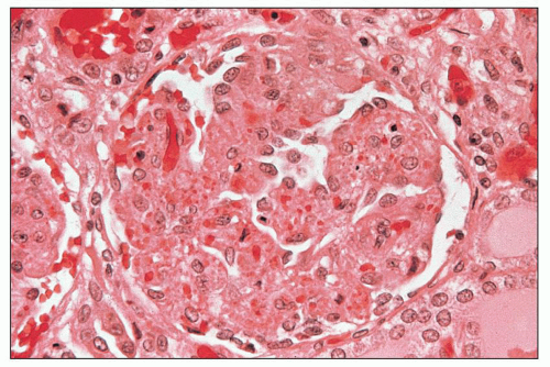
FIGURE 18.1 Hemolytic-uremic syndrome, classic form. The glomerulus is slightly hypocellular, and most of the glomerular capillary lumina are closed due to thickening of the capillary walls. Red blood cells and fragmented red blood cells are seen in the mesangial areas. The specimen is from a child with STEC infection. This type of glomerular change is typical during the acute stage of the disease. (H&E) (Courtesy of Dr. Vivette D’Agati.)

In the early stages, the glomeruli may show thickening of the capillary walls, caused mainly by expansion (swelling) of a thin layer between the endothelial cells and the underlying basement membrane. Severe swelling of the glomerular endothelial or interposed mesangial cells with subendothelial widening may occlude the capillary lumina (Fig. 18.1). The term bloodless is used to characterize the glomeruli with complete or near complete closure of the capillary lumina (Fig. 18.2). Separation of the endothelium from the underlying basement membrane and production of new basement membrane-like material by endothelial or interposed mesangial cells result in the occasional double contour appearance of the glomerular capillary walls best seen with silver or periodic acid-Schiff (PAS) stains (Fig. 18.3). Although double contours are generally considered to be a feature of a more advanced disease, they can also be seen during the relatively early stages of TMA. The glomerular capillary lumina may have fragmented red blood cells, fibrin, and platelet thrombi (Fig. 18.4). Fibrin is sometimes clearly detectable beneath the glomerular capillary endothelial cells. Larger localized areas of fibrin may be seen in the glomerular capillary tufts, particularly in continuity with thrombus or fibrinoid necrosis in the afferent arteriole as it enters the glomerulus (Figs. 18.5 and 18.6). The glomeruli may also be congested, containing red blood cells in dilated capillary loops, especially in cases with severe vascular involvement (Fig. 18.7). This feature is sometimes designated “glomerular paralysis.” This change is typical in patients in the early stages of cortical necrosis; frank glomerular necrosis usually develops later. Small crescents may occasionally be present. An increased number
of polymorphonuclear leukocytes in the glomerular capillaries can be seen in some cases; this feature may be prominent in patients with classic HUS (311). Capillary thrombi, endothelial swelling, and congestion were identified as the typical glomerular findings in patients with severe classic HUS (311). The glomerular capillary thrombi in TTP are usually not very extensive (Fig. 18.8). On occasion, the majority of the glomeruli may be affected, but in general, the glomerular changes in TTP are usually not as dramatic as in HUS.


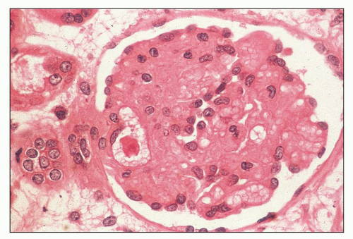
FIGURE 18.2 Hemolytic-uremic syndrome, atypical form. “Bloodless” glomerulus. The glomerular capillary walls are thickened, and the mesangial areas blend with the capillaries. (H&E) (From Kern WF, et al. Atlas of Renal Pathology. W.B. Saunders Company, Philadelphia, 1999, with permission.)


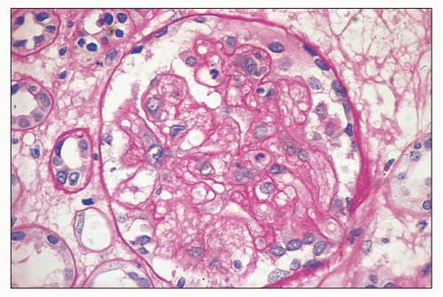
FIGURE 18.3 Hemolytic-uremic syndrome, atypical form. The mesangial areas of the glomerulus have fibrillary appearance. Focal reduplication of the glomerular capillary basement membranes is also seen. A few intracapillary polymorphonuclear leukocytes are present. The specimen is from an adult patient without known etiology of HUS. (Periodic acid-Schiff reaction.)


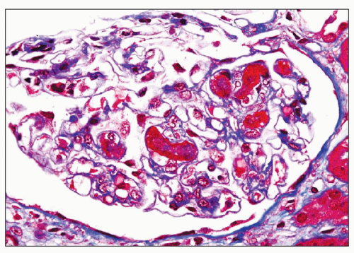
FIGURE 18.4 Thrombotic microangiopathy, associated with cyclosporine administration. Some of the glomerular capillary lumina are occluded by thrombi. No additional significant pathologic changes are present. (Masson trichrome.)


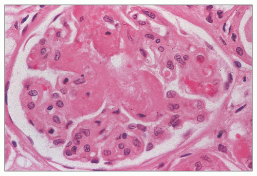
FIGURE 18.5 Hemolytic-uremic syndrome, atypical form. Some of the glomerular capillary tufts are permeated by eosinophilic acellular material. This change is often described as fibrinoid necrosis. Intraluminal thrombi are also present. (H&E) (From Kern WF, et al. Atlas of Renal Pathology. W.B. Saunders Company, 1999, with permission.)

Mesangial abnormalities may also be present in TMA during the early stages. A fibrillar or spongiform appearance of the glomerular mesangium is a characteristic feature, particularly in patients with narrowed arteries and arterioles (see Fig. 18.3). The reason for this fibrillar appearance is not obvious; collapsed glomerular capillary walls and mesangial edema may be contributing factors. Fibrin and fragmented red blood cells can also be seen in the mesangium (see Fig. 18.1). Mesangial cells, although often swollen and hypertrophic, are usually not increased in number during the acute phase of the disease. If mesangial cell proliferation occurs, it is usually slight, focal, segmental, and late.


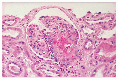
FIGURE 18.6 Thrombotic microangiopathy, secondary to abruptio placentae. The dilated vascular pole is occluded by a thrombus. The change is similar to that seen on Figure 18.10, but no significant chronicity with reduplication of the basement membranes is present. (H&E)


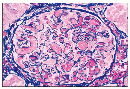
FIGURE 18.7 Thrombotic microangiopathy in a patient with primary antiphospholipid antibody syndrome. Some of the glomerular capillary lumina are occluded by fibrin thrombi; the rest of the capillaries are congested. Glomerular capillary congestion in HUS is often referred to as “glomerular paralysis.” Mesangiolysis is also apparent. (Methenamine-silver.)

Occasionally, mesangiolysis can also be seen. The term mesangiolysis was first used by Yajima (312) in 1956 in patients with nephritis associated with subacute bacterial endocarditis. However, the glomerular capillary cysts, one of the most typical features of mesangiolysis, were described earlier by Pearce (313) in an experimental model of glomerular lesion induced by Crotalus adamanteus venom. The term mesangiolysis refers to partial or complete dissolution of the mesangial matrix and cells. The affected glomerular lobules of mesangiolysis stain
poorly because of mesangial edema. The borders of the dissolving mesangium are hazy, and the mesangial matrix is difficult to identify. No associated inflammatory reaction or fibrin deposition is usually seen (314). Eventually, the glomerular basement membranes become unanchored from the underlying dissolving mesangial mass, leading to markedly dilated, sometimes cystic capillaries (Fig. 18.9). A particularly severe and widespread form of mesangiolysis can occur in aHUS after bone marrow transplantation or mitomycin therapy. However, in classic HUS, mesangiolysis has rarely been described. Mesangiolysis can also be seen in diabetes mellitus, various forms of glomerulonephritis, and transplant glomerulopathy (315). “Healing” of mesangiolysis may lead to proliferating or sclerosing glomerular changes as the disease progresses. In the late stage of mesangiolysis, the mesangium may be thickened by pale fibrillary (sclerotic) material. This process of healing and sclerosing may lead to a distinctive pattern of glomerular sclerosis (“bland sclerosis”) characterized by loss of glomerular cells and capillary lumina but with at least partial preservation of the lobular architecture.


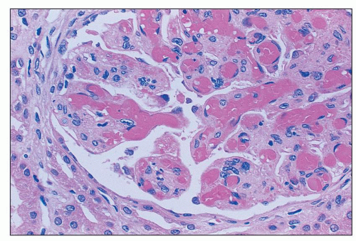
FIGURE 18.8 Thrombotic thrombocytopenic purpura. Most of the glomerular capillary lumina are occluded by homogenous eosinophilic thrombi. The extent of the glomerular capillary thrombosis in TTP is variable; however, it is often mild and patchy. The specimen is from a 40-year-old obese African American female who died a few days after initial presentation. Severe ADAMTS13 deficiency and a strong ADAMTS13 inhibitor were demonstrated in her serum. (H&E)


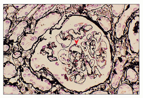
FIGURE 18.9 Thrombotic microangiopathy, associated with mitomycin administration. Ectatic glomerular capillary lumina are present as a result of mesangiolysis. Focal reduplication of the glomerular capillary basement membranes is also seen (arrowhead). (Methenamine-silver.)

Ischemic-type glomerular injury, characterized by collapse of the glomerular capillary tuft and thickening and wrinkling of the capillary basement membranes, during the acute stage of TMA usually indicates severe vascular lesions such as (a) arteriolar or arterial thrombi, (b) acute thickening of the arterial or arteriolar intima, or (c) coexistent chronic hypertensive vascular disease (arteriosclerosis). Focal necrotizing glomerular lesions can also be seen in TMA, albeit rarely. If present, they are usually small, affecting only a few capillary loops or a segment of the glomerulus. Sometimes, the necrotizing lesion is associated with arteriolar thrombosis and/or fibrinoid necrosis of the arteriolar wall.

In the more advanced stages of TMA, glomerular changes differ from those seen early in the course of the illness. This stage is characterized by mostly chronic-type glomerular changes such as mesangial widening resulting from matrix accumulation (mesangial sclerosis), thick capillary walls with occasional double contours, segmentally sclerotic lesions, and chronic ischemic glomerular injury. Activated mesangial cells can migrate along the glomerular capillary walls between the glomerular capillary endothelium and the lamina densa of glomerular basement membranes, with resulting mesangial cell interposition. Production of glomerular basement membrane-like material by glomerular capillary endothelial and migrating interposed mesangial cells results in a double contour (“tram-tracking”) appearance of the glomerular capillary walls (Figs. 18.10 and 18.11; see Fig. 18.3). The double contour is composed of new (inner) basement membrane and the original (outer) basement membrane. In the more advanced stage of TMA, the double contours with the mesangial sclerosis and occasional mild hypercellularity may give rise to a pattern of glomerular injury reminiscent of a membranoproliferative glomerular lesion (see Fig. 18.11). Double contours of the glomerular capillary walls may apparently persist for several months or years.


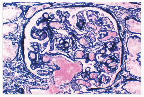
FIGURE 18.10 Thrombotic microangiopathy, postpartum. The dilated infundibulum is occluded by homogenous eosinophilic material (intraluminal thrombus). Extensive reduplication of the glomerular capillary basement membranes indicates developing chronicity. (Methenamine-silver.)


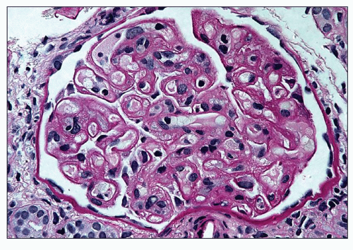
FIGURE 18.11 Thrombotic microangiopathy with extensive reduplication of the glomerular capillary basement membranes. This pattern is usually interpreted as chronic or advanced stage of TMA and may resemble a membranoproliferative pattern of glomerular injury. It should be emphasized that such extensive reduplication of the basement membranes as shown on the photograph is rarely seen. (Periodic acid-Schiff reaction.)


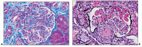
FIGURE 18.12 Thrombotic microangiopathy superimposed on lupus nephritis. A: The glomerulus is hypercellular with closure of some of the glomerular capillary lumina. There is a small thrombus in the arteriolar lumen. (Masson trichrome.) B: The arteriolar lumen is occluded by a thrombus. The glomerular capillary lumina are congested, but no capillary thrombi or cellular proliferation is present. Although this patient’s serum was positive for antiphospholipid antibodies, a similar picture can also be seen in patients who are antiphospholipid antibody negative. (Methenamine-silver.)

Segmentally sclerotic glomerular lesions can occasionally be seen in cases with evolving chronicity of TMA. The lesions closely resemble those seen in idiopathic focal segmental glomerulosclerosis with focal and segmental collapse of the glomerular capillary lumina, mesangial matrix accumulation, and visceral epithelial cell hyperplasia overlying the sclerotic segments. These segmentally sclerotic changes may represent healed necrotizing glomerular lesions. Alternatively, chronic sclerosing-type glomerular injury with segmental features can also develop as part of evolving chronicity affecting the glomeruli.

Chronic ischemic-type glomerular injury is characterized by thickening and wrinkling of the glomerular capillary basement membranes, simplification of the glomerular tuft, widening of the Bowman space between the collapsed glomerular loops and the Bowman capsule, and collagen accumulation internal to the Bowman capsule replacing the Bowman space. The ischemic changes may affect the glomeruli globally or segmentally. Simplification refers to shrinkage of the glomerular capillary tuft accompanied by an apparent decrease in the number of normal glomerular lobules and by apparent loss of the mesangial matrix and cells. Accumulation of PAS-negative collagen material inside the Bowman capsule usually begins at the hilar region of the glomerulus, and eventually the collagen may involve the entire circumference of the Bowman space. If the changes progress to complete glomerular ischemic obsolescence, the glomeruli appear as small, hypocellular, compact eosinophilic masses (“tombstones” or globally sclerotic glomeruli). However, with PAS stain, the collapsed PAS-positive glomerular tuft can easily be distinguished from PAS-negative collagenization of the Bowman space. In TTP, focal glomerular capillary wall thickening, mild proliferation, and sclerotic changes may be observed, but overall the glomerular changes are minor when compared to classic HUS. In patients who have TMA superimposed on glomerulonephritis (e.g., lupus glomerulonephritis), the glomeruli may be markedly hypercellular in addition to the typical changes of HUS (Fig. 18.12).


Arteries and Arterioles

Various changes occur in the renal arteries and arterioles. Arteriolar and arterial changes are more common in patients with aHUS and TTP (87,309). In the early stages, renal arterioles show swelling of the endothelial cells and subendothelial space. The arteriolar lumen may be severely narrowed and, sometimes, fragmented red blood cells are seen in the thickened arteriolar wall. Infiltration of the arteriolar wall by fibrin may occur, a change often referred to as fibrinoid necrosis (Fig. 18.13). The term fibrinoid necrosis is probably a misnomer because little evidence indicates that cellular necrosis is a constant feature of the lesion. Fibrinoid necrosis is thought to be related to increased vascular permeability and nonspecific trapping of plasma proteins, including fibrin in arteriolar walls. Fibrinoid
necrosis tends to occur only at the hilum of the glomerulus, and it may only involve the thickened intima of the arterioles. More often, however, the media of the arteriole is also affected. Unlike in true leukocytoclastic vasculitis, acute inflammatory cell infiltrate is rarely seen in fibrinoid necrosis with TMA. As the disease progresses, the involved arterioles tend to become hyalinized, losing the staining reactions for fibrin. Hyalinized arterioles show homogenous eosinophilic, refractile, strongly PAS-positive acellular material accumulated in the intima or media. Fibrin thrombi may also be seen in the afferent arterioles, and these may continue into the glomerular capillary tuft (see Figs. 18.10 and 18.12). One of the most conspicuous features of TTP is the presence of eosinophilic, platelet-rich granular thrombi in terminal renal interlobular arteries, or more commonly, in afferent arterioles. The most common site is the junction of the afferent arteriole and the glomerular tuft, sometimes called the infundibulum (Fig. 18.14). The thrombotic material in the lumen may merge with the arteriolar wall; therefore, it is often difficult to distinguish between fibrinoid necrosis of the arteriolar wall and the fibrin thrombus in the lumen. Sometimes both fresh and organizing thrombotic lesions can be recognized. Aneurysmal dilatation of arterioles sometimes takes place, particularly in the hilar region of the glomerulus, in patients with TTP (4,316). In addition, the arterioles may show proliferation of cells assumed to be endothelial cells; this change was first reported by Baehr et al. (2) and was later confirmed by several authors. The cellular proliferation in the arterioles is sometimes a prominent feature, and the collections of cells, often concentrically arranged, may attain the size of glomeruli. Capillary channels with or without an edematous extracellular matrix can sometimes be recognized. Lipidcontaining macrophages may be present in the intima of small renal arteries undergoing proliferative changes. Because these proliferations resemble glomeruli, they are referred to as glomera, or glomeruloid structures (317). Glomeruloid structures are typically described in TTP but may also be seen in HUS, SLE, and various forms of glomerulonephritis (317).


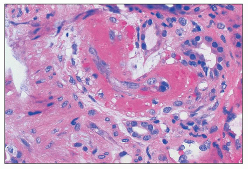
FIGURE 18.13 Thrombotic microangiopathy with arteriolar fibrinoid necrosis in a patient with systemic lupus erythematosus (SLE). In the setting of SLE, this finding is also referred to as lupus vasculopathy. Note the lack of inflammatory reaction in the vessel wall. Such finding can also be associated with intraluminal thrombosis affecting arterioles and/or glomerular capillary lumina. (H&E)

Interlobular renal arteries may show two major changes. One of the early abnormalities is swelling of the intima, which may be accompanied by a suffusion of red blood cells (some of which may be fragmented) or fibrin. Fibrin may appear deep in the intima and may permeate the wall extensively; as in arterioles, this lesion is also called fibrinoid necrosis. The second lesion is intimal swelling, which is usually sparsely cellular, containing mainly lucent amorphous material with a mucoid appearance (Fig. 18.15). This change is usually designated mucoid intimal hyperplasia; it may be severe, with marked narrowing of the lumen. Often, one sees a rapid proliferation of cells in the intima that might be regarded as organization of the intimal edema. The proliferating intimal cells are myointimal cells and are responsible for the cellular intimal thickening later during the course of the disease (Fig. 18.16). The cellular intimal proliferation may give rise to a pattern of change referred to as an “onion skin” lesion, which consists of concentric, ring-like layers of myointimal
cells and delicate connective tissue fibrils. The arterial changes may result in severe narrowing or occlusion of the vascular lumen with consequent reduction in blood flow. These severe vascular changes are associated with a poor prognosis (318) and are responsible for the secondary ischemic glomerular changes such as shrinkage of glomerular capillary tufts and wrinkling and thickening of the capillary walls. Fibrous replacement of the thickened intima is a later change in the interlobular arteries. Occasionally, thrombosis and recanalization can also be seen in the interlobular arteries (Fig. 18.17). Changes similar to those seen in the interlobular arteries can also be present, although less frequently, in the larger (arcuate and intralobar) arteries. Especially in older patients with preexisting chronic hypertension, the acute vascular lesions of TMA may be superimposed on chronic vascular changes such as intimal fibroplasia, medial hypertrophy, or arteriolar or arterial hyalinosis.


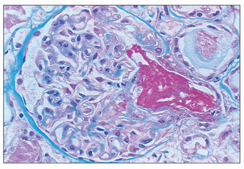
FIGURE 18.14 Thrombotic thrombocytopenic purpura. The infundibulum (i.e., vascular pole) is occluded by a large thrombus. The glomerular capillary walls are thickened; however, the glomerular capillary lumina are patent. (Masson trichrome.)


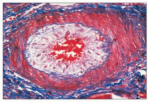
FIGURE 18.15 Thrombotic microangiopathy secondary to malignant hypertension. The small interlobular artery shows the edematous intima containing few myointimal cells (“mucoid intimal hyperplasia”). The patient presented with severe (“malignant”) hypertension and acute renal failure. (Lendrum stain.)


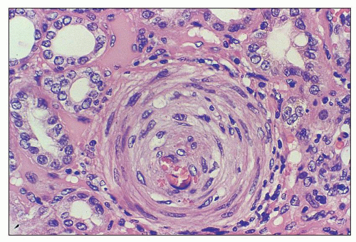
FIGURE 18.16 Hemolytic-uremic syndrome, atypical form. Prominent circumferential intimal cellular proliferation in a small interlobular artery in a nephrectomy specimen from a 5-year-old boy with aHUS. The child had severe hypertension. (H&E)


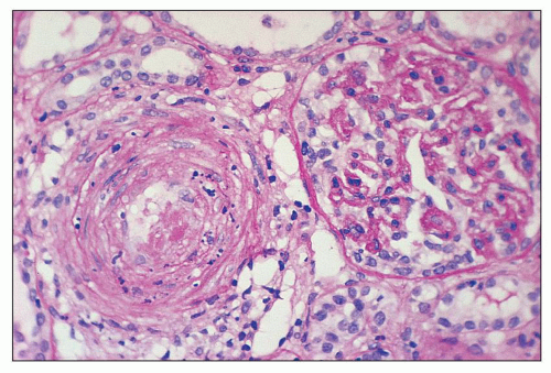
FIGURE 18.17 Hemolytic-uremic syndrome, atypical form. The interlobular artery shows luminal thrombus with nuclear debris in the arterial wall. The glomerulus exhibits ischemic features with thickening and wrinkling of the glomerular capillary basement membranes. The specimen is from an adult patient without known etiology of HUS. (Periodic acid-Schiff reaction.)


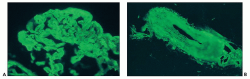
FIGURE 18.18 Hemolytic-uremic syndrome (TMA). The glomerular capillary walls and lumina (A) and the arterial wall (B) show strong fibrinogen positivity. The patient presented with severe (“malignant”) hypertension, acute renal failure, MAHA, and thrombocytopenia. (Direct immunofluorescence.)


Tubules

The tubules frequently contain hyaline casts and red blood cells. Frank tubular epithelial necrosis may occur or acute tubular necrosis as is seen with ischemia may be present. Iron pigment and hyaline droplets in the cytoplasm of the proximal convoluted segment may be observed, with varying degrees of tubular loss. In later stages, tubular atrophy may be seen. In those cases with cortical necrosis, small patchy infarcts or larger necrotic areas are seen. Rarely, calcifications of the cortex can be widespread in chronic cases of cortical necrosis.


Interstitium

The interstitium may be edematous or fibrous, and in some cases, it contains mild mononuclear cell infiltration. Large numbers of red blood cells are present in the interstitium in areas of cortical necrosis.


Immunofluorescence Microscopy

In the glomeruli, the usual finding is the presence of fibrinogen or fibrin along the capillary loops in a continuous, broken linear or granular pattern; fibrinogen or fibrin is found less frequently in the mesangium (Fig. 18.18A). The deposits along the capillary walls may be accompanied by IgM, by C3, less frequently by IgG and only rarely by IgA (Fig. 18.19). Intracapillary thrombi also contain fibrinogen or fibrin and fibrin fragments.

Arterioles and small arteries often exhibit fibrinogen or fibrin in their walls, usually in a subendothelial position (see Fig. 18.18B). IgM positivity was described in vessel walls, as were C3, C1q, and IgG and IgA. Intravascular thrombi also show positive fluorescence for fibrinogen or fibrin.

Although the morphologic changes of TMA are similar in both TTP and HUS, some studies suggest that the composition of microthrombi in TTP might be different from those in HUS. On autopsy material from 23 patients with TTP, Asada et al. (319) showed strong immunohistochemical staining in the renal vascular thrombi as well as in the subendothelial hyaline deposits with factor VIII-related antigen, but only weak staining with fibrinogen or fibrin. This staining pattern was in contrast to the immunohistochemical results of thrombi in patients with disseminated intravascular coagulation in which strong fibrinogen
or fibrin and weak factor VIII-related antigen staining was observed. Electron microscopic analysis showed numerous platelets in the glomerular capillary thrombi in TTP. The authors concluded that thrombi in TTP are composed of platelets. The strong subendothelial factor VIII-related antigen positivity was interpreted by these investigators as suggesting that the hyaline deposits are not a result of increased vascular permeability, but rather platelet thrombi incorporated into the vascular wall. Similarly, a more recent autopsy study of 25 patients with TTP and 31 patients with HUS demonstrated the histochemical and immunohistochemical differences in thrombi (320). HUS lesions contained a large component of fibrin, highlighted by phosphotungstic acid-hematoxylin stain, but only a few platelets or vWF (320,321). On the other hand, TTP-associated arterial thrombi had abundant platelets that stained for antibody to factor VIII. The TTP microthrombi were also rich in vWF but contained very little fibrin (322). Despite these apparent differences in HUS and TTP microthrombi, given the extensive clinical overlap between aHUS and TTP, the histologic accuracy and validity of these findings are unclear.


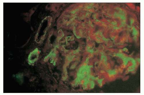
FIGURE 18.19 Thrombotic microangiopathy. IgM glomerular positivity in a patient with postpartum HUS. The arteriolar wall is also positive. (Direct immunofluorescence.)


Electron Microscopy

In the glomeruli, the most consistent change is a thickening of the capillary wall resulting from widening of the subendothelial space and swelling of the endothelial cells. The acellular subendothelial “fluff” is pale and rarefied and contains irregular collections of electron-dense material (Fig. 18.20). This subendothelial material in the lamina rara interna of the glomerular basement membrane is usually granular and has a variable electron density. Sometimes, it has a fibrillar or beaded appearance, but usually it lacks the periodicity and electron density of fully developed fibrin. The exact nature of the subendothelial material is unknown, but it is thought to represent the breakdown products of intravascular coagulation or cell debris organized into the capillary wall. Signs of endothelial damage may occur early during the course of TMA, with features such as swelling, localized areas of detachment of the endothelial cytoplasm from the basement membrane, and cytolysis. One may see intracapillary thrombi composed of amorphous osmiophilic material admixed with fibrin, platelets, and deformed red blood cells (Fig. 18.21). The platelets may be numerous and may fill the capillary lumen. Platelets can sometimes be seen between the endothelial cells; occasionally, the remnants of platelets are visible within the lamina densa of the glomerular capillary basement membrane. However, platelets are evanescent and may not be identified. Electron-dense fibrillar fibrin wisps are sometimes a conspicuous feature in the lumina and rarely in the subendothelial space. The swollen mesangial matrix appears as a meshwork filled with electron-dense, finely granular or fibrillar material similar to the subendothelial changes. Mesangiolysis may be seen, and this progressive disintegration results in capillary ectasia. One sees frequent visceral epithelial foot process effacement.

In the older lesions, wrinkling and collapse of the glomerular basement membranes may occur and are conspicuous in some cases. Multiple layers of material resembling basement membrane are often seen in the capillary wall with cellular (mesangial) interposition (Fig. 18.22). Small, rounded particles that are 40 nm in diameter and are limited by a thin membrane have been described in the cytoplasm of the endothelial cells.

Arterioles and arteries show changes in the endothelial cells similar to those seen in the glomeruli. One sees swelling, cytolysis, and detachment of the endothelium from the underlying structures with a widening of the intima. The intima has a lucent appearance with strands or granules of greater electron density (Fig. 18.23). Structures consistent with fibrin are found at various depths of the vessel wall; luminal thrombi made up of platelets, fibrin, and electron-dense material may be present. In the later stages, elongated myointimal cells abound in the thickened intima.

Jun 21, 2016 | Posted by in UROLOGY | Comments Off on Thrombotic Microangiopathies

Full access? Get Clinical Tree

Get Clinical Tree app for offline access