Membranous Glomerulonephritis


Membranous Glomerulonephritis


Glen S. Markowitz

Vivette D. D’Agati


BACKGROUND


Terminology and Synonyms

Synonyms for MGN include membranous nephropathy, membranous glomerulopathy, and, less commonly, epimembranous nephropathy. The term membranous glomerulonephritis (MGN) is most frequently used but is not without problems in that this entity lacks the inflammatory features typical of a glomerulonephritis. Nonetheless, this term continues to be preferred and can be justified based on the progressive nature of this entity, its apparent autoimmune pathogenesis, and the critical roles of antigen-antibody immune complex formation, serum complement fixation, and generation of the C5b-9 membrane attack complex (MAC).


Primary and Secondary MGN

MGN can be divided into primary and secondary forms of disease (Table 7.1). The term primary idiopathic MGN is frequently employed, but this term is likely nearing extinction because the majority of such cases are now understood to be mediated by an autoantibody to phospholipase A2 receptor (PLA2R) expressed on podocytes. Thus, the term primary MGN may soon be replaced by the more pathogenetically
specific term primary PLA2R-associated MGN (5). While the majority of cases represent the primary disease, MGN may occur secondary to many other conditions. With some exceptions, the pathology of primary and secondary MGN is often similar or even identical; thus, the diagnosis of primary MGN requires careful exclusion of the known secondary etiologies. In a review of nine series published between 1975 and 1989, Glassock (6) found that 23% of cases of MGN were secondary with the prevalence higher (35%) in children younger than 16 years and adults older than 60 years as compared to adults from 16 to 60 years of age (20%). The most common secondary etiology of MGN is systemic lupus erythematosus (SLE; i.e., membranous lupus nephritis [LN]). Most cases of secondary MGN relate to autoimmune/collagen vascular disease, infection, neoplasia, or therapeutic agents (i.e., drug-induced MGN) (6). New associations continue to appear, and critical evaluation is necessary before accepting an etiologic relationship. The well-established secondary etiologies of MGN covered in this chapter include therapeutic agents (gold, penicillamine, mercury, captopril, and nonsteroidal anti-inflammatory drugs [NSAIDs]), malignancy, infections (hepatitis B virus [HBV], hepatitis C virus [HCV], and syphilis), and miscellaneous conditions (sarcoidosis, IgG4-related disease [IgG4RD], allogeneic transplantation, and autoimmune thyroiditis). Chapter 14 covers MGN associated with SLE and other autoimmune/collagen vascular disease. Primary MGN is the focus of this chapter, with an understanding that it is critical to exclude secondary etiologies due to their different prognostic and therapeutic implications.


TABLE 7.1 Best established secondary etiologies of MGN


Therapeutic agents


Gold salts


Penicillamine


Bucillamine


Captopril


Mercury


NSAIDs


Tiopronin


Trimethadione


Malignancy


Mainly carcinomas, most commonly of lung, gastrointestinal, prostatic, or breast origin


Infection


HBV


HCV


Syphilis


Filariasis


Hydatid disease (echinococcosis)


Autoimmune rheumatologic conditions


SLEa


Rheumatoid arthritisa


Sjögren syndromea


Mixed connective tissue diseasea


Autoimmune thyroid disease (Graves disease, Hashimoto thyroiditis)


Additional conditions associated with MGN


Sarcoidosis


Allogeneic hematopoietic SCT


IgG4RD


Guillain-Barré syndrome


Chronic inflammatory demyelinating polyneuropathy


a Covered in Chapter 14.


PRIMARY MEMBRANOUS GLOMERULONEPHRITIS


Clinical Features

Primary MGN is seen throughout adult life, with a peak incidence in the fourth and fifth decades (7,8,9,10,11,12). MGN is less common in children and, in this age group, secondary forms of disease are more commonly seen (6,13,14,15). Males are more frequently afflicted in both adults (7,8,10,11,12,16) and children (15), and the proportion of male patients ranges from 57% to 72%, with a mean of 64%, in six large MGN databases (2,7,9,10,16). Within these databases, the mean age of presentation ranges from 40 to 62 years (7,8,9,10).

MGN is the most common cause of the nephrotic syndrome in Caucasian adults (17,18). However, focal segmental glomerulosclerosis (FSGS) is a more common cause of the nephrotic syndrome in African Americans (17,18) and, in some studies, has surpassed MGN in the overall population (17,19,20). Furthermore, when patients with primary glomerular disease who do not meet the full criteria for nephrotic syndrome are considered, MGN is less prevalent than FSGS and IgA nephropathy (IgAN) (17,19,20,21). The nephrotic syndrome is present at the time of presentation in approximately 75% of patients with MGN (7,9,10), although this percentage depends on the degree of screening for proteinuria in a particular patient population. In six large studies, a mean 24-hour urine protein of 6.5 g/d and a mean serum albumin of 2.5 g/dL were seen (7,8,9,10,16).

While nephrotic syndrome is the most common presentation of MGN, renal insufficiency, hematuria, and hypertension may also be seen. In six large databases from five different countries, the mean creatinine at presentation ranged from 0.9 to 1.3 mg/dL (mean of means: 1.08 mg/dL) and the mean creatinine clearance varied from 76 to 107 mL/min/1.73 m2 (mean of means: 88 mL/min/1.73 m2) (7,8,9,10,16). Renal insufficiency was seen in approximately 25% of patients with MGN at the time of presentation (7,8,9,10). Microscopic hematuria is present in approximately half of the patients with MGN. In contrast, gross hematuria is extremely rare, and its presence should prompt a search for an alternative etiology of gross hematuria in the kidney or elsewhere within the genitourinary tract. Similarly, hypocomplementemia is not a feature of primary MGN and, when present, secondary etiologies of MGN should be carefully excluded, in particular membranous LN (14). At the time of presentation, hypertension is present in at least half of the patients with MGN (7,9,10,16) Similar to most etiologies of the nephrotic syndrome, and unlike minimal change disease, there is a loss of selectivity of proteinuria in patients with MGN (22).

MGN has also been studied in particular age groups and ethnic populations. As previously noted, primary MGN is a
much less common etiology of nephrotic syndrome in children, accounting for only 3% of renal biopsies in a recent large pediatric biopsy series (23). Secondary forms of MGN are more common in the pediatric population (6) and most often relate to SLE (14) or infection, in particular HBV (13). At the opposite extreme, MGN is the most common etiology of nephrotic syndrome among patients above the age of 60, accounting for 32% of cases (24). This also holds true when only considering patients above the age of 80 (25). There is also significant experience with MGN in pregnant women who often experience a doubling of 24-hour urine protein, new onset of hypertension and, in some cases, renal insufficiency (26). The hypertension and worsening proteinuria often but not always reverse following parturition (26).


Pathologic Findings


Gross Pathology

Gross evaluation of the kidneys does not play a significant role in establishing the diagnosis of MGN. In the rare case where gross evaluation is possible (i.e., autopsy), the early appearance of kidneys with MGN is largely unremarkable. Similar to other patterns of glomerular disease, cases that run their course to end-stage renal failure are characterized by a contracted parenchyma with a granular surface, and these changes are more prominent in individuals with hypertension. Ehrenreich and Churg (27) described 10 autopsied cases of histologically confirmed MGN. In patients dying at the height of the nephrotic syndrome without renal insufficiency, the kidneys were large and pale with combined weights from the upper limit of normal (300 g) to 450 g or more.


Light Microscopy


GLOMERULI

The GBM is the principal site of pathology in MGN. Within a biopsy, the GBM changes are homogeneous, typically showing little variation among glomeruli. However, the pathology is dynamic, and as it evolves, the appearance of the GBM varies over time in each individual and between different patients. The resulting pathologic spectrum of glomerular capillary wall changes can be seen by light microscopy but is best understood and staged at the ultrastructural level (27). The light microscopic findings in MGN may be subtle, especially in early cases, and even with optimally stained thin sections, diagnostic changes may not be obvious by light microscopy alone. In these cases, immunofluorescence and EM readily establish the diagnosis.

In MGN, the glomeruli range in size from normal to enlarged and often appear normocellular. In the earliest stage of MGN, the GBMs appear normal in thickness and contour, resulting in no detectable abnormalities at the light microscopic level. More commonly, early cases will exhibit good preservation of the glomerular architecture, but the glomeruli appear stiff and inflated (Fig. 7.1), giving them an exaggerated appearance of normality. Even at this early stage, the podocytes typically appear swollen with enlarged cell bodies. As the disease progresses, the glomerular capillary walls appear thicker and more rigid than normal in PAS-stained histologic sections (Figs. 7.2 and 7.3), and the GBM becomes intensely eosinophilic and refractile in hematoxylin and eosin-stained sections (Fig. 7.4). Eventually, thickening of the GBM, increased mesangial matrix, and accumulation of immune deposits give the glomerulus a solid appearance, and in advanced cases, segmental scarring and global glomerulosclerosis develop. Even when renal insufficiency develops, unscarred glomeruli maintain their characteristic appearance.


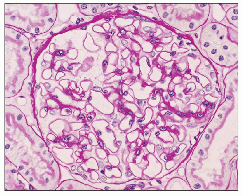
FIGURE 7.1 Histopathology of early MGN (stage I). The glomerulus exhibits no apparent abnormalities by light microscopy. The glomerular basement membrane appears thin and delicate, without evidence of thickening or spike formation. (PAS, ×200.)

The spectrum of GBM changes seen in MGN is best demonstrated by the use of special stains. Although hematoxylin and eosin stain can show overall capillary wall thickening, it is
not an optimal stain for demonstration of GBM pathology because it does not distinguish well between the cell cytoplasm, immune deposits, and extracellular matrix. The GBM changes in MGN are best appreciated using the JMS stain (27) but are also well visualized with the PAS stain. By light microscopy, the earliest change in MGN is mottling of and small depressions in the GBM seen in en face sections stained with JMS (Fig. 7.5). This appearance is subtle and results from alterations in the GBM caused by the subepithelial deposits. More commonly, the JMS stain will reveal further developed changes of MGN represented by small projections (spikes) of silver-positive material (Fig. 7.6) composed of type IV collagen and noncollagenous extracellular matrix proteins, including laminin, heparan sulfate proteoglycan, and vitronectin (28,29,30). Initially, the spikes may be small and segmental, and a careful search under oil immersion is necessary to demonstrate them. Cases such as this, with only focal spikes, may represent either an earlier or a milder form of the disease (15), but immune deposits in cases with segmental spikes usually are more diffusely distributed than indicated by the light microscopic changes. As the lesion progresses, the spikes become larger, thicker, and diffuse (Fig. 7.7). Eventually, the GBM becomes more prominently expanded by a thick band of argyrophilic material containing abundant nonargyrophilic “holes” and imparting a vacuolated appearance (Fig. 7.8). The “holes” represent a normal-thickness GBM beneath the deposits surrounded by a heaped-up matrix around the deposits. In many cases, combinations of these various abnormalities including spikes, holes, and complex thickening of the GBM are present simultaneously in the same biopsy. The nonargyrophilic material deposited within the GBM and on its epithelial side stains red with the trichrome stain, while the surrounding GBM stains blue or green depending on the method used (Fig 7.9). The deposits
also stain blue in toluidine blue-stained 1-µm sections of plastic-embedded tissue. However, both the trichrome and toluidine blue demonstration of deposits are inconsistent, and these stains may be negative even in obvious cases of MGN. Thus, the more consistent histologic changes in MGN are thickening and remodeling of the GBM itself, seen best with the JMS and, to a lesser extent, PAS stains.


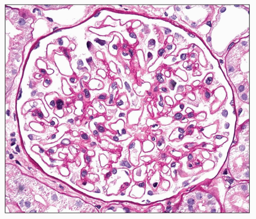
FIGURE 7.2 Histopathology of MGN with mild glomerular basement membrane (GBM) thickening (stage II). The GBM exhibits mild diffuse thickening with spike formation. Diffuse swelling of visceral epithelial cells is noted. (PAS, ×400.)


FIGURE 7.3 Histopathology of GBM thickening in MGN. There is marked global thickening of the glomerular basement membrane, which has a vacuolated appearance. (PAS, ×400.)


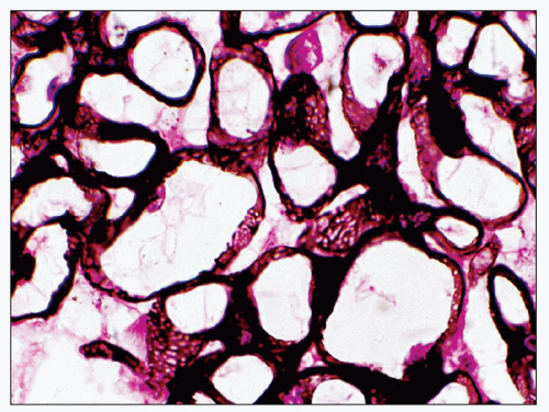
FIGURE 7.4 Histopathology of MGN. With the hematoxylin and eosin stain, the GBM has a thick and rigid appearance in this example of stage III MGN. Mild diffuse mesangial sclerosis is present. (H&E, ×400.)


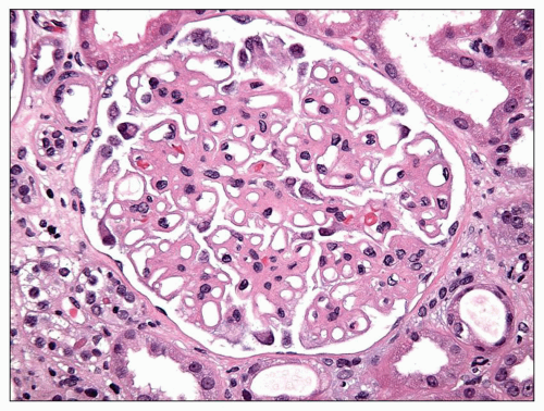
FIGURE 7.5 Histopathology of stage I MGN. In stage I MGN, the JMS stain may demonstrate small holes or depressions in the GBM. (JMS, ×600.)


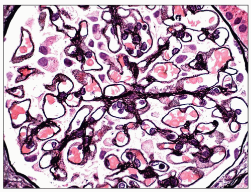
FIGURE 7.6 Histopathology of early stage II MGN. In early stage II MGN, the JMS stain demonstrates short GBM spikes. (JMS, ×400.)


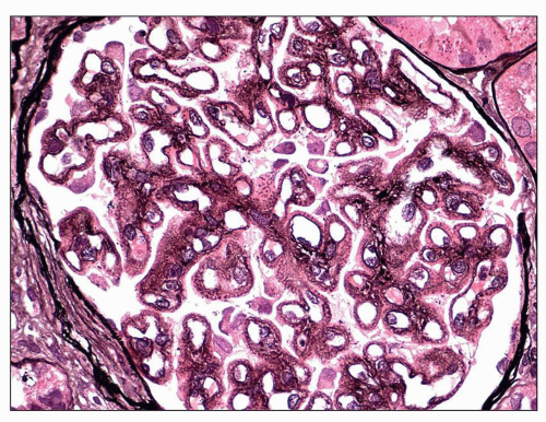
FIGURE 7.7 Histopathology of stage II MGN. In this better developed example of stage II MGN, the JMS stain exhibits more prominent GBM spike formation with intervening silver-negative subepithelial deposits (JMS, ×400.)

Endocapillary proliferation, GBM duplication, mesangial hypercellularity, fibrinoid necrosis, and crescent formation are uncommon features of primary MGN. When present, these findings suggest either a secondary form of MGN or the concurrence of MGN and an additional glomerular disease process. Thus, the presence of such atypical findings requires careful evaluation for a systemic disease and appropriate serologies before concluding that the patient has primary MGN. Each of these findings is considered separately in this section or the sections that follow.


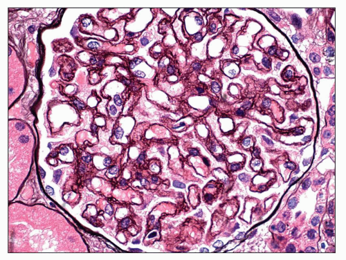
FIGURE 7.8 Histopathology of stage III MGN. In stage III MGN, the JMS stain reveals a globally vacuolated glomerular basement membrane (GBM) due to the presence of GBM spikes and overlying neomembrane formation. (JMS, ×400.)


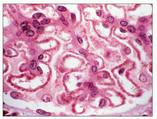
FIGURE 7.9 Light microscopy of subepithelial deposits in MGN. The Masson trichrome stain demonstrates fuchsinophilic deposits along the subepithelial aspect of the glomerular basement membrane. (Masson trichrome stain, ×600.)

The glomeruli in primary MGN typically are enlarged (31) and exhibit an increase in total glomerular area and mesangial volume, including the number of mesangial cells (28,31). In routine histologic sections, mesangial hypercellularity is a subtle finding, and well-defined mesangial hypercellularity is uncommon (Fig. 7.10). When present, the finding of mesangial hypercellularity may suggest a secondary form of MGN related to SLE or HBV infection (32,33) or the possibility
of coexistent IgAN (34,35). A secondary form of MGN (or coexistent IgAN) is particularly likely when the mesangial hypercellularity is accompanied by mesangial immune deposits (32,33,34,35,36).


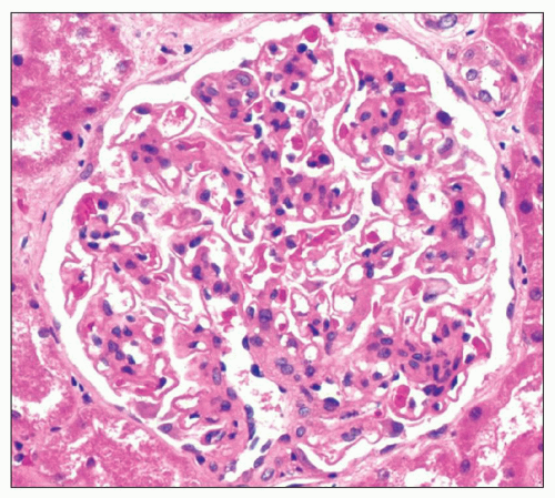
FIGURE 7.10 Mesangial hypercellularity in MGN. In this example of MGN, glomerular basement membrane thickening is accompanied by diffuse mesangial hypercellularity with up to 12 cells per mesangial area. (H&E, ×400.)

While mesangial hypercellularity can be seen in primary MGN, endocapillary proliferation and GBM duplication are generally considered to be incompatible with the diagnosis of primary MGN. GBM duplication, which is typically accompanied by glomerular lobulation and described as a “membranoproliferative” feature, can coexist with findings of MGN. This constellation of mixed membranous and membranoproliferative changes is most commonly encountered in the setting of LN (i.e., class IV and V), in patients with HBV or HCV infection, or as the idiopathic entity of membranoproliferative glomerulonephritis (MPGN) type 3, Burkholder subtype (37). The combination of mixed membranous and endocapillary proliferative changes is less common but similarly should raise the possibility of changes related to SLE, HBV, or HCV. In one study, the finding of an increased number of inflammatory cells within the glomeruli, in the absence of endocapillary proliferation or membranoproliferative features, favored a secondary form of MGN related to malignancy over primary MGN (38).

Lesions of FSGS are present in approximately 20% of renal biopsies with primary MGN (range 13% to 42%) (39,40,41,42) (Fig. 7.11). As compared to biopsies with MGN in the absence of FSGS, the finding of FSGS lesions with MGN (FSGS-MGN) is associated with a greater degree of proteinuria (39,42) and renal insufficiency (39,40,41,42) and a higher incidence of hematuria (39,40,41,42) and hypertension (39,40,42). Histologically, the lesions of FSGS form discrete segmental scars associated with hyaline and/or lipid insudation, adhesions to Bowman capsule, and swelling of overlying epithelial cells, resembling changes in FSGS not otherwise specified (NOS) or occasionally the glomerular tip lesion variant (43). FSGS-MGN is associated with a greater degree of tubulointerstitial scarring (39,40,41,42) and a higher stage of MGN (40,41). Most importantly, in the setting of MGN, the finding of FSGS is associated with a worse prognosis and can be viewed as a marker of chronicity and a negative prognostic indicator (39,41,42). Of note, rare cases exist in which the clinical and pathologic findings in FSGS-MGN suggest two separate and distinct glomerular disease processes, MGN and primary FSGS (44).


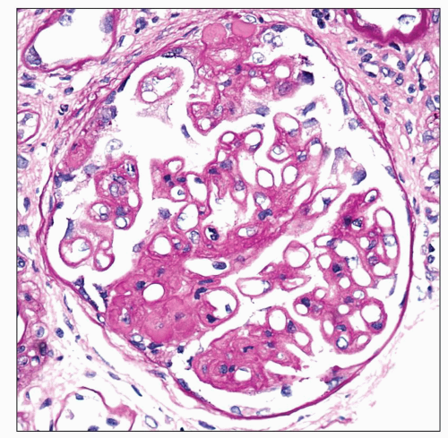
FIGURE 7.11 Focal segmental glomerulosclerosis in MGN. Findings of MGN are accompanied by two discrete lesions of segmental sclerosis with capsular adhesion. (PAS, ×400.)


TUBULES AND INTERSTITIUM

A spectrum of tubular and interstitial changes may be encountered in patients with MGN. Similar to other etiologies of nephrotic syndrome, proximal tubules often contain protein and lipid resorption droplets. Tubules may exhibit acute tubular injury characterized by luminal ectasia, cytoplasmic simplification and vacuolization, irregular luminal contours, loss of brush border, prominent nucleoli, and mitotic or apoptotic figures. These acute degenerative changes may result from severe, unremitting proteinuria or may relate to alternative factors such as drug-induced injury, ischemia, or prerenal hemodynamic changes resulting from intravascular volume depletion. As the glomerular scarring progresses, tubular atrophy and interstitial fibrosis ensue, and the extent of these findings correlates with decreased renal survival (8,28,45,46) (Fig. 7.12). This tubulointerstitial scarring likely results in large part from glomerulosclerosis, reduced efferent arteriolar blood flow, and resultant post-glomerular ischemia. In some instances, the degree of tubular atrophy and interstitial fibrosis significantly exceeds the extent of glomerulosclerosis (45). The pathogenesis of the tubulointerstitial scarring in this setting is incompletely understood. Although cell-mediated immunity may play a role, the absence of extraglomerular immune deposits argues against an immune complex mechanism. Magil (45) examined the extent of tubulointerstitial disease in MGN patients with no or mild vascular disease and fewer than 10% obsolescent glomeruli and found that the degree of tubulointerstitial scarring correlated
with the 24-hour urine protein excretion, serum albumin, and the percent of glomeruli with visceral epithelial cell protein resorption droplets, leading him to conclude that proteinuria per se played a role in the development of tubulointerstitial injury. A discussion of the role of proteinuria in the pathogenesis of tubular atrophy and interstitial fibrosis is included in the section on Etiology and Pathogenesis.


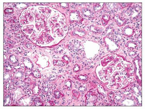
FIGURE 7.12 Interstitial fibrosis and tubular atrophy in MGN. In this example of MGN, the majority of tubules appear atrophic and are accompanied by diffuse interstitial fibrosis. (PAS, ×200.)

Interstitial inflammation is commonly seen in MGN, is usually mild, and is typically most prominent in areas of tubular atrophy and interstitial fibrosis. The interstitial infiltrates are mainly composed of monocytes and T cells with a predominance of CD4+ T cells, suggesting a pathogenic role for cell-mediated immunity (47). Interstitial foam cells are seen in approximately 16% of biopsies with MGN and originate from macrophages (48) (Fig. 7.13). The finding of interstitial foam cells does not correlate with proteinuria or hyperlipidemia (48). In rare instances, the degree of interstitial inflammation is out of proportion to the degree of tubulointerstitial scarring and is accompanied by tubulitis and interstitial eosinophils, leading to a diagnosis of coexistent acute interstitial nephritis (AIN). Potential secondary etiologies of both AIN and MGN include SLE, Sjögren syndrome, sarcoidosis, and treatment with NSAIDs (49,50).


BLOOD VESSELS

Arteriosclerosis and arteriolosclerosis are commonly present in renal biopsies with MGN and are often a reflection of the patient’s age and the presence of systemic hypertension. That said, these vascular lesions have also been shown to correlate with decreased renal survival (8). Occasionally, fibrin thrombi may be seen in blood vessels or glomeruli in biopsies with MGN, where their presence should increase the suspicion for the possibility of renal vein thrombosis (RVT). Additional findings that may suggest RVT include prominent congestion of glomerular or interstitial capillaries and a disproportionate degree of interstitial edema (51). Vascular inflammation is not a feature of MGN and, when present, should raise the possibility of a systemic vasculitis.


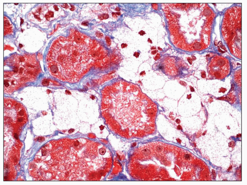
FIGURE 7.13 Interstitial foam cells in MGN. Interstitial foam cells expand the interstitium between tubules. (Trichrome stain, ×600.)


Immunopathology

Immunopathologic studies have shown that histologic changes seen in the GBM by light and electron microscopy in MGN are due to deposits of immunoglobulin and complement components. These immune deposits are best demonstrated by immunofluorescence and correspond to the ultrastructural finding of electron-dense deposits that lie between GBM spikes. The typical immunofluorescence finding in MGN is global deposits of immune reactants that follow the contour of the GBM (Fig. 7.14). Since the deposits lie at the subepithelial aspect of the GBM and project toward the urinary space, they typically have a granular appearance and appear relatively discrete and uniform. In contrast, a minority of cases are characterized by small, confluent deposits that produce a pseudolinear appearance (Fig. 7.15). In rare instances, the deposits are sparse and only segmentally distributed; this mainly occurs very early in the course of disease or in a resolving phase, although rare cases of segmental MGN have been described (52). Mesangial deposits are present in less than 10% of cases of primary MGN (53) but are important to identify as their presence favors a secondary form of disease. Mesangial deposits can be difficult to differentiate by immunofluorescence from subepithelial deposits that follow the GBM reflection around mesangial areas (33).

The principal and invariable component of the subepithelial immune deposits in primary MGN is IgG (Fig. 7.16), with a composition that includes both kappa and lambda light chains. Staining for complement component C3 is also usually present, seen in 85% of cases in one large series (33). Staining for IgM, IgA, and C1q is present in 47%, 16%, and 23% of cases, respectively (33). The intensity of staining for IgG is greatest in virtually all cases, and staining for IgM, IgA, and C1q is typically of no more than 1+ intensity (scale +/-, 1+ to 3+). The intensity of staining for C3 is more variable and typically less than that of IgG; few cases may have equivalent staining to IgG. There are rare cases of MGN in which the deposits are monoclonal and exhibit light chain restriction and IgG subclass restriction, most commonly IgG1-kappa (54,55).
Not surprisingly, these cases can be associated with hematologic malignancy (54,55).


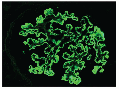
FIGURE 7.14 Immunofluorescence staining in MGN. Staining for IgG reveals intense, granular global subepithelial positivity involving the glomerular capillary walls. (×400.)


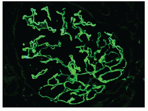
FIGURE 7.15 Immunofluorescence staining for IgG in early MGN. In early MGN, the subepithelial deposits may appear small and confluent, imparting a pseudolinear appearance. (IgG, ×400.)

Immunopathologic findings are often helpful in distinguishing primary MGN from membranous LN and, to a lesser extent, other secondary forms of disease (33). In comparison to primary MGN, membranous LN is characterized by a greater prevalence of positive staining for IgM (62% vs. 47%), IgA (38% vs. 16%), C3 (95% vs. 85%), C1q (67% vs. 23%), and, in particular, intense, ≥2+ staining for C1q (67% vs. 11%) (33). Combined staining for IgG, IgM, and IgA carries a sensitivity of 29% and a specificity of 87% for the diagnosis of membranous LN versus primary MGN, while intense, ≥2+ staining for C1q has a sensitivity of 67% and a specificity of 88% (33). The classic description and hallmark of membranous LN is “full house” staining for immunoglobulins and complement, an expression derived from the game of poker, which refers to the presence of three of a kind (three immunoglobulins: IgG, IgM, and IgA) and two of a kind (complement components: C3 and C1q). Importantly, many of these findings can also be seen in other secondary forms of MGN. For instance, staining for IgG, IgM, IgA, C3, and C1 are each individually present in greater than 50% of biopsies with MGN secondary to HBV infection (32).


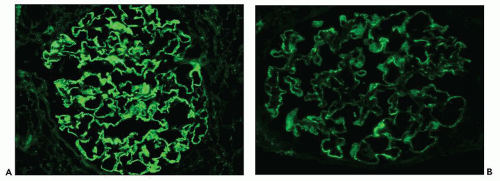
FIGURE 7.16 Comparative immunofluorescence findings in MGN. A: Staining for IgG reveals intense granular global positivity involving the glomerular basement membranes. B: In the same biopsy, a similar distribution but less intense staining for C3 is present along the GBM. (A and B: ×400.)

There are four distinct subclasses of IgG (IgG1, IgG2, IgG3, and IgG4), which nonetheless exhibit 95% homology with one another (56). The numbering of the subclasses reflects their relative prevalence in human serum, with IgG1 representing 66% of circulating IgG and IgG4 comprising only 4%. The small differences in amino acid sequence have significant functional consequences. For instance, IgG3 has the greatest ability to activate complement, the highest affinity for Fc receptors on phagocytic cells, and a significantly shorter serum half-life than the remaining three subclasses (7 vs. 21 days) (56). IgG1 also effectively fixes complement and binds to phagocytic cells. IgG4, the least prevalent subclass of IgG, is the only subclass that does not significantly activate complement (56). IgG4 has the unique property of weak disulfide bonds between the two heavy chains, allowing for dissociation into ½ IgG4 molecules composed of a single light and heavy chain and then reassembly with alternative ½ IgG4 molecules to form a heterobivalent IgG4 with two distinct specificities (57). Because the resultant IgG is bispecific, cross-linking of antigens is extremely unlikely, leading to the belief that IgG4 may serve to down-regulate the immune response (56,57). IgG4 is thought to play an important role in multiple disease processes, including IgG4RD and, more notably, MGN.

The predominant IgG subclass in the subepithelial deposits of primary MGN is IgG4 (58,59,60,61,62,63) (Table 7.2). While this is a well-established finding, the reasons remain somewhat unclear. IgG4 is not significantly increased in the serum of patients with MGN (61,62,63). The IgG4 immune response is thought to require prolonged antigenic exposure (57) and involves T-helper (Th)2 cell-mediated B-cell stimulation (64). It is possible that the IgG4 in MGN represents a protective response against cross-linking of antigens by other subtypes of
IgG, for instance IgG1 (64). This possibility is supported by the observation that C3 is present in the subepithelial deposits in 85% of cases of MGN (33), a finding that cannot be explained by the presence of IgG4 alone, unless it is capable of activating complement through the lectin pathway (64,65). A recent study reconfirmed the dominance of IgG4 in primary MGN but noted that IgG1 predominated in early, stage I disease, suggesting that an IgG subclass switch occurs early in the course of primary MGN (66).


TABLE 7.2 Staining for IgG subtypes in primary and secondary forms of MGN


No. of patients


IgG1


IgG2


IgG3


IgG4


Primary MGN (58,59,60,61,62,63)


108


1+ to 2+


1+


+/-


2+ to 3+


Recurrent MGN in the allograft (67)


7


2+


2+


1+


3+


Membranous LN (59,60,62,63,69)


54


2+ to 3+


2+


2+ to 3+


1+


De novo MGN in the allograft (63)


16


3+


2+


1+


MGN secondary to malignancy (58)


10


3+


2+


1+


2+


MGN secondary to mercury exposure (68)


11


3+


NA


+/-


1+


Degree of positivity reflects all studies referenced and reflects both frequency and intensity of positivity.


Scoring graded on a scale of -, +/-, 1+, 2+, and 3+.


NA = not available.


While IgG4 is the predominant subclass in the subepithelial deposits of primary MGN, IgG1 is also present in the majority of cases and is often accompanied by small amounts of IgG2 and IgG3 (58,59,60,61,62,63) (see Table 7.2). Not surprisingly, IgG4 is also the dominant subclass when MGN recurs in the allograft (67). In contrast, IgG1 tends to predominate in MGN secondary to malignancy (58) or mercury exposure (68) and in de novo MGN in the renal allograft (63). In membranous LN, IgG1, IgG2, or IgG3 may predominate and all three typically stain more intensely than IgG4, although IgG4 is present in the majority of cases (59,60,62,63,69). Staining for IgG subtypes has been proposed as a modality to differentiate primary and secondary forms of MGN. In our experience, this approach is of somewhat limited utility due to the extensive overlap in staining patterns seen in Table 7.1 as well as the significant subjectivity in grading of positivity. Nonetheless, this approach is likely to be helpful when the main diagnostic considerations are primary MGN and membranous LN. Fortunately, staining for the PLA2R has emerged as a far more effective strategy to differentiate primary and secondary forms of disease (70,71,72,73).

Given that primary MGN is causally linked to the development of anti-PLA2R antibodies and that renal expression of the PLA2R is limited to podocytes, it is not surprising that extraglomerular deposits are extremely uncommon in primary MGN. For instance, in two series involving 142 and 26 patients with primary MGN, extraglomerular deposits were not identified in any patient (33,74). In contrast, the finding of extraglomerular deposits favors a secondary form of the disease. In particular, extraglomerular deposits involving tubular basement membranes (TBMs) are present in 30% to 50% of cases of membranous LN (33) and are often accompanied by interstitial and vessel wall deposits (Fig. 7.17). We have reported a series of three patients with MGN and prominent Bowman capsular and TBM deposits in which a secondary etiology was not identified (75).

There are reports of children with anti-TBM nephritis and concurrent MGN. This entity is mainly seen in male children between the age of 2 months and 10 years (76,77), and an X-linked pattern of inheritance has been reported in two families with four affected male offspring (78). Clinical presentation typically includes proteinuria, renal insufficiency, and Fanconi syndrome, and serum studies may document the presence of anti-TBM antibodies. In additional to findings of MGN, pathologic evaluation reveals tubulointerstitial nephritis (TIN) with linear staining of TBMs for IgG, kappa, and lambda by immunofluorescence, and EM often demonstrates electron-dense deposits within TBMs. Indirect immunofluorescence applying the patient’s serum to normal kidney reveals linear staining of TBMs and may be used to confirm the diagnosis. The anti-TBM antibodies seen in this condition cross react with the TBM of multiple animal species (79). By Western blot, the antibodies react with a 58-kDa noncollagenous glycoprotein component of TBMs (79) that exhibits 30% homology with human preprocathepsin B (80) and interacts directly with type IV collagen and laminin, suggesting a role in tubular epithelial cell adhesion to TBMs (81). The 58-kDa
anti-TIN antigen has not been shown to be expressed in podocytes, although possible cross-reactivity of anti-TBM antibody with a podocyte antigen has been proposed (77). Prognosis for this condition is poor, with frequent progression to end-stage renal disease (ESRD).


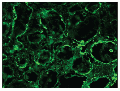
FIGURE 7.17 Tubular basement membrane deposits in MGN. In this patient with MGN, immunofluorescence reveals granular immune deposits within tubular basement membranes. The patient was subsequently found to have systemic lupus erythematosus. (IgG, ×400.)

A small subset of patients with MGN exhibit mesangial deposits that, in contrast to the subepithelial deposits, stain intensely for IgA with minimal to absent staining for IgG. These cases represent MGN with coexistent mesangial IgAN (34,35,82) (Fig. 7.18). Patients with combined MGN and IgAN are typically adults who present with proteinuria and hematuria. Given the separate and distinctive pathogenesis of these two disease entities and rarity of this combination, a unified mechanism of disease is unlikely. Combined MGN and IgAN appear to be common in patients with hepatitis B infection, in patients with Asian ethnicity, and in the allograft, where IgAN recurs in a large percentage of cases and MGN is among the most frequent de novo form of glomerular disease (34).


Electron Microscopic Findings

EM plays a central role in establishing the diagnosis of MGN, as it demonstrates both the immune complex deposits identified by immunofluorescence and the GBM “spikes” seen best by light microscopy. EM also is needed to accurately determine the stage of MGN.

MGN is defined by the presence of subepithelial deposits that form on the outer aspect of the GBM, beneath the podocyte. The deposits appear to sit on the GBM and are accompanied by a spectrum of GBM changes ranging from intervening projections of the extracellular matrix (“spikes”) to areas where the GBM projections surround and encase the deposits, creating the appearance of a newly formed, overlying “neomembrane.” The deposits can range from electron dense to pale and electron lucent, consistent with focal resorption. In the absence of neomembrane formation, the deposits are in direct contact with and may indent the cytoplasm of the overlying podocytes. Podocytes exhibit a spectrum of reactive changes including condensation of actin filaments, increased cytoplasmic organellar content, lipid and protein resorption droplets, microvillous transformation, and foot process effacement. Animal models have shown that at a molecular level, podocytes exhibit loss of nephrin expression, dissolution of the actin cytoskeleton, and loss of slit diaphragm integrity (reviewed in (83)).


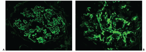
FIGURE 7.18 MGN with IgA nephropathy. A: Staining for IgG reveals granular global glomerular capillary wall positivity in a subepithelial distribution. B: Staining for IgA in the same biopsy reveals abundant mesangial deposits that are confined to the mesangium and do not appear to involve the peripheral capillary walls. (A and B: ×400.)

The range of ultrastructural findings seen in MGN led Ehrenreich and Churg (27) in 1968 to propose a morphologic classification that describes a pathologic sequence of subepithelial immune deposit formation, reactive GBM changes, and, in some cases, subsequent resorption of deposits and GBM repair (Fig. 7.19). This classic publication describes four sequential stages of MGN that provide general information about the relative age of the glomerular lesions.

Stage I MGN is characterized by subepithelial electron-dense deposits that are typically small, sparsely distributed, and by definition devoid of significant intervening GBM spike formation, although small depressions in the GBM may be noted (27) (Fig. 7.20). In the majority of cases, no significant abnormalities are identified by light microscopy with the exception of cases in which focal depressions of the GBM may be noted with the JMS stain or fuchsinophilic deposits are apparent with the trichrome stain. The diagnosis of stage 1 MGN is easily established by immunofluorescence and EM. Given that stage 1 represents the earliest changes of MGN, it is not surprising that, unlike the other stages, the deposits may be only segmentally distributed. Scanning EM of acellular glomeruli shows shallow depressions on the epithelial side of the GBM (84) (Fig. 7.21).

Stage II MGN is characterized by subepithelial electron-dense deposits that appear larger than the deposits in stage 1 and by definition are surrounded by intervening projections of the GBM, referred to as GBM “spikes” (27) (Fig. 7.22). By light microscopy, there is global thickening of the GBM. GBM spikes are best visualized with the JMS stain but are also apparent with the PAS stain. Similar to stage 1, the
deposits typically stain intensely by immunofluorescence. The three-dimensional appearance of the deposits can be partially appreciated by scanning EM of acellular glomeruli, where the elongated deposits are surrounded by a complex anastomosing network of basement membrane projections (84) (Fig. 7.23).


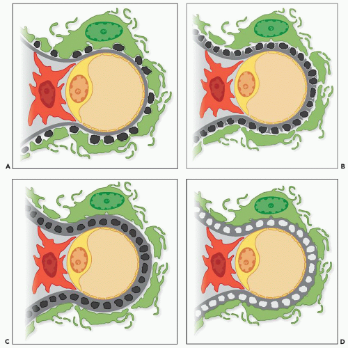
FIGURE 7.19 Depiction of the four stages of MGN as described by Ehrenreich and Churg. A: Stage I MGN is characterized by interspersed subepithelial deposits without intervening GBM spike formation. B: In stage II MGN, the subepithelial deposits are more numerous and more closely approximated with intervening GBM spikes. C: In stage III MGN, the GBM spikes extend over and encircle the deposits, incorporating them into the glomerular capillary walls. D: In stage IV MGN, the deposits lie within a mottled, irregularly thickened GBM and appear electron lucent, consistent with resorption. The irregular thickness of the GBM is consistent with extracellular matrix remodeling.

In stage III MGN, a new layer of GBM is laid down over the subepithelial deposits and connects the GBM spikes (27). As a result, the deposits appear to sink into the GBM (85) and are often described as having an intramembranous appearance, lying beneath a neomembrane (Fig. 7.24). In stage III, the deposits can be electron dense, similar to stages I and II, or may exhibit diminished electron density, indicating that they are undergoing resorption and incorporation into the GBM. The intensity of immunofluorescence staining is variable in stage III, as less intense staining is seen in cases in which the
deposits have a more electron-lucent appearance. By light microscopy, GBM thickening is most pronounced in stage III, and the JMS stain typically shows a complex, vacuolated GBM, while the deposits usually are not visible with the trichrome stain. Although the staining intensity may be reduced, the deposits are usually demonstrable by immunofluorescence. The findings of pure stage III MGN suggest a single generation of deposits, presumably formed over a similar time period. In contrast, most cases of stage III MGN have a mixed appearance with fresh deposits layered over older intramembranous deposits, suggesting a dynamic process of ongoing deposit formation, incorporation, and resorption. In such cases, it may be possible to assign a dominant stage based on the major pattern present. In many cases, two stages coexist, making it most appropriate to designate both stages (such as mixed stage II to III) (Fig. 7.25). By scanning EM, the complex network of GBM projections seen in stage II MGN is replaced by bridged segments of the neomembrane in stage III that have a relatively smooth appearance (84) (Fig. 7.26).


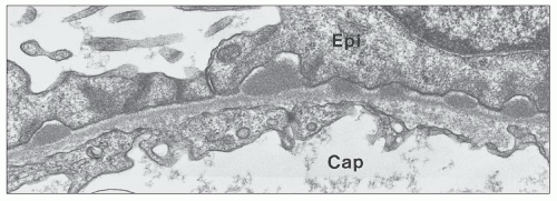
FIGURE 7.20 Electron micrograph of stage I MGN. In stage I MGN, there are small, interspersed subepithelial deposits without significant GBM spike formation. (×20,000.)


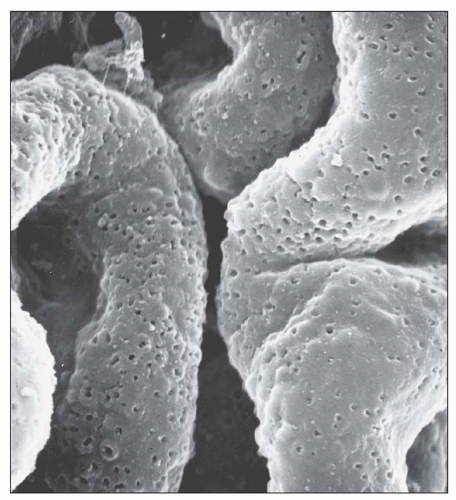
FIGURE 7.21 Scanning electron micrograph of an acellular glomerulus from a patient with stage I MGN. The cells were removed from the tissue by sequential treatment with detergents and deoxyribonuclease. The GBM contains scattered shallow pits representing the sites of immune complex deposits removed in the preparation of the tissue. (×5,000.) (Courtesy of Dr. Stephen M. Bonsib.)

In stage IV MGN, the subepithelial deposits lie within the GBM and no longer have an electron-dense appearance (27). In contrast, the deposits either are electron lucent or have a similar electron density to the surrounding GBM owing to remodeling of the extracellular matrix. These findings are irregularly distributed, leading to a GBM that ranges from markedly expanded to more normal in thickness. In some cases, the main finding is a layer of intramembranous vaguely electron-lucent material that may more closely approximate the subendothelial than the subepithelial aspect of the GBM (Fig. 7.27). By light microscopy, the GBM appears thickened and GBM vacuolations may be appreciated with the JMS and PAS stains. Ehrenreich and Churg also described a stage V of MGN characterized by capillary collapse, sclerosis, capsular adhesions, and crescents. In current clinical practice, these stage V changes, which unlike the previous stages are not based mainly on EM findings, are largely viewed as part of MGN stage IV. In stage IV (and stage V), immunofluorescence staining for IgG is typically of low intensity and can be absent, making the diagnosis of MGN difficult. In these instances, the finding of focal areas more typical of MGN II or III is of great assistance in establishing the diagnosis.

Although it represents a significant advance in our understanding of the morphogenesis of MGN, the Ehrenreich and Churg (27) classification of MGN into four stages of disease

has certain limitations. First, the staging system provides insight into the evolution of disease but should not imply that all cases necessarily progress from stage I to stage IV. Second, it is often difficult to classify a case of MGN into a discrete stage because multiple stages may overlap, particularly when a fresh generation of deposits develops over an older layer of intramembranous deposits (85,86). Third, the morphologic stages do not correlate well with the level of proteinuria, renal function, or outcome. Fourth, progression from one stage to another can be associated with clinical improvement or worsening of disease. For example, transition from stage II to III to IV may be seen in the setting of resolution of clinical disease, while a patient may progress to advanced renal failure with biopsy findings of only stage II to III. Finally, clinical remission may be associated with no change in the appearance of the deposits, evolution to stage IV, or, less commonly, complete resolution with restoration of a normal glomerular architecture (27,87,88,89,90).


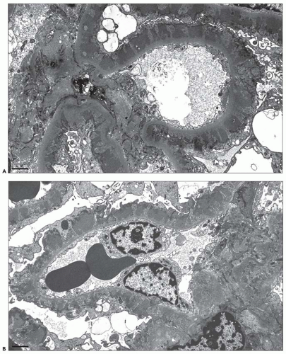
FIGURE 7.22 Electron micrograph of stage II MGN. Images (A) and (B) show the typical findings of stage II MGN, including global subepithelial electron-dense deposits separated by intervening GBM spikes. (A and B: ×6000.)


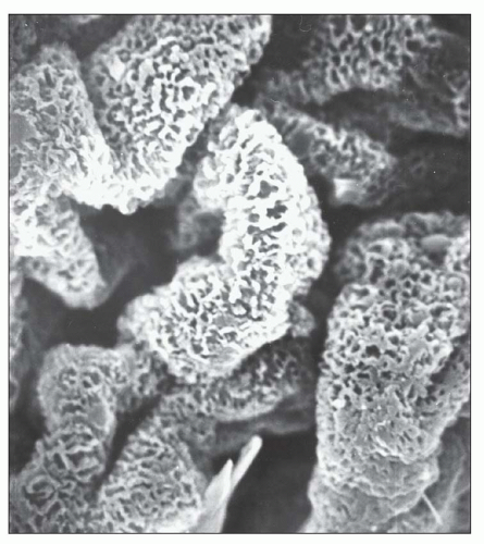
FIGURE 7.23 Scanning electron micrograph of an acellular glomerulus prepared as described in Figure 7.21 from a patient with stage II MGN. The projections of basement membrane material are seen as a complex anastomosing network on the outer surface of the GBM. (×4,000.) (Courtesy of Dr. Stephen M. Bonsib.)

Subepithelial deposit formation in MGN is a dynamic and potentially reversible process. The limitations of the Ehrenreich and Churg classification stem from the fact that the four stages are likely accurate for a single generation of deposits but do not address the complexity of ongoing and simultaneous deposit formation, incorporation into the GBM, and in many cases, resorption of the deposits, leading to GBM remodeling and potential healing. The clinical and pathologic resolution of disease is likely most dependent upon diminishing the rate of antibody production and deposit formation. Stated differently, outcomes are likely dependent on creating an imbalance such that the rate of healing and remodeling outpaces the rate of deposit formation. Along these lines, it has been suggested that the degree of thickening of the GBM indicates a more prolonged disease course (85), and the finding of homogeneous, synchronous deposits (i.e., deposits of a single stage) has been shown to have a significantly better prognosis than heterogeneous deposits (i.e., multiples phases of deposits) (91) (Fig. 7.28). Along the same lines, another method to prognosticate in MGN may be to measure the percentage of deposits that have an electron-lucent appearance, indicating at least partial resorption; in our experience, cases with a preponderance of electron-lucent deposits tend to have lesser degrees of proteinuria and are often in a state of at least partial remission. Future studies may be helpful to address this issue.

While the subepithelial deposits are globally distributed in the majority of cases of MGN, there are multiple reports of segmental MGN (52,92,93,94,95) (Fig. 7.29). The term “segmental MGN” should be reserved for cases with a segmental distribution of the deposits by light microscopy, immunofluorescence, and EM. The pathogenetic significance of segmental MGN, as compared to the more commonly encountered global lesion, is not well understood. It is likely that some of the cases, in particular those that exhibit stage I changes, represent an early manifestation of the usual form of (global) MGN, and it has been proposed that other cases may represent a resolving phase of MGN (92). Obana et al. (93) compared 27 children with global MGN to 11 children with segmental MGN and found that the segmental lesion was associated with a higher incidence of C1q positivity by immunofluorescence and mesangial deposits by EM but a similar clinical presentation and outcome. Interestingly, only 3 of the 11 patients with segmental MGN had stage I changes, and 2 patients underwent repeat biopsy at 3 years and were found again to have segmental deposits, arguing in favor of segmental MGN being a distinctive variant of MGN rather than an early manifestation of global MGN (93). Based on the limited information available, segmental MGN is most commonly reported in children (92,93,94). There are reports of segmental MGN in adults and, in most cases, the findings are superimposed on another pattern of glomerular injury. Bertani et al. (52) described four adult cases of segmental MGN, all of which coexisted with other glomerular lesions including minimal change disease (two patients), diabetic glomerulosclerosis, and hereditary nephritis. In a recent report on MGN with antineutrophil cytoplasmic antibodies (ANCA)-associated necrotizing and crescentic glomerulonephritis, the membranous changes were only segmentally distributed in 6 of 13 cases (95).

The electron-dense deposits in primary MGN are usually amorphous and finely granular, with notable exceptions (96,97,98,99,100,101). Kowalewska et al. (97) described 14 patients with a variant of MGN in which the subepithelial deposits exhibit a unique, microspherical substructure with a mean diameter of 85 nm (Fig. 7.30). Although these structures are the approximate size of nuclear pores, the authors were unable to identify nuclear pore antigens as a source for the spherules. Clinical presentation and outcomes were similar to other cases of primary MGN with the exception of associations with autoimmune diseases (SLE or Sjögren syndrome) in two patients. There are
reports of cases in which light microscopy and immunofluorescence strongly suggest the diagnosis of MGN, but EM reveals deposits that exhibit a fibrillar or microtubular substructure (96,98,99,100,101). Based on their ultrastructural appearance, these cases are best considered as examples of fibrillary glomerulonephritis and immunotactoid glomerulopathy, respectively, and should not be considered as variants of MGN (96,98,99,100,101). Podocyte infolding glomerulopathy is a recently described entity that resembles MGN by light microscopy and may or may not exhibit granular positivity for IgG by immunofluorescence (102,103,104). In contrast to MGN, EM reveals podocyte infolding with microspherical and microtubular structures within the GBM. The majority of cases of protein infolding glomerulopathy have occurred in Japan, and many of the patients have evidence of SLE (102,103,104). Although the morphogenesis of this lesion is unknown, the irregular outer contours of the GBM suggest that podocyte cytoplasmic fragments and cell membranes may become trapped in the course of deposit resorption and matrix remodeling. Finally, organized electron-dense deposits can be seen in membranous LN, and occasional cases of apparently primary MGN have unexplained organized deposits.


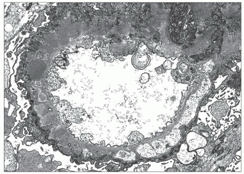
FIGURE 7.24 Electron micrograph of stage III MGN. In this example of stage III MGN, the subepithelial deposits are separated by intervening GBM spikes and accompanied by overlying neomembrane formation. Some of the deposits appear electron lucent consistent with partial resorption. (×6000.)

Mesangial electron-dense deposits are uncommon in primary MGN (33,105) and, when present, a secondary form of MGN should be carefully excluded (Fig. 7.31). Follow-up is necessary in MGN patients with mesangial deposits because SLE may manifest after a latent period of several years (14,33). Shearn et al. (105) found mesangial deposits in 9 of 107 cases (8.5%) of MGN in which SLE was not detected during a mean follow-up period of 9.8 years. In a series of 53 children with primary MGN followed for a mean of 53 months who did not have evidence of SLE or HBV, the Southwest Pediatric Nephrology Study Group (SPNSG) (14) reported an incidence of mesangial deposits of 31%, which is significantly higher than has been reported in adults. Jennette et al. (33) found mesangial deposits in 11% of 142 patients with primary MGN compared with 96% of 28 patients with membranous LN. Lai et al. (32) found mesangial deposits in 20 of 26 (76.9%) patients with membranous LN and 11 of 22 (50%) patients with MGN secondary to HBV infection. In addition to SLE and HBV infection, mesangial deposits have been described rarely in patients with MGN related to metastatic carcinoma and following treatment with penicillamine (36).


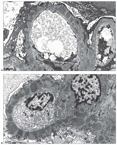
FIGURE 7.25 Electron micrographs of MGN stage II to III. In these two examples of stage II to III MGN, there are global subepithelial deposits, global intervening GBM spikes, and segmental overlying neomembrane formation. Thus, a mixture of stage II and stage III changes is present. (A: ×8000; B: ×10,000.)


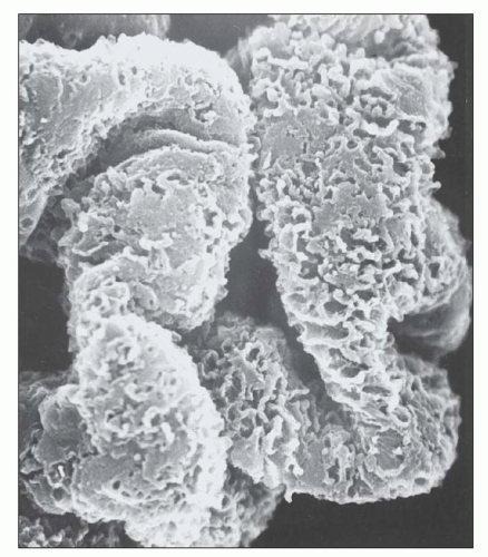
FIGURE 7.26 Scanning electron micrograph of an acellular glomerulus prepared as described in Figure 7.21 from a patient with stage III MGN. The anastomosing ridges of basement membrane material that surround and project above the electron-dense deposits (removed in this material by processing) show areas where they bridge over the deposits and form smooth plaques on the external surface of the GBM. (×5000.) (Courtesy of Dr. Stephen M. Bonsib.)

Subendothelial and extraglomerular deposits are also rarely encountered in the setting of MGN and, when present, a secondary form of disease is strongly favored (Fig. 7.32). Jennette et al. (33) described subendothelial deposits in 61% of 28 patients with membranous LN but only 3% of 142 patients with primary MGN. Similarly, TBM deposits were identified in 32% of 28 patients with membranous LN versus none of the 142 patients with primary MGN (33) (see Fig. 7.17). The SPNSG found subendothelial deposits in 9 of 9 children with membranous LN, as compared to 13% of 45 children with primary MGN (14). Lai et al. (32) found subendothelial deposits in 21 of 26 (80.8%) patients with membranous LN and 15 of 22 (68.2%) patients with MGN secondary to HBV infection. Another ultrastructural finding that favors a secondary form of MGN is endothelial tubuloreticular inclusions (Fig. 7.33), which are identified in 32% to 73% of cases of membranous LN (19,89) and 13.4% of cases of MGN secondary to HBV infection (32) but were absent in all 142 patients with primary MGN in at least one series (33).


Combined Membranous and Crescentic GN

The subepithelial deposits and associated GBM changes that characterize MGN do not typically result in GBM rupture or crescent formation. Nonetheless, crescents are encountered in a small percentage of cases of MGN and, when present, should raise the possibility of concurrent SLE, anti-GBM disease, or systemic vasculitis and should lead to a serologic workup that includes testing for antinuclear antibodies, anti-GBM antibodies, and ANCA (Fig. 7.34). The topic of mixed membranous and proliferative/crescentic forms of LN is covered in Chapter 14.

The first case of combined membranous and crescentic GN due to the presence of anti-GBM antibodies was reported by Klassen et al. in 1974 (106). In 1984, Pettersson et al. (107) reviewed 11 similar cases and observed that the relationship between MGN and anti-GBM disease fell into three temporal patterns: anti-GBM disease followed by MGN, MGN followed by anti-GBM disease, and patients in whom MGN and anti-GBM disease were simultaneously detected. The topic of MGN and anti-GBM disease has been the subject of more recent reviews (108,109). At present, there are 28 reports of this dual glomerulopathy (108) including 5 patients in whom MGN preceded the development of anti-GBM disease, 5 patients who initially presented with anti-GBM disease and subsequently developed MGN, and 18 patients who were simultaneously diagnosed with both entities. Based upon scant data, patients with anti-GBM disease before MGN tend to be younger and have a better prognosis than those with MGN before anti-GBM disease (108). Among the 28 patients, clinical recovery was noted in 11 patients, including 4 with anti-GBM disease before MGN and 7 who were simultaneously diagnosed.

The pathogenesis of MGN with anti-GBM disease is obscure. It has been proposed that MGN may damaged the GBM and expose cryptic epitopes that incite anti-GBM antibody formation (106), and the converse mechanism may be operative in cases of MGN that follow Goodpasture syndrome, in which the anti-GBM antibodies may incite an immune response to a podocyte or planted antigen. Interestingly, exposure to mercuric chloride in Brown Norway rats leads to a biphasic immune response with initial development of anti-GBM antibodies and subsequent development of proteinuria and MGN (110). The diagnosis of MGN with anti-GBM disease may be challenging because the granular subepithelial deposits of MGN may obscure the linear anti-GBM antibody staining. As a result, testing for anti-GBM antibodies should be performed in all patients with MGN and significant crescent formation.

The first case of membranous and crescentic GN related to ANCA seropositivity was reported in 1993 in a patient with Wegener granulomatosis (111). In 1997, Tse et al. (112) reported 10 cases of “vasculitic GN” in patients with MGN. All 10 biopsies revealed MGN with cellular and/or fibrous crescents. ANCA serologies were positive in only four of the nine patients who were tested, raising the question of whether the crescents can be attributed to a vasculitic mechanism in the absence of ANCA seropositivity. MGN and “vasculitic GN” were simultaneously diagnosed in nine patients, while one had MGN with a subsequent crescentic transformation (112). The authors of this study reviewed the relevant literature at the time and found 10 similar cases, although only one had documented ANCA seropositivity (111).

In 2009, Nasr et al. (95) described 14 cases of MGN with ANCA-associated necrotizing and crescentic GN.
ANCA serology was positive by indirect immunofluorescence or ELISA in all cases and was identified as P-ANCA with specificity for myeloperoxidase (MPO) in most cases. The cohort consisted with 8 men and 6 women with a mean age of 59 years. The mean serum creatinine was 4.4 mg/dL, and 12 of 14 patients presented with acute kidney injury (AKI). The mean 24-hour urine protein was 6.5 g/d, all of the patients had evidence of hematuria, and six had extrarenal manifestations of vasculitis (95). The diagnosis of MGN and ANCA-associated GN were established simultaneously in 13 patients, while one had been previously diagnosed with MGN. On pathologic evaluation, the findings of MGN were typically stage I or II and were only segmentally distributed in six cases. Twelve of thirteen patients with available follow-up were treated with steroids and cyclophosphamide; among these patients, 7 had stabilization or improvement in renal function, 1 had worsening renal function, and 4 progressed to ESRD (95).


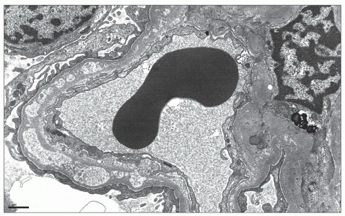
FIGURE 7.27 Electron micrograph of stage IV MGN. In this example of stage IV MGN, there is global remodeling of the glomerular basement membrane which contains intramembranous lucencies consistent with resorbed deposits. (×6000.)

Rare cases of MGN with crescents are seen in the absence of ANA, ANCA, or anti-GBM antibody seropositivity. Crescentic transformation should be considered in the differential diagnosis of the patient with MGN who develops a rapid decline in renal function with a nephritic urine sediment.


Etiology and Pathogenesis

The finding of subepithelial immune deposits defines MGN and raises the age-old question of whether circulating immunoglobulins traverse the GBM to bind to antigens that are inherent to the glomerulus and expressed on the podocyte surface or to circulating antigens that have become “planted” in the subepithelial region. Prevailing data suggest that both are correct depending on the etiology; primary MGN appears to represent an autoimmune response to endogenous antigens expressed on the podocyte cell membrane, whereas “planted” antigens likely mediate secondary forms of disease.

Great insights into MGN came from the 1959 landmark work of Heymann and Hackel (113), who immunized Sprague-Dawley rats with a crude preparation of the fractionated renal cortex (FX1a) containing the brush border of proximal tubules and produced an experimental model of MGN in which subepithelial deposit formation preceded the onset of nephrotic syndrome (114). Injection with fractionated liver, muscle, or lung did not produce the same effect, leading the authors to conclude that an “autosensitization mechanism” was central to the pathogenesis of this not yet named pattern of glomerular injury that was associated with nephrotic syndrome. It was shown subsequently that the subepithelial deposits in this model of active Heymann nephritis were composed of antigens and antibodies (115) and that passive transfer of heterologous antibodies produced in another species, such as sheep, to naive rats created a more rapid and therefore manipulable model, known as passive Heymann nephritis (PHN) (116).

A period followed in which there was an intense search for the antigenic target of the anti-rat kidney tubular antibody that is the central to the pathogenesis of PHN (117). In 1978, it became apparent that the FX1a antigen was

localized to the subepithelial aspect of the GBM and that the Heymann nephritis model resulted from in situ antigen-antibody complex formation involving an endogenous antigen, rather than circulating immune complexes containing tubular proteins that deposited in the subepithelial region (118,119). In a series of elegant experiments published in 1982, Kerjaschki and Farquhar determined that glycoprotein 330 (gp330) expressed on the proximal tubular brush border was the critical component of FX1a that induced PHN. Specifically, injection of isolated gp330 was able to induce PHN, while injection of FX1a depleted of gp330 did not produce this model (120). As a result, gp330, also referred to as megalin, was identified as the target antigen in Heymann nephritis.


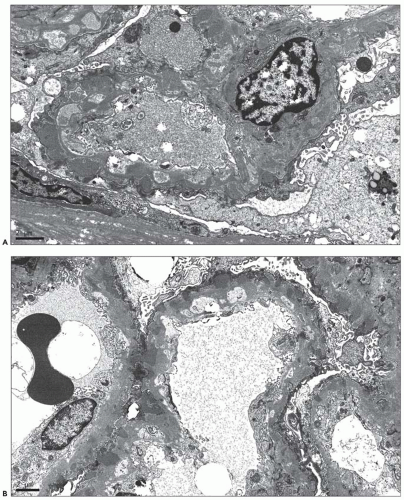
FIGURE 7.28 Heterogeneous deposits in MGN. In these two examples of MGN with heterogeneous deposits, the glomerular basement membrane is expanded by multiple generations of deposits. The deeper layer of intramembranous deposits appears electron lucent consistent with ongoing resorption. The outer layer of newly formed deposits appears electron dense. (A: ×8000; B: ×6000.)


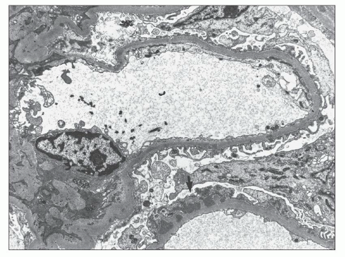
FIGURE 7.29 Electron micrograph of segmental MGN. In segmental MGN, there are segmental subepithelial deposits with intervening GBM spikes (arrow). The adjacent glomerular capillary exhibits only a rare, minute deposit. (×8000.)

Megalin/gp330 is expressed in the proximal tubular brush border of the rat, as well as the clathrin-coated pits at the base of the podocyte foot processes (120,121). Rats immunized with gp330 derived from proximal tubules produce antibody that cross-reacts with gp330 expressed on podocyte foot processes, leading to capping and shedding of antigen-antibody complexes into the subepithelial space. Megalin/gp330 is a large transmembrane glycoprotein that is a member of the lowdensity lipoprotein (LDL) receptor gene family and serves as an endocytic receptor for many ligands including apolipoproteins E and B (122,123). In the setting of PHN, antimegalin antibodies block the uptake of these apolipoproteins (124), leading to lipoprotein accumulation, lipid peroxidation, and adduct formation on matrix proteins of the GBM, a sequence of events that contributes to the development of proteinuria (125).

Human and rat megalin exhibit 77% amino acid sequence homology (122) and play a similar role in protein trafficking within the proximal tubule. The critical distinction is that megalin/gp330 is not expressed by human podocytes and is not present in the subepithelial deposits of primary MGN in man. Therefore, while megalin/gp330 is the “Heymann antigen” in the rat model, it is not the human Heymann antigen. The main historical significance of PHN is that it is a valuable experimental model for studying the molecular and cellular mechanisms of glomerular injury in MGN and pointed to the podocyte as a potential source of the target antigen in human disease (126).

The PHN model has elucidated the role of serum complement in the pathogenesis of MGN (126,127,128,129,130,131,132,133,134,135). In 1980, Salant et al. (132) treated rats with cobra venom factor to deplete C3 complement levels prior to injecting anti-FX1a; the rats developed pathologic changes of PHN, including anti-FX1a antibodies within the subepithelial deposits, but did not develop significant proteinuria. Subsequent studies showed a similar effect of C6 depletion (127), and isolated perfused rat kidneys with PHN develop pathologic changes but no proteinuria in the setting of C8-deficient plasma; with restoration of C8 levels, proteinuria develops (129). These studies demonstrated that proteinuria is complement dependent, with C5b-C9, the MAC, playing a central role. In MGN, MAC inserts into the cell membrane of podocytes, producing sublytic injury and inducing multiple cellular responses including synthesis of reactive oxygen species and proteases that degrade and disrupt the GBM, podocyte cytoskeletal alterations including nephrin-actin dissociation and loss of slit diaphragm integrity (133,135), up-regulation of transforming growth factor-β (TGF-β), and podocyte apoptosis and detachment (130). MAC formation also has been implicated in the tubulointerstitial changes that accompany the unremitting proteinuria in progressive MGN (136).

Only gold members can continue reading. Log In or Register to continue

Stay updated, free articles. Join our Telegram channel

Jun 21, 2016 | Posted by in UROLOGY | Comments Off on Membranous Glomerulonephritis

Full access? Get Clinical Tree

Get Clinical Tree app for offline access