Pyelonephritis and Other Direct Renal Infections, Reflux Nephropathy, Hydronephrosis, Hypercalcemia, and Nephrolithiasis


Pyelonephritis and Other Direct Renal Infections, Reflux Nephropathy, Hydronephrosis, Hypercalcemia, and Nephrolithiasis


Helen Liapis

Joseph P. Gaut

John E. Tomaszewski

Lois J. Arend


PYELONEPHRITIS AND OTHER INFECTIONS


Introduction, Terminology, and Historical Perspective

Pyelonephritis is a bacterial urinary tract infection (UTI) affecting the kidney parenchyma, calyces, and pelvis. It occurs in two forms, acute and chronic, and may be found with or without obstruction of the urinary tract (obstructive and nonobstructive pyelonephritis).
While bacteria cause immunologic glomerular injury and glomerulonephritis, this topic is not discussed here (see Chapter 10).

The infecting organisms are thought to be of intestinal origin and reach the kidney from the lower urinary tract by an ascending route. Rarely, bacteria in the bloodstream may colonize the kidney (1). Most UTIs are caused by Gram-negative enteric organisms and can be symptomatic or asymptomatic, classified as uncomplicated or complicated (Table 24.1). Uncomplicated UTIs occur in otherwise healthy individuals. Women are affected more than men, and the most common bacteria are Escherichia coli (E. coli) (80%) (1,2,3,4). Uncomplicated UTI is characterized by frequency, urgency, dysuria, or suprapubic pain. Acute nonobstructive pyelonephritis occurs in the same population that experiences acute uncomplicated UTI and is characterized by costovertebral angle pain and tenderness, often with fever. Complicated UTIs occur in immunocompromised patients, those undergoing catheterization, or those with functional or anatomical abnormalities and involve either the bladder or the kidneys. These infections are typically polymicrobial and include Proteus mirabilis, Klebsiella pneumoniae, and Pseudomonas aeruginosa, among others. The infectious landscape changes depending on the patient’s age and the presence of comorbid conditions such as diabetes, spinal cord injury, and immunologic abnormalities. Even though E. coli accounts for the majority of UTIs, in the elderly, polymicrobial infections with Gram-positive organisms are present in approximately one third of cases. Uncomplicated UTIs in children are most commonly caused by Enterobacteriaceae and may predispose them to adult disease (5,6). UTIs can have serious complications such as premature delivery in pregnant women, sepsis in the elderly, and renal scarring in children (1,2,3,4,5,6).


TABLE 24.1 Clinical syndromes of acute pyelonephritis


Uncomplicated pyelonephritis


in women


Subclinical pyelonephritis


Acute symptomatic pyelonephritis


Pyelonephritis in pregnant women


Recurrent pyelonephritis


In men (<50 years old)


Subclinical pyelonephritis


Acute symptomatic pyelonephritis


Complicated pyelonephritis


structural or functional abnormalities


Prostate disease (benign prostatic hyperplasia, prostatitis)


Obstruction


Calculi


Neurologic disease


Vesicoureteral reflux


Urologic manipulation


Intubated drainage (bladder catheter)


Urinary instrumentation (cystoscopy)


Renal transplantation


Underlying disease


Diabetes


Renal failure


Immunosuppressed or immunodeficient


Cystic renal disease


In 2007, UTIs accounted for 8.6 million ambulatory care visits in the United States (7,8). About 25% of affected women develop recurrent UTI within 6 to 12 months and can be either a “relapse” or “reinfection.” Relapse is defined as a recurrent UTI occurring after therapy and is due to persistence of the pretherapy bacteria. “Reinfection” is recurrent UTI with an organism originating outside the urinary tract, either a new bacterial strain or a strain previously isolated that persisted in the colonizing flora of the gut or vagina. Relapse of UTI requires appropriate management and prophylactic treatment according to clinical practice guidelines (9,10). The kidneys may be involved in such cases, increasing the risk of developing hypertension or renal failure.

Infecting organisms may also reach the kidney via blood-borne invasion. The most common organism infecting the kidney by this route is Staphylococcus aureus. This infection consists of vast numbers of minute abscesses scattered throughout the parenchyma, particularly the cortex. The terms multiple cortical abscesses, diffuse suppurative nephritis, and diffuse bacterial nephritis are used for this picture. Staphylococcus aureus has the ability to localize, proliferate, and incite an acute inflammatory reaction in an unobstructed kidney. In contrast to ascending infection, in blood-borne infections, minimal inflammatory changes are found in the pelvis and calyces; those that are present are secondary to the cortical infection.

Acute UTIs and the causative role of E. coli were established over 100 years ago; the terms cystitis and pyelitis were introduced at that time (11,12). Thiemich’s important observation that the renal parenchyma was frequently infected in what had previously been regarded as pyelitis gave birth to the concept of acute pyelonephritis (13). A full appreciation of the side effects of acute kidney infection came later and not until 1917 when Löhlein defined the clinical and pathologic features of the pyelonephritic kidney in three young women who died of uremia, two of them with hypertension (14).

In the context of pyelonephritis, it is important to define the term bacteriuria. Bacteriuria simply means that there are organisms in the specimen of urine tested but does not necessarily imply infection of the kidney. It may be found in patients with and without renal involvement. Furthermore, bacteriuria may result from contamination during collection. As might be expected, the urine passed at the beginning of micturition is most likely to be contaminated. Midstream specimens of urine are less likely to be contaminated and are therefore used to determine whether the bladder urine is infected. Suprapubic aspiration of urine avoids contamination by the urethral flora. Although this is an invasive procedure, it is frequently employed in infants. To distinguish between genuinely infected urine and contaminated urine, it is necessary to perform quantitative bacterial counts. Kass (15) demonstrated that bacterial counts of more than 100,000 colony-forming units (cfu) per milliliter of urine usually represent genuine infection. This is referred to as significant bacteriuria. Kass was careful to point out that a lower figure could indicate true infection under such conditions as rapid urinary flow, when urine pH is low or when bacteriostatic drugs are being used. To these can be added other factors, but over the years, the figure of 100,000 cfu/mL of urine has provided a workable basis for the determination of significant bacteriuria. A figure of 100 cfu/mL has been proposed for the specific instance of women with acute dysuria and frequency (16).

In certain patients, particularly women, significant numbers of bacteria are found in the urine on routine testing despite the fact that there are no clear clinical manifestations. The name asymptomatic bacteriuria or covert bacteriuria is given to this
situation. There is an increased prevalence of asymptomatic bacteriuria in older patients; in men, it is commonly caused by prostatic enlargement and loss of bactericidal activity of prostatic secretions (17). Poor bladder emptying due to uterine prolapse is considered important in women (18,19). Neuromuscular disease, increased instrumentation, and catheter use contribute in both sexes. Asymptomatic bacteriuria, or asymptomatic UTI, is defined by the U.S. Preventive Services Task Force as isolation of a specified quantitative count of bacteria in an appropriately collected urine specimen obtained from a person without symptoms or signs referable to urinary infection (20). Asymptomatic bacteriuria is due to bacteria that lack virulence factors (discussed under pathogenesis).

UTI associated with catheter use has caused considerable confusion as to the appropriate management and/or treatment in the elderly, and several authoritative guidelines are published recently to reinforce understanding of the definition of asymptomatic bacteriuria and symptomatic UTIs (Table 24.1) (21,22,23,24).

Lastly, the term “pyuria” refers to the presence of increased numbers of polymorphonuclear leukocytes in the urine and constitutes evidence of an inflammatory response in the urinary tract (20).

Distinct types of chronic pyelonephritis such as xanthogranulomatous, emphysematous pyelonephritis, and malakoplakia are discussed separately because of their unique pathology and clinical presentation.


Acute Pyelonephritis


Clinical Presentation

In a classic case of lower UTI in the adult, the patient presents with rapid onset of chills, fever, lumbar tenderness, dysuria, and urinary frequency. Hypertension is usually absent. Symptoms such as frequency, urgency, suprapubic discomfort, and flank pain should lead to screening. Young children may present with non-specific symptoms, such as poor feeding, vomiting, irritability, jaundice (in newborns), or fever alone, and a broader approach to screening may be appropriate (2,5). Acute renal failure occasionally occurs particularly in the elderly (25). Although bacteremia causes chills, it seldom causes more serious side effects, such as disseminated intravascular coagulation. The urine contains organisms in excess of 100,000 cfu/mL, and white blood cells (pyuria) and white blood cell casts are present in the sediment. White blood cell casts are significant, implying inflammation in the kidney. Proteinuria may or may not be present and is seldom heavy. Macroscopic or microscopic hematuria may develop as a result of small hemorrhages in the renal pelvis or bladder.

The clinical diagnosis of acute pyelonephritis is relatively crude and imprecise. It is often impossible to distinguish between acute pyelonephritis and infections confined to the lower urinary tract on purely clinical grounds (2). Various ancillary laboratory tests including tests for impaired urine concentration, increased serum concentration of C-reactive protein, and estimation of the lactic dehydrogenase concentration in the urine are not very useful. Urine dipstick for leukocyte esterase and nitrites and standard microscopy on a centrifuged specimen are very helpful; the so-called enhanced urinalysis that combines high-power microscopy with a hemacytometer and Gram stain of unspun urine for organisms have high predictive value (95%) (5). These tests are often complemented by various imaging techniques to add diagnostic precision. Ultrasonography (increased renal size), intravenous urography (renal enlargement with reduced nephrogram), and radionuclide methods using gallium 67 citrate and iodine 131 Hippuran (defective uptake) have been used with varying degrees of success (26,27,28). Computed tomography (CT) is currently the preferred method for assessing the extent of parenchymal involvement and possible complications of pyelonephritis such as parenchymal or perinephric abscess or emphysematous pyelonephritis, which are clinically difficult to diagnose in some cases and in atypical infections (26,27,28). In addition, CT can provide physicians with much more information about underlying abnormalities, such as stones, and congenital urologic anomalies. Unenhanced CT is excellent for identifying stones, gas, or obstruction; contrast-enhanced CT is better for acute pyelonephritis, which usually manifests as wedge-shaped (streaky) areas of low enhancement extending from the papilla to the renal cortex. The striated enhancement represents tubular obstruction by inflammatory cells and the associated edema of the renal parenchyma (Figs. 24.1 and 24.2).


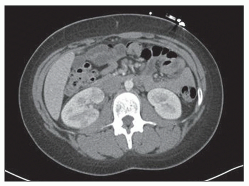
FIGURE 24.1 Contrast-enhanced transverse CT shows multiple wedge-shaped areas of lower attenuation with effacement of the corticomedullary junction in the right kidney. These findings are highly suggestive of pyelonephritis. (Courtesy of Cary Siegel, Mallinckrodt Institute of Radiology, St. Louis, Missouri.)


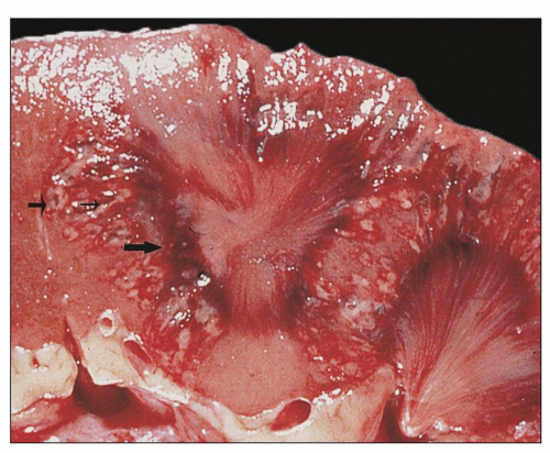
FIGURE 24.2 Acute pyelonephritis. Cortical abscesses are apparent and straight yellow streaks (thin arrows) and hyperemia in the medulla (thick arrow).


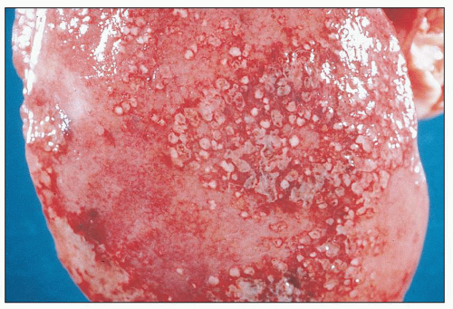
FIGURE 24.3 Acute pyelonephritis. Cortical abscesses produce discrete or confluent, raised, yellowish-white, rounded nodules with surrounding hyperemia on the subcapsular surface.


Gross Pathology and Light Microscopy

Acute pyelonephritis seen by the pathologist grossly is limited to surgically resected obstructed kidneys. Obstructive acute pyelonephritis presents as an enlarged kidney with a bulging cut surface. The cortex contains whitish areas of acute infection. Between these are scattered, small, discrete, whitish-yellow abscesses with a hemorrhagic rim. These small abscesses, measuring up to several millimeters in diameter, are seen particularly well on the subcapsular surface (Fig. 24.3). In some cases, almost the entire cortex is white and swollen. The pelvis and calyces are dilated. The mucosal surfaces are often congested, with thickening of the pelvic wall. In cases of severe obstruction, the renal parenchyma may be thinned with blunted papillae and the pelvis filled with pus. This situation is referred to as pyonephrosis (Fig. 24.4).

In addition to the cortical changes, the medulla shows characteristic straight, whitish-yellow streaks corresponding to pus-filled collecting ducts (see Fig. 24.2). Papillary necrosis may be present, particularly in diabetic patients with severe, often terminal, acute renal infection. Secondary involvement of the kidney occurs via a hematogenous route. Regardless of whether it is the obstructive or nonobstructive type, the histology of acute infection is similar. Tubules are extensively destroyed by acute, neutrophil dominant, inflammation (Fig. 24.5). Neutrophils may also fill the medullary collecting ducts. Acute inflammatory changes are seen in the pelvic and calyceal epithelium (Fig. 24.6). They are generalized in obstructive forms but restricted to the involved calyceal systems in nonobstructive forms. Papillary necrosis may be seen in severe terminal renal infections with obstruction and diabetes (2,3). An important feature of acute nonobstructive pyelonephritis is the way large areas of parenchyma are spared from infection.


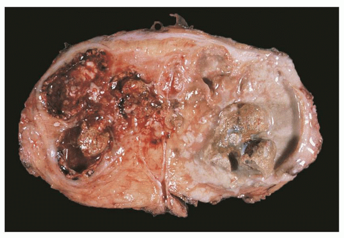
FIGURE 24.4 Pyonephrosis. The kidney is converted into a pus-filled sac, with little identifiable parenchyma. The mucosa of the collecting system is focally hemorrhagic and covered by creamy exudate; it contains several calculi.


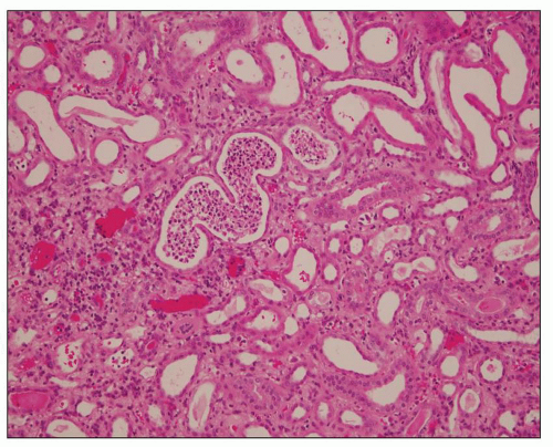
FIGURE 24.5 White cell casts in acute pyelonephritis. (H&E; ×400.)


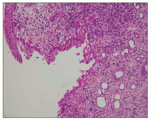
FIGURE 24.6 Acute pyelitis; neutrophils erode the lining epithelium forming microabscesses. (H&E; ×200.)


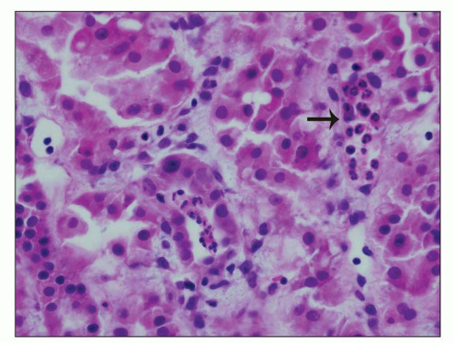
FIGURE 24.7 In acute pyelonephritis, neutrophils appear first in peritubular capillaries (arrow). (H&E; ×400.)

Initially, the inflammatory exudate consists of neutrophils, first appearing in the intertubular capillaries (Fig. 24.7). This is followed by capillary wall rupture, with leakage of fluid and cells into the interstitium (Fig. 24.8) (29). Chronic inflammatory cells, such as macrophages, lymphocytes, and plasma cells, appear within a few days of the start of infection as neutrophils disappear fast (30,31). Glomeruli are surprisingly resistant to damage (Fig. 24.8). Although some glomeruli are secondarily involved by inflammation—invasive glomerulitis—the vast majority remain unscathed (32).

Small interstitial capillaries are also involved in acute pyelonephritis as mentioned earlier; they are occluded by leukocyte plugs. Larger vessels contain acute inflammatory cells adhering to the endothelium. Endothelial cell necrosis, vascular wall fragmentation and remodeling around the endothelium, adherent degranulated neutrophils containing phagocytosed bacteria can be seen ultrastructurally. The perivascular interstitial matrix is filled with collagen fibrils or fibrin deposits. Reparative changes including capillary neovascularization are not infrequent (29). In addition to small-vessel damage, the renal vein or artery may undergo thrombosis in severe acute pyelonephritis (33).


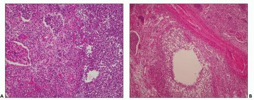
FIGURE 24.8 Acute pyelonephritis. A: Pools of neutrophils destroy tubules; glomeruli are remarkably unaffected. B: Abscess formation. (H&E; ×200.)

A diagnosis of pure acute pyelonephritis in native or allograft renal biopsies is currently infrequent. Diagnostic histopathologic findings are similar to obstructive pyelonephritis in surgically resected kidneys and consist of significant interstitial inflammatory infiltrate likely occupying about 50% of biopsy surface area and intratubular white cell casts. These findings in the appropriate clinical scenario, such as fever and positive urinary cultures, pose no diagnostic difficulty. Problems arise when tubular inflammation is focal, blood or urine cultures are negative, patients are partially treated prior to biopsy, or atypical infectious microorganisms underline the process. Focal intratubular neutrophils or white cell casts can be found in other diseases, for example, acute interstitial nephritis, cast nephropathy, and acute tubular necrosis (ATN). In allograft renal biopsies, the differential diagnosis includes tubulointerstitial rejection and acute allergic—drug-induced—interstitial nephritis (34,35,36). Acute cellular rejection is mediated by mononuclear lymphocytes infiltrating the tubular epithelium. Neutrophils, if present, are rare and only usually do not form tubular casts. Currently, most kidney transplant recipients are preemptively treated with antibiotics, usually vancomycin, to prevent UTIs; thus, acute pyelonephritis is an infrequent complication in this patient population (36).

A particularly challenging situation is excluding ATN associated with neutrophilic infiltrates. For example, white cell casts may be present in renal biopsies from patients presenting with acute renal failure and negative blood/urine cultures; multifocal ATN is the only finding in some cases. The role of infection in causing acute renal failure and presumed ATN has been addressed in the literature, but renal biopsy findings are not adequately documented (37). ATN is associated with an inflammatory response that includes monocyte/macrophage
and neutrophil recruitment to the kidney, which may worsen renal injury, but there is considerable controversy on the pathology of ATN in humans compared to animal models (38). Recent studies suggest that interstitial injury activates innate immunity and the inflammasome, a new concept to explain inflammatory responses in kidney disease in various conditions including ATN and obstruction (39,40). Innate immune response in the context of kidney infection is discussed further under pathogenesis of pyelonephritis.


Specific Forms of Acute Pyelonephritis


Diffuse Suppurative Pyelonephritis

This lesion is caused by blood-borne infection of the kidney, as opposed to ascending infection. It is typically caused by S. aureus, an organism that can localize in the kidney without obstruction. It may also be seen with E. coli bacteremia, but only when there is obstruction to urinary outflow. The source of infection is often nosocomial. Staphylococcus aureus is the most common organism. Immunosuppressed patients are particularly vulnerable to staphylococcal infections (34,35). Moreno et al. (34) studied 75 episodes of bacteremia or fungemia in renal transplant recipients. They found that the kidney and urinary tract was the site of infection in 21 cases (28%), and staphylococci were the most common infectious organisms. Patients presented with fever, lumbar pain, symptoms of lower UTI, and renal failure (34).

Grossly, the kidneys are enlarged. The subcapsular surface is studded with numerous whitish-yellow abscesses, often with red rims. The abscesses vary in size; some are as small as a pinhead, but others may measure up to half a centimeter across (Fig. 24.9). Abscesses are usually discrete, but some are confluent. The cut surface bulges because of the accompanying interstitial edema; some are rounded and similar to those seen on the subcapsular surface, but others are wedge shaped with the apex pointing inward. Most abscesses are in the cortex, although some are in the medulla, particularly the outer part. Whitish streaks are often seen in the medulla, representing pus-filled collecting ducts. The pelvis and calyces are not usually dilated. Both kidneys are affected and equally enlarged.


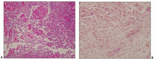
FIGURE 24.10 Suppurative pyelonephritis from a 68-year-old man with diabetic end-stage kidney disease. A: Massive neutrophil influx obscures and destroys proximal tubules. (H&E; ×200.) B: Gram-negative bacteria admixed with neutrophils in tubules and the interstitium. (Gram stain × 400.)


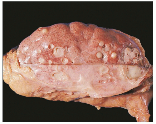
FIGURE 24.9 Diffuse suppurative nephritis. The subcapsular surface shows numerous discrete and focally confluent, whitish-yellow abscesses of variable size.

Histologically, abscesses consist of large numbers of interstitial neutrophils, with extensive destruction of tubules, particularly the proximal convoluted segments (Fig. 24.10A). Glomeruli, arteries, and arterioles are usually undamaged, although microabscesses may occasionally be seen in the glomeruli. Undamaged tubules may be filled with neutrophils, accounting for the linear streaking seen grossly in the medulla. Organisms are readily evident (Fig. 24.10B). In contrast to acute pyelonephritis, there are no, or only a few, inflammatory cells beneath the calyceal and pelvic epithelium (35). Immunocompromised patients may have a modified response
to hematogenously spread infection to the kidney. For example, the case shown in Figure 24.11 is from a 68-year-old man with history of weight loss, fatigue, weakness for 2 years, history of colon cancer, and documented blood infection. The glomeruli are filled with periodic acid-Schiff (PAS)-positive rod-shaped Tropheryma whippeli bacteria, better seen by electron microscopy (Fig. 24.12). Acute pyelonephritis was evident in the surrounding renal parenchyma, but the glomeruli are devoid of inflammation in spite of the presence of bacteria.


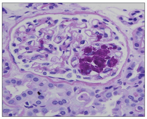
FIGURE 24.11 Acute pyelonephritis from an immunocompromised patient with Tropheryma whippeli infection. PAS-positive organisms are found within the glomerular tuft. (PAS; ×400.)


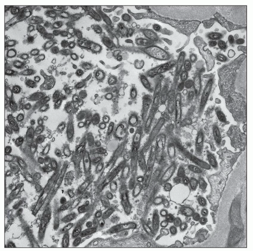
FIGURE 24.12 Transmission electron micrograph demonstrates the rod-like Tropheryma whippeli organisms within the glomerulus. (Courtesy of Carrie Phillips, Indiana University School of Medicine, Indiana.)


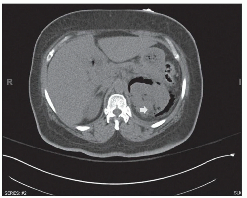
FIGURE 24.13 Emphysematous pyelonephritis. The patient was a 58-year-old woman with history of diabetes who presented with flank pain. CT coronal reconstruction shows gas in the parenchyma of the left kidney and extensive perinephric gas (arrow). (Courtesy of Sanjeev Bhalla, Mallinckrodt Institute of Radiology, Washington University School of Medicine, St. Louis, MO.)


Emphysematous Pyelonephritis

Emphysematous pyelonephritis consists of a severe suppurative infection of the kidney accompanied by gas formation in the pelvicalyceal region (emphysematous pyelitis), the kidney parenchyma, and sometimes in the perirenal tissue. Parenchymal abscesses and infarction, with gas formation in necrotic areas or papillary necrosis and vascular thromboses, are common (Figs. 24.13 and 24.14). Obstruction was recorded in 40% of cases. Ninety-five percent of patients with emphysematous pyelonephritis have diabetes (42). Drug abuse,
neurogenic bladder, alcoholism, and anatomic anomalies have also been found in association with this disease (43). Women are affected more often than are men, with a mean age in the sixth decade. Typically, only one kidney is involved, usually the left (3). Rarely, both kidneys are affected (3). Emphysematous pyelonephritis is associated with a 21% mortality rate (44). Patients may have nonspecific clinical symptoms including chills, fever, flank pain, nausea, vomiting, abdominal pain, and pyuria. Patients may initially present with thrombocytopenia, acute renal failure, disturbance of consciousness, and shock, which are risk factors for poor outcome and mortality (44). Escherichia coli is the most common organism encountered, but others, such as Klebsiella pneumoniae, Enterobacter spp., Proteus mirabilis, Candida spp., and Cryptococcus neoformans, have been described. Diagnosis can be made by CT, which also provides information for classifying the extent of the intrarenal and extrarenal disease, which has prognostic and therapeutic importance (2,3,26,27,44). Emphysematous pyelonephritis is a very serious condition that requires prompt and energetic treatment. Operative and nonoperative treatment with antibiotics is currently employed avoiding nephrectomy. The allograft kidney is rarely affected with only about 20 cases of emphysematous pyelonephritis reported in the literature (43). A new radiologic classification is proposed taking into account the extent of gas accumulation in the kidney (class 1 to 4) to help guide appropriate and timely management of patients (44). The pathogenesis of the condition is not clear, but many features are similar to those described in S. aureus infection, suggesting a blood-borne infectious etiology. Four factors involved in pathogenesis include gas-forming bacteria, high tissue glucose, impaired tissue perfusion, and a defective immune response (44,45,62). Successful medical therapy is possible in some cases (45).


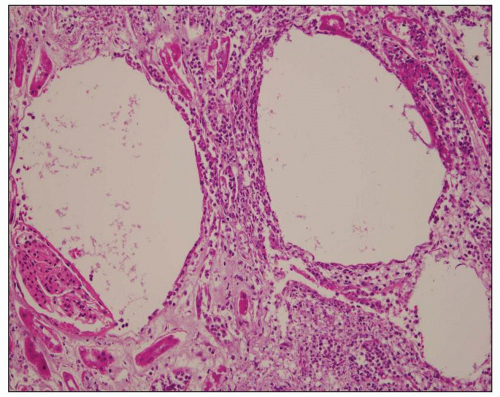
FIGURE 24.14 Emphysematous pyelonephritis. The patient was a 68-year-old diabetic woman. Gas formation in necrotic tissue produces circular spaces resembling pulmonary emphysema. (H&E; ×200.)


Sepsis and kidney injury

Sepsis is currently defined as a systemic inflammatory response associated with a confirmed infection. There appear to be two phases in response to sepsis. First, a proinflammatory response results in hypotension and organ dysfunction. This is followed by an anti-inflammatory response causing immune depression (46). The mechanism of kidney injury in sepsis is multifactorial. Hemodynamic instability, diffuse intravascular coagulation, inflammatory mediators, and tubular obstruction secondary to tubular cell death are all believed to play a role (47). Patients present with acute renal failure, but the pathology of kidney injury in patients dying of sepsis is of lesser magnitude than is the associated degree of renal dysfunction (48). Tubular injury is typically manifested within the proximal tubules as cytoplasmic volume loss, focal cell detachment and sloughing, cytoplasmic blebbing, and loss of the brush border, findings characteristic of ATN. However, there is no evidence of acute pyelonephritis in most patients with sepsis. Suppurative and emphysematous nephritis are the extreme situations, while most commonly, sepsis is complicated by acute kidney injury (AKI), which can be a serious complication regardless (further discussed in Chapter 24).


Clinical Course, Prognosis, and Therapy of Acute Pyelonephritis

A detailed discussion of the clinical course, prognosis, and therapy of acute pyelonephritis is beyond the scope of this chapter. Outcome data and therapeutic approaches are influenced by the clinical syndromes of “uncomplicated” or “complicated” pyelonephritis. Patients with “complicated” pyelonephritis present with a wide range of structural or functional abnormalities of the urinary tract, with various underlying diseases rendering them more susceptible to infection, or with a renal infection following urologic manipulation. A number of sequelae can complicate the disease including stones, papillary necrosis, pyonephrosis, perinephric abscess, septicemia, and involvement of other organs, for example, the gallbladder (49). Important current issues are what the most common microorganisms causing acute pyelonephritis are and what are the best management and treatment strategies for antibiotic-resistant bacteria in the various age/sex groups (2,50,51).


Chronic Pyelonephritis


Definition and Controversies

The clinical and pathologic features of chronic pyelonephritis appeared over three decades following Löhlein’s 1917 article (14) on acute pyelonephritis (52,53,54). Recognition of a unilateral form of chronic pyelonephritis and its ability to produce hypertension (see Refs. (53,54)) firmly established chronic pyelonephritis as an important disease entity.

Regrettably, during the 1950s and 1960s, many pathologists diagnosed chronic pyelonephritis with such profligacy that it became the most abused term in the whole of renal medicine. The diagnosis was frequently made purely on parenchymal histologic changes, few of which, if any, were specific for the sequelae of infection. The essential “pyelo-” part of the term was completely ignored. It was not until pathologists, including Heptinstall (41,54), took notice of the radiologic observations of Hodson (55) that stricter criteria for the diagnosis of chronic pyelonephritis were imposed. Refinement of our diagnostic criteria has been aided by a better understanding of the mechanisms organisms use to reach the kidney and the pattern of infection produced. These findings in turn have led to problems in nomenclature.

The term chronic nonobstructive pyelonephritis gave rise to much confusion in the past. It is understood that vesicoureteral reflux (VUR) and infection may be the initial events leading to parenchymal damage. The possibility that reflux of sterile urine can initiate renal scarring has called into question the term chronic nonobstructive pyelonephritis, which by definition implies an infectious origin. Second, it has led to the introduction of the term reflux nephropathy (56) to describe the kidney with discrete scars in a lobar distribution. This term displaced “chronic pyelonephritis,” specifically the nonobstructive form. Since reflux nephropathy does not specify the origin of the scars, it can be used in potential cases of sterile reflux. It is also a more appropriate description of the kidney with severe reflux with generalized pelvic and calyceal dilatation, the so-called back-pressure type. The argument against its use is the exclusive emphasis placed on the mechanism whereby urine reaches the kidney, denying the role of infection in scar formation (discussed further later).


Gross Pathology and Light Microscopy

Depending on the site of obstruction, one or both kidneys may be affected. For example, only one kidney will be affected if the obstruction is above the vesicoureteral junction (Fig. 24.15), but both will be involved if the obstruction is below that level. When the renal capsule is stripped, coarse, depressed scars are evident on the cortical surface (Fig. 24.16). The pelvis is
dilated as are all the calyceal systems. The pelvic wall is thickened and granular and often shows signs of congestion owing to active infection. Some cases are associated with stones, typically found in the pelvis and calyces. As a consequence of generalized dilatation of the collecting system, it is common to find a thinned parenchyma, particularly in areas aligned with dilated calyces (Fig. 24.17B). Blunting of the papillae is almost invariably a feature. In some cases, large, discrete scars are seen, as in kidneys with the back-pressure type of reflux nephropathy. Often, however, scars are not apparent, and parenchymal thinning is uniform.


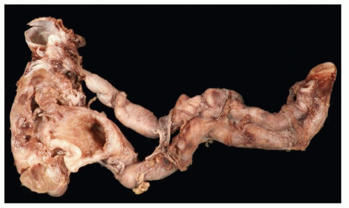
FIGURE 24.15 Chronic pyelonephritis in an 8-year-old girl with left duplicated ureter and multiple recurrent UTIs, but no evidence of renal dysplasia. The kidney is atrophic with thin cortex and dilated pelvis. The contralateral kidney was intact.

Chronic obstructive pyelonephritis shows changes in virtually the entire parenchyma: Tubular atrophy with zones of thyroidization, chronic inflammatory cells, and neutrophils are common in the interstitium, within tubules, and under the pelvic and calyceal mucosa (Fig. 24.17). As inflammation subsides, interstitial fibrosis develops particularly intensely around collecting ducts; a striped pattern of fibrosis—geographic maldistribution—and hyaline degeneration of the ducts of Bellini appear (Fig. 24.18). Lymphoid follicles are present in the parenchyma and under the epithelium of the pelvis and calyces (see Fig. 24.17B). In cases where not all of the parenchyma shows evidence of chronic infection, there may be obstructive atrophy with a paucity of inflammatory cells.


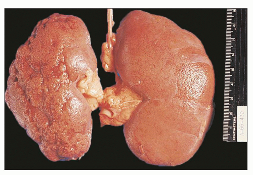
FIGURE 24.16 Chronic pyelonephritis in an adult. Irregular, coarse, depressed scars on the cortical surface of the left kidney are easily appreciated with the capsule stripped.


Specific Forms of Chronic Pyelonephritis


Xanthogranulomatous Pyelonephritis


CLINICAL PRESENTATION

Xanthogranulomatous pyelonephritis is a rare chronic debilitating inflammatory condition of the kidney characterized by focal or diffuse renal destruction. Xanthogranulomatous pyelonephritis manifests with various combinations of flank pain, a palpable mass, malaise, weight loss, fever, and sometimes nausea and vomiting. There is frequently anemia, leukocytosis, raised erythrocyte sedimentation rate, proteinuria, and white blood cells in the urine. Miscellaneous organisms, including E. coli, Proteus sp., Klebsiella sp., P. aeruginosa, and Enterococcus faecalis, may be cultured from the urine. In an appreciable number of cases, the urine is sterile (57). Because renal involvement is predominantly unilateral, significant renal failure is rare. Hepatic dysfunction has been reported (58). The reason for hepatic dysfunction is unclear, but abnormal test results often return to normal following removal of the damaged kidney.

Xanthogranulomatous pyelonephritis occurs most often in adult females by a ratio of almost 4:1. The peak incidence is in the fifth and sixth decades. However, it may appear at any age, and patients as young as 2 months (59) and as old as 94 years (60) have been reported. In the pediatric population, boys and girls are affected with equal frequency. In general, it is unilateral. Occasionally, xanthogranulomatous pyelonephritis is bilateral. Usually the entire kidney is involved (generalized form), but restricted forms are not uncommon (57,61).

Enlargement of the kidney, presence of stones or tumors in the renal pelvis, infected areas, and spread of infection to perinephric tissue may all be detected. A high degree of diagnostic precision has been achieved with computed tomography (Fig. 24.19) (2).

Urinary obstruction is an almost invariable feature of xanthogranulomatous pyelonephritis, commonly a result of stones. In large series, stones were found in 78% of cases (57). Stones are frequently of the large staghorn type. Other causes of obstruction include transitional cell tumors of the renal pelvis, congenital pelviureteric stenosis, tumors of the ureter, and postirradiation stricture. In some cases, no cause for the obstruction is apparent (57).


Gross Pathology and Light Microscopy

Grossly, xanthogranulomatous pyelonephritis shares many characteristics with true renal neoplasms in terms of its radiographic appearance and ability to involve adjacent structures (26,60,63). The kidney is enlarged, and adhesions to surrounding renal tissue and perirenal fibrosis are common. In severe cases, the inflammation may spread outside the kidney. Three stages are proposed: Stage I, the lesion is confined to the renal parenchyma; stage II, the lesion involves the perirenal space; and stage III, the lesion extends into perirenal and pararenal spaces (63). The pelvis is dilated and frequently contains staghorn calculi, necrotic material, and pus (Fig. 24.20). Papillae are frequently lost. The parenchyma is brownish and firm from the fibrosis and cellular infiltration. Similar to the
calyces, the parenchyma also contains foci of yellowish material. In cases with significant pelvicalyceal dilatation, there may be considerable cortical thinning.


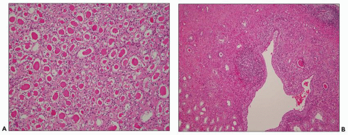
FIGURE 24.17 Chronic pyelonephritis. A: Tubule thyroidization composed of atrophic or dilated tubules with flattened epithelium containing eosinophilic, waxy casts. B: The cortex is thin above the dilated calyx. Lymphoid follicles in the cortex and in the pericalicial region, tubular atrophy and pericalicial fibrosis are present. (H&E; ×100.)

Microscopic examination reveals the yellow areas to consist of large, finely granular foam cells and smaller macrophages containing coarser granules. The large foam cells contain lipid (Fig. 24.21A) and stain positive with CD68. PAS-positive granules may be thinly scattered throughout the foam cells, but they are larger and more prominent in the smaller macrophages. In the outer parts of the yellow zone and the adjacent parenchyma are mononuclear cells, plasma cells, eosinophils, and fibroblasts. Some fibrosis is present in these outer areas as are occasional foreign body giant cells or necrotizing granulomas (Fig. 24.21B and C). Giant cells are found in proximity to cholesterol crystals, which are seen in some cases (see Fig. 24.21A). On the inside of the yellow zone, nearest to the calyx, there is necrotic debris with many neutrophils. Foci of calcification are not uncommon. The overlying cortex shows changes of chronic inflammation and, in some kidneys, microabscesses. Tubular loss is profound. There is interstitial fibrosis and marked interstitial chronic inflammation, composed of lymphocytes, large numbers of plasma cells, and frequent lymphoid follicles. Glomeruli are often normal, but occasionally, may be sclerotic. Thrombosis with recanalization of large veins has been reported in the parenchyma between the yellow areas and the cortex. A pathogenic role for venous obstruction has been suggested (60,63).


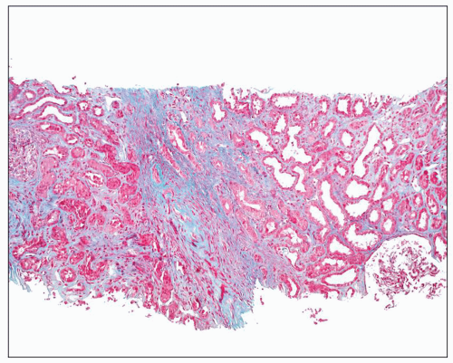
FIGURE 24.18 Chronic pyelonephritis in a 55-year-old man with creatinine of 15 mg/dL secondary to bilateral ureteral obstruction. Renal biopsy was performed to assess extend of damage. Sections show diffuse interstitial fibrosis in a stripped pattern, more pronounced around the collecting ducts. (trichrome stain × 200.)


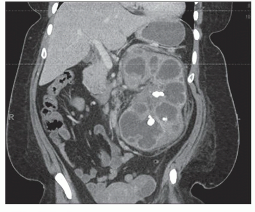
FIGURE 24.19 Xanthogranulomatous pyelonephritis. Contrast-enhanced CT scan demonstrates left kidney enlargement and distension of the collecting system by hypoattenuated material corresponding to inflammatory debris.


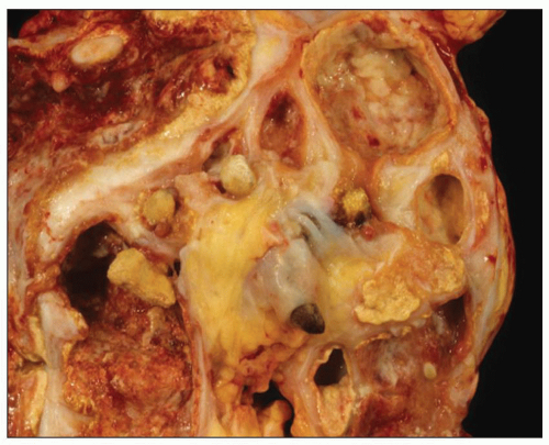
FIGURE 24.20 Xanthogranulomatous pyelonephritis. Friable, yellow tissue surrounds the dilated calyces. Numerous calculi are evident.

The focal variant, which may be mistaken grossly for a clear cell renal cell carcinoma, is restricted to a portion of the kidney with the rest of the kidney appearing normal. The involved areas show pathology identical to the generalized form. Oftentimes, focal xanthogranulomatous pyelonephritis is related to a stone that has formed in a dilated calyx or in cases of duplicated ureter. Segmental resection is curative in the focal form.


Pathogenesis

Xanthogranulomatous pyelonephritis is of infective origin. Its characteristic appearance is likely due to massive parenchymal necrosis and impaired urinary drainage, resulting in accumulation of foam cells. Ultrastructural studies of the lipid-laden macrophages have demonstrated intracellular bacteria within cytoplasmic autophagic vacuoles (64). The histologic appearance may be related to incomplete bacterial degradation and altered host responses.


Malakoplakia


CLINICAL PRESENTATION

Malakoplakia is an unusual inflammatory condition that occurs in the urinary tract, gastrointestinal tract (mainly the large intestine), testis, prostate, vagina, lung, bone, brain, and skin. Typically, malakoplakia manifests when the immune system is suppressed, for example, in transplant, diabetic, or alcohol-addicted patients. Credit for the first description is given to von Hansemann (65), although two cases had apparently been described earlier by Michaelis and Gutmann (66). Malakoplakia is well known to urologists, who are familiar with the characteristic small, discrete, yellowish-brown plaques or nodules seen on the bladder mucosa during cystoscopy. Similar nodules are found less frequently in the ureter, renal pelvis, and renal parenchyma. When the parenchyma is involved, the nodules are much larger, sometimes involving the entire organ. On renal imaging at various stages, kidneys appear enlarged or nodular mimicking malignancy (2,3,25,26).


FIGURE 24.21 Xanthogranulomatous pyelonephritis. A: Foamy cells, cholesterol crystals, and granular macrophages. B: Giant cells, mononuclear inflammatory cells, and (C) necrotizing granulomas are characteristic. (H&E; ×400.)

A 1993 review by Dobyan et al. indicated females are affected more often than males by a 4:1 ratio for malakoplakia in general and 3:1 for the renal form (67). The age ranges from childhood to the ninth decade, with a mean age for women of 45 years and a peak incidence in the sixth decade for men (67,68,69). Since the review by Dobyan et al., numerous case reports have been published both in children and adults. Unlike xanthogranulomatous pyelonephritis, which it resembles, malakoplakia often affects both kidneys, causing bilateral disease in about half of the cases.

Clinically, patients present with fever, loin pain, and history of UTI (67). Those with bilateral disease may present with acute renal failure. Signs of perinephric abscess may be noted. The urine contains protein, leukocytes, and sometimes red blood cells. Culture of the urine reveals E. coli in most cases. Urinary cytology contributes to the diagnosis by demonstration of characteristic polygonal, granular cells, and Michaelis-Gutmann bodies. Various imaging techniques are employed including CT (2,3). In their review, Dobyan et al. also discussed the role of renal biopsy and pointed out that of the 62 published cases of renal malakoplakia, 18 were accurately diagnosed by either open or needle biopsy. A review by Tam et al. (70) evaluated all cases reported since 1990. The authors emphasized the importance of renal biopsy in establishing the diagnosis. Early identification of malakoplakia allowed for effective therapy using fluoroquinolones, first used to treat malakoplakia in 1990. Renal malakoplakia has traditionally been associated with a substantial mortality rate (70%) and poor recovery of renal function (70). However, the survival rate has increased dramatically to >90% since the introduction of fluoroquinolones (70). Of the 25 cases with long-term
follow-up analyzed in the review by Tam et al., six developed significant renal impairment with three requiring renal replacement therapy.


Gross Pathology and Light Microscopy

Pathologic changes have been studied at autopsy, in surgically excised kidneys, and in biopsy specimens. On macroscopic examination, the kidney may show effects of obstruction caused with pelvicalyceal dilatation, pus in the pelvic cavity mimicking acute pyelonephritis, and thinning of the renal parenchyma. In contrast to xanthogranulomatous pyelonephritis, calculi are seldom noted in the pelvis and calyces. Perinephric abscesses were documented in 18% of the 62 cases reviewed by Dobyan et al. (67).

Parenchymal involvement consists of yellowish or tan variably sized nodules. These may remain discrete, coalesce to involve much of the renal substance, or undergo suppuration with abscess formation. The nodules are grossly visible on the subcapsular surface. On the cut surface, they can be seen extending down to the papilla. The lesions are sometimes confined to the papilla, which may show necrosis. Yellow areas lining dilated calyces are sometimes evident, but are not as frequent as in cases of xanthogranulomatous pyelonephritis. As is true of the latter condition, malakoplakia may be diffuse or focal. The diffuse variant is much more common.

Microscopic findings in malakoplakia vary and range from lesions mimicking nodular fasciitis, acute pyelonephritis, or fibromatosis. However, classic pathology consists of clusters of moderately large, polygonal cells with a foamy eosinophilic cytoplasm and compact, densely staining nuclei (Fig. 24.22). Within the cytoplasm of these cells are PAS-positive granules and larger inclusions, 4 to 10 µm in diameter, that stain strongly with hematoxylin. These larger inclusions—which may be homogeneous or laminated—are called Michaelis-Gutmann bodies. In most instances, they stain with PAS (maintained after treatment with diastase). Prussian blue and von Kossa stains for iron and calcium, respectively, also stain these inclusions. Less consistently, positive staining is obtained with oil red O, alizarin red, and Sudan black B. In addition to the histiocytic infiltrate, mononuclear and plasma cells are also seen. The tubules are severely damaged accompanied by an interstitial fibroblastic and collagenous reaction, which may show a superficial resemblance to a connective tissue neoplasm, ranging from nodular fasciitis to fibromatosis. Unless Michaelis-Gutmann bodies are diligently sought, the diagnosis of malakoplakia may be overlooked. In cases with significant pelvicalyceal dilatation, the unaffected parenchyma may show obstructive changes with features of acute pyelonephritis.


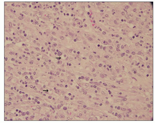
FIGURE 24.22 Malakoplakia. Tubules are destroyed by an interstitial inflammatory infiltrate composed of histiocytes. Numerous intracytoplasmic Michaelis-Gutmann bodies are shown (arrows). (H&E; ×200.)


Pathogenesis

The macrophages with PAS-positive granules and the Michaelis-Gutmann bodies have been studied extensively with the electron microscope in both renal and nonrenal cases (71). The PAS-positive granules correspond to phagolysosomes containing complex membranous whorls. The typical picture of the Michaelis-Gutmann body is a central core (often containing crystals), an adjacent lighter zone without crystals, and one or more exterior lamellar rings in which crystals are often found. Complex membranous whorls may be seen at the periphery. These observations led to the belief that Michaelis-Gutmann bodies form by aggregation of crystals upon a nidus of bacterial breakdown products in phagolysosomes. A bacterial origin is supported by studies in rats. Injection of a lipopolysaccharide (LPS) extract of E. coli cell walls (Boivin antigen) into kidney or testis reproduces many features of human malakoplakia (72). Injecting low concentrations of Boivin antigen causes macrophage infiltration with a granular cytoplasm. Higher concentrations produced structures similar to Michaelis-Gutmann bodies.

A large number of cases of malakoplakia have been associated with immune system abnormalities or immunosuppressive agents. A role for defective leukocytes and monocytes in the pathogenesis of malakoplakia has received considerable attention. A study of four patients treated with immunosuppressive agents (73) revealed that their leukocytes were unable to kill S. aureus and E. coli. When the immunosuppressive agents were withdrawn, malakoplakia improved. Abdou et al. (71) found that monocytes derived from a hypogammaglobulinemic patient with widespread malakoplakia had diminished bactericidal activity against E. coli. These cells had low levels of cyclic guanosine monophosphate. It was proposed that this resulted in decreased lysosomal degradation and an inability of the cells to release lysosomal enzymes. Cholinergic agonists reversed the impaired bacterial killing activity of this patient’s monocytes. Subsequent studies have supported the role of cholinergic agonists in treating malakoplakia (73).


UTI Pathogenesis

Our understanding of UTI pathogenesis has advanced dramatically over the last few decades by discoveries that have shed new light into the complex mechanisms and factors that determine susceptibility to and outcome of infection (74). Pathogenesis involves (a) bacterial virulence, (b) adherence and motility, (c) toxin production, and (d) host-pathogen interactions including evasion of host immune defenses (Table 24.2).


Bacterial Virulence

This special propensity of certain bacteria to cause pyelonephritis, for example, E. coli, is due to certain properties of the organisms, collectively known as virulence factors. Virulence factors are proteins, toxins, or a structure such as fimbria that enables bacteria to be harmful (74,75,76). Many novel, putative virulence factors in uropathogenic E. coli have been identified using polymerase chain reaction (PCR), including catecholate siderophore receptor (iroN), iron-regulated gene A homologue adhesin (iha), group II capsule (kpsMT), and outer membrane protease T (ompT) (77). It was apparent from early studies that E. coli invading the urinary tract had distinct characteristics and only a subset was capable of causing UTI. For example, based on serotyping, the most prevalent E. coli serotypes in patients with clinically diagnosed acute pyelonephritis are O1, O2, O4, O6, O7, O8, O16, O16/72, O18, O25, O50, and O75 (78). These strains were found less commonly in cystitis and least commonly in asymptomatic bacteriuria (79). The serotypes were determined by the components of the E. coli cell wall (capsule). The wall consists of several layers of polysaccharides such as the O antigen oligosaccharides, K antigens (Kapsel antigen) to distinguish them from the O antigens and the F antigens of fimbriae and flagella. Capsules represent the outermost bacterial layer and impart a mucoid appearance to colonies grown on soft agar. The capsular polysaccharides exhibit extraordinary diversity in structure—dozens are known—but the presence of certain common sugars forms the basis for serologic classification; for example, O, K, and H serotypes are involved in various pathologies. The explanation for the uropathogenicity of the various O serotypes has been the subject of active research that continues to evolve. A new terminology to define virulence is advancing (80). For example, it is now understood that the E. coli selection pattern of uropathogenic species is determined by genetic loci known as pathogenicity islands (defined blocks of DNA) (81,82,83,84,85). Escherichia coli strains 536 (81), IAI, and UMN026 (85) are found to have up to 13 pathogenicity islands (82,83). These sequences represent horizontal gene transfer (HGT)—also known as lateral gene transfer—(86) and can transfer genes from one species of bacteria to another. Gene transfer often involves plasmids and bacteriophages. Such genes include those that confer antibiotic resistance; therefore, HGT plays an important role in the maintenance and transmission of virulence and is the primary reason for bacterial antibiotic resistance (86).


TABLE 24.2 UTI pathogenesis: key facts


Bacterial factors


Host factors


Bacterial virulence


Antibody production against bacteria


Adhesion


pH (vaginal flora)


Motility


Tamm-Horsfall protein (THP)


Toxin production


CXCR1 gene polymorphisms


Intracellular communities (IBCs)


Neutrophils


Quiescent intracellular reservoirs (QIRs)


Innate immune response—IL, TLR


A putative uropathogenic island including a gene encoding uropathogenic-specific protein (USP) has been identified in E. coli strains isolated from patients with pyelonephritis (77).


Adherence and Motility: the Role of Fimbriae

Much attention has been given to the adhesion of bacteria to mucous membranes to explain colonization of the urinary tract. Adhesins include fimbriae, adherence pedestals (similar to fimbriae), and afimbrial adhesins, such as polymers, polysaccharides, lipoteichoic acid, and high molecular weight proteins. Whereas Gram-positive bacteria adhere more frequently via extracellular polysaccharides, Gram-negative bacteria utilize fimbriae (Fig. 24.23). Adhesion is affected by interactions between epithelial cell surface receptors and hair-like appendages termed fimbriae or pili found on the surface of the infecting organism. Fimbriae are broadly divided into two main groups, depending on the ability of mannose to interfere with their ability to attach to receptors. Thus, they are designated mannose sensitive or mannose resistant. Type 1 fimbriae are mannose sensitive, while P fimbriae and X fimbriae are mannose resistant. P fimbriae (so called because they attach to a digalactoside residue [Gal-Gal] related to P blood group antigens on human erythrocytes and uroepithelial cells) appear to be most important in
UTIs, especially with regard to renal involvement. The receptorbinding adhesin at the tip of P fimbriae is pap G. There are three classes of G-tip proteins (87). Class II-tip adhesin is associated with pyelonephritis and class III-tip adhesin with cystitis.


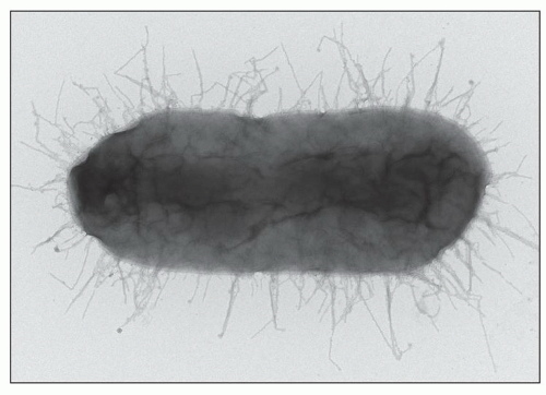
FIGURE 24.23 Uropathogenic E. coli bind to urinary epithelium with fimbriae (pili). P fimbriae (for pyelonephritis-associated pili) are important virulent factors; only bacteria featuring P pili cause pyelonephritis. (electron microscopy-negative stain × 20,000). (Courtesy of Scott Hultgren, Washington University School of Medicine, St. Louis, MO.)

Fimbriae of uropathogenic E. coli contain distinct gene clusters known as operons that code for proper assembly of the structural components of fimbriae and other proteins that control their function. For example, uropathogenic E. coli strain CFT073 has 13 fimbriae gene clusters including type 1, P, F1C, Dr, Auf, S, and M. P fimbriae are associated with human pyelonephritis but cause no disease in mouse models of UTI (88). Dr harboring E. coli adhere to bladder epithelium and enable cell invasion in vitro, in contrast to Auf fimbriae that play no role in colonization of the urinary tract in experimental animals. The genetically and chemically distinct forms of fimbriae are under the control of a promoter upstream of the genes of interest. When the promoter is on (in the correct orientation in the gene sequence), fimbriae form; when it is off (in the opposite direction), production of fimbriae is halted. The promoter is supervised by multiple enzymes, for example, Fim B and Fim E (89), and affected by local factors such as pH, oxygen supply, and presence of antibodies.

Motility is mediated by bacterial surface structures called flagella. Escherichia coli use flagella to ascend from the lower to upper urinary tract in a process that appears highly regulate. For example, E. coli flagellae are present in ascending infection and are decreased in chronic infection (90). Proteus mirabilis has a swarming mobility that transforms this bacterium to an elongated form enabling it to move across the surface of urinary catheters and cause invasive UTI (91,92).


TOXIN PRODUCTION

Escherichia coli produce three types of toxins: hemolysin, cytotoxic necrotizing factor 1 (CNF1), and autotransporter secreted toxins. Hemolysin toxins enable bacteria to insert into host cells including urothelial cells. Invasion triggers cytokine production leading to inflammatory response (93). CNF1 toxin is also implicated in invasion of host cells by altering their cytoskeleton and causing exfoliation of bladder epithelial cells, thus exposing submucosal tissue to bacteria (94). CNF1-positive E. coli stains cause more inflammation compared to bacteria lacking CNF1 (95).


Host-Pathogen Interactions


ANTIBODIES AGAINST BACTERIA

There is experimental evidence that antibodies can protect against bacteria invading the urinary tract. Their role in humans is more difficult to assess. Antibodies may be produced either locally or systemically (96,97). Experimental studies support the claim that finding antibody-coated bacteria in the urine in human patients indicates kidney infection, as opposed to lower UTI (98).

In human acute pyelonephritis caused by E. coli, antibodies are produced against O and K antigens (96) and fimbriae (99), principally as a systemic response. IgM is dominant in the early stages, followed by IgG and IgA. The urine of patients with pyelonephritis also contains IgG and IgA. IgA is probably produced by local mechanisms (17). The efficacy of these antibodies in humans is difficult to ascertain, but their presence explains the fact that recurrent UTIs are usually caused by different strains, the original strains having been eradicated by antibodies.


VAGINAL FLORA

Various mechanisms operate to prevent attachment of uropathogens to epithelium of the vagina, periurethral region, and urethra. Endogenous bacteria, such as lactobacilli in the vagina, protect by lowering local pH and by interfering with attachment via steric hindrance, competition for receptor sites, and inhibition of bacterial growth (100). More recently, using molecular-based techniques, healthy vaginal microflora was found to lack high numbers of many “good” Lactobacilli species. Instead, one or two lactobacilli from a range of three or four species are dominant, whereas other species are rare (101). It appears that the disease known as bacterial vaginosis is due to different bacterial profiles of greater microbial diversity than is evident from cultivation-dependent studies. These studies hold promise to solve the mystery of why a good number of women are prone to UTIs.


HOST DEFENSE MECHANISMS

Antiadherence mechanisms abound in the bladder. First, there is a layer of urothelial glycosaminoglycans (102), which prevents the attachment of pathogens. Second, there is the so-called slime or uromucoid layer (now recognized to be Tamm-Horsfall protein [THP]), which lines the bladder, shows great avidity for E. coli bearing type 1 fimbriae, and thereby protects the underlying mucosa (103). Experimental infection in mice supports an antimicrobial, protective role for THP, and in addition, it appears that THP also exerts immunoregulatory activity (103,104). THP knockout mice develop severe infection and lethal pyelonephritis in an experimental model of UTI (104). Furthermore, recent data demonstrate that THP links the innate immune response with specific THP-directed cell-mediated immunity via IL-1β release from inflammatory cells (monocytes) (105). Immunoglobulins in the urine of patients with acute pyelonephritis are capable of reducing adherence of E. coli to urothelial cells (17). Urine supports the growth of bacteria, but because of its relatively low pH, high osmolality, and high urea content, it is not the ideal culture medium (106). When glucose is present in the urine, as in diabetes mellitus, conditions are more propitious for bacterial growth. Voiding is also an important factor in preventing infection. Its importance becomes apparent when it is impaired, such as in obstruction of urinary outflow and VUR.

Persistent E. coli infection induces mucosal cells to produce interleukin-6 (IL-6) and IL-8, which initiate a local inflammatory response (100). Neutrophils abound in the acute phase and are seen adjacent to bacteria (Fig. 24.24); they kill bacteria by phagocytosis and release of intracellular proteolytic enzymes and reactive oxygen species. Also, neutrophils degranulate, releasing bactericidal enzymes extracellularly. Extracellular killing of bacteria by neutrophils is achieved with extracellular fibers that capture bacteria (107). The fibers are called neutrophil extracellular traps (NETs) and are only made by activated neutrophils. NETs contain neutrophil elastase, cathepsin G, and myeloperoxidase (MPO) and other enzymes derived from neutrophil granules. NETs target both gram-positive and gram-negative bacteria. It appears that the purpose of bacterial NETs is to prevent phagocytosis (107). While the aim is achieved efficiently, tissue damage ensues in part because of the toxic effects of granular enzymes to the tissue but also because NETs contain DNA including histone particles acting as immune modulators causing chronic damage similar to autoimmune diseases (40,107).


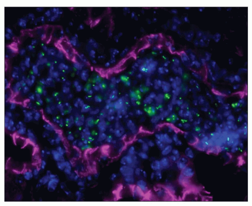
FIGURE 24.24 Experimental pyelonephritis: uropathogenic E. coli (green FITC) induce influx of neutrophils (Hoechst nuclear blue) in tubules (labeled pink with E-cadherin). Mouse kidney immunofluorescence × 63. (Courtesy of Indira Mysorekar, Washington University School of Medicine, St. Louis, MO.)


SUSCEPTIBILITY FACTORS

Blood group antigens and secretor status were found to correlate with increased susceptibility to UTI. Secretors differ from nonsecretors in their ability to secrete water-soluble blood group antigens. An increased risk of recurrent UTIs was found among women of blood groups B and AB who were nonsecretors (108). It has also been shown that women who were nonsecretors were unduly prone to renal scarring following recurrent UTIs (109). Uroepithelial cells from nonsecretors adhere with greater avidity to uropathogenic E. coli than do cells from secretors (110). Recent studies have focused on adhesive properties of bladder and kidney epithelial cells to evaluate how interactions between bacteria and host epithelia may facilitate bacterial adhesion and colonization (Fig. 24.25). For example, studies have shown that urothelial cells express a hyaluronic acid-binding protein, adhesion molecule CD44, which is constitutively expressed in the bladder and acts as a receptor for adhesion of bacteria such as Streptococcus. In the urinary bladder, it facilitates E. coli adhesion and accumulation. CD44 is not expressed on tubular epithelial cells. However, following tubular injury, CD44 is rapidly up-regulated at the tubular epithelial cell surface facilitating E. coli migration (111). Disruption of CD44 and hyaluronic acid in the bladder dramatically decreased bacterial overgrowth in spite of similar granulocyte and cytokine response in a murine UTI model, suggesting that this may be another approach to prevent and/or treat UTI (111).

The innate immune response of the host is important in the antibacterial defense mechanisms of the urinary tract, and normally, bacterial clearance proceeds without sequelae. Innate immune response is triggered by bacteria-induced influx of inflammatory cells (neutrophils and monocytes) and damage of epithelial cells that express toll-like receptors (TLRs). About 13 TLRs are identified; the first to be identified was TLR4, which recognizes LPSs from gram-negative bacteria (112,113).

A variety of host genetic susceptibility factors have been identified within the innate immune response molecules that are associated with increased incidence of acute pyelonephritis, for example, interferon regulatory factors (IRFs) and type 1 interferons. A strong association between decreased IRF3 promoter activity and acute pyelonephritis in humans was demonstrated. IRF3 is a gene encoding the IRF-3 protein. This protein is part of the TLR4 signaling pathway. Knockdown of Irf3 in mice impaired neutrophil bactericidal activity and resulted in severe disease with urosepsis and abscess formation (114). Other studies suggest a role for the CXCR1 gene, encoding the chemokine receptor type 1 protein, in acute pyelonephritis susceptibility. CXCR1 is important for neutrophil recruitment to sites of infection. Diminished expression of this protein was found in children prone to developing acute pyelonephritis (115,116). Single nucleotide polymorphisms in other genes involved in the innate immune response including CCL5, TLR4, VEGF, and CXCL8 were also found in association with human acute pyelonephritis (reviewed in Refs. (116,117)).


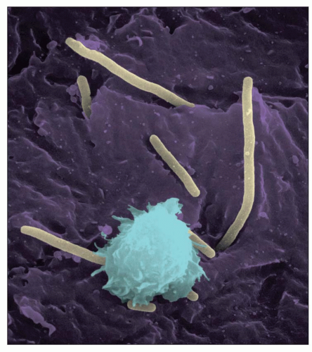
FIGURE 24.25 Scanning EM of urothelial mucosa with adherent bacteria reveals uropathogenic E. coli adhering to and colonizing the urinary bladder (magenta). Inflammatory cells (light blue) are recruited to contain bacterial invasion (pale yellow). (Courtesy of Chia Hung and Scott Hultgren, Washington University School of Medicine, St. Louis, MO.)


Bacteria and Host Immune Defenses

Breaking through mucosal barriers and immune defenses, bacteria employ ingenious approaches to succeed in establishing infection in the urinary tract. For example, E. coli expressing P fimbriae are shown to down-regulate the polymeric Ig receptor (pIgR) produced by renal epithelial cells, which transports IgA in the urinary tract (118). Decrease of pIgR correlates with IgA decrease in P fimbriae-positive E. coli-induced UTI in mice (118).


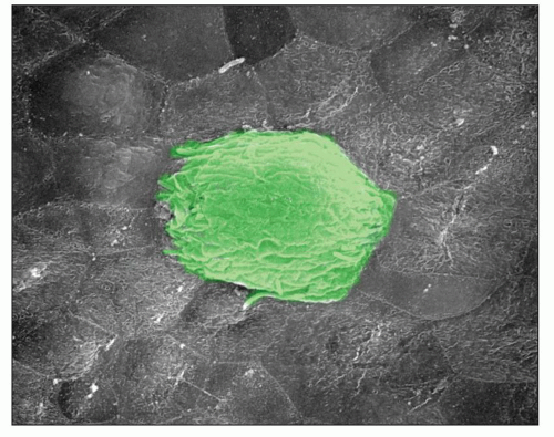
FIGURE 24.26 Scanning EM of an intracellular bacterial community (IBC) on the urothelial surface. Escherichia coli shelter from host defenses leading to persistent bacterial residence within the bladder epithelium. IBCs never form in the kidney; only form in the urinary bladder. Exfoliation of the superficial urothelial layer acts to reduce the bacterial load but facilitates chronic residence of small nests of bacteria that later reemerge to cause recurrent cystitis. (Courtesy of Indira Mysorekar.)

Further, uropathogenic E. coli evade immune response via intracellular bacterial communities (IBCs) (Fig. 24.26) and quiescent intracellular reservoirs (QIRs). IBCs are intracellular biofilms defined as assumption of a temporary multicellular lifestyle by a single-cell organism in which “group behavior” facilitates survival in adverse environments. Under certain conditions, IBCs are protected from host immune response such as neutrophil influx and antibiotics, in spite of shedding of epithelial cells triggered by the infection. It appears that inside the host cells, E. coli manage to subvert immune responses in part through autophagy, a physiologic self-cleaning mechanism; epithelial cells use autophagy to degrade toxic cytoplasmic substances, but microorganisms can utilize this mechanism to their advantage (119). Bacteria eventually exit their intracellular reservoirs seeding new colonies leading to persistent or recurrent infection.


Risk Factors


Obstruction to Urinary Outflow

Obstruction is a potent factor not only in initiating infection but also in causing it to persist and spread to the kidney. Obstruction below the bladder neck results in loss of the “flushing mechanism,” which, together with incomplete emptying of the bladder, permits bacterial growth in relatively static residual urine. The use of catheters is an additional hazard for introducing bacteria and for permitting bacterial growth (120). Urine flow obstruction complicates various cystic diseases as well, autosomal dominant polycystic kidney disease in particular, further discussed in Chapter 4.


Diabetes Mellitus

Diabetes mellitus is cited as a risk factor for UTIs and their side effects (121). Claims for certain acute infections of the upper urinary tract stand on solid foundations. For example, patients with diabetes are described as being more prone to cortical abscesses, perirenal abscesses, and emphysematous pyelonephritis (2,3). There is little evidence of an increased prevalence of true chronic pyelonephritis in the diabetic population.


Calculi

Calculi affect the host’s defense mechanisms in several ways. First, they cause obstruction, which may occur at various levels. Second, they may serve as a nidus for the persistence of infection, either because they act as an irritant or because they harbor organisms, making them difficult to eradicate (122). Calculi may aggravate infections and, in the case of struvite stones, be caused by infections. Struvite stones are composed of magnesium ammonium phosphate caused by bacterial urease activity, for example, in the presence of urea-splitting bacteria such as Proteus sp.


OTHER INFECTIONS

Renal infections are a major cause of morbidity and mortality particularly in immunocompromised patients such as AIDS and transplant recipients (123).


Mycobacterial Infections

After continued decline for three decades, pulmonary tuberculosis is again increasing. It is the leading cause of death worldwide from infectious disease (124). In Western countries, the prevalence of renal involvement among patients with tuberculosis is around 5% (125). In African countries, the prevalence of renal tuberculosis is probably higher. In Nigeria, the prevalence of renal tuberculosis among patients with pulmonary tuberculosis was found to be 9.5% if diagnosed using urine Ziehl-Neelsen stains, but the prevalence rate rose to 14% if a combination of urine stains, sterile pyuria, and tissue histology was used (124). Therefore, the diagnostic methods clearly influence the prevalence rate. Newer, more sensitive methods, including PCR, may improve diagnostic accuracy.

Immunosuppressed patients are more vulnerable to mycobacterial infections. Transplantation is a risk factor, particularly in developing countries (126). The human immunodeficiency virus (HIV) epidemic has had a significant impact on the spread of tuberculosis worldwide, particularly in Africa (123,124). Approximately 10% of tuberculosis cases worldwide were HIV related, but in regions of sub-Saharan Africa, the percentage was as high as 60%. There are two main types of renal tuberculosis: miliary and cavitary.


Miliary Tuberculosis of the Kidney (Disseminated Infection)

Renal involvement may result from hematogenous dissemination of a primary tuberculous infection, from an active pulmonary lesion, or from reactivation of a healed tuberculous lesion. Miliary tuberculosis of the kidneys is often clinically silent (27,127) and overshadowed by clinical manifestations of systemic infection. Grossly, the kidneys show white nodules (i.e., tubercles), which occur more often in the cortex than in the medulla, but they may occupy the entire kidney (Fig. 24.27). Microscopically, the early tubercle is a caseating granuloma consisting of epithelioid cells and neutrophils with central
caseous necrosis (Fig. 24.28). Organisms are usually found in such lesions. Often, a mononuclear infiltrate of lymphocytes, monocytes, and plasma cells is also present. The tubercle may be contained and heal, or the infection may expand. If the medulla is involved, the infection may reach the renal pelvis, allowing release of microorganisms into the urinary tract.


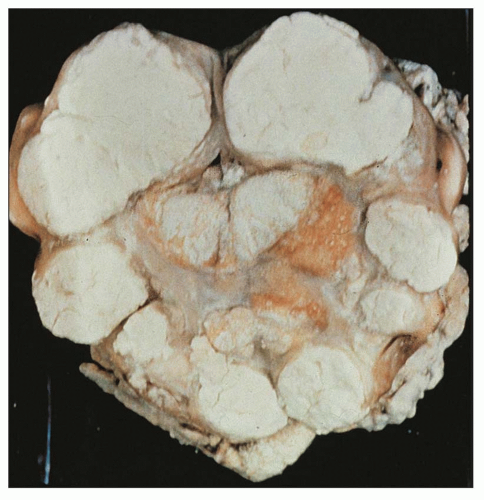
FIGURE 24.27 Tuberculous pyonephrosis. Kidney is filled with cheesy material.


Cavitary Tuberculosis of the Kidney (Localized Urinary Tract Infection)

A high proportion of men with renal tuberculosis have associated genital tuberculosis, particularly affecting the epididymis and, less frequently, the prostate (123). Genital tuberculosis is less common in women (if present, it is usually in the fallopian tubes). The lower urinary tract is commonly affected in cavitary renal tuberculosis raising the possibility of ascending infection. However, it is believed that descending spread to the urinary tract from a primary renal lesion is more likely. The renal medulla is preferentially involved by cavitary tuberculosis, where confluent epithelioid caseating granulomas will form larger and larger cavities, frequently associated with papillary necrosis (27). In cavitary renal tuberculosis, also called caseous and ulcerative, most of the symptoms result from lower urinary tract involvement, particularly the urinary bladder. The disease manifests with urinary frequency, dysuria, and hematuria. Commonly, sterile pyuria and microscopic hematuria are present. Renal function is typically preserved since unilateral renal involvement is common. Usually presents as a unilateral disease, but the contralateral kidney is involved to some degree (26,27). The diagnosis of renal tuberculosis requires a positive culture for mycobacteria, rhodamine-auramine fluorescence, or urine PCR for mycobacteria.


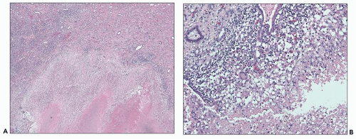
FIGURE 24.28 A: Caseating granulomas with central necrosis and epithelioid histiocytes at the periphery. B: Giant cells are appreciated. (H&E; A, ×100; B, ×400.) (Courtesy of Neeraja Kambham, Stanford University, California.)

Gross pathology of the kidneys shows either enlarged or decreased size. The surface shows irregular scarring. On cut section, the calyces and the pelvis are dilated or deformed, pelvicureteric constriction may occur, and parenchymal atrophy and foci of calcification are apparent. The lesion often begins in the renal medulla with involvement of the papilla by caseating necrosis (27). Extension of infection into the perinephric tissues may simulate invasive renal cell carcinoma (3). Segmental ureteral strictures may be seen with ureteral involvement. A combination of pelvicalyceal caseous necrosis and ureteral stenosis leads to tuberculous pyonephrosis. When this occurs, the renal parenchyma is replaced by caseous material, leaving rims of fibrous tissue imparting a loculated appearance to the organ. This condition is also known as “cement,” “putty,” or “chalk” kidney (see Fig. 24.27).

Microscopically, these lesions show central caseous debris surrounded by a peripheral granulomatous reaction (see Fig. 24.28). Mycobacteria are found in peripheral areas of caseation or cavitary lesions. Less involved areas may show
variable interstitial inflammation with lymphocytes and plasma cells amidst calcific foci, probably representing calcified tubercles.

In renal biopsies, tuberculosis is rarely seen in developed countries (128). Interstitial nephritis and epithelioid or caseating granulomas may be present, but stains for acid-fast bacilli are usually negative. Findings of acute interstitial and granulomatous nephritis in patients with documented extrarenal tuberculosis can be the result of drug reaction, and kidney biopsy should be performed to rule out active renal involvement or changing therapy. However, it is imperative to request a mycobacterial stain (acid-fast blue or Ziehl-Neelsen) if granulomas are present in the biopsy specimen, particularly if the patient’s immune system is compromised. Also, it is important to remember that in immunocompromised patients, atypical mycobacterial infections can occur, including infections with M. avium-intracellulare, which does not form typical granulomas. In such cases, abundant mycobacteria are present in foamy-appearing macrophages.


Mycobacterium leprae

Visceral involvement is more common in the lepromatous leprosy than in tuberculoid leprosy (129). Tuberculous (paucibacillary) leprosy is characterized by epithelioid granulomatous inflammation, whereas lepromatous leprosy (multibacillary) has a tendency toward diffuse infiltration of macrophages and foamy bacilli-laden cells. Renal injury is common in patients with erythema nodosum leprosum, a type of reaction typically associated with lepromatous leprosy (129).

Renal lesions are found in approximately 70% of autopsied patients with leprosy. The glomeruli, tubules, and interstitium may be involved causing glomerulonephritis, including amyloidosis and acute interstitial nephritis (130,131). Histologic examination may show endocapillary proliferation, mesangial proliferation, membranoproliferative glomerulonephritis (MPGN), and, less commonly, crescentic glomerulonephritis. In addition to glomerular pathology, chronic tubulointerstitial nephritis has also been observed in 3.8% to 54% of cases (131). Organisms are not detected in the kidney in such cases. The inflammatory infiltrate is mononuclear with associated interstitial fibrosis and tubular atrophy. Renal infection appears to occur in patients who have undergone prolonged chemotherapy for leprosy. Leproma caused by direct invasion of the renal parenchyma by M. leprae is rare. When present, bacteria can be identified in aggregations of macrophages or giant cells.


Pathogenesis

Granulomas are the hallmark of mycobacterial infection, but fungi, parasites, and even viruses cause granulomatous inflammation. The mycobacterial granuloma seems to be a host defense mechanism for walling off the bacilli, but microorganisms can survive within macrophages that compose the granulomas and persist in a latent form until immunosuppression and other triggers cause reactivation and dissemination. An understanding of the pathophysiology of granulomas is critical for the design of new drugs and vaccines. Animal models including mice, guinea pigs, rabbits, and monkeys, and in vitro systems were developed that reproduce granulomatous inflammation (132). In addition, the role of innate immunity was explored asking the question how mycobacteria manage to escape killing by immune cells. Master et al. (133) showed that Mycobacterium tuberculosis prevents inflammasome activation and IL-1β processing and that a functional M. tuberculosis zmp1 gene is required for this process. Experimentally induced infection of macrophages caused increased secretion of IL-1β and enhanced mycobacterial phagosome maturation into phagolysosomes, thus improving mycobacterial clearance by macrophages (40,114,133).


Fungal Infections

Primary fungal infections of the kidney are rare. The most common offending agents are Candida albicans and Candida glabrata. Such agents infect the lower urinary tract and may involve the kidneys through the ascending route. However, fungal infections of the kidney more commonly result from systemic fungemia, possibly of nosocomial origin and almost invariably occur in immunocompromised patients, particularly those undergoing cytostatic treatment for malignancies. In such cases, renal involvement is part of systemic fungal sepsis. Irrespective of the type of fungus infecting the kidney, the renal biopsy typically shows granulomatous inflammation. Therefore, when granulomatous inflammation is seen, a fungal infection should be considered in the differential diagnosis. Importantly, the PAS stain, excellent for detecting fungi, should be part of routine renal biopsy workup. Some fungi, for example, Aspergillus, tend to be angiocentric, as opposed to Candida, which tends to be glomerulocentric.


Candidiasis

Most infections by C. albicans are opportunistic. Infections originate from mucosal surfaces of the oral cavity, upper respiratory tract, digestive tract, and vagina, followed by hematogenous dissemination. The source of infection often is nosocomial (134). Candidiasis typically affects patients with prolonged hospitalization, with 50% of Candida bloodstream infections occurring in the ICU (134). The mean time of onset of systemic Candida infections is 22 days after hospitalization. Furthermore, when skin/mucosal barriers are breached by medical devices or surgery, it opens a portal of entry for pathogens like C. albicans. For instance, major abdominal surgery poses an increased risk for systemic Candida infections, which is underlined by the observation that in a cohort of 107 patients with candidemia, 50% underwent recent surgery (134).

The incidence of candidiasis varies. Of 102 episodes of nosocomial fungemia, C. albicans was detected in 74.5% (134). Presenting symptoms are those of severe renal infection, with low-grade fever, flank pain, costovertebral angle tenderness, hematuria, hypotension, progressive loss of renal function, and acute renal failure. Fungus balls may develop in the pelvis and calyces (135), and their passage may result in ureteral colic. Anuria occurs when fungus balls obstruct the ureters (135). Recovery of Candida from urine specimens, together with positive blood culture, suggests disseminated infection.

The kidneys may show little inflammatory response, or there may be extensive necrosis, or miliary abscesses (Fig. 24.29), and papillary necrosis. In those with diabetes, C. albicans infection may result in emphysematous pyelonephritis. Mycotic aneurysms may be present in glomerular capillaries and arterioles. However, in comparison with other fungi, invasion of blood vessels by Candida is less common, and cortical infarction
is rare (136). In tissue sections, pseudohyphae and rounded yeast forms, 2 to 4 µm in diameter, predominate (Fig. 24.30). Although Candida can be seen in sections stained with hematoxylin and eosin, they are more readily identified with PAS or Grocott methenamine silver stains.


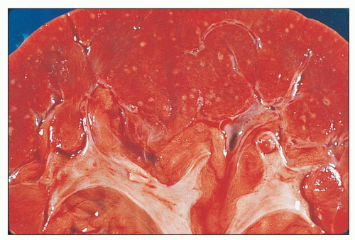
FIGURE 24.29 Candida septicopyemia with scattered small, yellowwhite miliary abscesses.

To colonize surface epithelia, Candida organisms adhere to epithelial cells through mannoproteins, hydrophobic forces (137), and proteins that bind iC3b receptors. The C. albicans cell wall can be divided into two distinct layers: the inner layer composed of polysaccharides like chitin, 1,3-b-glucans and 1,6-b-glucans, and the outer layer composed of proteins that are heavily mannosylated. These Candida molecules are recognized by Toll-like receptors (TLRs) and C-type lectins (CLRs) on the surface of antigen-presenting cells (APCs) (138). Unless phagocytosed, Candida organisms reach the subepithelial layer where through surface receptors bind to extracellular matrix components. During colonization and penetration, epithelial proliferation and T-cell-based inflammation are elicited. Animal models have shown that C. albicans can escape killing by mouse neutrophils but is efficiently cleared in human neutrophils probably due to higher levels of MPO activity and the presence of α-defensins (139). Only a minority of patients exposed to Candida develop disease. These include patients with defective T-cell function or immunosuppression who are unable to adequately contain the fungus. As a result, invasion of the vascular endothelium ensues. More recently, susceptibility to Candida was found to be linked to defective innate immunity either due to single gene mutations (rare) or due to common polymorphisms in pathogen recognition receptors such as TLRs (138).


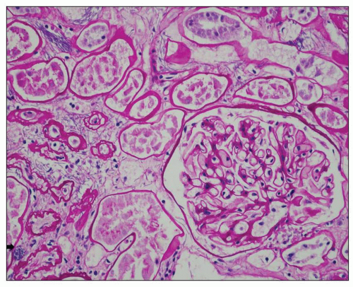
FIGURE 24.30 Renal candidiasis. Bundles of fungal pseudohyphae invade the interstitium and tubules; fungal spores are focally present (arrow); an inflammatory response is lacking. Autopsy kidney from a 49-year-old man who died abruptly from disseminated candidiasis involving multiple organs. (PAS; ×400.)


Candida glabrata

Candida glabrata (previously classified as Torulopsis glabrata) (140) is an opportunistic yeast-like fungus present in the normal microflora of the oropharynx, gastrointestinal tract, skin, urethra, and vagina. Of low virulence, C. glabrata is the second most common fungal pathogen of the urinary tract. The kidneys are usually involved as part of disseminated infections, but they may be the site of a primary infection through the ascending route, particularly in diabetic patients. Candida glabrata is also an important cause of nosocomial infection. Presenting symptoms are comparable to those caused by infection with C. albicans. Inflammatory changes resemble those caused by Candida albicans infection. Candida glabrata organisms may be seen in tissue sections as 2- to 4-µm, budding, round to oval, nonencapsulated, yeast-like organisms (Fig. 24.31).


Aspergillosis

Aspergillosis can be caused by various species of aspergilli, but the most common pathogen is A. fumigatus. The organism, present in nature, typically enters the host through respiratory
tract and mucosal surfaces, cutaneous wounds, or intravenous access lines. Renal aspergillosis is frequently the result of hematogenous dissemination, usually from invasive bronchial infection, necrotizing pneumonitis, or infarct by aspergilli. Patients receiving corticosteroids, neutropenic patients, diabetics, and immunocompromised patients (26) are particularly at risk. Less frequently, the kidneys may be involved through the ascending route (141). Renal parenchymal infections may produce symptoms comparable to those of acute pyelonephritis. The urinary tract may be obstructed by growth of mycelium, and fungus balls may be passed into the urine.


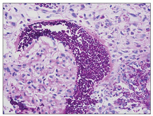
FIGURE 24.31 Renal allograft biopsy from a 50-year-old woman with end-stage kidney disease secondary to diabetes who presented with acute renal failure. Sections show PAS-positive yeast spores in Bowman space and the interstitium. Culture results indicated Candida glabrata. (Courtesy of Salinas-Madrigal, Saint Louis University, St Louis, MO.)

Renal involvement occurs in 30% to 40% of patients who die of disseminated aspergillosis (136). The involvement may be bilateral in systemic infections and in those with isolated renal involvement. Multiple small abscesses, a few millimeters in diameter and each surrounded by a red rim, are most common. However, extensive abscess formation with vascular invasion, thrombosis, and infarction may occur. Microscopic examination reveals inflammation with mononuclear cells and neutrophils. Abscess formation and infarcts containing typical septate branching hyphae are also seen. The fungus forms branching septate hyphae 3 to 5 µm wide best demonstrated with PAS or Grocott methenamine silver stain (Fig. 24.32). This morphology, however, is not specific to aspergillosis. Members of the genera Fusarium and Pseudallescheria show similar histomorphology.

The mechanism of Aspergillus infection involves adherence to epithelial surfaces and release neutrophil phagocytosis. Some of these released proteases may facilitate vascular invasion and thrombosis. The mechanism by which killing of Aspergillus takes place continues to be a subject of investigation, but there is some evidence to suggest that priming of neutrophils by formylated tripeptide followed by IL-8 enhances phagocytosis of serum-opsonized conidia from 15% to 55% (142) and distinct human monocyte subsets such as CD14(+)CD16(+) that secrete large amounts of tumor necrosis factor (TNF) are superior in fighting Aspergillus sp. compared to other monocytes (143).


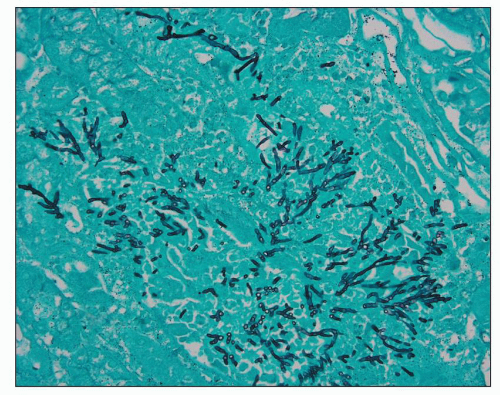
FIGURE 24.32 Renal aspergillosis. Large numbers of branching septate hyphae are illustrated. (methenamine silver × 380.)


Cryptococcosis

Cryptococcus neoformans, the agent of cryptococcosis, is a yeast-like fungus encountered in avian habitats, particularly those contaminated with pigeon dropping. The portal of entry to the host is the respiratory tract. Pulmonary infection is common, particularly in immunosuppressed and neutropenic patients. Hematogenous dissemination results in central nervous system and organ-based infection, including the kidneys. In renal transplant recipients, transmission may be donor derived (144). Renal involvement may be clinically silent or may manifest with costovertebral angle tenderness, pyuria, and gross hematuria. Yeast forms can be cultured from the urine and can be recognized in the urinary sediment by negative staining with India ink.

Renal involvement is found in about 50% of patients who die of disseminated cryptococcosis (136). Small parenchymal abscesses, or granulomas, with central necrosis involving the cortex and medulla have been described (136). The organism may elicit little inflammation despite extensive tubular destruction. Cryptococci can be identified in tissue sections as 4- to 20-µm-diameter spherical structures with a polysaccharide capsule (Fig. 24.33). The capsule is distinct, staining intensely with mucicarmine, Alcian blue, and PAS. Renal impairment has been described, but was attributed to coexistent disease.

Eliminating cryptococci from sites of infection involves growth inhibition, a process that depends on nitric oxide production by phagocytes, and phagocytosis by macrophages, a complement-dependent mechanism modulated by cytokines.


Histoplasmosis

Histoplasmosis, caused by Histoplasma capsulatum, is endemic in South and Central America and in the Ohio River and Mississippi River valleys in the United States (136). Infection is caused by inhalation of dust particles from soil contaminated with bird or bat droppings containing the fungus. The initial presentation resembles pulmonary tuberculosis. Disseminated histoplasmosis may result from a primary infection or from a reactivated, healed lesion during
immunosuppression. The disease is not contagious. Renal involvement is usually clinically silent, and compromise of renal function is uncommon.


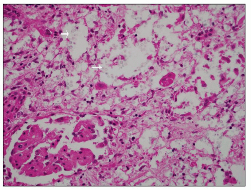
FIGURE 24.33 Renal involvement in systemic cryptococcosis. Yeast within glomeruli and tubules are separated by clear halos corresponding to their thick capsule. (H&E; ×200.)

The kidneys are involved in about 40% of patients as a result of progressive disseminated histoplasmosis. Less commonly, infection may be acquired during graft implantation. Grossly, lesions range from one or more well-circumscribed nodules to diffuse inflammation and necrosis. Papillary necrosis has also been described. Microscopically, small aggregates of yeast-laden macrophages may be present in all renal compartments, usually associated with granulomas. Parasitized macrophages show large numbers of round to oval yeast measuring up to 5 µm in diameter, usually identifiable with Grocott methenamine silver stain (Fig. 24.34). The histologic diagnosis of histoplasmosis can be confirmed by immunofluorescence with antibodies to polysaccharide antigens of H. capsulatum (136).


Coccidioidomycosis

Coccidioidomycosis, caused by Coccidioides immitis, is endemic in the southwest and western United States. Sporadic cases of disseminated coccidioidomycosis have been reported in nonendemic areas. In nature, the fungus is in a mycelial form, but it produces arthroconidia, which is the infective form. Inhalation of arthrospores causes symptomatic infection in about 10% of patients. Progressive pulmonary infections are rare. Systemic dissemination occurs in <1% of cases. Of these cases, patients are typically immunosuppressed, diabetic, or pregnant. Although not contagious, coccidioidomycosis is transmissible during autopsy procedures, presumably through aerosolization of endospores (145).

The kidneys are involved in one third of patients who die of disseminated infection (136). Grossly, minute granulomas and abscesses are present. The granulomas show caseous or suppurative necrosis. Coccidioides immitis organisms are easily found in active lesions within macrophages or giant cells as thick-walled spherules, about 100 µm in diameter, containing endospores that are 5 to 30 µm in diameter (136). Disease progression has been equated with attenuation of cellular immunity from antigen overload, suppressor cells, immune complexes, and immunosuppressive factors released from the fungus. Containment of infection depends on cellular immunity that, through release of lymphokines, enhances phagosome-lysosome fusion and killing of the fungus as described above for other fungi.


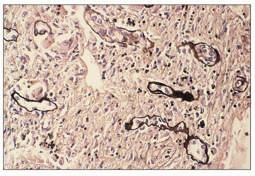
FIGURE 24.34 Histoplasmosis in a renal allograft. Yeast forms of H. capsulatum are highlighted with silver stain (arrows). (methenamine silver × 400.)


Blastomycosis

North American blastomycosis, caused by Blastomyces dermatitidis, is endemic in the Ohio and Mississippi River valleys and the southeastern United States (136). The fungus is a saprophytic budding yeast found in soils. Infection results from inhalation of infectious forms. Blastomycosis is prevalent among immunosuppressed patients. It is found four times more frequently in males than in females. Infection typically occurs between the ages of 30 and 50 years (136). Primarily, a pulmonary infection disseminates through blood to various organs, including the kidneys. Renal involvement is usually clinically silent. In severe infections, fever, weight loss, chest pain, cough, costovertebral angle tenderness, flank pain, renal insufficiency, and chronic discharging sinuses or subcutaneous abscesses have been reported. The diagnosis can be established through culture or by identifying the organism in fluids or tissue sections. Blastomycosis is not a transmissible disease.

Renal blastomycosis is estimated to occur in 25% of systemic infections. Involvement is often bilateral and varies from small, circumscribed nodules to diffuse inflammation and necrosis (136). The cortex is more often affected than the medulla. Perinephric abscesses and discharging sinuses may result from extension of the infection through the capsule. In tissue sections, granulomatous and suppurative lesions are seen. Occasional microabscesses and epithelioid caseating granulomas form, resembling tuberculosis. Blastomyces dermatitidis can be detected in either type of lesion as yeast cells with a double-contoured appearance. The cells measure 8 to 15 µm in diameter and demonstrate broad-based budding. Although organisms can easily be seen in routine hematoxylin and eosin-stained sections, the Grocott methenamine silver stain facilitates their detection. Antibodies against the cell wall polysaccharide antigen of B. dermatitidis are available and can be used for organism identification (136).


Paracoccidioidomycosis

Paracoccidioidomycosis (i.e., South American blastomycosis), caused by Paracoccidioides brasiliensis, is a chronic pulmonary disease endemic to Mexico and South and Central America. Pulmonary infection caused by inhalation of spores from P. brasiliensis tends to be progressive and followed by dissemination to mucous membranes, lymph nodes, and various organs. Although the presenting symptoms are usually pulmonary, the disease is often manifested by dissemination.

The kidneys are involved in 10% to 15% of cases of disseminated infection. Grossly, the renal cortex and medulla contain miliary necrotizing granulomas measuring a few millimeters (136). Organisms are found at the periphery of necrotic granulomas and within giant cells, easily identified in sections stained with hematoxylin and eosin. The Grocott methenamine silver stain brings out greater detail. Nonbudding forms, about 10 µm in diameter, predominate. Multiple buds from a single cell, 10 to 60 µm in diameter, are diagnostic, but less common. These resemble the steering wheel of a ship (136).


Mucormycosis

Mucormycosis (i.e., zygomycosis) is an opportunistic infection of the lungs and upper respiratory tract caused by fungi of the order Mucorales, whose most common pathogen is Rhizopus oryzae. Infection is acquired by inhalation of airborne spores. Disseminated infection occurs in immunocompromised and diabetic patients. Rarely, mucormycosis may only involve the kidney. Rhizopus oryzae frequently invades blood vessels and disseminates through the blood. Morrison and Mcglave (146) reported mucormycosis in 13 of 1500 bone marrow transplant recipients (0.9%), with kidney involvement in a single patient. Involvement of the urinary tract may be clinically silent or show signs of renal infection, including flank pain, dysuria, gross hematuria, and acute renal failure (136).

Renal involvement occurs in 50% of patients dying of disseminated mucormycosis (136). Thrombosis may occur, resulting in segmental or subtotal renal infarction. Involvement can be unilateral or bilateral. Microscopically, there is suppurative, necrotizing inflammation with thrombosis of interlobar and arcuate arteries. Granulomatous inflammation, fibrosis, and Langerhans-type multinucleated giant cells are seen. Hyphae can be detected in areas of acute inflammation or infarction. In tissue sections, the fungus has broad, nonseptate hyphae with right-angle branching (Fig. 24.35). Organisms can be identified with Grocott methenamine silver stain or fluoresceinated antibodies (136).


Viral Nephropathies


Viral Pathogenesis and Tropism in the Kidney

When encountering a host, eukaryotic cell viral pathogenesis requires an initiation phase in which the virus must first cross the cell membrane and enter the cell usually through a receptor-mediated process. The virus must then cross the nuclear envelope and enter the nucleus. During the replication phase, DNA synthesis, transcription, and translation of viral genes occur. Finally, during the release phase, assembly and maturation precedes the exit of the virus from the cell. Viruses have evolved several subversion mechanisms at all of these phases, which require the large-scale use of the host cellular machinery. Bruggeman (147) identifies and gives examples of the subversion mechanisms viruses use in chronic kidney disease (CKD) as (a) molecular mimicry, (b) hijacking strategies, and (c) transformers and oncogenes. Viruses must also evade the host immune response.


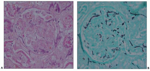
FIGURE 24.35 Renal mucormycosis involving glomeruli. A: Hyphae are broad and nonseptate with right-angle branching. (H&E; ×400.) B: Methenamine silver × 400. (Courtesy of Antony Chang University of Chicago, Chicago, Illinois.)

Cytomegalovirus (CMV) uses extensive molecular mimicry to avoid the host immune response. Of the 160 genes encoded by CMV, the majority are employed to manipulate the host immune system by mimicking cytokines, chemokines, chemokine receptors, and cytokine-binding proteins. As an example of immune molecular mimicry, CMV produces an IL-10 homologue (vIL-10), which suppresses the host proinflammatory cytokines and helps the virus evade the host immune response.

HIV-1 can “hijack” the NFκB signal transduction pathway in both immune and kidney cells. When IkB is phosphorylated and degraded, its inhibition of NFκB is removed, and NFκB can translocate to the nucleus and activate transcription. The net result is not only the vigorous transcription of viral genes but also activation of NFκB-dependent host genes, which leads to abnormal regulation of processes such as proliferation and apoptosis in the host kidney cells and may contribute to the pathogenesis of HIV-associated nephropathy (HIVAN). These altered processes yield a nonmalignant transformation of kidney epithelial cells, which in HIVAN may be represented morphologically by visceral epithelial hyperplasia and tubular ectasia.

Viruses can play a role in inducing cellular mechanisms of kidney injury and involve the kidney as an “innocent bystander” though a number of mechanisms (148). For example, TLRs on kidney dendritic cells can recognize virally
encoded molecules and facilitate a TH1 response at the locale of the renal parenchyma. The subsequent inflammatory response may injure the kidney. As a phenotypic disease example of virally induced innocent bystander damage, the hemophagocytic syndrome (HPS), in which nonmalignant proliferations of activated macrophages infiltrate many organs, can cause acute renal failure. Viral triggers such as Epstein-Barr virus (EBV), parvovirus, and CMV have been described in HPS. Heterologous immunity, in which established memory T-cell responses to a previously encountered pathogen, can have a major impact on the course and outcome of a subsequent infection with an unrelated pathogen. Heterologous immunity is dependent on the sequence of infections, the prior T memory network at the time of the infection, and can be either beneficial or detrimental to the host in transplantation settings (149).

Viruses may also perturb the T-lymphocyte-dependent B-lymphocyte response with subsequent B-cell hyperactivity and a loss in tolerance through mechanisms such as molecular mimicry and epitope spreading (150). In these situations, autoimmune or alloimmune antibodies are produced and may facilitate autoimmune disease or rejection.

Viral tropism to the kidney is determined by both virus and tissue factors. In cell culture, human CMV has differential tropism for primary human cells and cell lines (151). Differences in virus cell-binding factors may also influence tropism to the kidney. Once again with CMV, in a murine CMV model, homologues of the human CMV tegument phosphoprotein pp65, when mutated, will exhibit differential tropism among the liver, spleen, salivary gland, and kidney (152). The localization factors of virus and kidney cells impacting human disease are largely unexplored.


The Biology of Epstein-Barr Virus Infection

EBV infections in nonimmunosuppressed hosts are common. Most adults have serologic evidence of past infection. Primary infections may be manifested as infectious mononucleosis. Primary infections may also be silent with little in the way of clinical signs and symptoms. The major target for EBV is the B cell. EBV attaches to cells for which it is tropic through the CD21 receptor. Augmented GP110, which is the product of the EBV gene BALF4, may dramatically enhance viral tropism (153). In infected cells, the EBV virus may exist in a latent or a lytic state. A latently infected B cell may exist in one of four programs (154). Latency “0” is characterized by complete silencing of the viral genome. In healthy carriers of EBV, between 1 and 50 per million peripheral blood lymphocytes are infected. The infected cells of latency “0” are phenotypically similar to long-lived memory cells. Latency I expresses LMP2A (late membrane protein) alone or together with EBNA-1 (EBV nuclear antigen). In latency II, EBNA-1 and the three LMPs (LMP1, LMP2A, and LMP2B) are expressed in infected B cells that home to the germinal centers. In the latency III program, all nine of the latency genes (EBNA-1, EBNA-2, EBNA-3 [or EBNA-3A], EBNA-4 [or EBNA-3B], EBNA-5 [or LP], and EBNA-6 [or EBNA-3C] and the latent membrane proteins LMP1, LMP2A, and LMP2B) are expressed. Latency III is the “growth program,” which is associated with autonomous B-cell growth. The latency III program is expressed in the immunoblast-like cells of posttransplant lymphoproliferative disorders.

During early latency, EBNA-2 activates B lymphocytes through induction of CD23. Also in latency, the EBV LMP induces expression of CD23, ICAM1, and LFA-3. The highest-frequency RNA in late latency is EBER. Latently infected cells can be switched into a productive or lytic phase by a number of environmental stresses. The immediate early gene of the latent to lytic switch is BZLF-1. GP-350 is expressed in the late lytic phase. EBV mutants defective for lytic viral replication are unable to promote the induction of posttransplant lymphoproliferative disorder (PTLD) in mouse models, a phenomenon that has been ascribed to the decreased production of IL-6, IL-10, and angiogenic factors, such as VEGF (154,155). Thus, there may also be a significant role of lytic infection in the pathogenesis of PTLD.


Posttransplant Lymphoproliferative Disorders and EBV Infection

In latently infected immunocompetent hosts, B-cell proliferation is kept in check by cytotoxic T cells that recognize and kill infected B cells. In patients with a compromised T-cell immune system, EBV-activated B cells may proliferate without regulation, resulting in B-cell expansions and neoplasia. Posttransplant lymphoproliferative disorders are lymphoid proliferations, which occur in immunosuppressed patients including bone marrow and solid organ transplant recipients, patients treated with immunosuppression for autoimmune disease, and patients with inherited immune deficiencies. Longitudinal studies in PTLD patients show a progression from the restricted pattern of latency “0,” which is seen in healthy carriers to the broader patterns of EBV latency III coupled with lytic replication (156).

Among solid organ transplant recipients, patients receiving renal allografts have the lowest frequency of PTLD at <1%. Cardiac allografts (1% to 2%) and heart-lung or liver-bowel (5%) having increased frequencies of PTLD. Over 80% of PTLD cases are associated with EBV infection. In solid organs, most PTLD cases are of recipient cell origin with <10% of donor origin. This suggests a role of endogenous EBV reactivation in many of these patients. There are multiple risk factors for PTLD. The most important is seronegativity for EBV, which conveys a 24X risk. Other risk factors include CMV seromismatch and prior therapy with OKT3. A patient with all of these factors is estimated to have a 654X risk over the reference transplant population for the development of PTLD. Serologic testing for EBV is unreliable in immunosuppressed patients. In general, EBV viral loads are increased in PTLD. Many studies show elevated EBV viral loads as measured by PCR of blood (157,158). There is controversy over the standardization and reference ranges in viral load testing (159,160).

Frequent molecular changes in PTLD involving cellular genes include alterations of c-MYC, BCL-6, p53, aberrant promoter hypermethylation, and somatic hypermutations targeting multiple protooncogenes (161).


Pathology of PTLD

The lesions of PTLD are categorized by the World Health Organization (162) according to Table 24.3. Since some of the histopathologic features of rejection can overlap with PTLD in kidney tissue, particular attention to the morphology of “early lesions” and polymorphous PTLD is warranted. “Early lesions” are characterized by either marked plasmacytosis (in the absence of substantial chronicity) or an infectious mononucleosis-like reaction with a brisk lymphoid proliferation showing a mixture
of small lymphoid cells, plasma cells, and rare immunoblasts (Fig. 24.36). In “polymorphous PTLD,” the mixed morphotype of the infectious mononucleosis-type reaction is repeated but with a greater number of immunoblasts, occasional atypical immunoblasts, cells with irregular nuclei resembling centrocytes, increased mitoses, and frequently, necrosis (Fig. 24.37). PTLD involving renal allografts does not show the lymphocytic tubulitis or vasculitis of cellular rejection.


TABLE 24.3 World Health Organization categories of PTLD


Early lesions


Plasmacytic hyperplasia


Infectious mononucleosis-like


Polymorphic PTLD


Monomorphic PTLD


B-cell neoplasms


Diffuse large B-cell lymphoma


Burkitt/Burkitt-like lymphoma


Plasma cell myeloma


Plasmacytoma-like lesions


T-cell neoplasms


T/natural killer cell lymphomas


Peripheral T-cell lymphoma


Hepatosplenic T-cell lymphoma


Classic Hodgkin lymphoma


Renal Involvement in PTLD

In a representative series of 36 renal transplant (31 cases) and/or renal/pancreas (4 cases) transplant patients with a diagnosis of PTLD from the University of Pennsylvania (163), PTLD was diagnosed from 6 days to 10 years after engraftment (mean 509 days). Fourteen patients (40%) had been given 10- to 20-day courses of OKT3 or ALG prior to PTLD diagnosis. All 14 of these patients developed PTLD within 6 weeks of transplantation. Seven of ten patients tested showed recent EBV infection, and 7 of 15 patients tested had active CMV infection at the time of PTLD diagnosis.


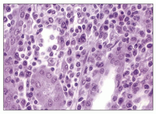
FIGURE 24.36 Early mononucleosis-like infiltrate of PTLD. The interstitial infiltrate is mostly mature lymphocytes. Only rare immunoblasts are seen. (H&E; ×27.)


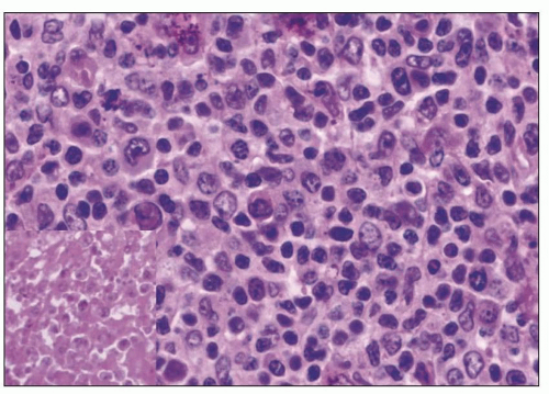
FIGURE 24.37 Polymorphous PTLD. The number of transformed cells is increased, with many immunoblasts. Focal necrosis is seen (inset). (H&E; ×27.)

Essentially all of the morphologies listed in Table 24.3 have been described in renal allografts. The recognition of the early lesions and polymorphic histologies of PTLD are particularly critical to the practicing nephropathologist in that the distinction from rejection is challenging. Furthermore, the therapeutic approaches to rejection and PTLD are markedly different making the distinction between the two conditions all the more important. In the University of Pennsylvania series (163), the initial PTLD diagnosis was classified as “early” lesion (13 patients) polymorphic PTLD (P-PTLD) (11 patients) and monomorphic PTLD (M-PTLD) (12 patients). Thirty-three lesions were B-cell proliferations, while the remaining two lesions were gamma-delta T-cell lymphomas. Two patients who initially presented with early lesions rapidly progressed to P-PTLD and then to M-PTLD. Thirty-two of the PTLDs, all of which were B cell in phenotype, were EBV positive by in situ hybridization. Seventeen patients (49%) presented with PTLD in the allograft. Of the 17 patients that presented with allograft PTLD, 9 showed moderate to marked interstitial hemorrhage. Six of these patients had renal biopsies prior to PTLD diagnosis. The renal biopsies prior to PTLD were all classified as moderate to severe acute rejection. While these biopsies showed acute rejection (tubulitis, vasculitis, and/or interstitial hemorrhage), closer examination also showed transformed lymphocytes, lymphocyte mitoses, and occasional immunoblasts in three patients. In situ hybridization showed EBV in these three biopsies. Thus, very early PTLD in allografts may coexist with rejection.

PTLD can present in renal allografts and may be confused with allograft rejection. Careful examination of the histology and EBV studies may be needed to distinguish PTLD from rejection. Allograft biopsies with dense lymphoid infiltrates should be evaluated for transformed lymphocytes, lymphoid mitoses, and immunoblasts. These findings may precede PTLD development. Allograft involvement by PTLD is also often associated with interstitial hemorrhage, a finding which is also seen in humoral rejection. Further analysis
for PTLD, including studies for EBV and C4d, should be undertaken in renal allograft recipients with dense lymphoid infiltrates showing atypia and/or interstitial hemorrhage.

Most cases of PTLD in renal transplants regress with reduction of immunosuppression; however, a minority of cases can progress to lethal lymphoma. The time from infection to fatal disease progression can be as little as a few weeks. The early recognition and diagnosis of PTLD can offer an opportunity for reduction of immunosuppression and reconstitution of the host immune system, which is necessary for control of the EBV infection.


Cytomegalovirus Nephritis

20 to 60% of renal transplant recipients were reported in the literature to suffer from CMV disease with clinical signs of fever, leukopenia, and organ dysfunction prior to 2000 (164,165). However, the disease is rare today after prophylactic and preemptive CMV therapies were implemented (166). CMV infection in a renal allograft recipient may be a primary infection, a reactivation infection, or a reinfection. Primary infections occur in previously uninfected patients who are seronegative for CMV. Blood products or the donated allografts are the usual sources of infection. Reactivation infection occurs in previously infected recipients who reactivate latent infections. Reinfection occurs when a seropositive recipient acquires a new strain of latent virus from a seropositive donor with subsequent reactivation. The most significant risk factor for primary CMV infection is receipt of a seropositive donor allograft by a seronegative recipient (165). Children are at the highest risk for primary infection. The extent of immunosuppression is the other risk factor for CMV infection. The total amount of immunosuppressive therapy and, in particular, use of antilymphocyte antibodies enhance the risk for CMV infection. Studies of the viral gene products targeted by the host immune responses have demonstrated that the human CMV 65-kDa tegument phosphoprotein pp65 (UL83) is the target of antibody (167), cytotoxic T-lymphocyte (CTL) (168), and lymphoproliferative (169) responses.

The histopathologic changes associated with CMV infection vary. Cowdry type A intranuclear CMV inclusions are the typical (CMV is the only virus to show intranuclear Cowdry type A and intracytoplasmic inclusions) and diagnostic finding, but interstitial inflammation, glomerulopathy, ATN, and other findings pose diagnostic dilemmas. A seminal article by Richardson et al. (170) described a glomerulopathy in renal allografts with endothelial swelling, hypertrophy and necrosis, obliteration of the capillary lumens, fibrillar deposits in the glomerular capillaries, mild segmental hypercellularity, mononuclear cell infiltration, but absent CMV inclusions. Since most of these patients had CMV viremia or CMV infection without viremia, the authors associated this pathology with CMV infection and not rejection. Tuazon et al. (171) described similar cases and noted a predominance of CD8+ cells in the glomeruli. Rao et al. (172) described a case of de novo immunotactoid glomerulopathy with resolution after recovery from a CMV infection. Others have questioned the causative role of CMV infection in such cases. Herrera et al. (173) in studying immunosuppressed patients with CMV infection found histologic lesions similar to those previously described; however, no parenchymal evidence for CMV infection of the kidney could be found by immunofluorescence or electron microscopy. The authors raised the possibility of damage secondary to antiendothelial antibodies and vascular rejection. Previous studies by Anderson et al. (174), using immunohistochemistry and in situ hybridization from CMV viremic allograft recipients with glomerulopathy, could find no evidence of CMV antigens or DNA. Rubin (175) has suggested that CMV-mediated injury may be indirect. Proinflammatory cytokines such as gamma interferon can up-regulate MHC in kidney. CMV infection may also increase both class I and II MHCs. Immunologically mediated injury resulting from enhanced MHC expression and increased targeting may produce the lesions associated with CMV glomerulopathy.

A second mechanism for CMV damage of kidney is more clearly related to direct viral infection. Payton (176) described CMV inclusions in glomerular and peritubular capillary endothelial cells as well as in tubular epithelial cells (Fig. 24.38). Cameron (177) described CMV inclusions in tubular epithelial cells in the absence of rejection. Birk and Chavers (178) reported CMV inclusion glomerulopathy in a young transplant patient. Such cases, while rare, document productive CMV infection in both glomerular and tubulointerstitial compartments in a small subset of patients. CMV infection may be diagnosed by serology, tissue examination through histology with immunofluorescence or immunohistochemistry, or CMV-PCR of buffy coat or tissue. Quantitative buffy coat CMV-PCR does not correlate well with the tissue presence of CMV inclusions; however, the frequency of discovering CMV infection in renal allografts may be increased by CMV-PCR techniques (179).


Adenovirus Infection

Adenoviruses belong to the family Adenoviridae, which include a group of DNA viruses such as polyoma viruses and herpes viruses. Adenoviruses are double-stranded DNA viruses. Adenovirus can cause respiratory infection, pharyngitis, keratoconjunctivitis, gastroenteritis, hepatitis, hemorrhagic cystitis, and nephritis. In immunocompetent hosts, many adenovirus infections are subclinical. Significant infections with
adenovirus are well described in immunosuppressed patients including solid organ and bone marrow transplant patients and may present as disseminated infection (180). Adenovirus infection may also present as acute hemorrhagic cystitis in renal transplant patients (181). Adenovirus viremia is cited in approximately 7% of renal transplant patients (182), and the virus is excreted in approximately 11% (183). Serotypes 7, 11, 34, and 35 constitute most of the cases (184). Adenovirus has tropism for epithelial cells via coxsackievirus and AdV receptors, class I human leukocyte antigen molecules, and sialoglycoprotein receptors (185); CD46; and fiber knob gene protein (186). Secondary interactions with integrins may be needed for virus internalization.

Only gold members can continue reading. Log In or Register to continue

Stay updated, free articles. Join our Telegram channel

Jun 21, 2016 | Posted by in UROLOGY | Comments Off on Pyelonephritis and Other Direct Renal Infections, Reflux Nephropathy, Hydronephrosis, Hypercalcemia, and Nephrolithiasis

Full access? Get Clinical Tree

Get Clinical Tree app for offline access