Renal Disease in Systemic Lupus Erythematosus, Mixed Connective Tissue Disease, Sjögren Syndrome, and Rheumatoid Arthritis


Renal Disease in Systemic Lupus Erythematosus, Mixed Connective Tissue Disease, Sjögren Syndrome, and Rheumatoid Arthritis


Vivette D. D’Agati

M. Barry Stokes


SYSTEMIC LUPUS ERYTHEMATOSUS


Historical Perspective

Systemic lupus erythematosus (SLE) is a chronic inflammatory autoimmune disease affecting multiple organ systems, including the skin, joints, heart, lung, kidneys, central nervous system, and serous membranes. The cutaneous manifestations of SLE were the first to be identified. As early as the 13th century, medical writings by Rogerius (c. 1230) and Paracelsus (c. 1500) proposed the Latin term lupus, meaning wolf, to describe the erythematous ulcerative lesions affecting the skin of the cheeks (1). However, it is Kaposi contention, in his lengthy treatise on lupus vulgaris (2), that even the lesions called herpes esthiomenos by Hippocrates in the fourth century BC included characteristic examples of this condition. Cazenave and Schedel are generally credited with the first illustration of discoid lupus erythematosus in 1838 (3), which depicts the bilateral butterfly distribution over the cheeks and bridge of the nose, a distribution evocatively portrayed as resembling bat wings by Jonathan Hutchinson (4) in 1870. It was not until the late 19th and early 20th centuries that the visceral manifestations of SLE were generally appreciated (5,6,7).

William Osler, in historic publications from 1895 through 1903, is credited with first synthesizing the diverse visceral manifestations of SLE into a loosely defined disease entity that included variable features of “gastrointestinal crises, endocarditis,
pericarditis, acute nephritis, and hemorrhage from the mucous surface.” In later writings, he emphasized the arthritis, pulmonary, and central nervous system manifestations while remarking that the arthritis was typically nondeforming. He observed the variable clinical presentation and remitting and relapsing nature of the condition: “Recurrence is a special feature of the disease and attacks may come on month after month or even throughout a long period of years… . The attacks may not be characterized by skin manifestations; the visceral symptoms alone may be present, and to the outward view, the patient may have no indications whatever of erythema exudativum” (5).

In 1923, Libman and Sacks (8) described four cases of “a hitherto undescribed form of valvular and mural endocarditis,” which we now recognize as the sterile thrombotic valvular manifestations of circulating lupus anticoagulant. The distinctive histologic features of the glomerulonephritis were first reported by Baehr et al. (9), who observed that the proliferative features were accompanied by “wire-loop” alterations of the glomerular capillary walls, which they described as a “peculiar hyaline thickening of the capillary walls which is striking even in sections stained with hematoxylin and eosin.”

The hypothesis that SLE constitutes an autoimmune disorder was spurred by three major independent laboratory findings in the early 20th century. First, false-positive serologic test results for syphilis using the Wassermann reaction were obtained for up to 35% of SLE patients who lacked clinical evidence of treponemal infection, sometimes preceding the development of clinical features of SLE. It would be many years before the significance of this finding as a manifestation of antiphospholipid (APL) antibody or lupus anticoagulant syndrome was recognized. Later, Hargraves made the highly important discovery of the LE cell in bone marrow preparations of patients with active SLE. He described two types: a mature neutrophil, which he called the LE cell, and a histiocyte, which he called a tart cell. Both contained phagocytosed nuclear material in which the chromatin pattern was sometimes still discernible (10). Similar LE bodies consisting of altered hematoxylin-staining nuclear material had previously been observed in the heart valves (11), lymph nodes, spleen, and kidneys of patients with active SLE. The cause of the LE phenomenon, the presence of antinuclear antibody (ANA), would not be identified for another decade. The most important historical event in the evolution of concepts about SLE was the discovery by Coons (12) in 1951 of the immunofluorescence technique employing fluoresceinlabeled antibodies and its subsequent application to the sera and tissues of SLE patients for the detection of ANAs by Friou (13) in 1958.

With the explosion of knowledge in the early 1970s about the human leukocyte antigen (HLA) system and the role of T and B cells in immunity, the groundwork was laid for investigations into the genetic, molecular, and cellular determinants of autoimmunity in SLE. Although there have been tremendous advances in our understanding of the basis of autoimmunity and the properties of nephritogenic immunoglobulins, a unifying concept of pathogenesis remains elusive.


Introduction to Clinical Features

Among the diverse organ systems involved in SLE, it is the renal complications that pose the greatest risk of morbidity and mortality and present the most demanding therapeutic challenge. The renal manifestations of SLE, called lupus nephritis, are highly pleomorphic with respect to their clinical and morphologic expressions. Clinically, renal involvement may occur at any time in the course of SLE, ranging from the earliest identifiable clinical onset of disease to years after the initial diagnosis. The onset of renal involvement is most common within the first year and clinically affects up to 50% of patients with SLE. Many studies suggest that a much higher percentage of patients would have morphologic evidence of renal disease if renal biopsies were performed systematically on all SLE patients and if histologic studies were supplemented with more sensitive immunofluorescence and electron microscopy studies.

Clinical features range from asymptomatic urinary findings of microhematuria and mild proteinuria to full-blown nephrotic syndrome or rapidly progressive renal failure. In adults and children (14,15,16), the renal course is frequently complicated by periods of remission and exacerbation that may occur unpredictably at any time in the course of the disease. Moreover, the nephritis has the ability to transform from one morphologic pattern to another spontaneously or after treatment. Because of this heterogeneity of clinical renal manifestations, the typical presenting clinical features will be described separately for each of the morphologic subtypes of lupus nephritis.


Introduction to Classification

The pathologic manifestations of lupus nephritis are extremely diverse and may affect any or all renal compartments, including glomeruli, tubules, interstitium, and blood vessels. The complexity of the protean renal manifestations of SLE can be most easily approached using the World Health Organization (WHO) classification as an organizational construct. The original WHO Classification was formulated in 1974 at a convocation of renal pathologists and nephrologists in Buffalo, NY, under the auspices of the WHO (17). The classification that they devised has been the framework most widely used by practicing clinicians and pathologists (18) (Table 14.1). In 1982, this original WHO classification was expanded and refined by the International Study of Kidney Disease in Children (ISKDC) (19), with further modifications in 1995 (20)
(Table 14.2). It defined six major classes and a large number of subclasses, with emphasis on distribution, activity, and chronicity of the lesions. Although much more detailed and precise, this revised classification was not as widely accepted because of its cumbersome reliance on subclasses. A third classification was proposed in 2003 by a large consensus conference of renal pathologists, nephrologists, and rheumatologists that was organized jointly by the International Society of Nephrology (ISN) and the Renal Pathology Society (RPS) (Table 14.3). This new classification, which has been entitled “ISN/RPS WHO classification revisited,” retains the simplicity of the original 1974 WHO classification while incorporating some of the refinements introduced by the 1982 revised WHO classification (21,22). The ISN/RPS system provides more detailed and more precise pathologic criteria for each class, which facilitates greater reproducibility among pathologists. This updated consensus classification is widely accepted and has superseded prior classifications.


TABLE 14.1 Original (1974) WHO classification of lupus nephritis


Class I


Normal glomeruli (by LM, IF, EM)


Class II


Purely mesangial disease


  1. Normocellular mesangium by LM but mesangial deposits by IF and/or EM


  2. Mesangial hypercellularity with mesangial deposits by IF and/or EM


Class III


Focal segmental proliferative glomerulonephritis (< 50%)


Class IV


Diffuse proliferative glomerulonephritis (≥50%)


Class V


Membranous glomerulonephritis


LM, light microscopy; IF, immunofluorescence; EM, electron microscopy.


TABLE 14.2 Modified (1982) WHO classification of lupus nephritis


Class I


  1. Normal glomeruli (by LM, IF, EM)


  2. Normal glomeruli by LM but deposits seen by IF and/or EM


Class II


Purely mesangial alterations (mesangiopathy)


  1. Mesangial widening and/or mild hypercellularity (+)


  2. Moderate hypercellularity (++)


Class III


Focal segmental glomerulonephritis (associated with mild or moderate mesangial alterations)


  1. With active necrotizing lesions


  2. With active and sclerosing lesions


  3. With sclerosing lesions


Class IV


Diffuse glomerulonephritis (severe mesangial, endocapillary, or mesangiocapillary proliferation and/or extensive subendothelial deposits). Mesangial deposits are present invariably and subepithelial deposits often and may be numerous


  1. Without segmental lesions


  2. With active necrotizing lesions


  3. With active and sclerosing lesions


  4. With sclerosing lesions


Class V


Membranous glomerulonephritis


  1. Pure membranous glomerulonephritis


  2. Associated with lesions of category II (a or b)


  3. Associated with lesions of category III (a, b, or c)a


  4. Associated with lesions of category IV (a, b, c, or d)a


Class VI


Advanced sclerosing glomerulonephritis


a Deleted from the 1995 Modified WHO Classification.


LM, light microscopy; IF, immunofluorescence; EM, electron microscopy.


Modified from Churg J, Sobin LH. Renal Disease. Classification and Atlas of Glomerular Disease. Tokyo: Igaku-Shoin, 1982.


All three classifications are based entirely on an assessment of the glomerular alterations. They rely heavily on the light microscopic findings, while simultaneously integrating information obtained by fluorescence and electron microscopic studies (Table 14.4). Accurate use of the WHO system is best achieved if the same pathologist studies the biopsy by the three modalities of light microscopy, immunofluorescence microscopy, and electron microscopy, especially because the glomerular sampling of relatively focal lesions may not be equally represented in tissue processed for the three techniques. The first step is to determine the presence or absence of glomerular hypercellularity in the mesangial and endocapillary zones and the extent of glomerular involvement. These findings are then interpreted in the context of the distribution of immune deposits in mesangial, subendothelial, and subepithelial locations as detected by light microscopy, immunofluorescence microscopy, and electron microscopy (Fig. 14.1). Because some lesions are focal, the accuracy of the WHO classification depends on the adequacy of the glomerular sampling. According to one study, at least 20 glomeruli were required for accurate classification of lupus nephritis (23).

Generalized adoption of the WHO system for classification of lupus nephritis in academic and community centers alike has had far-reaching impact. It has served to simplify the interpretation of the renal pathologic findings, which are notoriously pleomorphic, when comparing one biopsy to another and comparing neighboring glomeruli in a biopsy specimen. Use of this classification has facilitated the ease and reliability with which nephrologists and nephropathologists communicate information. Greatly improved standardization and reproducibility of biopsy interpretation between centers has been achieved. Most importantly, use of the WHO classification has provided the standardized nosology necessary for careful clinical pathologic studies, which have yielded valuable information about disease subsets with differing prognostic and therapeutic implications.


Role of Renal Biopsy

Renal biopsy plays an important role in the management of patients with SLE (24). In some patients, it is instrumental in establishing a diagnosis of SLE. This is especially common early in the disease, before overt extrarenal manifestations of SLE are evident. This scenario applies most frequently to patients with mesangial proliferative or membranous patterns who lack serologic markers of SLE and may present many months or even years before the American College of Rheumatology (ACR) criteria for SLE have been met. More typically, a diagnosis of SLE has already been made prior to renal biopsy, based on presenting clinical features and confirmatory serologic tests. Renal biopsy provides the most accurate window into the kidney, because it provides information about the class, severity, activity, and chronicity of the lupus nephritis that cannot be accurately predicted on the basis of clinical manifestations (25). Based on these findings, important decisions about therapy and prognosis are made.

Indications for renal biopsy vary among centers. Few clinicians would advocate baseline renal biopsy in a newly
diagnosed patient with SLE who lacks clinically apparent renal disease. However, because some cases of severe lupus nephritis are clinically “silent,” this liberal approach has some proponents (26). Most nephrologists and rheumatologists would agree that the new appearance of any significant marker of renal disease such as hematuria, proteinuria, nephrotic syndrome, or renal insufficiency at any time in the course of SLE is ample justification for renal biopsy. The recent ACR guidelines for management of lupus nephritis advocate that renal biopsy should be performed in any patient with clinical
evidence of renal involvement defined as one of the following: (a) increasing serum creatinine without compelling alternative causes (such as sepsis, hypovolemia, or medication); (b) confirmed proteinuria of ≥1.0 g per 24 hours or by spot urine protein:creatinine ratio; or (c) combinations of proteinuria ≥0.5 g plus hematuria defined as ≥5 RBCs per hpf or proteinuria ≥0.5 g plus cellular casts (27). Moreover, lupus nephritis is one of the few renal diseases for which “follow-up” renal biopsies are routinely performed in some centers 6 months or more after therapy to gauge the efficacy of treatment and guide further therapeutic management. Repeat renal biopsies are also frequently performed in any patient with a sudden change in renal findings (e.g., new onset of proteinuria, new activity of the urinary sediment, declining glomerular filtration rate [GFR]) that may presage a transformation in the class of lupus nephritis, or reactivation of disease requiring reinstitution or adjustment of therapy.


TABLE 14.3 ISN/RPS classification of lupus nephritis (LN) (2004)


Class I


Minimal mesangial LN


Normal glomeruli by LM, but mesangial immune deposits by IF


Class II


Mesangial proliferative LN


Purely mesangial hypercellularity of any degree or mesangial matrix expansion by LM, with mesangial immune deposits


There may be a few isolated subepithelial or subendothelial deposits visible by IF or EM but not by LM


Class III


Focal LNa


Active or inactive focal, segmental, and/or global endocapillary and/or extracapillary GN involving < 50% of all glomeruli, typically with focal subendothelial immune deposits, with or without mesangial alterations


III (A): purely active lesions: focal proliferative LN


III (A/C): active and chronic lesions: focal proliferative and sclerosing LN


III (C): chronic inactive with glomerular scars: focal sclerosing LN


Class IV


Diffuse LNa


Active or inactive diffuse, segmental, and/or global endocapillary and/or extracapillary GN involving ≥50% of all glomeruli, typically with diffuse subendothelial immune deposits, with or without mesangial alterations. This class is divided into diffuse segmental (IV-S) when > 50% of the involved glomeruli have segmental lesions and diffuse global (IV-G) when > 50% of the involved glomeruli have global lesions. Segmental is defined as a glomerular lesion that involves less than half of the glomerular tuft


IV-S (A) or IV-G (A): purely active lesions: diffuse segmental or global proliferative LN


IV-S (A/C) or IV-G (A/C): active and chronic lesions: diffuse segmental or global proliferative and sclerosing LN


IV-S (C) or IV-G (C): inactive with glomerular scars: diffuse segmental or global sclerosing LN


Class V


Membranous LNb


Global or segmental subepithelial immune deposits or their morphologic sequelae by LM and by IF or EM, with or without mesangial alterations


Class VI


Advanced sclerosing LN


Ninety percent or more of glomeruli globally sclerosed without residual activity


Indicate the proportion of glomeruli with fibrinoid necrosis and with cellular crescents.


Indicate and grade (mild, moderate, severe) tubular atrophy, interstitial inflammation, and fibrosis, severity of arteriosclerosis or other vascular lesions.


a Indicate the proportion of glomeruli with active and with sclerotic lesions.

b May occur in combination with III or IV, in which case both will be diagnosed; may show advanced sclerosis.


LM, light microscopy; IF, immunofluorescence; EM, electron microscopy; GN, glomerulonephritis.


TABLE 14.4 Integration of LM, IF, and EM findings by the WHO class


Light microscopy


Immunofluorescence


Electron microscopy


Class


MES


PCW


MES


PCW


MES


SENDO


SEPI


I


0


0


+


0


+


0


0


II


+


0


+


0


+


0


0


III


+


+


++


+


++


+


+/-


IV


++


++


++


++


++


++


+/-


V


+


++


+


++


+


+/-


++


LM, light microscopic [abnormalities]; IF, immunofluorescence [positivity]; EM, electron microscopic [location of deposits]; MES, mesangial; PCW, peripheral capillary wall; SENDO, subendothelial; SEPI, subepithelial.


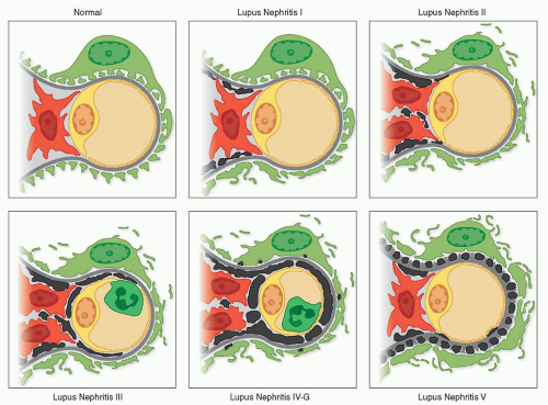
FIGURE 14.1 Normal capillary with intact podocyte foot processes and no electron-dense deposits. Class I lupus nephritis with minimal mesangial deposits, focal foot process effacement, and no mesangial hypercellularity. Class II lupus nephritis with substantial mesangial deposits, mesangial hyperplasia, and more extensive foot process effacement. Class III lupus nephritis with scanty subendothelial deposits, mesangial hyperplasia, endocapillary leukocytes, and extensive foot process effacement. Class IV-G lupus nephritis with numerous subendothelial and a few subepithelial deposits, mesangial hyperplasia, endocapillary leukocytes, and extensive foot process effacement. Class V lupus nephritis with numerous subepithelial and a few subendothelial and mesangial deposits and extensive foot process effacement.


Pathologic Findings


Gross Pathology

Since the advent of the modern therapeutic era, autopsy examination of the kidney from patients who have died of acute SLE has become a rare event. For descriptions of the gross pathology of the acute disease, it is necessary to study the writings of early pathologists such as Klemperer, who described enlarged, swollen kidneys with petechial hemorrhages and focal, shallow, superficial scars (28). In the modern era of immunosuppressive therapy, the appearance of the kidneys at autopsy usually reflects a combination of chronic and acute changes, with a contracted or swollen appearance.


Light Microscopy

Before considering the individual features that define the various classes of lupus nephritis, it is helpful to discuss the basic types of renal lesions that may be encountered in lupus. These
general remarks serve as a preface to the more detailed consideration of the WHO classification that follows. Many of these morphologic features pertain to more than one WHO class of lupus nephritis.


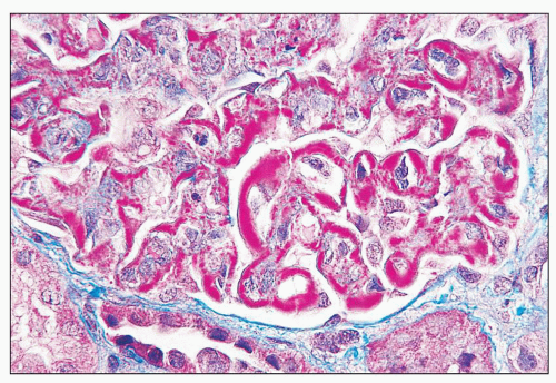
FIGURE 14.2 Lupus nephritis class IV. Trichrome stain highlights the presence of global subendothelial fuchsinophilic deposits. (Masson trichrome, × 500.)


GLOMERULI

The major histologic abnormalities of the glomerulus include immune deposits, glomerular proliferation, influx of leukocytes, glomerular necrosis, and scarring. Glomerular proliferation may be mesangial, endocapillary, and extracapillary.

Immune Deposits Although glomerular immune deposits are best identified by immunofluorescence and ultrastructural techniques, in lupus nephritis the glomerular immune deposits are also frequently identifiable by light microscopy because of their large size and widespread distribution. As in many other forms of glomerular disease, it is the distribution of deposits in the glomerular tuft that determines the proliferative response, and the glomerular lesions that result are therefore best interpreted in the context of the pattern of immune deposition (29). A number of stains, properly performed, are helpful in demonstrating deposits by light microscopy. Although deposits frequently have a glassy (i.e., hyaline), hypereosinophilic appearance with the hematoxylin and eosin (H&E) stain, they are often difficult to differentiate from the eosinophilic mesangial matrix, glomerular basement membrane (GBM), and cytoplasm of the indigenous glomerular cells without the use of special stains. Particularly helpful are the trichrome stain, which highlights the deposits as red (or fuchsinophilic) against the blue-staining glomerular matrix components (Fig. 14.2) and the Jones methenamine silver or combination methenamine silver-Masson Ponceau stain, which stain the deposits pink or red, respectively, against the black-staining mesangial matrix and GBM.


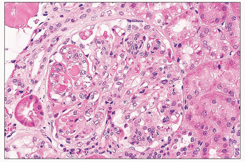
FIGURE 14.3 Lupus nephritis class IV. Glomerular capillary walls are segmentally thickened by wire-loop deposits. An intraluminal deposit forms a “hyaline thrombus” in one capillary, and there is global endocapillary proliferation. (H&E; × 320.)

The mesangium is the most common site for glomerular immune deposits and may be the sole location for renal deposits in some cases. Mesangial deposits in the absence of glomerular capillary wall deposits are the defining feature of class I and class II lupus nephritis. However, mesangial deposits are also found in combination with peripheral glomerular capillary wall deposits in class III, class IV, and class V lupus nephritis. Mesangial deposits and the mesangial proliferation that often accompanies them can be considered the common substratum on which these more complex classes of lupus nephritis are built.

Subendothelial immune deposits are regularly encountered in class III and class IV lupus nephritis. They typically are relatively focal and segmental in class III and more global and diffuse in class IV (especially when accompanied by global endocapillary hypercellularity). When large enough to completely involve the peripheral circumference of the glomerular capillary, they are referred to as classic wire loops, which produce a refractile thickening of the glomerular capillary wall in H&E-stained sections, as initially described by Klemperer et al. (28) (Fig. 14.3). Special stains reveal the deposits to be entirely or largely subendothelial, with preservation of an outer peripheral layer of GBM (Fig. 14.4). In florid examples, these massive subendothelial deposits may focally replace the GBM, producing intramembranous deposits best delineated by electron microscopy or extending through the GBM in continuity with overlying subepithelial deposits. In some cases, subendothelial deposits are enclosed by the formation of a subendothelial layer of neomembrane formation, producing a double
contour, similar to that observed in membranoproliferative glomerulonephritis. Not surprisingly, mesangial extension or “interposition” frequently accompanies this finding.


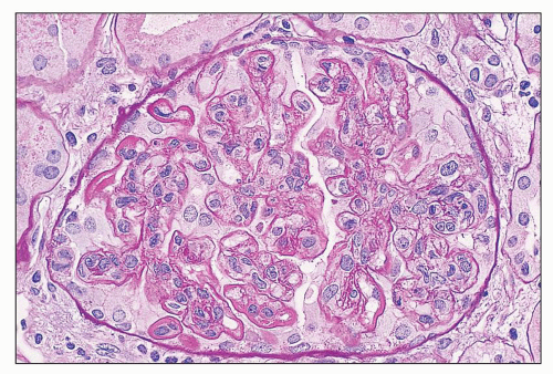
FIGURE 14.4 Lupus nephritis class IV. PAS stain highlights the thickening of the glomerular capillary walls by numerous subendothelial deposits. (PAS; × 500.)

Abundant, regularly distributed subepithelial deposits are the defining feature of membranous lupus nephritis (class V), but they may also be seen in association with class III or IV lupus nephritis, as examples of combined class III and V or combined class IV and V lupus nephritis. GBM spikes and a vacuolated texture where cut obliquely can usually be identified with the silver or periodic acid-Schiff (PAS) stain. Scattered subepithelial electron densities may also occur as a sporadic finding in lupus nephritis class III or IV, without the formation of a well-developed membranous pattern. In such cases, the occurrence of these sparse, irregularly distributed, subepithelial deposits is not considered sufficient to justify an additional classification of membranous lupus nephritis class V. The ISN/RPS classification requires subepithelial deposits involving greater than 50% of the tuft of greater than 50% of the glomeruli by light microscopy or immunofluorescence to make a diagnosis of class V plus class III or IV.


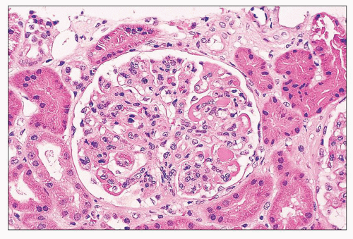
FIGURE 14.5 Lupus nephritis class IV. In addition to wire-loop deposits, there are segmental intraluminal deposits forming “hyaline thrombi.” (H&E; × 320.)


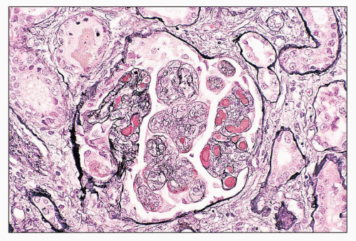
FIGURE 14.6 Lupus nephritis class IV. Silver stain is useful to highlight the intraluminal location of the glomerular capillary fibrin thrombi. Some GBMs appear double contoured. (Jones methenamine silver, × 320.)

Some patients with active class III or IV lupus nephritis have large intracapillary immune deposits forming hyaline thrombi (Figs. 14.5, 14.6, 14.7). This term is actually a misnomer, because these are not true fibrin thrombi but are instead massive intracapillary immune deposits with the same composition by immunofluorescence as the neighboring subendothelial deposits. Careful serial sectioning usually discloses that these intraluminal deposits without apparent glomerular capillary wall attachment are actually in continuity with large subendothelial deposits in a deeper plane of section. These hyaline thrombi are most common in class IV lupus nephritis, particularly in specimens with extensive wire-loop deposits. We have observed that capillaries with hyaline thrombi or large wire-loop deposits often have less exuberant endocapillary hypercellularity than neighboring capillaries, suggesting possible differences in their ability to incite an inflammatory glomerular response (Fig. 14.8).

Hyaline thrombi must be differentiated from true fibrin thrombi by special stains for fibrin (modified Fraser Lendrum stain and phosphotungstic acid-hematoxylin [PTAH] stain) and
by staining for fibrin-related antigens by immunofluorescence. Of these two histochemical stains, PTAH is less reliable, because it sometimes stains immune deposits and fibrin, whereas the Lendrum stain is the more sensitive and specific for fibrin. Of considerable help in differentiating true fibrin thrombi is the appearance of the material in H&E-stained sections. Fibrin often has a darkly eosinophilic fibrillar appearance, whereas hyaline thrombi of the immune deposit type are more lightly eosinophilic, with a homogeneous, glassy, smooth texture.


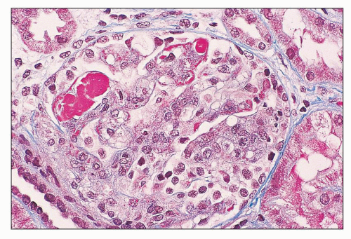
FIGURE 14.7 Lupus nephritis class IV. The hyaline thrombi stain red against the blue-staining glomerular capillary walls. (Masson trichrome, × 500.)

Kant et al. (30) extensively studied glomerular thrombi in SLE and concluded on the basis of immunofluorescence and clinical data that there are two different mechanisms of thrombosis in lupus nephritis. Thrombosis may complicate any severe active lupus nephritis through complement activation and by triggering the coagulation cascade. Alternatively, glomerular capillary thrombosis (plus arterial and arteriolar thrombosis) may occur as a manifestation of lupus anticoagulant/APL antibody syndrome (discussed in section Thrombotic Microangiopathy). Thrombosis caused by APL antibody may occur as a pure thrombotic microangiopathy or superimposed on virtually any class of lupus nephritis.


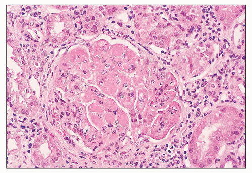
FIGURE 14.8 Lupus nephritis class IV. Extensive intraluminal and subendothelial deposits form “hyaline thrombi” and wire loops. Endocapillary proliferation is less conspicuous in the capillaries with voluminous deposits. (H&E; × 320.)

Mesangial and Endocapillary Proliferation Mesangial hypercellularity and matrix expansion are the first observable response to mesangial deposits. Mesangial hypercellularity and increased mesangial matrix are the only detectable glomerular abnormalities at the light microscopic level in lupus nephritis class II, where they may be diffuse or focal, and segmental or global in the individual glomerulus. Usually, the mesangial immune deposits seen by fluorescence and electron microscopy are more diffuse and regular in distribution than the mesangial proliferative response. There is often a poor correlation between the size and extent of the mesangial immune deposits and the severity of the mesangial hypercellularity. Mesangial hypercellularity is the defining histologic feature of class II lupus nephritis, but it also occurs as an underlying finding in classes III, IV, and V. Like mesangial immune deposits, it is a common base on which the other classes are founded.

Endocapillary hypercellularity can be defined as a proliferation of endothelial cells and mesangial cells together with infiltrating leukocytes (including mononuclear or polymorphonuclear leukocytes) that significantly narrows or occludes the glomerular capillary lumen. Endocapillary hypercellularity is typically focal and segmental in distribution in class III but diffuse and global in class IV disease (Fig. 14.9). Other than neutrophils, the nature of the endocapillary cells is often impossible to determine by light microscopy without the aid of immunohistochemistry using specific markers for monocytes, T lymphocytes, mesangial cells, and endothelial cells. Cells of macrophage/monocyte lineage are particularly numerous in lupus glomerulonephritis and are only exceeded in number in examples of cryoglobulinemic glomerulonephritis (31). They tend to parallel the quantity of immune deposits, complement activation, and proliferative response but do not correlate well with renal function at biopsy or outcome (32,33). On the other hand, the number of macrophages (as well as tubular macrophages) in a second renal biopsy taken 6 months following therapy has been found to correlate well with outcome (34). In computing an activity index, Balow and Austin (35,36,37) use exudation of more than two neutrophils outside the glomerular capillaries per glomerulus as one of the features of glomerular activity but do not consider lymphocyte or monocyte infiltration. Because neutrophil exudation outside the glomerular capillaries is rare, except in areas of necrosis, this criterion weights even more heavily the importance of necrotizing lesions in the activity index.

Necrosis Glomerular necrosis is a feature of class III or IV lupus nephritis and is never observed in pure mesangial proliferative (class II) or membranous (class V) lupus nephritis. It consists of a focus of smudgy fibrinoid obliteration of the glomerular tuft, which is often associated with any or all of the following: deposition of fibrin, GBM rupture or gap formation, and apoptosis of infiltrating neutrophils forming pyknotic or karyorrhectic nuclear debris (Figs. 14.10 and 14.11). The latter change was referred to as “nuclear dust” in the older literature but is now understood to represent a form of apoptosis, which can be confirmed by in situ end labeling of DNA (38). By electron microscopy, the cells undergoing this form of programmed cell death have been identified as infiltrating neutrophils, as well as indigenous glomerular cells (38).


FIGURE 14.9 Lupus nephritis class IV. There is relatively uniform diffuse and global endocapillary proliferation. (H&E; × 100.)

Necrotizing lesions are typically segmental in distribution, but more than one glomerular lobule may be affected, particularly in diffuse proliferative lupus nephritis class IV. As in other forms of glomerulonephritis in which necrotizing lesion are common (e.g., pauci-immune crescentic glomerulonephritis), there is evidence of neutrophil infiltration, and early cellular crescents frequently directly overlie the affected lobules. One study found glomerular necrosis to correlate with lower serum complement (CH50) levels and more severe proteinuria in a large group of patients with class III and IV lupus nephritis (39). In a large Chinese study, necrotizing lesions strongly correlated with anti-C1q antibodies, as well as anti-dsDNA antibodies (40).

Hematoxylin Bodies Hematoxylin bodies are the tissue equivalent of the LE body and consist of naked nuclei whose chromatin has been altered by binding to circulating ANA. They are thought to occur after nuclear exposure to the ambient circulation in the course of individual cell death. Owing to their nuclear origin, they are Feulgen positive. Hematoxylin bodies are most common in necrotizing glomerular lesions. They are considered the only truly pathognomonic lesion in lupus nephritis but are extremely uncommon, affecting approximately 2% of lupus biopsy specimens (41). In H&E-stained sections, hematoxylin bodies consist of rounded, smudgy, lilac staining (amphophilic) structures that are generally smaller than normal nuclei (Fig. 14.12). Hematoxylin bodies may be isolated or clustered. They usually have indistinct borders and appear to merge into the background tissue with surrounding flecks of hematoxyphilic material. Their lilac, hyaline coloration, and smudgy borders allow them to be distinguished from the more darkly basophilic, smaller, and distinctly punctate nuclear fragments observed in foci of karyorrhexis or pyknosis.


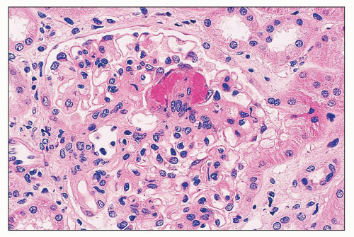
FIGURE 14.10 Lupus nephritis class III. There is segmental fibrinoid necrosis with neutrophil infiltration and pyknosis. (H&E; × 500.)

Cellular Crescents Cellular crescents are a feature of active lupus nephritis that may be encountered frequently in class III or IV lupus nephritis, but never (by definition) in pure class II or class V disease (Fig. 14.13). Crescents may be seen in membranous lupus nephritis in mixed class V and III or class V and IV lesions. Cellular crescents commonly overlie the necrotizing lesions, but they may also occur in glomeruli with nonnecrotizing segmental or global endocapillary hypercellularity. In their activity index formulation, Balow and Austin
(35,36,37) define cellular crescents as “aggregates comprising two or more layers of proliferating visceral and parietal epithelial cells with infiltrating mononuclear cells lining one fourth or more of the interior circumference of the Bowman capsule.” This reasonable definition allows differentiation of crescents from the single layer of reactive hyperplastic visceral epithelial cells commonly encountered in glomeruli with membranous features or undergoing sclerosis. Cellular crescents of inflammatory origin must be differentiated from the epithelial cell proliferation that accompanies the collapsing variant of focal segmental glomerulosclerosis (FSGS) (collapsing glomerulopathy), which has been described in some patients with SLE (see Lupus Podocytopathy).


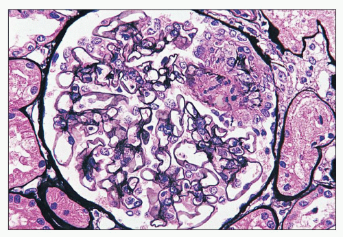
FIGURE 14.11 Lupus nephritis class III. The segmental necrotizing lesion displays segmental rupture of the GBM. (Jones methenamine silver, × 500.)


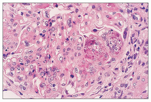
FIGURE 14.12 Lupus nephritis class IV. Several glomerular capillaries contain hematoxylin bodies consisting of smudged, coarsely clumped basophilic material. Another lobule contains karyorrhectic nuclear debris. (H&E; × 500.)


GLOMERULAR SCARRING

Glomerular obsolescence is a feature of chronic glomerular injury that may result from severe or prolonged glomerular damage in the course of class III, IV, or V lupus nephritis. Although it has not been well studied, it is unlikely that extensive glomerulosclerosis supervenes on pure class II lupus nephritis unless there is transformation into one of the more progressive classes. Focal glomerular sclerosis in a patient with lupus may occur in the course of aging or as a complication of hypertensive arterionephrosclerosis and does not always imply scarring in the course of an immunologically mediated injury. The ISN/RPS classification advocates that such nonspecific glomerulosclerosis be excluded, whenever possible, from the computation of the total percentage of glomeruli affected by scarred lupus nephritis (21,22,42). In class III lupus nephritis, the glomerular scarring is often initially focal and segmental, mirroring the distribution of the proliferative and necrotizing lesions. Serial biopsies can demonstrate an evolution from focal segmental necrotizing glomerulonephritis to focal segmental sclerosing glomerulonephritis, sometimes with associated fibrous crescents or synechiae overlying the sclerotic segments. Similarly, in chronic class IV lupus nephritis, the glomerular scarring is typically more global and diffuse, although segmental sclerosis may affect some glomeruli (Figs. 14.14 and 14.15).


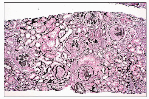
FIGURE 14.13 Lupus nephritis class IV. A severe example with extensive cellular crescents. Although the glomerular tufts are compressed, endocapillary proliferation is evident. (Jones methenamine silver; × 80.)


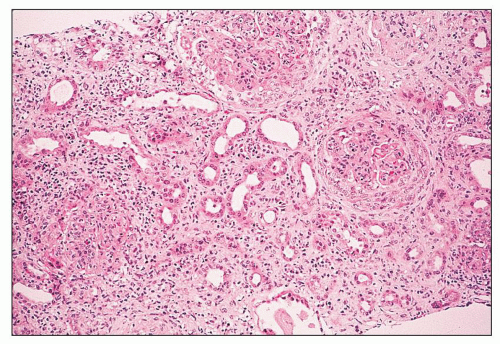
FIGURE 14.14 Lupus nephritis class IV. Initial renal biopsy shows diffuse endocapillary proliferation with focal crescents and severe interstitial inflammation. (H&E; × 80.)


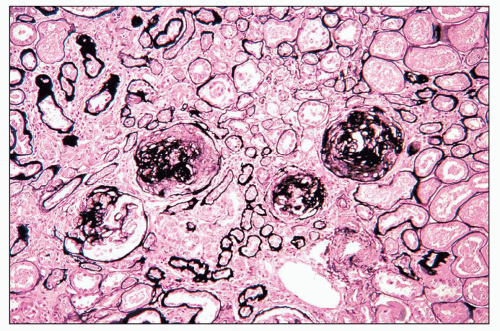
FIGURE 14.15 Lupus nephritis class IV. Despite aggressive therapy, repeat renal biopsy 2 years later shows progression to segmental and global glomerulosclerosis with focal fibrous crescents. There is marked reduction in the degree of interstitial inflammation. (Jones methenamine silver, × 80.)


In some patients with class V lupus nephritis, there is progression to chronic renal failure without clear evidence of transformation into a proliferative phase consistent with combined class III or IV disease. This progressive course, which may affect up to 20% of class V patients, can be considered analogous to the situation in primary membranous glomerulonephritis, which progresses to end-stage renal failure in up to 25% of patients within 10 years of initial clinical detection. In a small percentage of lupus patients, an initial renal biopsy performed at the outset of detectable renal disease reveals an advanced picture of diffuse and global glomerular sclerosis affecting 90% or more of glomeruli. Called lupus nephritis class VI in the ISN/RPS classification (21,22), this picture is usually the sequela of a previous class IV lupus nephritis. In some patients, the phase of active and potentially treatable glomerulonephritis is clinically occult. At this stage, residual but scanty immune deposits may still be detectable in the sclerotic glomerular tuft by immunofluorescence and electron microscopy.

Because lupus nephritis is a typically remitting and relapsing condition with repeated episodes of reactivation, it is not uncommon to detect sclerosing and active proliferative lesions side by side in renal biopsy specimens, especially in patients with longstanding lupus and a history of active lupus nephritis. When active and chronic scarred lesions are detected in an initial renal biopsy specimen at the apparent outset of disease, the disease onset antedated first clinical recognition.


TUBULES AND INTERSTITIUM

Tubulointerstitial lesions may be encountered in all classes of lupus nephritis. Even in class I, in which glomeruli appear normal by light microscopy, there may be mild tubulointerstitial disease resulting from arteriosclerosis associated with aging or hypertension that cannot be directly attributed to lupus nephritis. In specimens with only class I or II glomerular changes, the presence of focal active tubulointerstitial lesions typical of class III should raise the possibility of unsampled focal class III glomerular lesions that are not in the planes of section available for examination. Tubulointerstitial disease is most commonly encountered in class IV lupus nephritis but also occurs frequently in class III and to a lesser degree in class V, with lowest frequency found in classes I and II. The types of lesions that affect the tubulointerstitial compartment can be divided into acute and chronic subgroups, and those related to an interstitial inflammatory process or resulting largely from prolonged nephrotic proteinuria.

In patients with nephrotic-range proteinuria, whether in the setting of class III, class IV, or class V lupus nephritis, the tubules may show nonspecific changes common to many conditions with heavy or longstanding glomerular proteinuria. These predominantly affect the proximal tubules and consist of intracytoplasmic lipid resorption droplets that appear as clear vacuoles in H&E-stained sections because of removal of the lipid in the course of tissue processing and protein resorption droplets that appear eosinophilic and strongly PAS positive and usually trichrome red. The latter change has been referred to as “hyaline degeneration” of the proximal tubules, although this is not really a degenerative process but one of active resorption by the proximal tubules of filtered albumin and lipoproteins. In some cases with longstanding nephrotic syndrome, especially in the setting of a membranous lesion, interstitial foam cells may also be found.

Active (or acute) tubulointerstitial lesions most common in class IV and class III lupus nephritis include interstitial inflammation and edema, which may coexist with the more chronic lesions of tubular atrophy and interstitial fibrosis. The interstitial infiltrates consist predominantly of mononuclear leukocytes, including lymphocytes, monocytes, and plasma cells. Neutrophils and eosinophils are seldom identified. There are rare reports of giant cell reaction to deposits involving the tubular basement membranes (43). These leukocytic infiltrates range from sparse to dense and diffuse and are sometimes associated with lymphocytic infiltration of the tubules (i.e., tubulitis) and tubular epithelial degenerative and regenerative changes. Rarely, hematoxylin bodies ingested by neutrophils may be identified in the tubular lumen (44). This active and severe tubulointerstitial damage is most common in severe diffuse proliferative lupus nephritis class IV. Casts of neutrophils, erythrocytes, and shed tubular epithelial cells are readily identified in active class III or IV lupus nephritis. Intratubular oval fat bodies consisting of lipid-laden desquamated epithelial cells are most common in cases with severe nephrotic proteinuria, usually in the setting of class V membranous lupus nephritis or mixed proliferative and membranous glomerulonephritis (i.e., class V and IV or class V and III).

Several investigators have correlated the immunophenotype of the interstitial inflammatory infiltrates with other histopathologic and clinical parameters (31,32,45,46,47). Most infiltrating mononuclear leukocytes are T lymphocytes, with lesser numbers of monocyte/macrophages, B lymphocytes, and natural killer cells. The relative proportions of CD4 (helper/inducer) and CD8 (suppressor/cytotoxic) T cells vary, with some investigators finding a predominance of CD8+ cells in most patients (31,46) and others a predominance of CD4+ cells (45) or a roughly equal balance (32). Alexopoulos et al. (32) suggested that this disparity may be related in part to treatment, because corticosteroids preferentially reduce CD8+ cells, thereby shifting the CD4:CD8 tissue ratio. Nevertheless, there is general agreement that the percentage of interstitial CD8+ cells is greater than in other forms of glomerular disease such as IgA nephropathy and membranous glomerulonephritis (48,49). One study (46) found the tissue CD4:CD8 ratio to correlate with activity index, whereas others have been unable to demonstrate such a correlation (32). One group identified a correlation between the chronicity index and the extent of interstitial infiltration by T lymphocytes and monocytes (32).

In a recent detailed study, CD4+ T cells were prominent in two thirds of lupus nephritis biopsies and tended to be distributed as broad periglomerular aggregates intermixed with CD8+ T cells (50). By contrast, CD8+ T cells were present in all samples and tended to adhere to the Bowman capsule and infiltrate the tubular epithelium (as CD8 tubulitis). Their differentiation into CD28null memory-effector phenotype suggests participation in adaptive immunity. Study of the T-cell receptor (TCR) β-chain clonotypes revealed oligoclonal T-cell infiltration of the kidney. The clones identified in the kidney represent a small subset of the many clones present in the peripheral circulation, supporting their selection and involvement in adaptive immune responses. Furthermore, when repeat biopsies were performed in individual patients, there was a persistence of the same CD8+ T-cell clones over years, suggesting that they mediate progressive renal injury (50). The intrarenal B-cell population has also been shown to be clonally
expanded and organized into aggregates with focal germinal centers containing follicular dendritic cells (51). The B cells have an immunoglobulin (Ig) repertoire consistent with somatic hypermutation and organ-intrinsic adaptive immune responses. The close proximity of B and T cells suggests that in addition to in situ antibody secretion, resident B cells are contributing to local inflammation by presenting major histocompatibility complex (MHC) class II-restricted antigens to T cells (51). Intrarenal production of anti-dsDNA antibody by resident plasma cells has also been described in the New Zealand Black (NZB)/W murine model of lupus nephritis, indicating that the kidney can be a site of autoantibody formation (52). IL-17-producing double-negative (CD4/CD8) T cells have also been identified in the renal interstitium (53). Several studies using immunostaining for macrophages confirmed the large number of interstitial and intratubular macrophages in diffuse proliferative lupus nephritis and found good correlations between tubular macrophages and serum creatinine, proteinuria, and progression to renal insufficiency (34,54). Urinary biomarkers that reflect the severity and activity of interstitial inflammation in human lupus biopsies include monocyte chemotactic protein 1, hepcidin (produced by monocytes in response to INFα), and liver-type fatty acid-binding protein (produced by the proximal tubule in response to injury) (55).

The strong predominance of T cells among the infiltrating interstitial cells in lupus nephritis suggests a role for cellular immunity in the development of the tubulointerstitial damage. The predominance of CD8 cells in the areas of tubulitis and greater numbers of natural killer cells in biopsies with tubulointerstitial immune deposits raise the question of possible cellular cytotoxicity in response to local autoantigens of renal or systemic origin (32). The presence of follicular structures with germinal centers strongly correlates with tubular basement membrane deposits, suggesting that germinal centers and T- and B-cell aggregates select for cells that locally secrete pathogenic antibodies in the tubulointerstitium (51). The in situ antigens that promote these responses remain unknown but are likely to be important determinants of disease severity. The up-regulation of MHC class II antigen (DR) on the tubular epithelial cells and many of the interstitial leukocytes in human (32,45) and murine lupus nephritis (56,57) also suggests that tubular cells may become activated in lupus and stimulate the influx of activated mononuclear cells. Similar tubular DR expression has been reported in transplant rejection and other renal conditions manifesting tubulitis. Up-regulation of costimulatory molecule CD40 has also been demonstrated in the renal epithelial cells and infiltrating leukocytes of murine and human lupus nephritis (58,59).

Tubulointerstitial immune deposits can be detected by fluorescence and electron microscopy in approximately 50% of patients. They occur more frequently in diffuse proliferative lupus nephritis than in the focal proliferative variant, but they may also occur in some patients with membranous and mesangial proliferative forms. The precise location of these deposits is best elucidated by electron microscopy. They consist of granular electron-dense deposits that involve tubular basement membranes, interstitial capillary basement membranes, and interstitial collagen. They are most frequently associated with the tubular basement membrane, within the tubular basement membrane itself or more commonly abutting the membrane at the interstitial interface (Fig. 14.16). Only rarely are they seen along the epithelial aspect of the tubular basement membrane. These tubulointerstitial deposits may involve any segment of the nephron and often lack a conspicuous leukocytic response. New layers of tubular basement membrane or interstitial capillary basement membrane may encircle the deposits, producing a lamellated network of basement membrane material (Fig. 14.17).
By immunofluorescence, the deposits have a granular to semilinear texture that has a tendency to outline the profiles of tubular basement membranes and interstitial capillaries or to form punctate aggregates in the interstitial collagen (Fig. 14.18). Interstitial capillary immune deposits are particularly specific for lupus nephritis (Fig. 14.19).


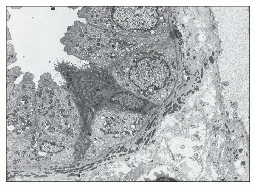
FIGURE 14.16 Lupus nephritis class IV. The electron micrograph shows electron-dense deposits within a tubular basement membrane and, to a lesser extent, in the adjacent interstitial collagen. (× 2000.)


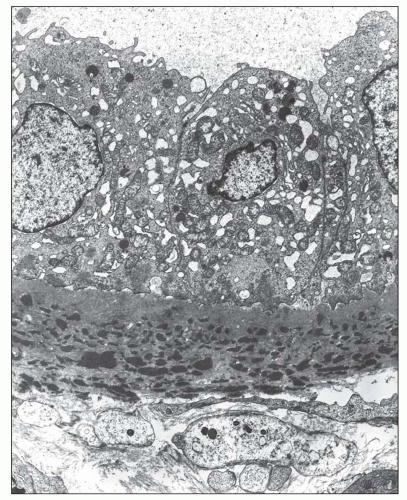
FIGURE 14.17 Lupus nephritis class IV. High-power view shows a lamellated network of tubular basement membrane splayed around the tubular basement membrane deposits. (Electron micrograph, × 4000.)


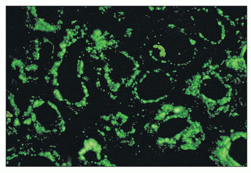
FIGURE 14.18 Lupus nephritis class IV. The immunofluorescence micrograph shows abundant granular deposits of IgG within the tubular basement membranes and interstitium. (× 200.)

The composition of the tubulointerstitial deposits varies. Most have positive staining for IgG, with other immunoglobulins detected less frequently (60,61). In some biopsies, there is positivity for IgG, IgM, and IgA in the tubulointerstitial deposits, and in others, only a single immunoglobulin class may be found. There even may be differences in the composition of IgG subclasses between the tubulointerstitial, vascular, and glomerular compartments of a given biopsy (62). Complement components are associated with the immunoglobulins in most cases and occasionally may be detected in their absence (63), suggesting a role for antibody-independent complement activation.


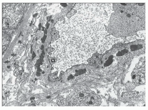
FIGURE 14.19 Lupus nephritis class IV. This electron micrographs shows numerous electron-dense deposits within the wall of an interstitial capillary. (× 6000.)

In general, the presence of tubulointerstitial deposits correlates well with glomerular lesions, becoming progressively more frequent with increasing glomerular endocapillary hypercellularity (classes III and IV) and being relatively infrequent with membranous (class V) or mesangial proliferative lesions (class II) (47,64). Interstitial inflammation tends to be particularly severe in crescentic forms of lupus nephritis (47). Park et al. (64) and Jeruc et al. (47) made the important observation that there is no correlation between the prevalence of tubulointerstitial deposits and the presence and severity of interstitial inflammation, which may be severe in the absence of detectable deposits. For example, among the 103 patients studied by Park et al. (64), 76 had interstitial infiltrates, but only 32 had tubulointerstitial deposits, and interstitial inflammation without tubulointerstitial deposits was found in all classes of renal lesions. Clearly, mechanisms other than a simple inflammatory response to tubulointerstitial deposits are operant in most cases.

The severity of tubulointerstitial inflammation correlates broadly with glomerular inflammatory lesions (64). It constitutes one of the best morphologic correlates with the degree of renal insufficiency (32,64) and is an accurate prognosticator of subsequent decline in renal function (64). Tubular atrophy, at least in part the result of interstitial inflammation, is one of the strongest predictors of renal failure (36), as it is in many other glomerular diseases. Similarly, increased interstitial volume, which directly parallels the development of tubular atrophy, closely correlates with impaired renal function (65,66). Staining with picro-Sirius red followed by quantitative morphometry and computer imaging has allowed more accurate measurements of interstitial matrix volume, which was a strong predictor of doubling of serum creatinine (67). Morphometric measurement of chronic renal damage using an interactive image analysis system that captures silver-stained images and outlines zones of chronic glomerular and tubulointerstitial injury predicted renal outcome better than WHO class (68). Renal tubular dysfunction has been demonstrated in some patients with lupus nephritis (69,70), and reports include hyperkalemic distal renal tubular acidosis (RTA) with hyporeninemic hypoaldosteronism (71,72,73).

In most instances of tubulointerstitial deposits, the associated tubular atrophy, interstitial fibrosis, and inflammation accompany severe glomerular lesions. There are rare cases in which severe tubulointerstitial damage occurs in the presence of only trivial glomerular lesions (74,75,76,77,78,79), sometimes leading to acute renal failure. The presence of abundant tubulointerstitial deposits in most of these patients attests to their pathogenetic relationship to lupus rather than representing a superimposed, unrelated tubulointerstitial process (reviewed in (78)). In some of these cases, the tubulointerstitial damage is at least partially reversible with immunosuppressive therapy (76,78,79,80,81). There are rare reports of interstitial nephritis induced by anti-tubular basement membrane deposits, producing a linear fluorescence with antisera to IgG and C1q along the tubular basement membranes and the Bowman capsule (82).


VASCULAR LESIONS

Vascular lesions are commonly encountered in renal biopsy specimens from patients with SLE and may assume a variety of morphologic forms (83,84), including uncomplicated vascular immune deposits, noninflammatory necrotizing vasculopathy,
thrombotic vasculopathy, true inflammatory vasculitis, and nonspecific arteriosclerosis (Table 14.5). Because assessment of vascular lesions is not factored into the ISN/RPS classification of lupus nephritis or formulation of the activity and chronicity index according to the National Institutes of Health (NIH) criteria, renal vascular lesions run the risk of being overlooked. This is especially the case when vascular lesions are focally distributed. To accurately classify renal vascular lesions, vessels must be systematically examined by light, immunofluorescence, and electron microscopy. The morphologic findings must then be analyzed in the context of particular clinical syndromes that may complicate SLE, including APL antibody syndrome (alternatively known as anticardiolipin syndrome), thrombotic thrombocytopenic purpura, renal vein thrombosis (RVT), and accelerated hypertension.


TABLE 14.5 Vascular lesions in lupus nephritis


Arteriosclerosis and arteriolosclerosis


Uncomplicated vascular immune deposits


Noninflammatory necrotizing vasculopathy (so-called lupus vasculopathy)


Thrombotic microangiopathy


Associated with HUS/TTP syndrome


Associated with APL antibodies


Associated with scleroderma/mixed connective tissue disease


Necrotizing vasculitis (PAN type)


HUS, hemolytic uremic syndrome; TTP, thrombotic thrombocytopenic purpura; PAN, polyarteritis nodosa.


Klemperer reported a high incidence of renal vascular lesions in his autopsy-based study of SLE. The lesions predominantly involved small arteries and arterioles with fibrinoid alteration of the intima and subendothelium (28). Grishman and Venkataseshan (85) observed renal vascular lesions in the autopsy specimens of 8 (33%) of 24 patients who died in the presteroid era and 5 (25%) of 26 who died in the modern era, but with a much lower incidence in 19 (6.9%) of 276 renal biopsy specimens. A greater incidence of renal vascular lesions in autopsy (7 of 20) than biopsy (10 of 200) specimens was also described in an earlier report (86) and may be related to differences in sample size, severity, duration, and treatment of the lupus nephritis. Most studies are in agreement that the presence of renal vascular lesions of the thrombotic, necrotizing, or vasculitic type adversely affects renal outcome.

Attention to the importance of vascular lesions in SLE has been renewed in recent years (83,84,85,87,88,89). In a large Italian study of 285 patients with lupus nephritis from 20 nephrology centers, renal vascular lesions were found in 79 cases (27.7%) and included lupus vasculopathy (27 cases), hemolytic uremic syndrome/thrombotic thrombocytopenic purpura (HUS/TTP), or malignant hypertensive lesions (24 cases), vasculitis (8 cases), and arteriosclerosis or arteriolosclerosis (20 cases) (84). Renal vascular lesions were associated with a higher rate of progression to renal failure. The 5- and 10-year renal survival rates were 74.3% and 58.0%, respectively, for patients with renal vascular lesions, compared with 89.6% and 85.9% for those without renal vascular lesions (84). A recent prospective study also found renal vascular lesions to be associated with hypertension and reduced renal function at the time of biopsy, and both variables predicted poor renal outcome in multivariate analyses (87). A large Chinese study of 341 patients with lupus nephritis found renal vascular lesions in 279 (82%), including 253 with uncomplicated vascular immune deposits, 82 with arteriosclerosis, 60 with thrombotic microangiopathy, 13 with noninflammatory necrotizing vasculopathy, and 2 with true arteritis (89). This group found that a statistical model that included vascular lesions was a superior predictor of outcome than traditional NIH activity and chronicity indices (89).


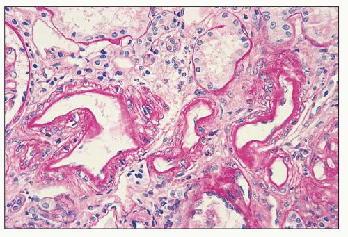
FIGURE 14.20 Lupus nephritis class IV. Intimal immune deposits in the walls of interlobular arteries have caused slight thickening of the intimal basement membrane without significant luminal narrowing. (PAS; × 320.)

Uncomplicated Vascular Immune Deposits The most common renal vascular lesion in SLE is immune complex deposition in the walls of small arteries and arterioles; deposition occurs to a lesser extent in veins (84,85,90,91). The affected vessels usually appear normal by light microscopy. Rarely, thickening of the subendothelial zone or medial intercellular zones by glassy hyaline eosinophilic material is observed by light microscopy, but the vessel lumen usually is not compromised (Fig. 14.20). Diagnosis requires the demonstration of granular deposits of immunoglobulin (IgG, IgM, and IgA in various combinations), often associated with C1q or C3 (Fig. 14.21). Finding IgM or C3 alone is insufficient to diagnose this condition, because these components are frequently nonspecifically trapped in vessels with the ordinary arteriosclerosis or arteriolar hyalinosis that accompanies the more chronic inactive forms of lupus nephritis. By electron microscopy, granular, electron-dense deposits are identified in the intimal and perimyocyte matrix (Fig. 14.22). As in glomerular immune deposits, some of these vascular immune deposits may exhibit an organized fingerprint substructure (83). Such vascular immune deposits may occur in classes II through V of lupus nephritis, but they are most common in the more active proliferative classes (III and IV), especially in association with tubulointerstitial deposits. Uncomplicated vascular immune deposits are usually clinically silent, and they have not been found to confer a higher risk of hypertension or progressive renal disease.


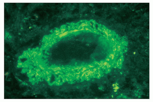
FIGURE 14.21 Lupus nephritis class IV. The fluorescence micrograph shows abundant granular staining for IgG within the intima and media of an interlobular artery. (× 400.)

Noninflammatory Necrotizing Vasculopathy Noninflammatory necrotizing vasculopathy (i.e., lupus vasculopathy) is far less common than simple vascular immune deposits (83). It affects predominantly preglomerular arterioles and, to a lesser extent, interlobular arteries, especially in the setting of severe active class IV lupus nephritis. The affected vessels are severely narrowed and sometimes occluded by abundant intimal and luminal deposits of glassy eosinophilic material that may extend into the media (Fig. 14.23). This material is usually fuchsinophilic in trichrome-stained preparations and may demonstrate focal reactivity for fibrin with the Lendrum and PTAH stains (Fig. 14.24). The endothelium is often swollen or denuded, and there is smudgy degeneration and loss of medial myocytes, although without inflammatory infiltration of the vessel wall.

Bhathena coined the term lupus vasculitis (92), but we prefer the term lupus vasculopathy to depict this lesion because of the absence of vascular infiltration by inflammatory cells (83). Immunofluorescence shows variable staining of the vessel intima and lumen for immunoglobulins (e.g., IgG, IgM, IgA), complement components (C3 and C1q), and fibrin-related antigens (Fig. 14.25). Electron microscopy usually reveals endothelial loss with mural deposits of granular, electron-dense material with the combined appearance of immune deposits and insudated plasma proteins, sometimes associated with fibrillar fibrin. Degenerative changes of the medial myocytes may be seen adjacent to these deposits but without leukocyte infiltration. These morphologic features strongly suggest that the combined processes of vascular immune deposition and intravascular coagulation contribute to their morphogenesis. Severe hypertension is not invariably associated with these lesions and is unlikely to be the primary etiologic factor. However, severe hypertension is common and probably exacerbates the vascular changes (85,93). In most series, lupus vasculopathy carries an ominous prognosis, with frequent, severe renal insufficiency, active urinary sediment, active lupus serologies, hypertension, and rapid progression to renal failure.


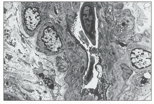
FIGURE 14.22 Lupus nephritis class IV. The electron micrograph shows small, granular, electron-dense deposits within the subendothelial basement membrane of an interlobular artery. (× 2875.)


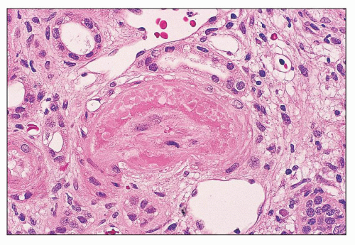
FIGURE 14.23 Lupus vasculopathy. The lumen of an arteriole is severely narrowed by intimal deposits of intensely eosinophilic material suggestive of fibrin admixed with more lightly eosinophilic immune deposits. The endothelium is necrotic; there is no inflammation of the vessel wall. (H&E; × 500.)

Thrombotic Microangiopathy Thrombotic microangiopathy may be difficult to differentiate from the necrotizing, noninflammatory lupus vasculopathy described in the previous section (83). It may occur in association with distinct clinical syndromes such as HUS/TTP, lupus anticoagulant/APL syndrome, overlap with scleroderma, or malignant hypertension. However, in a significant number of patients, thrombotic lesions are found in renal biopsies of patients with SLE who lack a recognizable systemic thrombotic process. Thrombotic vascular lesions most commonly affect preglomerular arterioles and interlobular arteries. Unlike lupus vasculopathy, they are also frequently associated with signs of glomerular thrombosis, including glomerular fibrin thrombi, mesangiolysis, double contours of the GBM enclosing subendothelial electron lucent flocculent material, and ischemic glomerulosclerosis
(Fig. 14.26). By light microscopy, the affected vessels are occasionally narrowed or occluded by intraluminal fibrin and platelet thrombi, which may be associated with endothelial swelling and denudation (Fig. 14.27). The products of coagulation may penetrate the intima and sometimes contain fragmented erythrocytes. There is no overt leukocyte infiltration of the media, although a rare neutrophil or lymphocyte may be entrapped in the luminal thrombosis. In the interlobular arteries, the intima may have a more mucoid, edematous appearance, with “onion skin” myointimal proliferation. By fluorescence microscopy, the affected vessels usually reveal intense, dominant staining for fibrin-related antigens, with variable positivity for IgM and C3. In contrast to lupus vasculopathy, IgG is usually absent from these lesions. Recently, C4d has been identified as a potential marker of glomerular thrombotic microangiopathy in lupus nephritis, irrespective of whether there was an associated APL antibody (94). The incidence of thrombotic lesions resembling HUS/TTP was 8% of lupus nephritis patients according to a collaborative Italian study (84). Their presence was associated with reduced (66.3%) 5-year renal survival compared with 89.6% for lupus nephritis patients without renal vascular lesions. They may be superimposed on virtually any class of lupus nephritis including classes II (95), III (95), and IV (96).


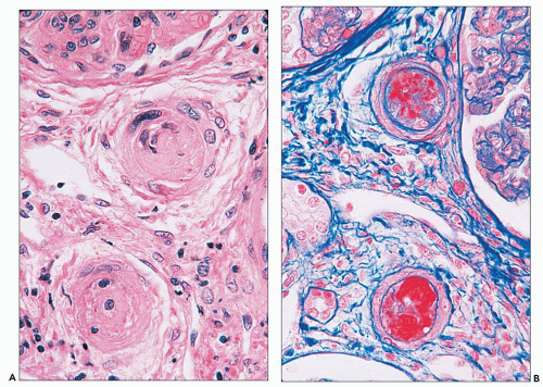
FIGURE 14.24 Lupus vasculopathy. A double panel shows occlusion of preglomerular arterioles by eosinophilic deposits (A) that stain positive with Lendrum stain for fibrin (B). (× 500.)


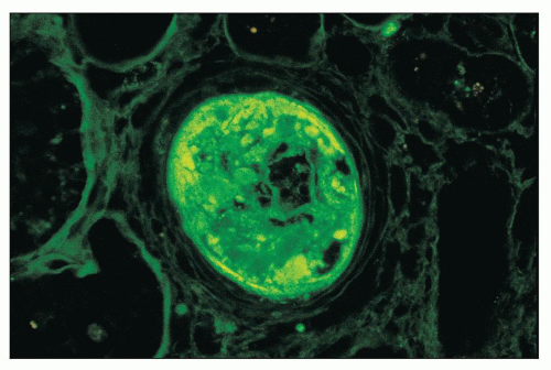
FIGURE 14.25 Lupus vasculopathy. An interlobular artery shows staining for IgA within its expanded intima. Similar deposits were seen for IgG, IgM, C3, C1q, and fibrinogen. The vessel lumen is nearly occluded. (× 600.)

Syndrome of Hemolytic Uremic Syndrome or Thrombotic Thrombocytopenic Purpura Some patients with biopsy features of thrombotic microangiopathy have a clinical syndrome of HUS or TTP, which may precede, be contemporaneous with, or follow clinical onset of SLE (83,97). Symptoms related to TTP include fever, malaise, petechial and purpuric
skin lesions, abdominal pain and bleeding, and neurologic symptoms, including reduced consciousness, seizures, transient pareses, and coma (95,98,99). Most well-documented cases have evidence of thrombocytopenia and microangiopathic hemolytic anemia. The results of other coagulation studies (e.g., prothrombin time, partial thromboplastin time, fibrin degradation products) have been normal, and the lupus anticoagulant is absent in most patients (83,95). Some cases have been linked to autoantibody to the von Willebrand factor cleaving protease ADAMTS-13 (100,101). An association with decreased serum complement factor H levels in some individuals suggests a role for dysregulation of the alternative complement pathway (101,102). Renal manifestations of the TTP-like process range from asymptomatic urinary abnormalities to mild reduction in renal function to fulminant oligoanuric renal failure requiring dialysis. Hypertension has been absent in most cases (95,97,103) but present in others (84). The TTP has responded to the same therapeutic strategies used in idiopathic TTP. In the modern era, plasma exchange and plasma infusion have achieved greatly improved survival compared with older forms of therapy, including steroids, antiplatelet agents, and splenectomy.


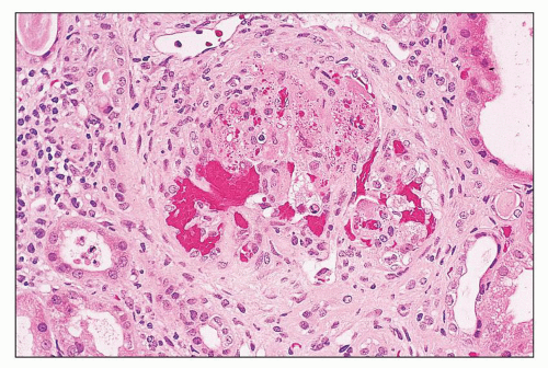
FIGURE 14.26 Thrombotic thrombocytopenic purpura-like syndromein SLE. This glomerulus displays features of acute thrombotic microangiopathy, including marked glomerular capillary congestion, endothelial swelling and necrosis, and glomerular capillary thrombosis with entrapment of fragmented erythrocytes (i.e., schistocytes). (H&E; × 320.)


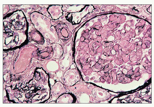
FIGURE 14.27 Thrombotic thrombocytopenic purpura-like syndrome in SLE. The preglomerular arteriole and many glomerular capillaries are occluded by fibrin thrombi. The arteriolar and glomerular endothelium appears denuded. (Jones methenamine silver, × 320.)

Lupus Anticoagulant or Antiphospholipid Syndrome Although Conley and Hartmann first described a lupus inhibitor in 1951, it was only in the 1980s that the lupus anticoagulant or APL syndrome was fully recognized as a major complication of SLE and related collagen-vascular diseases. Patients with this condition may have SLE, a lupus-like condition that fails to fulfill ACR criteria for SLE (i.e., usually low-titer ANA with negative results for anti-DNA antibody), or primary APL syndrome (104,105,106,107,108). Slow recognition of the nature and clinical features of the APL syndrome is related in part to the diverse organ system involvement, which overlaps between specialties, including hematology, rheumatology, neurology, nephrology, and obstetrics (109,110). The kidney is commonly affected in this condition, with overt renal manifestations in approximately 25% of patients (109). APL antibodies have been detected in 25% to 50% of SLE patients, although a smaller percentage develops any clinical signs or symptoms of thrombosis (111,112). In an analysis of 29 published series comprising greater than 1000 patients with SLE, there was a 34% incidence of lupus anticoagulant and 44% incidence of anticardiolipin antibodies (113).

The clinical features of APL syndrome are diverse and include superficial and deep venous thromboses, spontaneous abortions (from placental thrombosis), pulmonary hypertension, cerebral infarcts and transient ischemic attacks, Budd-Chiari syndrome, livedo reticularis, cardiac valvular disease, adrenal hemorrhage, thrombocytopenia, and vague constitutional symptoms (114,115,116,117). Renal manifestations range from microthrombosis of glomerular capillaries and arterioles to thrombosis of intraparenchymal arteries to thrombosis of the main renal artery and vein, with secondary renovascular hypertension, renal cortical necrosis, and infarction (63,106,118,119,120,121,122,123,124,125,126) (Figs. 14.28 and 14.29). Thrombi may be in various stages of organization and recanalization. Some cases are associated with ischemic subcapsular cortical scars (127). These features may produce a spectrum of renal clinical manifestations, including asymptomatic hematuria and mild proteinuria, hypertension (ranging from mild to malignant range), mild to severe renal insufficiency, nephrotic-range proteinuria, and rapidly progressive renal failure (126,128,129,130,131,132,133). Some patients with SLE develop catastrophic antiphospholipid syndrome (CAPS), also known as Asherson syndrome, which frequently involves the kidney and is life threatening (134). CAPS is defined as follows: (a) clinical evidence of involvement of at least three organ systems over a period of less than 1 week, (b) histologic confirmation of thrombosis in at least one organ system, and (c) documented APL antibody, which is usually present at high titer.

In 1981, Kant et al. (30) first called attention to the strong association between the existence of a circulating lupus anticoagulant and the occurrence of glomerular capillary thrombosis, which often could not be explained by the activity of the glomerulonephritis. In 1988, Kincaid-Smith described
a syndrome of pregnancy-associated thrombotic microangiopathy in 12 young women with APL syndrome, 8 of whom had no evidence of SLE and 4 of whom had SLE, including 2 with proliferative glomerulonephritis (135). D’Agati et al. (129) stressed that the occurrence of thrombotic microangiopathy in APL syndrome could not be accounted for by secondary intravascular coagulation caused by active proliferative and necrotizing lupus nephritis.


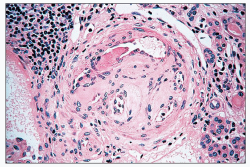
FIGURE 14.28 Lupus anticoagulant syndrome in SLE. An interlobular arteries contains fresh and recanalized thrombus. (H&E, × 400.)

In SLE, thrombotic microangiopathy related to APL syndrome may occur in a variety of classes of lupus nephritis. In a study of 26 patients with APL antibodies and renal disease but failing to fulfill ACR criteria for SLE, there was a particular predominance of membranous lupus-like nephritis with or without features of associated thrombotic microangiopathy on renal biopsy (136). In these 26 cases, renal biopsy disclosed pure thrombotic microangiopathy in 4 cases, thrombotic microangiopathy combined with lupus-like nephritis in 11 cases, and lupus-like nephritis alone in 4. The lupuslike nephritis consisted of membranous glomerulonephritis in 11, mesangial proliferative in 3, and focal proliferative in 1 (136). In a study by Daugas et al. (127), renal lesions of APL syndrome were detected in 32% of lupus patients with renal biopsies, superimposed on or independently of lupus nephritis. The renal biopsy findings of APL nephropathy were statistically associated with lupus anticoagulant, but not with anticardiolipin antibodies, and were independent risk factors for the development of hypertension, elevated serum creatinine, and increased interstitial fibrosis (127). Moroni et al. (137) prospectively followed 111 patients with lupus nephritis for mean of 173 months and found an overall prevalence of APL antibody in 26% of patients; the presence of an APL antibody was associated with worse renal survival. In a study of 151 lupus patients with or without APL antibody, renal biopsy features of thrombotic microangiopathy were identified in 40% of lupus patients with APL antibody compared to only 4% of lupus patients without APL antibody (138). There was a higher frequency of hypertension and renal insufficiency in those with nephropathy related to APL antibody. Interestingly, some patients with apparently primary APL syndrome evolve into SLE over time, suggesting these conditions are related (139).


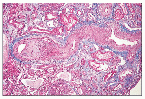
FIGURE 14.29 Lupus anticoagulant syndrome in SLE. An interlobular artery is narrowed by organizing thrombus. There is adjacent tubular atrophy and interstitial fibrosis. (Masson trichrome; × 200.)

The APL antibody syndrome is caused by antibodies to a family of naturally occurring phospholipids (including cardiolipin, phosphatidylcholine, phosphatidylserine, phosphatidylinositol, and others) and/or to the β2-glycoprotein 1 (β2GP-1) to which they bind (140). Laboratory diagnosis is made by demonstration of a positive lupus anticoagulant or demonstration of APL or β2GP-1 antibodies by enzyme-linked immunosorbent assay (ELISA).

The lupus anticoagulant test depends on the prolongation of clotting tests such as the activated partial thromboplastin time, kaolin clotting time, or dilute Russell viper venom time owing to interference with the phospholipid component of the prothrombin activator complex, consisting of factors Xa, V, Ca2+, and phospholipid. LAC activity is defined as an unexplained prolongation of the activated partial thromboplastin time that is not reversed when the patient’s plasma is diluted 1:1 with normal platelet-free plasma (a procedure that would be expected to reverse clotting caused by factor deficiencies) (141). This occurs because the patient’s plasma contains an inhibitor. Although the presence of a lupus anticoagulant prolongs phospholipid-dependent coagulation tests in vitro, it rarely causes bleeding problems. Thus, in so far as the “lupus anticoagulant” promotes coagulation in vivo, it is something of a misnomer.

ELISA for APL antibodies uses a phospholipid matrix of hexagonal-phase phospholipids (142), which more closely resemble the structure of endothelial membrane-associated phospholipids, or a platform of β2GP-1 itself. Some ELISAs are able to distinguish APL antibody reactive to phospholipids alone from antibody to phospholipids that requires the presence of β2GP-1 and from antibody to β2GP-1 that is independent of the presence of phospholipids. ELISA for APL antibodies is reported as specific titers and an immunoglobulin class (IgG, IgM, or IgA), both of which may have prognostic importance. In SLE patients, high-titer IgG APL antibodies correlate best with thrombotic episodes and obstetrical complications, although even moderate titers of IgG APL antibody may give rise to severe thrombotic events. In general, the titers of APL antibodies do not correlate with anti-DNA antibody titers or complement levels. IgM APL antibodies are less
closely linked to APL syndrome and may be a manifestation of drug-induced APL antibody, particularly from procainamide. IgA APL antibody has been linked to a higher incidence of thrombocytopenia in some reports. Many patients have APL antibodies of more than one class. False-positive Venereal Disease Research Laboratory (VDRL) test results have been obtained from APL antibodies reactive with the cardiolipin and phosphatidylcholine component of beef heart used in this assay.

The pathogenesis of thrombosis in patients with APL syndrome is uncertain. In many patients, the binding of anticardiolipin antibody to phospholipid appears to depend on a plasma cofactor, β2GP-1, which has a high affinity for anionic phospholipids (143,144,145). The means by which this interaction promotes coagulation may involve direct endothelial damage or platelet activation. There is also evidence that these antibodies bind to lipid surfaces that have been altered by oxidants, thereby exposing novel epitopes that are themselves immunogenic (146). Altered endothelial cell functions may result, promoting thrombosis or decreased fibrinolysis through reduced production of free protein S, decreased activation of protein C by thrombomodulin, reduced prostacyclin, and reduced prothrombinase activity.

Optimal therapy for SLE patients with APL antibodies, with and without thrombotic events, has not been clearly defined. Most clinicians advocate high-dose anticoagulation with heparin followed by long-term warfarin for patients with APL antibodies who have suffered thrombotic complications to prevent recurrent thromboses. More controversial are the large numbers of lupus patients with APL antibody who have not manifested clinical features of APL syndrome. Options include no specific therapy or aspirin alone; a role for prophylactic anticoagulation has not been defined (147). Although steroid or cytotoxic therapy may alter APL antibody titers, immunosuppressive therapy does not reduce the risk of thrombosis (140).

Renal Vasculitis True inflammatory vasculitis in which there is leukocyte infiltration of vessel walls, often accompanied by necrosis, is the least common vascular lesion encountered in renal biopsies from lupus patients (83). This form of renal vasculitis, which is histologically indistinguishable from that seen in polyarteritis nodosa or microscopic polyangiitis, is so rare in lupus nephritis that some have questioned whether it may not represent an overlap with some other forms of vasculitis. Some of these patients have clinical evidence of systemic vasculitis, but others appear to have renal-limited vasculitis. We have seen an example of necrotizing inflammatory vasculitis in the kidney and probable cerebral vasculitis with associated seropositivity for antineutrophil cytoplasmic antibody (ANCA). This form of renal vasculitis has not been studied systematically, owing to the rarity of the lesion and the fact that most of the reported patients antedate the availability of serologic testing for ANCA. This form of renal vasculitis was observed in 1 (1.8%) of 56 patients reported by Appel et al., 1 (0.3%) of 326 reported by Grishman et al., 8 (2.8%) of 285 reported by Banfi et al., and 2 of 279 (0.7%) reported by Wu et al. (18,84,85,89). Morphologically, these lesions are the only form of lupus-associated vascular disease in which there is true inflammatory infiltration of the intima and media by neutrophils and mononuclear leukocytes, often accompanied by fibrinoid necrosis and rupture of elastic lamellae (Fig. 14.30).


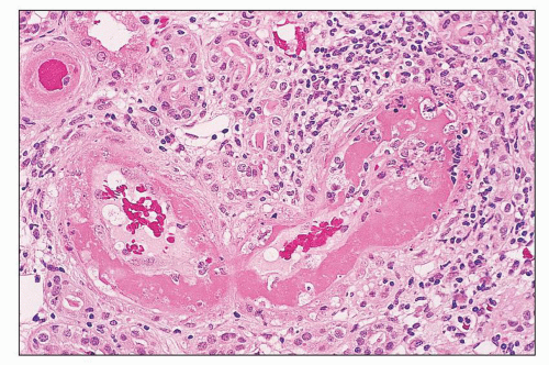
FIGURE 14.30 Acute necrotizing vasculitis in SLE. This patient with class IV lupus nephritis and multisystem vasculitis had necrotizing, inflammatory vasculitis of several small interlobular arteries. There is transmural infiltration by neutrophils and lymphocytes, with abundant intimal fibrin and necrosis of the media. (H&E; × 320.)

Immunofluorescence discloses strong staining for fibrin-related antigens, with weak and more variable staining for immunoglobulin and complement, probably representing nonspecific trapping of plasma proteins in areas of necrosis. Electron microscopic descriptions of these lesions are lacking, and it is unknown whether affected vessels contain electron-dense immune deposits.

This lesion may occur in the setting of any class of active or inactive lupus nephritis. Because of its extremely poor prognosis and need for aggressive immunosuppressive therapy, the occurrence of true inflammatory vasculitis in a renal biopsy specimen should be promptly reported.


Immunofluorescence Microscopy

Lupus nephritis is one of the few diseases of the kidney in which immune deposits can be detected in all renal compartments, including the glomeruli, tubules, interstitium, and blood vessels. The immunofluorescence staining pattern is often extremely helpful in confirming a diagnosis of lupus nephritis when the diagnosis of SLE may be in doubt at the time of biopsy.

Although individual patterns of immunofluorescence staining are highly variable and are discussed in more detail in the section on the various ISN/RPS classes, some general remarks are applicable to all classes. Data from four different series giving the frequency of positivity of different antisera are listed in Table 14.6 (18,148,149,150). IgG is found almost universally. There are codeposits of IgM and IgA in most specimens. The corresponding presence of both light chains indicates the polyclonal nature of the immunoglobulins. Some investigators have identified a more intense staining of the IgG and IgA deposits with antisera to λ than κ (151), although this λ dominance is less pronounced than that observed in IgA nephropathy (152). Others have observed a κ predominance or equal intensity of light chain staining in lupus nephritis (152). C3 is the most frequent complement component, followed by C1q, which usually stains very intensely, sometimes in the absence of C3 and C4 (152). C4 stains least frequently and often most faintly. The presence of early complement components C1q
and C4 attests to the activation of complement by the classic pathway (153). The staining pattern is called “full house” (meaning 3 of a kind and 2 of a kind in poker parlance) when deposits containing all three immunoglobulin classes (IgG, IgM, and IgA) and both complement components (C3 and C1q) are present.


TABLE 14.6 Incidence of positive glomerular immunofluorescence for different antisera from four series


Antiserum


Appel et al. (18)


Cameron et al. (741)


Hill et al (149)


Sinniah and Feng (150)


Total


Percentage positive


IgG


24/27


38/41


56/64


32/32


150/164


91.5


IgM


26/29


29/41


41/64


16/32


112/166


67.5


IgA


13/28


23/41


41/64


21/32


98/165


59.4


C1q


24/31


21/32


30/40


75/103


72.8


C4


18/31


20/53


38/85


44.7


C3


26/29


32/41


47/63


29/32


134/165


81.2


Fibrin(ogen)


6/13


12/40


17/62


29/32


64/147


43.5


Properdin can also be identified in the glomerular deposits (154), suggesting that complement is also activated through the alternative pathway. Membrane attack complex (C5 through C9) has also been identified in the glomerular deposits (155). Fibrin-related antigens are commonly intensely positive in areas corresponding to necrotizing lesions and in the urinary space in association with active crescents (Fig. 14.31). Weaker positivity for fibrin may also persist in older fibrous crescents. In contrast to the intense and discretely localized positivity for fibrin in necrotizing glomerular lesions, it is not uncommon to observe a more generalized and weaker positivity for fibrin outlining the glomerular tuft in cases of diffuse proliferative glomerulonephritis without necrotizing features by light microscopy. It is likely that fibrin is deposited in the course of activation of the coagulation cascade through glomerular capillary immune deposition, leukocyte infiltration, and endothelial activation. Because antibodies to fibrin-related antigens do not differentiate fibrin I, fibrin II (cross-linked fibrin), fibrinogen, and fibrin degradation products, more specific antisera can be used to differentiate between these coagulation products (156,157). These studies show that fibrinogen may be detected in glomeruli without histologically identifiable thrombi and that the presence of cross-linked fibrin correlates best with active necrotizing lesions (157). C4d is a newly identified biomarker associated with glomerular thrombosis in lupus nephritis (94) but can also colocalize with immune deposits.


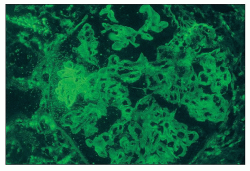
FIGURE 14.31 Lupus nephritis class III. There is strong staining for fibrin/fibrinogen in the distribution of a segmental necrotizing lesion. (Immunofluorescence micrograph, × 500.)

Another commonly observed phenomenon in immunofluorescence studies is the detection of tissue ANA in renal biopsies (158). Although ANA is detectable by indirect immunofluorescence using tumor cell lines as substrate in up to 98% of lupus patients, for unknown reasons only a fraction of patients with lupus exhibit ANA reactivity in their own renal biopsies. Reactivity consists of positive staining of the renal epithelial nuclei with antisera to IgG (in a homogeneous, speckled, or rim pattern similar to that observed in serum ANA testing) (Fig. 14.32). This phenomenon has long been considered an artifact of the cryostat sectioning allowing ambient ANA in the patient’s serum to bind to the nuclei exposed in the course of sectioning, rather than representing an in vivo phenomenon. ANA reactivity is revealed by adding fluoresceinated
rabbit antihuman IgG in a standard fashion. However, experimental data on murine lupus suggest that ANA may penetrate and bind to the nuclei of intact cells and therefore may occur in vivo (159,160). In cases with strong tissue ANA (particularly speckled type), the diffuse nuclear staining for IgG may interfere with detection of true immune deposits in glomeruli and the tubulointerstitium, requiring careful integration with the results of immunofluorescence staining for the other immune reactants.


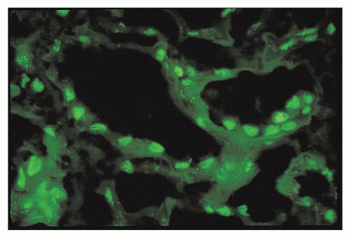
FIGURE 14.32 Lupus nephritis class IV. The immunofluorescence micrograph shows tubular ANA reactivity in this cryostat section stained for IgG. This finding has been referred to as “tissue ANA.” (× 600.)


Electron Microscopy

The electron microscopic appearances of the kidneys from SLE patients are exceedingly diverse. Deposits in the glomeruli may range from sparse and small to abundant and large. The mildest glomerular alteration consists of small, discrete, mesangial electron-dense deposits (in lupus nephritis class I or II). In specimens with severe glomerular immune deposition, electron-dense deposits consist of copious mesangial deposits and involve subendothelial, intramembranous, and subepithelial locations, even to the point of obliterating the glomerular capillary lumina in the more dramatic examples of diffuse proliferative glomerulonephritis (Fig. 14.33). Many researchers (18,161,162) have emphasized that voluminous deposits in all these locations simultaneously are seldom found in conditions other than lupus nephritis, with the exception of some forms of primary and secondary membranoproliferative glomerulonephritis type 3.

Of the types of deposits found in lupus nephritis, the subendothelial deposits correlate best with clinically active disease. These subendothelial deposits distributed in a diffuse and global pattern are the ultrastructural hallmarks of the most ominous pattern of lupus nephritis class IV, whereas they are more focal or segmental in class III. The various patterns of immune deposit formation in the glomerulus will be described later in the section on WHO classification of glomerular lesions.

In lupus nephritis, electron-dense deposits are predominantly finely granular in texture, but in a few cases the deposits may be partially organized, exhibiting a fingerprint, microtubular, or lattice-like substructure (Figs. 14.34 and 14.35). This substructure usually does not affect all deposits completely or uniformly but is identified focally in just a portion of an otherwise granular deposit, especially in the larger intraluminal or subendothelial deposits. Fingerprint substructure, first described by Grishman et al. (163), consists of curved arrays of from two to six parallel dark bands of various lengths alternating with light bands arranged around a center, producing a configuration that mimics a fingerprint (Figs. 14.36 and 14.37). The diameter of the bands varies from 10 to 15 nm, and the distance from the center of one band to the center of the next gives a periodicity of 22 to 29 nm. These bands are usually curved, but some may be straight and seemingly tubular. Cross-striations are sometimes discernible between the bands (Fig. 14.37). These structures somewhat resemble the annulartubular deposits regularly identified in mixed cryoglobulinemia (see Chapters 22 and 23), suggesting that they represent cryoglobulins. Type 3 cryoglobulins containing two polyclonal immunoglobulins of various classes are common in SLE.


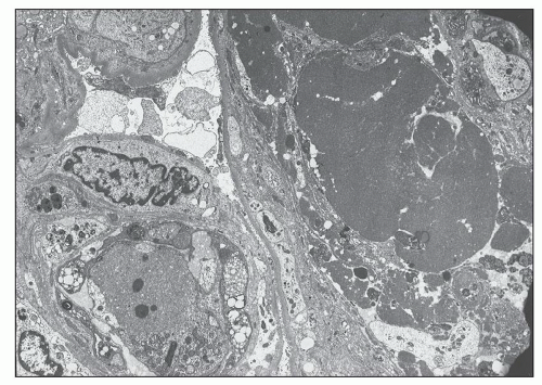
FIGURE 14.33 Lupus nephritis class IV. Electron micrograph shows luminal obliteration by a massive glomerular capillary electron-dense deposit corresponding to a “hyaline thrombus.” (× 2300.)


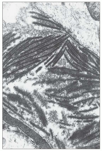
FIGURE 14.34 Lupus nephritis class IV. Electron micrograph shows an organized mesangial electron-dense deposit with tubulofibrillar substructure resembling that seen in cryoglobulinemia. (× 80,000.)

Although this hypothesis has not been studied systematically in a large number of patients, cryoglobulinemia was documented in three of five lupus patients with glomerular fingerprint deposits (164). Several cases of lupus nephritis

with a mixed IgG-IgA-IgM cryoglobulin exhibited the same fingerprint substructures on ultrastructural study of the cryoprecipitate and the glomerular deposits (164,165). Moreover, fingerprint substructures similar to those in SLE have also been described in some cases of monoclonal IgG cryoglobulinemia (166). It appears that the fingerprint deposits are not specific for SLE but reflect the composition of the associated immune deposits.


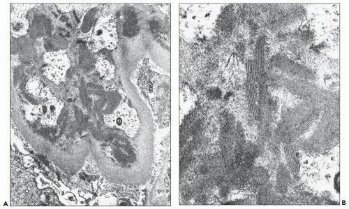
FIGURE 14.35 Lupus nephritis class IV. A large subendothelial deposit displays an organized substructure composed of parallel linear arrays resembling those seen in some forms of cryoglobulinemia. (Electron micrographs; A, × 10,000; B, × 30,000.)


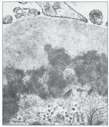
FIGURE 14.36 A subendothelial electron-dense deposit displays a fingerprint-whorled, lamellated substructure. (× 40,000.) (From Churg J, Grishman E. Ultrastructure of immune deposits in renal glomeruli. Ann Intern Med 1972;76:479.)


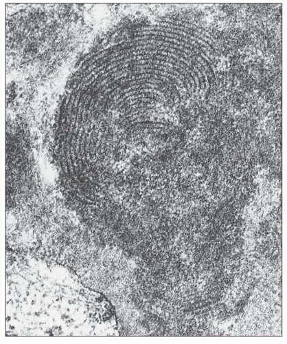
FIGURE 14.37 Electron micrograph of a subendothelial deposit with a fingerprint substructure shows it to be composed of curvilinear, concentric, membranous structures with fine cross-hatching. The remainder of the deposit has a granular texture. (× 100,000.)


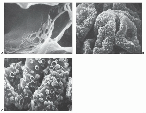
FIGURE 14.38 A: SEM from a patient with the membranoproliferative variant of lupus nephritis. Notice the web-like network on the inner aspect of capillary basement membrane, which probably represents subendothelial extension of mesangial matrix. (× 5500.) B: SEM of a glomerulus from a patient with membranous lupus nephritis shows discrete crater-like deformities, some containing “balls” of immune complex material. (× 687.) C: Higher-power view of the glomerulus in (B) shows crater-like deformities and immune complex-like balls in greater detail. (× 5500.) (From Weidner N, Lorentz WB. Scanning electron microscopy of the acellular glomerular and tubular basement membrane in lupus nephritis. Am J Clin Pathol 1986;85:135.)

Schwartz et al. (167) pointed out that the subepithelial deposits seen in diffuse proliferative lupus nephritis are often different from those in membranous lupus nephritis. In the membranous form, the subepithelial deposits tend to be more uniform in size, consistency, and distribution. In contrast, they are often irregularly distributed and heterogeneous in size in the diffuse proliferative form, in which one loop may have many small subepithelial deposits and an adjacent loop may have none at all. Moreover, in lupus nephritis, it is common for some subepithelial deposits to traverse the full thickness of the GBM in continuity with underlying subendothelial deposits, a phenomenon not observed in primary membranous glomerulonephritis. Fingerprint substructure may also be identified in the subepithelial deposits of lupus nephritis but not in primary membranous glomerulonephritis.

The use of scanning electron microscopy has illustrated the diversity and complexity of the GBM alterations in lupus nephritis. Using a technique to extract the cells and deposits from the GBMs, Weidner and Lorentz (168) examined the naked GBM in several classes of lupus nephritis. In focal and diffuse proliferative lupus nephritis they found small craters on the GBM, sometimes in clusters corresponding to the sites of small subepithelial and incorporated intramembranous deposits. In some cases with membranoproliferative features, extensive web-like arrays of basement membrane were found on the inner aspect of the capillary walls, corresponding to sites of GBM remodeling and subendothelial extension of mesangium into the peripheral capillaries (Fig. 14.38). The most dramatic alterations of GBM texture were identified in the forms of membranous lupus nephritis. Depending on the stage of development, the membranous alterations produced fields of crater-like pitting of the GBM or nodular plaques consisting of incorporated deposits (see Fig. 14.38). More advanced cases exhibited a complex moth-eaten appearance corresponding to intramembranous deposits with extensive remodeling of the GBM.

The ultrastructural morphogenesis of hematoxylin bodies was elegantly described by Cohen and Zamboni (169) (Fig. 14.39). They consist of two major components of probable nuclear and cytoplasmic origin. The first, which probably is nuclear, occupies a central position and ranges from irregular aggregates of marginated and coarsely clumped chromatin to more spheroid masses of moderately electron-dense, finely granular, or amorphous material. These masses are sometimes lobulated, suggesting a possible neutrophilic origin. This central
component is partly or completely enveloped by membranes. The second component, presumed to be cytoplasmic in origin, consists of aggregates of vesicles, vacuoles, glycogen granules, and other spheroidal or rod-shaped granules, some of which exhibit swollen cristae identifiable as degenerating mitochondria. In some cases, the characteristics of the granules are identical to those of specific granules of neutrophils. These cytoplasmic components are surrounded by a continuous or fragmented membrane, which probably represents the original plasma membrane. These structures are enclosed within large phagocytic vacuoles of cells that appear to be mesangial or monocytic in origin. Adjacent mesangial deposits are often identified surrounding the phagocytic mesangial cell. The morphologic appearance of these structures is similar to the description of the LE cell by Maldonado et al. (170). Grishman and Churg (171) published electron microscopic studies of hematoxylin bodies that occurred in arterial walls in the absence of inflammation.


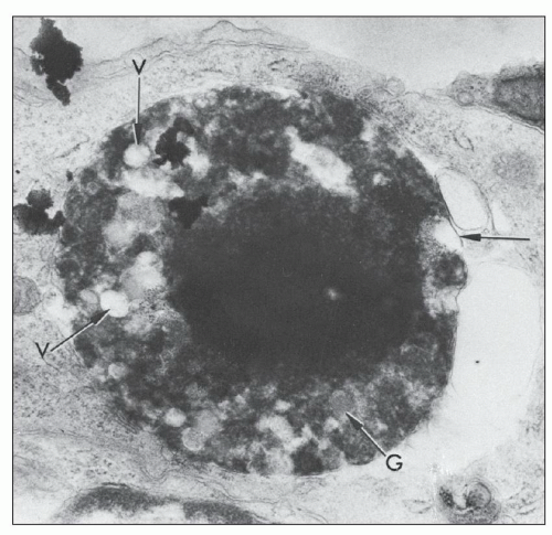
FIGURE 14.39 Lupus nephritis class IV. Electron micrograph of a hematoxylin body with a densely rounded central core of altered nuclear material and surrounding cytoplasmic elements consisting of degenerating organelles, all contained within a discontinuous membrane (arrows). V, vacuoles; G, granules. (× 21,402.) (From Cohen AH, Zamboni L. Ultrastructural appearance and morphogenesis of renal glomerular hematoxylin bodies. Am J Pathol 1977;89:105.)

Another common and characteristic ultrastructural finding in lupus nephritis is intracellular tubuloreticular inclusions (TRIs) (172) (Fig. 14.40). These structures are most often identified in glomerular endothelial cells, in which they may reach large size and commonly number several per glomerulus. They are also readily detected in the endothelium of interstitial capillaries and arteries of the kidney. Rarely can they be identified in glomerular epithelial or mesangial cells. TRI also occur in affected and normal skin in chronic discoid and systemic lupus, in synovium, and in lymphocytes (173,174,175). These structures were first reported in the kidney biopsy of patients with SLE by Gyorkey et al. (176) in 1969 and consist of 23-nm interanastamosing tubular structures measuring 8 nm in their inner diameter and up to 100 nm long. They are always located in dilated cisternae of endoplasmic reticulum. When first reported, their resemblance to myxovirus particles suggested that they might represent viral particles (176,177). However, Schaff et al. (178) demonstrated that the inclusions were not digestible by ribonuclease or deoxyribonuclease and had chemical properties suggestive of phospholipid and acid glycoproteins, a composition not consistent with actual virions. These structures are inducible in normal lymphocytes on exposure to interferon-α in vitro, earning them the eponym interferon footprints (179).


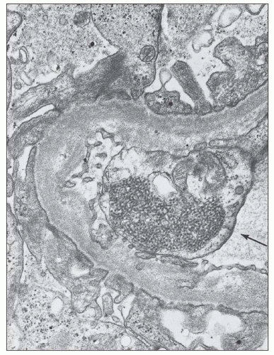
FIGURE 14.40 The electron micrograph shows TRIs within the endothelial cell (arrow) of a glomerular capillary and scattered, subepithelial, electron-dense deposits. (× 14,000.) (Courtesy of Dr. Antonovych, Armed Forces Institute of Pathology, Washington, DC.)

The precise cellular function of these structures remains unknown. Although they are identifiable in most cases of lupus nephritis, TRI are not specific for SLE (180). They are common in patients with human immunodeficiency virus (HIV) infection and to a lesser extent other collagen-vascular diseases (181). They may even be identified in a few healthy individuals. In some patients with apparent primary membranous glomerulonephritis, the finding of TRI may presage development of overt SLE months to years later (182,183,184).

In cases with significant glomerular proteinuria (most common in class V but also observable in class IV and class III nephritis), the podocytes display a variety of cytoplasmic
alterations that are common to many other glomerular diseases manifesting altered glomerular permeability. These ultrastructural alterations include variable foot process effacement, condensation of cytoskeletal microfilaments, microvillous transformation, and cellular hypertrophy with increased organelles, including endoplasmic reticulum, mitochondria, and membrane bound vesicles, some of which contain electron-dense material suggestive of resorbed proteins.


Classification of Lupus Nephritis


Historical Perspective

Because of the wide variety of renal manifestations of SLE, several classifications of lupus nephritis have been proposed. The first to gain wide acceptance were those of Pollak et al. (185) in Chicago and the New York group of Baldwin et al. (186). These early classifications were based entirely on the light microscopic appearance of the glomeruli and essentially recognized three major categories: focal proliferative lupus nephritis (i.e., lupus glomerulitis), diffuse proliferative lupus nephritis (i.e., active lupus glomerulonephritis), and membranous glomerulonephritis. These seminal classification systems of the 1960s failed to acknowledge the existence of milder mesangial forms of the disease because the existence of the mesangial cell was not yet fully appreciated. It was not until 1977 that Baldwin et al. (93) added a fourth category of mesangial lupus nephritis. The subclassification of mesangial lupus nephritis into forms with mesangial hypercellularity and mesangial deposits (IIb) compared with those with mesangial deposits but lacking mesangial hypercellularity (IIa) was solidified by Appel et al. (18) in their 1978 study of the natural history and biopsy features of a large population of patients with lupus nephritis.

The original WHO classification, which arose from a meeting of renal pathologists and nephrologists in Buffalo in 1974, was first published in preliminary form in 1975 by McCluskey (17), with further refinements in 1978 (18) (see Table 14.1). It was further modified by the Pathology Advisory Group of the ISKDC during a meeting in Paris in 1980 to define a number of subclasses (see Table 14.2). This modified classification, published in 1982, was the first to receive the formal imprimatur of the WHO (19). The use of numerous subcategories and the handling of mixed classes made this classification cumbersome to use. Most controversial was the subdivision of the membranous category into designations Va through Vd. Many renal pathologists preferred to treat lesions in class Vc as mixed classes III and V lupus nephritis and those in class Vd as mixed classes IV and V lupus nephritis. This bias is justified by the ample clinical evidence that patients in class Vc and Vd do not behave as membranous disease but have a poor prognosis even worse than that of uncomplicated diffuse proliferative lupus nephritis (187). Accordingly, subgroups Vc and Vd were deleted from the 1995 modified classification (20).


TABLE 14.7 Frequencies of histologic patterns of lupus nephritis (ISN/RPS classification)


Histologic type (%)


Study


Number of patients


Class I


Class II


Class III


Class IV


Class IV-S


Class IV-G


Class V


Class VI


Markowitz (234)


640


< 1%


7.8%


23.7%


53.1%


15%


< 1%


Kojo (221)


99


3%


13%


9%


66%


20%


46%


8%


1%


Yokoyama (215)


60


15%


17%


13%


38%


10%


28%


17%


Seshan (193)


541


1%


10%


20%


37%


16%


21%


29%


3%


The ISN/RPS 2003 WHO classification revisited was formulated by a group of 23 individuals including renal pathologists, nephrologists, and rheumatologists at Columbia University in May 2002 (see Fig. 14.1 and Table 14.3) (21,22). The meeting was spurred by a universally perceived need to reexamine the existing classifications, eliminate ambiguities, clarify and standardize definitions, and facilitate uniformity in reporting. The 2003 classification has the advantage of defining very precisely the distinctions between each class and the threshold for the diagnosis of each. Features of activity and chronicity are clearly delineated. In many ways, it is a return to the simplicity of the original WHO classification, while incorporating modern refinements in our understanding of activity and chronicity. Inconsistencies, such as the designation of a normal biopsy as a form of class I lupus nephritis, were eliminated. Mesangial lesions are clearly separated into class I and class II depending on the absence or presence of histologically identifiable mesangial expansion. Classes III and IV are separated unequivocably based on the percentage of glomeruli affected by active and chronic lesions. The classification requires that sclerotic glomeruli representing scarred lesions of lupus nephritis be factored into the total number of glomeruli affected. Probably the most controversial aspect is the introduction of a subdivision of class IV based on whether the lesions are predominantly segmental or global, which was designed to test the significance of this differentiation. The threshold for the diagnosis of membranous class V as an isolated or additional class (superimposed on a proliferative class) is clearly defined. And finally, the threshold for a diagnosis of class VI is delineated with the qualification that there should be no residual activity. The classification requires that the diagnostic line include entries for the attendant tubulointerstitial and vascular lesions.

Table 14.7 details the incidence of the various classes of lupus nephritis in renal biopsy specimens using the ISN/RPS classification in several large series, comprising 1341 subjects in total. The average frequencies were class I, 1.3%; class II, 9.3%; class III, 20.6%; class IV, 46.6%; class V, 20.3%; and class VI, 1.6%. Although there is some variability between series, the diffuse proliferative category is the most frequent, accounting for 37% to 66% of patients who have had biopsies. Membranous lesions are less common, averaging 20.3% (range, 8% to 29%).


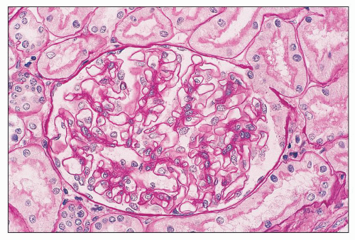
FIGURE 14.41 Lupus nephritis class I. The glomerulus is normal in cellularity, and the GBMs are unremarkable. (PAS; × 500.)


Pathologic Findings by ISN/RPS Classification With Clinical Correlates


CLASS I: MINIMAL MESANGIAL LUPUS NEPHRITIS

According to the ISN/RPS classification, class I denotes normal glomeruli by light microscopy with mesangial immune deposits detected by immunofluorescence and/or electron microscopy. This is the mildest glomerular lesion in lupus nephritis. In the original WHO classification, class I had been defined as a normal renal biopsy from a patient who fulfills ACR criteria for SLE. There are few examples of class I lupus nephritis so defined because these patients generally have no clinical renal abnormalities and are not referred to a nephrologist for biopsy. Because normal biopsies in SLE are so rare and to classify them as a form of “lupus nephritis” is a contradiction in terms, this normal category was eliminated from the ISN/RPS classification and replaced with the original WHO class IIa.

There are no glomerular histologic abnormalities, and the glomerular tuft is normocellular (Fig. 14.41). By immunofluorescence, there are immune deposits limited to the mesangium (Fig. 14.42). The mesangial deposits tend to be small and vary from segmental to global in distribution. By electron microscopy, small electron-dense deposits are present in the mesangium (Fig. 14.43). No electron-dense deposits are identified involving the peripheral glomerular capillary walls.


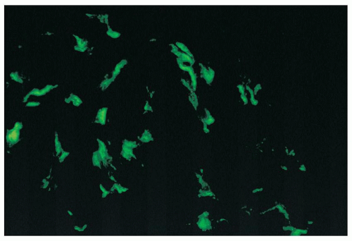
FIGURE 14.42 Lupus nephritis class I. Immunofluorescence microscopy shows delicate mesangial positivity for immunoglobulin G, although no abnormalities were seen by light microscopy. (× 600.)

Patients with class I lupus nephritis usually have minimal urinary findings of microhematuria or mild subnephrotic proteinuria. Renal function is normal. Despite the very mild clinical renal abnormalities, the systemic manifestations of lupus and lupus serologies may be active.


CLASS II: MESANGIAL PROLIFERATIVE LUPUS NEPHRITIS

According to the ISN/RPS schema, class II is defined as purely mesangial hypercellularity of any degree and/or mesangial matrix expansion by LM with mesangial immune deposits. There may be rare isolated minute subepithelial or subendothelial deposits by immunofluorescence or electron microscopy that are not visible by light microscopy. In the original (1974) WHO classification, class II had been subdivided into class IIa and IIb according to the absence or presence of mesangial hypercellularity, respectively. In the ISN/RPS classification, the original WHO class IIa has become class I and the original WHO class IIb has become class II.


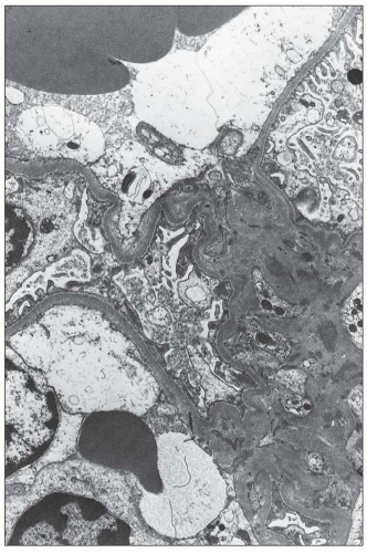
FIGURE 14.43 Lupus nephritis class I. The electron micrograph shows small, mesangial, electron-dense deposits. No deposits involve the peripheral GBMs, and foot processes are intact. (× 2500.)


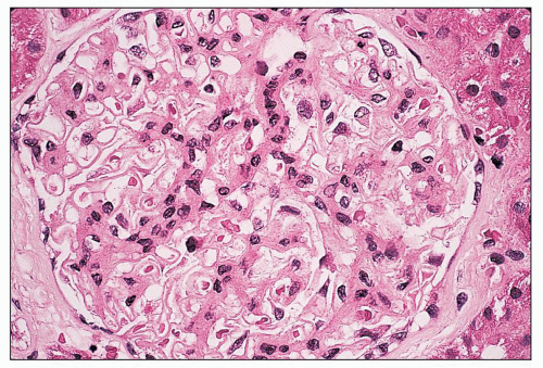
FIGURE 14.44 Lupus nephritis class II. There is mild, global, mesangial hypercellularity with thin capillary loops. (H&E, × 500.)

By light microscopy, there is mesangial proliferation of any severity (Fig. 14.44). Mesangial hypercellularity is defined as ≥3 mesangial cells in mesangial areas away from the vascular pole, assessed in 3-µm-thick histologic sections. The mesangial proliferation is usually mild to moderate and does not significantly compromise the glomerular capillary lumen. It may vary in distribution from focal to diffuse and may involve the glomerular tuft segmentally or globally. Variable increase in mesangial matrix may accompany the mesangial hypercellularity. Usually, the mesangial deposits are not large enough to be identified by light microscopy. In some cases, however, large mesangial deposits expand the mesangium and impart a glassy, hypereosinophilic appearance to the mesangial matrix. Masson trichrome stain may reveal large mesangial immune deposits as fuchsinophilic (red) zones in the blue/green mesangial matrix.

Cases of lupus nephritis with severe, but purely mesangial, hypercellularity, without obliteration of the capillary lumina, may pose difficulties in classification. Proper classification requires careful assessment of the immunofluorescence and electron microscopic findings. If the immune deposits are limited to the mesangium, even cases of severe diffuse mesangial proliferation should be classified as class II. If significant subendothelial deposits are present by immunofluorescence and/or electron microscopy, or if subendothelial deposits are visible by light microscopy, the case should be classified as focal proliferative if the subendothelial deposits are focally distributed and diffuse proliferative if diffusely distributed. Unfortunately, some early series included examples of segmental endocapillary extension of the mesangial proliferation (148,149) or subendothelial extension of paramesangial deposits visible by light microscopy (188) under the rubric of class II nephritis. In modern series, such cases would more accurately be considered examples of mild or early class III.

Immunofluorescence microscopy typically reveals immune deposits of IgG (18,148,150) and more variably IgM and IgA (93,149), as well as C3 and C1q outlining the mesangial axis, with sparing of the peripheral glomerular capillary wall (Fig. 14.45). These mesangial deposits may be sparse and finely granular or large and globular. Usually, the pattern of deposition by immunofluorescence is more diffuse and regular than the distribution of mesangial hypercellularity seen by light microscopy. Glomerular staining for fibrinogen is usually negative.


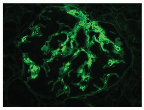
FIGURE 14.45 Lupus nephritis class II. Immunofluorescence microscopy shows deposits of IgG confined to the mesangium. (× 500.)

As determined by electron microscopy, the mesangial deposits range from small to large and may be segmental or global in distribution (Fig. 14.46). In mild cases, they are often confined to the paramesangial region, subjacent to the GBM reflection. When the mesangial deposits are more abundant, they are widely distributed throughout the full thickness of the mesangial matrix.

Most examples of class II lupus nephritis have exclusively mesangial deposits. However, in practice, some cases of purely mesangial proliferative lupus nephritis will manifest rare small subendothelial electron-dense deposits, especially as extensions from the paramesangial region. These cases raise the question of unsampled focal proliferative (class III) lupus nephritis or at least the possibility of imminent transformation to class III or class IV. Such nonconforming cases are
problematic and were not adequately addressed by the original WHO classification system. According to the ISN/RPS classification, cases of mesangial proliferative lupus nephritis with rare minute subendothelial or subepithelial deposits (visible only by IF and/or EM) should be classified as class II (21,22). Most practicing renal pathologists deal with these atypical features by indicating in a note that the rare small subendothelial or subepithelial deposits observed in a minority of capillaries suggest the possibility that the glomerulonephritis may evolve into focal proliferative or membranous glomerulonephritis in the near future and that the patient should be carefully monitored. However, the presence of any sizeable (visible by LM) or numerous subendothelial or subepithelial deposits in an otherwise mesangial proliferative glomerulonephritis exceeds what is acceptable in pure class II disease and warrants a designation of class III, IV, or V depending on their distribution and quantity.


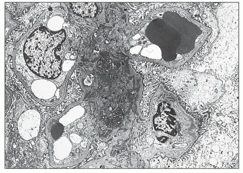
FIGURE 14.46 Lupus nephritis class II. The electron micrograph shows mesangial electron-dense deposits accompanied by mild mesangial hypercellularity. There are no deposits involving the peripheral glomerular capillary walls. (× 2000.)

Tubular, interstitial, and vascular lesions are typically minimal in class II nephritis. The presence of significant tubulointerstitial or vascular disease should raise the question of a superimposed process, such as interstitial nephritis, hypertensive arterionephrosclerosis, or thrombotic microangiopathy secondary to lupus anticoagulant syndrome or associated TTP-like syndrome.

The clinical renal manifestations of class II lupus nephritis are mild. Fewer than 50% of patients have mild hematuria or proteinuria, which generally does not exceed 1 g in 24 hours (18,149,150,189). The nephrotic syndrome is virtually never observed (unless there is superimposed podocytopathy resembling minimal change disease). Renal insufficiency is uncommon, and mildly reduced creatinine clearance can be demonstrated in less than 15%. Despite the relatively mild glomerulonephritis, serologic tests for SLE may be strongly positive. For example, in two combined series, anti-DNA antibody was detectable in 22 of 28 patients (148,149), with reduced C3 in 6 of the same 28 patients (148,149). Cameron reported C4 to be more regularly reduced than C3 in patients with class II nephritis. Some refinement of these data is available in series in which the mesangial lesions are subclassed as those with and without mesangial proliferation (original WHO class IIb vs. IIa) (18,149,189). Patients with original WHO class IIa nephritis are more likely to have totally normal renal function without proteinuria or abnormal urinary sediment than those with class IIb.


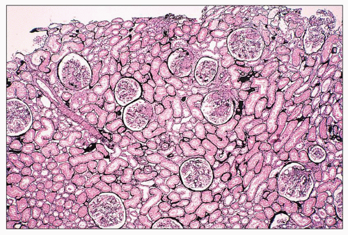
FIGURE 14.47 Lupus nephritis class III. A low-power view shows the focal and segmental distribution of the endocapillary proliferation, with some overlying crescents. Endocapillary proliferation affected less than 50% of the total glomeruli in this biopsy. (Jones methenamine silver stain; × 40.)

Mesangial lupus nephritis usually has an excellent prognosis, with a 5-year renal survival rate of greater than 90%. Although in many patients it is a stable lesion that may persist for years without change in clinical renal findings, in other patients it may undergo transformation to a more ominous class, which is often heralded by a change in the level of proteinuria, development of a more active urinary sediment, or reduction in renal function. Transformations to diffuse proliferative glomerulonephritis (93,149,162,190,191,192,193) are the most common, followed by transformations to focal proliferative or membranous glomerulonephritis (18,149,193). In some patients, a mesangial proliferative glomerulonephritis may represent a phase in the regression of focal or diffuse proliferative lesions, especially after treatment (193).


CLASS III: FOCAL LUPUS NEPHRITIS

According to the ISN/RPS and WHO classifications, class III lupus nephritis consists of a focal segmental or global endocapillary or extracapillary glomerulonephritis that affects less than 50% of glomeruli (Fig. 14.47). The endocapillary proliferation is usually focal and segmental. There are typically focal subendothelial immune deposits, with or without mesangial alterations. Lesions may be active or chronic. In determining the percentage of glomeruli affected, both active and chronic lesions must be taken into account. Although most active glomerular lesions are endocapillary proliferative in nature, class III includes glomerular lesions that are membranoproliferative pattern, extracapillary proliferative, or consist of wire-loop deposits without associated proliferation. For this reason, the ISN/RPS classification prefers the broader term “focal lupus nephritis” to the more restrictive term “focal proliferative lupus nephritis” used in the original WHO classification.

The endocapillary proliferative lesions may be an isolated finding. More typically, they occur on a background of mesangial hypercellularity that may be focal or diffuse (Fig. 14.48). Correspondingly, all glomeruli are involved, as detected by immunofluorescence, with generalized mesangial immune deposits, regardless of the focal endocapillary proliferative
pattern by light microscopy. The segments with endocapillary hypercellularity exhibit severe narrowing or occlusion of the glomerular capillaries by proliferation of endothelial and mesangial cells with a variable infiltration of mononuclear and polymorphonuclear leukocytes. This may be the only histologic finding in mild cases. More active examples may demonstrate any or all of the histologic features of activity previously discussed in a focal and segmental distribution. These include fibrinoid necrosis, karyorrhexis or pyknosis, rupture of the GBM, wire-loop deposits, intraluminal hyaline thrombi, and overlying cellular crescents (Fig. 14.49). Hematoxylin bodies are sometimes identified in the necrotizing lesions. Typically, foci of focal necrosis heal by progression to segmental or global scars with associated fibrous crescents or small synechiae to the Bowman capsule. Glomerular capillary walls in the “uninvolved” segments are usually thin and delicate.


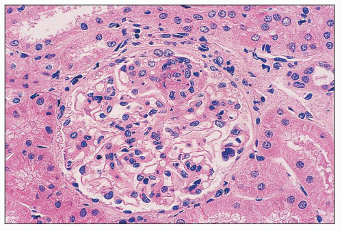
FIGURE 14.48 Lupus nephritis class III. There is segmental obliteration of glomerular capillary lumina by endocapillary proliferation, including infiltrating leukocytes, with associated fibrinoid necrosis. The adjacent lobules display mild mesangial hypercellularity. (H&E; × 400.)

Class III biopsy specimens often display focal tubulointerstitial disease, including patchy interstitial edema, inflammation, and tubular atrophy. In the chronic phase, tubular atrophy and interstitial fibrosis are more pronounced and usually parallel the distribution of the glomerulosclerotic lesions. Such evolution may be seen on repeat biopsy, during the natural course of disease, or after treatment. In many cases, active and chronic lesions may coexist, especially in chronic cases with recent clinical reactivation.


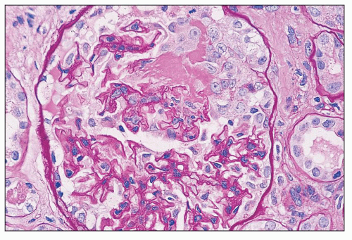
FIGURE 14.49 Lupus nephritis class III. There is segmental endocapillary proliferation and necrosis, with focal rupture of GBMs and an adjacent cellular crescent. (PAS, × 500.)


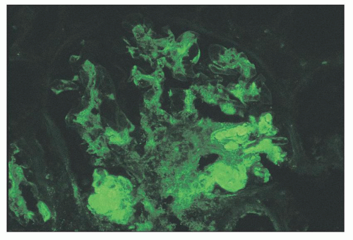
FIGURE 14.50 Lupus nephritis class III. The immunofluorescence micrograph shows segmental heavy IgG deposits in several glomerular capillary walls and lumina. The adjacent lobules have mesangial deposits. (× 500.)

Immunofluorescence microscopy reveals diffuse and global mesangial deposition in all glomeruli with more focal and segmental subendothelial capillary wall deposits, a pattern that mirrors the distribution of glomerular proliferative lesions by light microscopy (Fig. 14.50). The subendothelial deposits often exhibit a semilinear, comma-shaped appearance with smooth outer contour owing to conformity to the delimiting GBM (Fig. 14.51). Small numbers of subepithelial deposits may appear as more granular, rounded deposits. IgG, IgM, and IgA may be present (18,93,194), but IgG is the most constant and usually the most intense. C3 and C1q are also commonly seen. Strong staining for fibrin is identifiable in the necrotizing lesions and crescents. Immune deposits are frequently identifiable in the tubulointerstitial compartment and arterial walls.


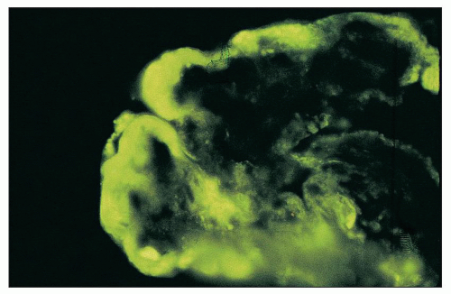
FIGURE 14.51 Wire-loop deposit. By immunofluorescence, there is a large subendothelial deposit that conforms to the contour of the GBM, producing a smooth comma-shaped outer contour. (× 1000.)


By electron microscopy, mesangial deposits are readily identifiable. Segmental subendothelial deposits are usually demonstrable in the glomeruli with segmental endocapillary proliferative lesions, but they may be absent if the more severely affected glomeruli are not sampled in the tissue processed for electron microscopy. Scattered subepithelial deposits are also frequently seen, often in an irregular distribution. Although there is some disagreement on this point (191), some researchers have suggested that class III biopsies with substantial capillary wall subendothelial or subepithelial deposits are often predictive of future transformation to diffuse proliferative or membranous glomerulonephritis, respectively (192,195).

Some investigators have called attention to a subgroup of class III lupus nephritis with extensive segmental necrosis and crescents but with little or no identifiable subendothelial deposits (196,197,198,199). It has been suggested that the glomerular immune complex load is insufficient to account for the severe active lesions, raising the possibility that they may have a natural history and pathogenesis akin to vasculitis and corresponding pauci-immune focal necrotizing and crescentic glomerulonephritis. Circulating ANCA have been identified in some of these patients (198), although ANCA serologies have not been investigated in any systematic way.

Class III lupus nephritis has a heterogeneous clinical picture (200). About 50% of patients have an active urinary sediment (e.g., hematuria, leukocyturia, cellular casts), and 25% to 50% have proteinuria, which may be accompanied by the nephrotic syndrome in up to one third of patients. Renal insufficiency, however, is uncommon, affecting only 10% to 25% of patients. Hypertension occurs in up to one third of patients, initially or over the course of follow-up (18,93,148). Serologic abnormalities are common in this class, with anti-DNA antibody and reduced complements detected in more than one half of patients (18,148,149).

The course and prognosis of class III lupus nephritis are variable. Initial experience suggested a favorable picture with no histologic progression and little renal functional deterioration (185,186,201,202,203). However, many subsequent studies have reported a 5-year renal survival rate of 85% to 90%, indicating progression to severe, irreversible renal damage in a small percentage of patients (204,205). Repeat renal biopsies have indicated that this poor outcome usually results from progression from class III to class IV, which many consider movement along a disease continuum rather than a real change in the quality of the glomerular lesions. Mahajan et al. (195) and Zimmerman et al. (192) have emphasized that patients with disease that progresses to diffuse proliferative glomerulonephritis often have a higher initial level of proteinuria than those whose disease remains stable. Transformations from class III to class V (membranous) have also been described. These transformations often are heralded by an abrupt increase in proteinuria, sometimes with the development of the nephrotic syndrome.


CLASS IV: DIFFUSE LUPUS NEPHRITIS

Class IV denotes diffuse segmental or global endocapillary or extracapillary glomerulonephritis involving ≥50% of glomeruli. Typically, there are diffuse subendothelial immune deposits, with or without mesangial alterations. Lesions may be active or inactive (sclerosing), and both types of lesions should be accounted for when determining the percentage of total glomeruli affected by glomerulonephritis. As for class III, class IV includes glomerular lesions that are membranoproliferative pattern, extracapillary proliferative, or consist of wire-loop deposits without proliferation, thereby justifying “diffuse lupus nephritis” as the preferred designation over the older term “diffuse proliferative lupus nephritis” used in the original WHO classification. The ISN/RPS classification subdivides class IV into those cases with diffuse segmental (IV-S) versus diffuse global (IV-G) distribution (Figs. 14.52 and 14.53). The designation diffuse segmental (IV-S) is used if greater than 50% of the involved glomeruli have segmental lesions; similarly, diffuse global (IV-G) applies if greater than 50% of the involved glomeruli have global lesions.


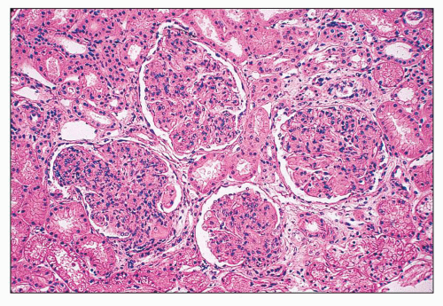
FIGURE 14.52 Lupus nephritis class IV-G. There is diffuse and global endocapillary proliferation involving all the glomeruli in this biopsy. (H&E, × 180).

Many investigators consider class III and IV lupus nephritis as ends of a pathologic continuum, such that the two classes differ from each other quantitatively but not qualitatively. However, as discussed in detail later in this chapter, some investigators propose that class IV-S is pathogenetically distinct from lupus nephritis that has predominantly endocapillary
hypercellularity rather than predominantly segmental necrosis. All the lesions of active glomerular disease described for class III (e.g., nature of the endocapillary cells, fibrinoid necrosis, karyorrhexis and pyknosis, neutrophil infiltration, wire-loop deposits, hyaline thrombi, hematoxylin bodies, crescents) also apply to class IV. The two classes are distinguished by definition based on the percentage of glomeruli affected.


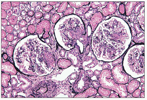
FIGURE 14.53 Lupus nephritis class IV-S. There is diffuse segmental glomerular proliferation involving more than 50% of the total glomeruli in this biopsy. (Jones methenamine silver, × 180.)


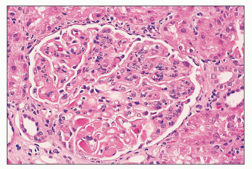
FIGURE 14.54 Lupus nephritis class IV. There is global narrowing of glomerular capillaries by mesangial and endocapillary proliferation. Wireloop deposits and hyaline thrombi are segmentally distributed. (H&E; × 500.)

In most examples of class IV lupus nephritis, especially class IV-G, the lesions tend to be diffuse and global, although segmental lesions may affect some glomeruli (Figs. 14.54 and 14.55). Occasionally, several glomeruli with only mesangial proliferative features among many other severely affected glomeruli are found. Typically in class IV-G, the subendothelial and mesangial deposits are larger and more abundant than in class III and class IV-S and usually stain more intensely by immunofluorescence (Figs. 14.56 and 14.57). As in class III, lobules lacking endocapillary proliferation and subendothelial deposits usually display mesangial deposits and some degree of mesangial hypercellularity. In some patients, proliferation is distributed uniformly throughout most of the glomeruli, but in others, there may be considerable variation in the severity of proliferation from one glomerulus to the next or even between adjacent lobules of an individual glomerulus.


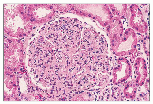
FIGURE 14.55 Lupus nephritis class IV. The endocapillary proliferation is global and includes many infiltrating neutrophils. (H&E, × 500.)


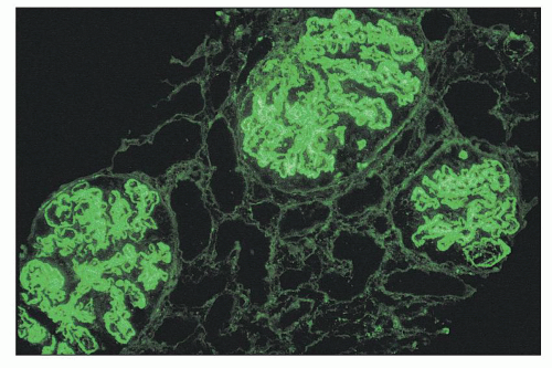
FIGURE 14.56 Lupus nephritis class IV. The low-power immunofluorescence micrograph shows intense, diffuse staining for IgG in the glomerular mesangium and peripheral capillary loops, consistent with a subendothelial distribution. (× 120.)

Schwartz et al. (187) described a category of “severe segmental glomerulonephritis” in which the glomerular inflammation was predominantly diffuse but segmental. This category would now be designated as IV-S in the ISN/RPS classification. This category had an outcome measured in short-term renal survival that was intermediate between the classic focal and diffuse proliferative groups (187).

In class IV, all the histologic features of active lupus nephritis reach their most florid expression. In severe examples, there may be abundant wire-loop deposits and necroses, and it is in class IV that hematoxylin bodies are most likely to be encountered. In addition to classic diffuse endocapillary proliferative glomerulonephritis, the modified WHO classification recognized several morphologic variants of class IV that may
pose difficulty in diagnosis (19,20). These include severe mesangial proliferative glomerulonephritis with diffuse subendothelial deposits and mesangiocapillary (or membranoproliferative) pattern glomerulonephritis and extensive subendothelial deposits with minimal glomerular hypercellularity (Fig. 14.58). Class IV-G disease has diffuse distribution of subendothelial deposits by immunofluorescence and electron microscopy, although the pattern of proliferation that accompanies these deposits varies considerably.


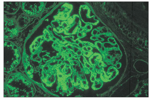
FIGURE 14.57 Lupus nephritis class IV. There are abundant deposits of IgG in the mesangium and the peripheral capillary walls. Most of the glomerular capillary wall deposits appear subendothelial, with more segmental subepithelial deposits. (Immunofluorescence micrograph, × 600.)


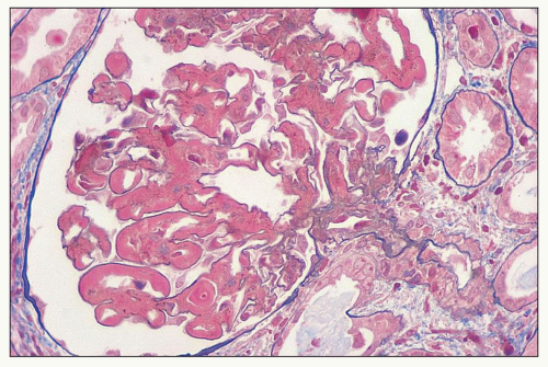
FIGURE 14.58 Lupus nephritis class IV. This example has diffuse wire-loop deposits without appreciable endocapillary proliferation. (Masson trichrome, × 600.)

In the severe mesangial proliferative subgroup, which is uncommon, the light microscopic findings frequently suggest class II, and it is not until immunofluorescence and electron microscopy are performed that the true diagnosis of class IV nephritis is apparent. In the variant with diffuse subendothelial deposits but minimal mesangial or endocapillary hypercellularity, the correct designation as class IV is usually apparent even by light microscopy, because the subendothelial deposits are large enough to be visible as extensive wire loops. Diffuse distribution of subendothelial deposits that can be detected by light microscopy, regardless of the pattern of proliferation, is indicative of class IV lupus nephritis.

The membranoproliferative pattern subgroup of class IV is characterized by widespread circumferential or partial mesangial interposition with double-contoured capillary loops owing to subendothelial neomembrane formation (Fig. 14.59). This pattern typically causes accentuation of the glomerular lobularity that is indistinguishable histologically from primary membranoproliferative glomerulonephritis type 1. If numerous subepithelial deposits are also present, the findings closely resemble membranoproliferative glomerulonephritis type 3 of Burkholder (see Chapter 8). It has been suggested that these membranoproliferative variants of class IV lupus nephritis usually lack necrotizing features (18), but others (149) have not found any difference in the frequency of necroses or crescents in this group compared with other examples of diffuse proliferative lupus nephritis.

Immunofluorescence studies reveal diffuse mesangial and widespread (but more irregular) capillary wall staining of immune reactants in class IV, especially class IV-G (86,149,150,162,194,206,207), although there are less immune reactants in class IV-S (214). The capillary wall staining is predominantly subendothelial, but irregularities in the outer contour of the deposits often attest to the presence of scattered subepithelial deposits as well (Fig. 14.60). If the subepithelial deposits are regular and diffuse in distribution (involving at least 50% of the glomerular capillary surface area of at least 50% of glomeruli), a designation of mixed class IV and V (i.e., diffuse proliferative and membranous) lupus nephritis is warranted (21,22). IgG is universally present, although codeposits of IgM or IgA are commonly but less consistently found. In contrast to IgA nephropathy, IgA may be equal to IgG in intensity but is rarely dominant over IgG in intensity (152). Most commonly, the intensity of IgA is less than that of IgG (149,207). In most cases, both C1q and C3 are codeposited, although the intensity of C1q staining is frequently stronger than C3. Properdin is usually present (154) in the same distribution as the other complement components. Staining for fibrinogen is strong in the distribution of crescents and necrotizing lesions, often obliterating
the tuft architecture (Fig. 14.61). However, more delicate semilinear staining for fibrinogen of about 1+ intensity may be seen more diffusely in areas of nonnecrotizing endocapillary proliferation in active glomerulonephritis.


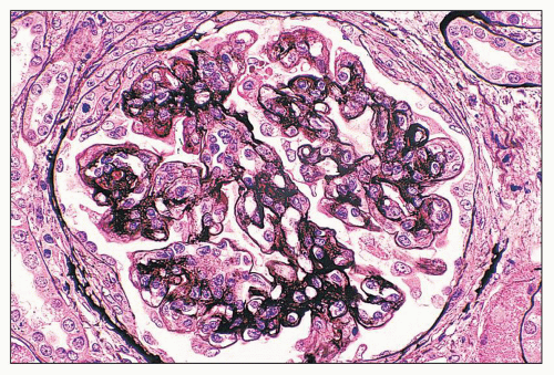
FIGURE 14.59 Lupus nephritis class IV. Membranoproliferative variant with lobular accentuation, nodular mesangial expansion, and numerous double contours of the GBM. (Jones methenamine silver; × 500.)


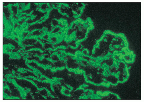
FIGURE 14.60 Lupus nephritis class IV. By immunofluorescence, granular deposits of C1q outline the mesangial areas and capillary loops. The peripheral capillary wall deposits are predominantly subendothelial, with minute, more irregular, subepithelial deposits. (× 700.)


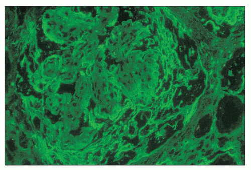
FIGURE 14.61 Lupus nephritis class IV. The immunofluorescence micrograph shows staining for fibrinogen in the periphery of the glomerular capillaries and the overlying crescent. (× 600.)

Electron microscopy confirms and sometimes amplifies the light microscopic and fluorescence microscopic assessment of extensive mesangial and subendothelial deposits, with lesser subepithelial deposits (Fig. 14.62). It is in this form of lupus nephritis that the fingerprint substructure is most commonly identified (208). Fibrin tactoids may be identified in necrotizing lesions (Fig. 14.63).

Vascular lesions occur most frequently in the diffuse proliferative group, although they affect less than 50% of cases. Uncomplicated vascular immune deposits detected by immunofluorescence or electron microscopy are the most common lesion and are usually unaccompanied by detectable vascular changes by light microscopy. These usually consist of granular vascular deposits of IgG, IgM, IgA, C3, and C1q in the subendothelial basement membrane and perimyocyte membranes. Only a subset of patients with severe, active class IV disease has histologically identifiable lupus vasculopathy with obliterative noninflammatory arteriolar lesions containing abundant intimal and intraluminal immune deposits. True arteritis resembling polyarteritis nodosa or microscopic polyangiitis is rare. Thrombotic microangiopathy affecting small arteries, arterioles, and glomerular capillaries may occur in association with a circulating lupus anticoagulant or APL antibody or as a manifestation of TTP-like syndrome.


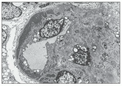
FIGURE 14.62 Lupus nephritis class IV. The electron micrograph shows a circumferential, subendothelial, electron-dense deposit that has been incorporated into the glomerular capillary wall by subendothelial neomembrane formation. There is marked mesangial expansion by mesangial proliferation and increased matrix containing many granular, electron-dense deposits. (× 2875.)

Tubular and interstitial lesions are nearly universal in diffuse proliferative lupus nephritis. These range from immune deposits detectable only by immunofluorescence and electron microscopy in the interstitium and tubular basement membranes to histologically identifiable interstitial inflammation, edema, fibrosis, and tubular atrophy (Fig. 14.64). The activity and chronicity of these lesions usually correlates with the activity and chronicity of the glomerular lesions. These changes reflect secondary atrophy of the dependent nephron following glomerulosclerosis (i.e., process of nephron dropout) as well as active immunologically mediated tubular damage.

Segmental and global glomerulosclerosis are the inevitable consequence of active necrotizing lesions with crescent formation. Glomerulosclerosis occurs at a slower tempo, even in cases without overt necrotizing or crescentic lesions, as evidenced by repeat biopsies performed to monitor treatment response and prognosis. In such cases, repeat biopsy may show regression to a less active mesangial proliferative form with focal segmental and global sclerosis. At this stage, as detected by immunofluorescence and electron microscopy, small residual capillary wall deposits and ultrastructural basement membrane irregularities such as thickening, lamellation, mesangial interposition, and organized, partially resorbed intramembranous deposits usually attest to the previous presence of more active class IV disease.

Patients with class IV lupus nephritis have the most severe and active clinical renal presentation. Not surprisingly, they constitute the largest percentage of patients in most clinical series of severe lupus nephritis based on renal biopsy. Proteinuria is universal, and up to 50% of patients may have the nephrotic syndrome, initially or manifesting later in the course (18,93). Baldwin’s experience (93) is typical in that 41 of 44 patients with diffuse proliferative lupus nephritis had nephrotic proteinuria, in 26 as an initial finding, and in 15 during the course of the renal disease. Hematuria occurs to variable degrees in 80% to 90% of patients (18,93,148), with frequent detection of red blood cell casts and associated leukocyturia in very active cases. Between 30% and 40% of patients has hypertension at disease outset (93,148). Renal insufficiency of various degrees of severity is detected in greater than 50% of patients by determinations of the GFR, although serum creatinine levels may be in the normal range. The serum creatinine levels are typically low in young women with little muscle mass and are a less sensitive marker of renal function than the GFR. Serologic test results for lupus indicate active disease in 50% to 90% of patients (148,149). ANA is detectable in greater than 98%, but anti-dsDNA antibody is less often detectable. Serum levels of complements C3 and C4 are reduced in approximately two thirds of cases (148). Complement levels may normalize with
steroid therapy, even when brief and inadequate to control the disease. Thus, the percentage of untreated patients with hypocomplementemia is considerably higher, 87% in one series (149). In untreated patients, the quantity of subendothelial deposits was found to correlate roughly with the reduction in serum C3 levels (209).


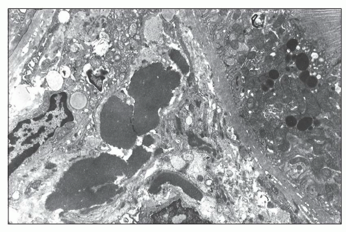
FIGURE 14.63 Lupus nephritis class IV. The electron micrograph from a fibrinoid necrotizing lesion shows endothelial necrosis with subendothelial deposition of abundant fibrillar fibrin and scant granular, electron-dense immune deposits. Neutrophils and monocytes infiltrate the glomerular capillary lumen. (× 2200.)

The series (18,149,150) that separate the membranoproliferative pattern from other forms of diffuse proliferative lupus nephritis are in agreement that the level of proteinuria is greater in the membranoproliferative group. In the series of Sinniah and Feng (150), patients with a membranoproliferative pattern had a higher incidence of the nephrotic syndrome (11 of 19 patients) than those with a usual diffuse proliferative pattern (2 of 15 patients). Appel et al. (18) found that patients with the membranoproliferative variant tend to be persistently hypocomplementemic throughout their course, whereas those with diffuse proliferative lesions of the usual type frequently experience a normalization of the serum complement during therapy. Hill et al. (34) found that patients with a membranoproliferative pattern on first biopsy tended to respond to therapy; however, the persistence of this pattern or its new appearance on repeat biopsy 6 months following therapy was associated with a poor outcome.


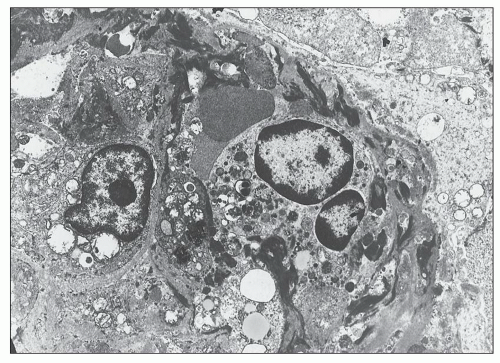
FIGURE 14.64 Lupus nephritis class IV. Massive electron-dense deposits are present in the interstitial collagen adjacentto a tubule. (× 2000.)

The category IV-S was introduced because of evidence from the Chicago group that this subgroup has worse longterm outcome than IV-G, suggesting important prognostic differences (210,211). Renal survival at 10 years was 75% for IV-G (n = 35) compared to 52% for IV-S (n = 24), which the authors refer to as class III ≥50% (211). During the course of follow-up, 60% of patients with IV-G entered remission compared to only 38% of patients with IV-S (211). The authors proposed that the diffuse segmental lesion, particularly one with necrotizing features unaccompanied by endocapillary proliferation, is a particularly ominous histologic form and may involve pathogenetic mechanisms similar to those in pauci-immune necrotizing glomerulonephritis and vasculitis. Roles for ANCA, antiendothelial antibody, and anticardiolipin antibody have been proposed, but none of these has been studied systematically. The ISN/RPS classification has been used by a number of groups to test the significance of class IV-S versus IV-G (Table 14.8 and summarized in Markowitz and D’Agati (212)). By contrast, no significant differences in outcome were demonstrated by the Boston group after an average follow-up period of 38 months for the IV-S group and 55 months for the IV-G group (213). However, there were some interesting differences in presenting clinical and pathologic features. Greater serologic activity (lower C4 level) was observed in the IV-S group, which also had more frequent segmental fibrinoid necroses and crescents, although the latter did not reach statistical significance. In contrast to the observations by the Chicago group (211), the necrosis in IV-S was accompanied by endocapillary proliferation in most glomeruli, arguing against a distinct pathogenetic mechanism of injury (213). The IV-G group had higher presenting serum creatinine levels, diastolic blood pressures, and more frequent wire-loop deposits (213). Transformation from IV-S to IV-G was observed in two of three repeat biopsies from the IV-S group, suggesting that the segmental phenotype is not immutable, but may evolve into
a more global pattern of involvement over time (213). Hill et al. (214) studied 15 French patients with IV-S and 31 with IV-G and found that at baseline, patients with IV-G have more proteinuria, renal insufficiency, anemia, and hypocomplementemia. Morphologic differences include more membranoproliferative features, wire-loop deposits and hyaline thrombi, greater IF positivity involving the peripheral capillary walls, and less fibrinoid necrosis in IV-G than IV-S. Interestingly, repeat biopsies showed both types of interconversion (IV-S to IV-G in three patients and IV-G to IV-S in seven patients). The French group also observed no significant difference in 10-year survival between class IV-S and IV-G lesions at first biopsy (survival rates 65% vs. 60%, respectively). Interestingly, when repeat biopsies were performed 6 months following therapy, second biopsies with IV-G had a worse outcome than those with IV-S (214). A Japanese study also failed to find significant outcome differences in class IV-S versus IV-G (215). A recent meta-analysis (216) has analyzed the results of eight published studies addressing differences in IV-S versus IV-G (213,214,215,217,218,219,220,221) and concluded that there was no significant difference in renal outcome (doubling of serum creatinine or ESRD) between these histologic forms. These data call into question the validity of a classification that subcategorizes class IV based on the proportion of segmental and global lesions. Moreover, the IV-S category is relatively infrequent, comprising a minority of class IV biopsies (13% to 34%; see Table 14.8). Based on these data, future iterations of the ISN/RPS classification may opt to eliminate this distinction.


TABLE 14.8 Comparison of ISN/RPS class IV subgroups


Reference


N


Frequency


Clinical presenting features and pathology


Response to treatment or outcomes


IV-G


IV-S


Mittal (213)


33


66%


33%


IV-G had higher sCR and HTN


IV-S had more fibrinoid necrosis and lower C4


No difference


Yokoyama (215)


23


74%


26%


IV-G had lower CH50 levels


Trend to more ESRD in IV-S (P = 0.1495)


Hill (214)


46


67%


33%


IV-G had more renal insufficiency, lower C3 and CH50, more proteinuria, more immune deposits, less fibrinoid necrosis


No significant difference in 10-year renal survival


Kim (1073)


42


71%


29%


IV-G had higher proteinuria and lower anti-dsDNA antibody titers


IV-S had higher complete response rates to cyclophosphamide (67% vs. 33%)


Hiramitsu (219)


55


75%


25%


IV-G had more nephrotic syndrome (81% vs. 43%)


Trend to worse outcomes in IV-G, P = 0.68 (related to chronicity)


Kojo (221)


65


69%


31%


IV-G had more endocapillary proliferation and wire-loop deposits


Trend to worse outcomes in IV-G, P = 0.433


Grootscholten (218)


72


79%


21%


IV-G had more HTN, lower C3 and C4, higher sCR


No difference


Yu (220)


172


87%


13%


IV-S had less proteinuria, lower sCR, higher C3 more ACL, more ANCA, less anti-C1q, more fibrinoid necrosis


No difference


Schwartz (217)


93


66%


34%


Not examined


No difference


N, number of patients; sCR, serum creatinine; HTN, hypertension; ESRD, end-stage renal disease.


Some patients with class IV lupus nephritis have normal renal function and inactive urinary sediment in the face of biopsy findings of active glomerular lesions, as part of the spectrum of “silent lupus nephritis” (discussed in Other Renal Manifestations of SLE below) (222,223,224,225,226).


CLASS V: MEMBRANOUS LUPUS NEPHRITIS

Class V designates membranous lupus nephritis, which is defined by subepithelial immune deposits or their morphologic sequelae as seen in the various stages of primary membranous glomerulonephritis. According to the INS/RPS classification, a diagnosis of class V is based on the presence of global or segmental continuous granular subepithelial immune deposits (21,22). The membranous alterations may be present alone or on a background of mesangial hypercellularity and mesangial immune deposits. Any degree of mesangial hypercellularity may occur in class V. There may be few small subendothelial immune deposits identified by immunofluorescence and/or electron microscopy, but not by light microscopy. Because scattered subepithelial deposits may also be encountered in class III and class IV lupus nephritis, the threshold for an additional diagnosis of membranous lupus nephritis in a proliferative class is membranous involvement of greater than 50% of the tuft of greater than 50% of the glomeruli by light microscopy or immunofluorescence (21,22).


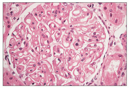
FIGURE 14.65 Lupus nephritis class V (modified Va). There is global thickening of glomerular capillary walls without mesangial proliferation. (H&E, × 500.)

In the modified (1982) WHO classification, membranous lupus nephritis was subdivided into four subclasses, designated Va through Vd (19). It is important to be familiar with these categories because older outcome studies frequently employ these designations. Class Va denotes pure membranous lupus nephritis without associated mesangial proliferation, a glomerular lesion indistinguishable morphologically from primary membranous glomerulonephritis (Fig. 14.65). Class Vb can be considered a form of class Va plus class II in that the lesion manifests the typical peripheral capillary wall features of membranous glomerulonephritis together with mesangial alterations. The latter may consist of mesangial deposits alone without mesangial hypercellularity (superadded IIa) or mesangial deposits accompanied by histologically identifiable mesangial expansion by increased mesangial cell number or matrix (superadded class IIb) (Fig. 14.66). By contrast, the ISN/RPS classification does not subdivide membranous glomerulonephritis into subgroups based on mesangial hypercellularity, and any membranous glomerulonephritis is designated simply as class V irrespective of the severity of mesangial hypercellularity or the presence of mesangial immune deposits (21,22).


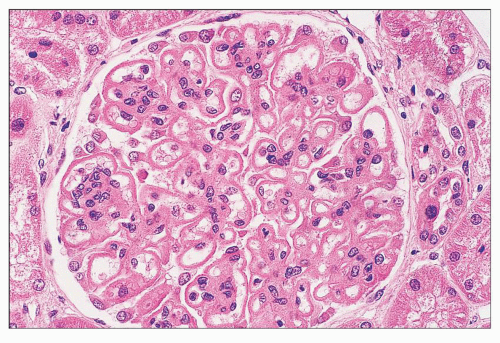
FIGURE 14.66 Lupus nephritis class V (modified Vb). There is regular thickening and rigidity of the glomerular capillary walls accompanied by global mesangial hypercellularity. (H&E; × 500.)


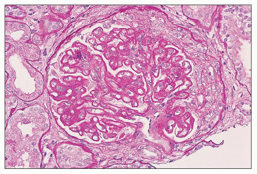
FIGURE 14.67 Lupus nephritis classes III and V (modified Vc). There is segmental endocapillary proliferation with an overlying crescent. The patent glomerular capillaries display chain-like thickenings of the glomerular capillary walls typical of membranous glomerulopathy. (PAS, × 400.)

The modified (1982) WHO classification also recognized class Vc (combined classes V and III) (Fig. 14.67), in which there are typical features of focal and segmental endocapillary proliferative glomerulonephritis superimposed on the membranous pattern, and class Vd (combined classes V and IV), in which there is superimposed diffuse endocapillary proliferative and membranous lupus nephritis (Fig. 14.68). A major disadvantage of this classification was its placement of classes Vc and Vd lesions under the membranous heading. This placed undue emphasis on the membranous component by implying that it is the dominant and most clinically relevant lesion and detracted from the more serious proliferative component. For this reason, classes Vc and Vd were eliminated from the 1995 Revised WHO classification (20). This approach is amply supported by clinical-pathologic studies
demonstrating that class Vd has an extremely poor prognosis, even worse than pure diffuse proliferative class IV (187,227,228). According to the ISN/RPS classification, as in the original WHO classification, the designation mixed class III and class V replaces the Vc lesion. Similarly, a designation of mixed class IV and class V replaces the Vd lesion. In the ISN/RPS schema, the additional designation of class V in the setting of class III or IV requires membranous involvement of at least 50% of glomerular capillary surface area of at least 50% of glomeruli by LM and/or IF.


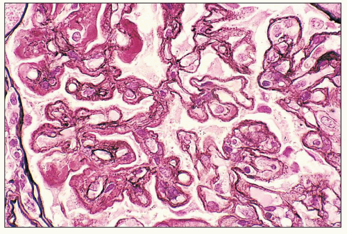
FIGURE 14.68 Lupus nephritis class IV and V (modified Vd). There is mixed, diffuse proliferative and membranous glomerulonephritis with complex thickening of the glomerular capillary walls by double contours enclosing subendothelial deposits and well-developed subepithelial spikes. (PAS-methenamine silver-Masson Ponceau stain, × 600.)


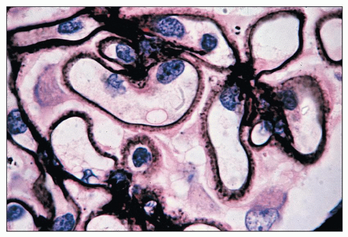
FIGURE 14.69 Lupus nephritis class V. Jones methenamine silver stain highlights the spiking of the GBMs. (× 1000.)

By light microscopy, the peripheral glomerular capillary wall alterations display a spectrum and evolution similar to primary membranous glomerulonephritis. In early stages, the glomerular capillary walls may appear normal in thickness and texture by light microscopy, and subepithelial deposits may only be detected by immunofluorescence and electron microscopy. At this stage, the glomerular capillaries may have a rigid, ectatic appearance with visceral cell swelling. Well-established membranous lesions are typically characterized by uniform and diffuse thickening of the glomerular capillary walls with well-developed spikes of the GBM that are best demonstrated with the silver stain (Fig. 14.69). In older lesions, the deposits may become largely resorbed and overlaid by neomembrane formation producing a vacuolated GBM profile (analogous to stages 3 and 4 of primary membranous glomerulonephritis). This newly laid down basement membrane seems to have a different composition from the original GBM, consisting only of laminin without type IV collagen (229).

By immunofluorescence, IgG is found in virtually all specimens in the distribution of the subepithelial deposits (Fig. 14.70). Subepithelial deposits also frequently stain for IgM, but staining for IgA is somewhat more variable and fainter (18,149,230). C3 is often demonstrable. One study found membranous lupus nephritis to have stronger and more prevalent staining for C1q compared with primary membranous glomerulonephritis (158), but another study of membranous lupus nephritis in children was unable to identify a difference in C1q staining between cases of primary membranous glomerulonephritis and membranous lupus nephritis (184). A background of mesangial immune deposits is commonly observed (Fig. 14.71). Mesangial deposits are more likely to contain IgM than IgG, and they usually have associated C3, although C3 may be absent in some specimens (227).


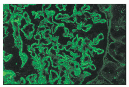
FIGURE 14.70 Lupus nephritis class V (modified Va). There are delicate subepithelial immune deposits staining for IgG. No mesangial deposits are observed. (Immunofluorescence micrograph, × 600.)

By electron microscopy, the subepithelial deposits range from small to large but usually involve the majority of capillaries. In some patients, the membranous changes are well developed but involve less than 50% of capillaries, in which case the term segmental membranous glomerulonephritis may be used. As the disease progresses, the same ultrastructural stages seen in primary membranous glomerulonephritis may evolve. GBM spikes often separate the subepithelial deposits. In more chronic cases, the deposits become overlaid by neomembrane and later become resorbed and relatively electron lucent. There is extensive foot process effacement in the distribution of the subepithelial deposits. Mesangial deposits are demonstrable in most cases and vary in quantity (Fig. 14.72). Their presence is helpful to differentiate membranous lupus nephritis from primary membranous glomerulonephritis, in which less than 10% of cases have associated mesangial deposits (231). Scattered, small subendothelial deposits are
also common but are not accompanied by endocapillary proliferation. Schwartz et al. (227) found them in eight of nine cases of lupus membranous nephritis (Va and Vb), and the Southwest Pediatric Nephrology Study Group found them in seven of nine cases (184). The presence of subendothelial deposits, which are rare in primary membranous glomerulopathy, is a particularly sensitive ultrastructural feature to distinguish membranous lupus nephritis from the primary form (158). Endothelial TRIs, a common feature of all ISN/RPS classes of lupus nephritis, are readily identified in class V. Tubulointerstitial deposits occur in approximately 25% of cases (64). Tubular atrophy and interstitial fibrosis usually parallel the distribution and severity of the glomerulosclerotic lesions.


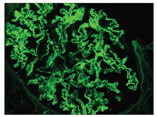
FIGURE 14.71 Lupus nephritis class V (modified Vb). There are heavy mesangial immune deposits of IgG with more delicate granular subepithelial deposits. (Immunofluorescence micrograph, × 600.)


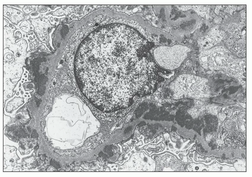
FIGURE 14.72 Lupus nephritis class Vb (membranous). The electron micrograph shows numerous subepithelial electron-dense deposits, some of which are surrounded by spiked projections of GBM. There are abundant mesangial electron-dense deposits. The glomerular capillary lumen is patent, and foot processes are extensively effaced. (× 3300.)

Rare cases of lupus nephritis mimicking a membranous pattern by light microscopy demonstrate trapping of podocytic cytoplasmic fragments and cell membrane projections within the GBM. The resulting distinctive ultrastructural appearance has been called “podocytic infolding glomerulopathy” and has been reported primarily from Japan (232,233). Such cases may have weak or no staining for Ig. Although the morphogenesis of this unusual lesion remains unclear, it likely involves remodeling of the GBM following resorption of subepithelial deposits.

Membranous lupus nephritis class V accounts for 8% to 29% of biopsy series using the ISN-RPS classification (193,215,221,234). In older series using the modified WHO classification, virtually all patients with pure membranous lupus nephritis (Va and Vb) have proteinuria at presentation and 59% to 70% have the nephrotic syndrome (18,93,235,236). In some, the nephrotic syndrome is lacking at presentation but supervenes over the course of follow-up (93,230). Hematuria is found in about one half of the patients with red blood cell casts in 10% (230). Hypertension is detectable in about one fourth of the patients. Renal insufficiency is uncommon at the time of presentation with a frequency ranging from 0% in Donadio’s experience to 25% in the series of Baldwin (93,230). Renal insufficiency and active urinary sediment are more common in patients with combined endocapillary proliferative and membranous lesions (Vc or Vd) than pure membranous forms (Va or Vb).

It has long been recognized that the membranous form of lupus nephritis differs significantly from the proliferative classes III and IV with regard to presenting serologic findings and multisystem disease manifestations. Patients with class V are more likely to present with renal disease before other systemic features of lupus are apparent. For example, among 60 patients with membranous lupus nephritis biopsied at our center, 32% of patients presented with renal-limited disease manifestations such as proteinuria, whereas 60% presented with predominant extrarenal manifestations. Only 22% of these patients fulfilled four or more ACR criteria for SLE at the time of initial biopsy. Patients with membranous lupus nephritis were more likely to be ANA negative (35%) and normocomplementemic (37%) at presentation than patients with class III or IV lesions. Serum complement levels of CH50 or C3 are lowered in 65% to 75% of patients (18,149,230,237). In a significant number of patients with class V lupus nephritis, the renal disease may precede by months or years a clinical diagnosis of SLE (18,149,230,237). Many of these patients are initially diagnosed as having “idiopathic” membranous glomerulonephritis because they lack serologic markers of SLE (238). However, careful review of their renal biopsies often discloses one or more telltale features such as mesangial hypercellularity, mesangial immune deposits, focal small subendothelial deposits, tissue ANA, strong staining for C1q, full house immunofluorescence, TRIs, and, occasionally, tubulointerstitial deposits or vascular immune deposits, betraying a secondary membranous glomerulonephritis related to SLE (158).

Two groups performed a systematic study of the biopsy features that help to differentiate membranous lupus nephritis from primary membranous glomerulopathy (158,184). The presence of subendothelial deposits, tubulointerstitial deposits, and tissue ANA appear to be the most sensitive and specific for SLE, and any combination of features increases the accuracy of a diagnosis of membranous lupus nephritis (158). A positive immunofluorescence stain for anti-PLA2R in the distribution of the subepithelial deposits has not been reported in membranous lupus nephritis (239,240,241) and strongly favors a diagnosis of nonlupus, primary membranous glomerulonephritis (241).

Patients with membranous lupus nephritis may develop RVT as a complication of their nephrotic syndrome and its associated hypercoagulable state (242,243). It is uncertain whether a circulating lupus anticoagulant may also predispose to this complication (120,123). Some patients have no clinical findings referable to RVT. Others may present with flank pain, gross hematuria, increased proteinuria, reduction in the GFR, oliguria, or signs of pulmonary embolus. The symptoms of pulmonary embolus, including dyspnea, shortness of breath, and tachypnea, may be mistaken for symptoms of lupus pleuritis or infectious pneumonia. The diagnosis of RVT can be confirmed radiographically by the use of renal venography, which demonstrates a filling defect in the renal vein, often extending into the inferior vena cava and with loss of the usual washout (i.e., “streamer effect”) of unopacified blood. Doppler ultrasonography and magnetic resonance imaging have greatly facilitated the diagnosis of RVT without
subjecting the patient to radiopaque contrast materials, which are potentially nephrotoxic.


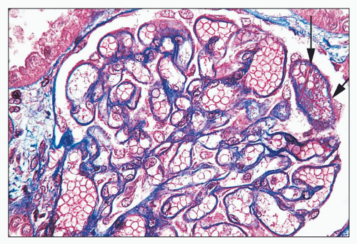
FIGURE 14.73 Lupus nephritis class V with superimposed RVT. Histologic features pointing to RVT include the separation of tubules by interstitial edema, marked glomerular capillary congestion, and fibrin strands within a glomerular capillary lumen (s). (Masson trichrome stain, × 500.)

Clues to a diagnosis of complicating RVT may be evident in renal biopsies of membranous lupus nephritis. Their astute identification by the pathologist is vital to ensure prompt clinical recognition of RVT and rapid institution of anticoagulation. A diagnosis of possible acute RVT should be suspected in any biopsy specimen with membranous lupus nephritis with diffuse interstitial edema, interstitial microhemorrhage, excessive glomerular capillary congestion, fibrin thrombosis, or neutrophil margination in glomerular capillaries (Fig. 14.73). Chronic RVT may be signaled by the presence of diffuse tubular atrophy and interstitial fibrosis that appears disproportionately severe relative to the degree of glomerular sclerosis.


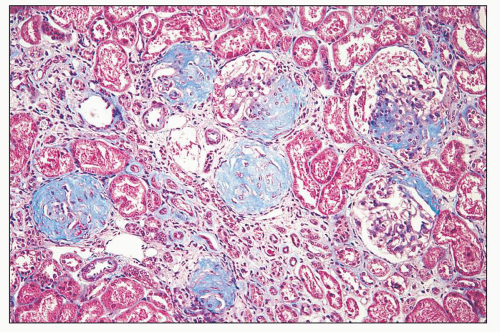
FIGURE 14.74 Lupus nephritis class VI. Extensive glomerular sclerosis shows vestiges of fibrous crescents. The global sclerosis affected more than 90% of glomeruli in this biopsy. Several glomeruli pictured here are segmentally sclerotic. Atrophic tubules alternate with groups of compensatorily hypertrophied tubules. (Masson trichrome, × 80.)


CLASS VI: ADVANCED SCLEROSING LUPUS NEPHRITIS

The modified WHO classification introduced a sixth class in which the findings are those of extremely chronic and advanced glomerulonephritis with widespread glomerular scarring affecting most glomeruli (19,20) (Fig. 14.74). The ISN/RPS schema defines class VI more precisely as advanced sclerosing lupus nephritis with global glomerular sclerosis affecting ≥90% of glomeruli without residual activity (21,22). These examples of class VI lupus nephritis undoubtedly represent the advanced phase of class IV nephritis in most cases. Severe tubular atrophy, interstitial fibrosis, inflammation, and arteriosclerosis usually accompany the glomerular sclerosis. In some of these cases, the changes are so advanced and nonspecific that it is difficult to ascertain (other than by the clinical history of SLE or documented findings in a prior renal biopsy) a diagnosis of chronic lupus nephritis by objective morphologic criteria. In such cases, glomeruli with the less advanced sclerosis often display residual features of mesangial hypercellularity. Discontinuities in the Bowman capsule associated with subcapsular fibrous proliferations are helpful to identify old fibrous crescents.

Small granular immune deposits are usually still detectable by immunofluorescence or electron microscopy in the thickened and sclerotic GBMs, in the fibrotic tubulointerstitial compartment, or in vessel walls.

In advanced lupus nephritis, it is common to observe focal segmental sclerosing features and visceral epithelial cell reactivity (e.g., hypertrophy, hyperplasia, intracytoplasmic protein resorption droplets) that mimic the changes seen in primary FSGS. These changes probably represent the effects of podocyte depletion, glomerulosclerosis, and nonimmunologic progression of renal disease mediated by hyperfiltration in remnant nephrons. This interpretation is supported by the almost invariable presence of glomerular hypertrophy in the remaining glomeruli.

Patients with class VI lupus nephritis have severe renal insufficiency, with variable subnephrotic proteinuria and relatively inactive urinary sediment. Hypertension is common. At this stage, lupus serologies may be inactive (i.e., “burnt-out” lupus). In this phase, it is inappropriate to treat the renal disease with immunosuppressive therapy, and maneuvers designed to reduce intraglomerular pressures, such as by the administration of angiotensin-converting enzyme inhibitors, are usually initiated to allay nonimmunologic progression of renal disease. Preparation for inevitable renal replacement therapy should be initiated.


TRANSFORMATIONS IN WORLD HEALTH ORGANIZATION CLASS

Lupus nephritis is not static but has the capacity to transform from one class to another, spontaneously or after treatment (41). Because of this potential for transformation, which may occur unpredictably at any time in the course of disease, patients with SLE must be monitored closely. Transformations often are heralded by sudden worsening of proteinuria, development of a nephrotic syndrome, increased activity of the urinary sediment, or a sudden decrease in the GFR (18,191,192,195,244). Some investigators have observed that patients who transform are often younger (18) and often have worse lesions than most other patients in their class of lesions (18,195).


TABLE 14.9 Class tranformations on repeat biopsies in 427 cases of lupus nephritis


Repeat biopsy class N (% of reference biopsy class)


N


II


III


IV


V


III + V


IV + V


VI


Reference biopsy class in first biopsy


I


2


1 (50%)


1 (50%)


II


45


3 (7%)


11 (24%)


14 (31%)


6 (13%)


7 (16%)


4 (9%)


III


59


6 (10%)


9 (15%)


20 (34%)


8 (14%)


7 (12%)


1 (2%)


1 (2%)


IV


185


24 (13%)


22 (12%)


69 (37%)


22 (12%)


17 (9%)


10 (5%)


19 (10%)


V


53


2 (4%)


2 (4%)


8 (15%)


21 (40%)


13 (25%)


3 (6%)


3 (6%)


III + V


37


1 (3%)


9 (24%)


8 (22%)


14 (38%)


5 (14%)


IV + V


42


2 (5%)


4 (10%)


7 (17%)


9 (21%)


4 (10%)


11 (26%)


1 (2%)


N, number of biopsies.


Refs. (193, 245, 1074, and 1075).


Transformations are relatively common, as outlined in Table 14.9. They occurred in 11 of 56 patients followed by Appel et al. (18), 15 of 88 patients reported by Baldwin et al. (93), and 14 of 90 patients reported by Mahajan et al. (195), for an average transformation rate of 13%. Recent series suggest higher rates of transformation. Seshan et al. (193) found maintenance of the same ISN/RPS class in only 9% of biopsies that were initially diagnosed as class II, 28% of class III, 28% of class IV-G, 33% of class IV-S, and 30% of class V, with the remainder transforming. A recent Chinese series of 156 patients described a change in histologic pattern in 58% of biopsies with pure proliferative pattern (class III or IV), 50% of biopsies with pure membranous pattern (class V), and 60% of biopsies with mixed proliferative and membranous pattern (classes III and V or classes IV and V) (245). Virtually all directions of transformation have been reported, including focal to diffuse (191,192,193,244), focal to membranous (193,195), diffuse to membranous (162,193,206), diffuse to mesangial (193,246), membranous to diffuse (149,193,227,230), and membranous to membranous with focal proliferative lesions (18). Probably the most commonly reported transformation is class III to IV lupus nephritis, which many prefer to consider movement along a disease continuum rather than a true transformation. Approximately 30% to 40% of class III cases have been reported to transform to class IV (18,191,192,193,195,244). Patients with extensive subendothelial deposits appear to be particularly at risk. Transformation of class IV to class V after treatment has also been described (204). After treatment, diffuse proliferative lupus nephritis frequently transforms to a mesangial proliferative pattern, although ultrastructural examination usually discloses residual irregularities of the peripheral GBM consistent with resorbed, organized subendothelial deposits (193,246) (Fig. 14.75).


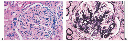
FIGURE 14.75 The initial renal biopsy of diffuse proliferative glomerulonephritis with marked endocapillary proliferation (A) regressed to a mesangial proliferative pattern with focal synechiae (B) following aggressive therapy. (A, H&E; B, Jones methenamine silver, × 500.)


Activity and Chronicity Index

From the earliest days of steroid therapy, it has been known that immunosuppressive agents are capable of reducing the amount of immune deposition in the kidney and the degree of glomerular necrosis and proliferation (162,185,202,247,248,249). However, it was equally appreciated that reduction in the histologic activity of the lesion was not always accompanied by
clinical improvement (249,250). It was presumed that there were some lesions, notably glomerular sclerosis, tubular atrophy, and interstitial fibrosis that are irreversible and may progress despite improvement in the proliferative and necrotic lesions. For more than five decades, investigators have attempted to analyze renal biopsy specimens of lupus nephritis with respect to active and chronic features as predictors of outcome and guides to therapy. The rationale is a simple one and based on the premise that active lesions are potentially treatable, whereas chronic lesions represent irreversible damage.

Pirani et al. (185) were the first to attempt to systematically separate “active” lesions from sclerosing lesions. Morel-Maroger et al. (244) carried this concept further to evaluate the efficacy of corticosteroid therapy in a large series of lupus patients with repeat biopsies. They classified the following as active lesions: fibrinoid necrosis, endocapillary proliferation, cellular crescents, nuclear debris, hematoxylin bodies, wire loops, hyalin thrombi, acute tubular lesions, and necrotizing angiitis. Chronic lesions included primarily glomerular sclerosis and interstitial fibrosis. From the number and severity of the different histologic lesions, they were able to develop an activity and chronicity index. Among 20 patients with exclusively active lesions on initial biopsy, 15 showed marked clinical and morphologic improvement after treatment. In contrast, the sclerosis worsened in 14 of 15 patients who had significant chronic features on initial biopsy. High-dose steroids in this latter group were ineffective in slowing the progression of sclerosing lesions in 8 of the 14 patients, although treatment did diminish the active lesions. This tendency for active lesions to be more amenable to therapy than chronic lesions was confirmed in a later study by Striker et al. (203).

The concept of activity and chronicity indices (AI and CI) was adopted in the studies of Austin et al. at the NIH (35,36,251). They modified the features of activity used by Morel-Maroger by adding glomerular leukocyte exudation and interstitial inflammation and deleting renal vasculitis (35). Their CI included fibrous crescents and tubular atrophy in addition to glomerular sclerosis and interstitial fibrosis (35) (Table 14.10). According to their system, the activity index is graded on a scale of 0 to 24 by calculating the sum of individual scores (0 to 3+) for each of six histologic parameters, including glomerular endocapillary proliferation, glomerular neutrophil infiltration, wire-loop deposits and hyaline thrombi, glomerular karyorrhexis and fibrinoid necrosis, cellular crescents, and interstitial inflammation. Glomerular features (including endocapillary proliferation, wire-loop deposits, necrosis, and cellular crescents) are graded as follows: 0, absent; 1+, less than 25% of glomeruli affected; 2+, 25% to 50% of glomeruli affected; 3+, greater than 50% of glomeruli affected (35,36). Neutrophil exudation is defined as more than two neutrophils per glomerulus and scored as mild (1+), moderate (2+), and severe (3+). Fibrinoid necrosis and cellular crescents are weighted double because of their more ominous prognostic importance. For interstitial inflammation, the scoring is as follows: 0, absent; 1, mild; 2, moderate; and 3, severe. A similar score for chronicity is computed by summing the individual scores (0 to 3+) for each of the following: glomerular sclerosis, fibrous crescents, tubular atrophy, and interstitial fibrosis. For glomerular sclerosis, segmental or global glomerulosclerosis in less than 25% of glomeruli is graded as 1+, in 25% to 50% of glomeruli as 2+, and greater than 50% as 3+. Similarly, fibrous crescents involving less than 25% of glomeruli are graded as 1+, 25% to 50% as 2+, and greater than 50% as 3+. Interstitial fibrosis and tubular atrophy are graded as mild (1+), moderate (2+), or severe (3+).


TABLE 14.10 Activity and chronicity indices


Index of activity (0,1,2,3,4,5,6,7,8,9,10,11,12,13,14,15,16,17,18,19,20,21,22,23,24)


Endocapillary hypercellularity


(0-3 +)


Leukocyte infiltration


(0-3 +)


Subendothelial hyaline deposits


(0-3 +)


Fibrinoid necrosis/karyorrhexis


(0-3 +) × 2


Cellular crescents


(0-3 +) × 2


Interstitial inflammation


(0-3 +)


Index of chronicity (0,1,2,3,4,5,6,7,8,9,10,11,12)


Glomerular sclerosis


(0-3 +)


Fibrous crescents


(0-3 +)


Tubular atrophy


(0-3 +)


Interstitial fibrosis


(0-3 +)


Austin HA III, Muenz LR, Joyce KM, et al. Prognostic factors in lupus nephritis. Contribution of renal histologic data. Am J Med 1983;75(3):382-391; Austin HA III, Muenz LR, Joyce KM, et al. Diffuse proliferative lupus nephritis: identification of specific pathologic features affecting renal outcome. Kidney Int 1984;25(4):689-695.


Using these indices in a group of patients with diffuse proliferative disease, Austin et al. (36) found the AI to be moderately predictive of outcome, with an AI greater than 12 associated with a 60% 10-year survival. However, none of the elements of AI was individually predictive. In their hands, the CI was more predictive of renal outcome than AI, such that a CI of ≤1 had a 100% 10-year survival, a CI of 2 or 3 had 68% 10-year survival, and a CI of ≥4 indicated a 32% 10-year survival. These findings suggested that higher CI has a graded effect on outcome and that even low levels of chronicity have prognostic significance. In contrast to AI, all the elements of CI were individually predictive of renal failure, particularly tubular atrophy. The NIH group found the combination of cellular crescents and moderate to severe interstitial fibrosis to be a particularly sensitive predictor of the subgroup at risk to double serum creatinine levels (251).

Hill et al. (252) proposed a more comprehensive biopsy index that incorporates histologic features of activity and chronicity involving all renal compartments, as well as immunofluorescence findings. This detailed index consists of four components: glomerular activity index (total possible score 24), tubulointerstitial activity index (total possible score 21), chronic lesions index (total possible score 15), and immunofluorescence index (total possible score 96). This index is unique for the inclusion of many features of tubulointerstitial activity (such as tubular cell flattening, nuclear activation and intraluminal macrophages) and scoring of glomerular monocytes. The comprehensive biopsy index proposed by Hill et al. (252) demonstrated a statistically higher correlation with clinical presenting features and outcome than the NIH activity index or chronicity index. Correlations with outcome were even greater when the index was computed for second biopsies performed 6 months following treatment of diffuse lupus nephritis, indicating its ability to identify recalcitrant disease activity that has not responded to therapy. While the index is useful for
research purposes, many consider it to be too intricate and time consuming to be applied to routine biopsy work-up.

The value and reproducibility of renal AI and CI in lupus nephritis is controversial. Several groups have found good correlations between CI and renal outcome (253,254,255). Wallace et al. (255) confirmed the earlier observations of Morel-Maroger (244) and found that AI improved in 84% of patients after treatment, whereas the CI worsened in 71%. They also observed that normalization of C3 levels was associated with reduction in AI but that prolonged depression of C3 was associated with worsening of the CI.

Other investigators have failed to corroborate a strong predictive value for the AI and CI (256,257). Appel et al. (204) found no correlation between these indices and outcome in a series containing all classes of renal lesions, although they conceded that these indices might be of greater value if only patients with diffuse proliferative disease were considered. Schwartz et al. (256), in a large study of patients with diffuse proliferative disease, found that the AI did not distinguish between those with eventual renal failure or adverse outcome and those without. They also found that there was no single value of CI that separated those destined to progress to renal failure from those with good outcome. Their data reveal that increasing CI is a measure of increasing probability of poor renal outcome but that minimal levels of chronicity cannot reliably predict adverse outcomes. Schwartz et al. (258) also found that there was poor interobserver and intraobserver reproducibility of the AI and CI, suggesting that it is influenced by subjectivity. When four highly experienced, university-based renal pathologists scored 83 biopsies of lupus nephritis, the mean activity indices ranged from 9.64 to 12.89 and mean chronicity indices from 2.84 to 4.61. The AI, which factors six variables, was consistently less reproducible than the CI (258). Wernick et al. also studied the reproducibility of the AI and CI used by the NIH group among five experienced pathologists, including four community hospital based and one from a university medical center (259). Pairs of pathologists gave scores within 1 point for chronicity and within 2 points for activity index in only 50% of cases. Moreover, repeated readings by the same pathologist at an interval of 8 to 9 months yielded CI scores that were greater than 1 point discordant in 45% of cases and AI scores that were greater than 2 points discordant in 43% of cases.

These studies underscore that there is no cutoff for AI and CI that reliably predicts outcome and that absolute reproducibility of these indices is not possible. Nevertheless, these data should not detract from the general observation that active and chronic renal lesions behave differently, especially in response to immunosuppressive therapy, and should be factored into any formulation of appropriate treatment. Although the value of these indices is controversial, most nephrologists find an overall assessment of activity and chronicity useful, especially when repeat biopsies are performed in individual patients to monitor evolution and response to therapy. In addition to the ISN/RPS classification, our biopsy reports routinely include an AI and a CI and designate in the microscopic description the percentage of glomeruli with active and chronic lesions. More important than the actual numerical value is the description of the type of active lesions and proportion of glomeruli affected. Most pathologists agree that certain lesions, such as necrotizing lesions and cellular crescents, are less reversible than other active lesions, such as subendothelial wire loops or neutrophil infiltration. The ISN/RPS classification specifies that the proportion of glomeruli with active and sclerosing lesions, and specifically those with fibrinoid necrosis or cellular crescents, should be indicated in each biopsy report. It also encourages, but does not mandate, the use of the NIH (or other) index for systematic quantitation of activity and chronicity (21,22). The ISN/RPS classification also incorporates designations of activity and chronicity into the diagnostic line of the renal biopsy report by applying the symbols (A) for active, (A/C) for mixed active and chronic, and (C) for chronic after diagnoses for lupus nephritis class III or IV. In this way, a diagnosis of lupus nephritis class IV (A/C) indicates a diffuse proliferative and sclerosing glomerulonephritis with both active and sclerosing features, whereas a designation of class III (A) indicates focal proliferative lupus nephritis that is purely active and (C) indicates focal sclerosing lupus nephritis that is purely chronic.


Reproducibility of the ISN/RPS Classification

Several studies have confirmed the improved interobserver reproducibility of the ISN/RPS classification compared to earlier WHO classifications (215,260). A large study involving 20 centers in the UK classified cases of lupus nephritis by the ISN/RPS classification and the modified WHO (1982) classification (260) and concluded that the ISN/RPS classification was more reproducible. They ascribed this to clearer separations between the classes and the elimination of subgroups of class V. Interestingly, the percentage of cases of class IV increased from 23% by the WHO 1982 classification to 46% by the ISN/RPS classification, with fewer diagnoses of class III and class V. This increase could be attributed primarily to the elimination of class Vd as a subgroup of membranous lupus nephritis and the inclusion of sclerotic glomeruli in the assessment of total glomeruli affected. Other studies have shown that the classification is useful to predict outcome based on its incorporation of modifiers reflecting activity (A) and chronicity (C) (219).


Extrarenal Clinical Manifestations

SLE is extremely diverse in its clinical manifestations, which affect a large number of organ systems with highly variable initial presentations and evolution over time (261). Constitutional symptoms such as fever, malaise, and weight loss are common presenting features. Fever secondary to active disease was documented in over 80% of patients with SLE in the early 1950s, but only 41% of patients in a study conducted from 1980 to 1989 (262), possibly reflecting earlier disease recognition and greater use of antipyretic medications in the modern era.

Involvement of the skin and mucous membranes occurs in 55% to 90% of patients (262,263) and includes butterfly rash over the cheeks and bridge of the nose, oral or nasal ulcers, discoid lupus, and subacute cutaneous lesions. The butterfly rash, detectable in approximately one half of patients, often appears after sun exposure. Presumably, ultraviolet B radiation exposure damages cellular proteins and DNA, releasing subcellular antigenic particles such as nucleosomes (264). Release of cytokines such as interleukin-1 (IL-1) in areas of sun damage may potentiate the immune response. Moreover, ultraviolet light promotes binding of anti-Ro, anti-La, and anti-RNP to keratinocytes (265). Discoid skin lesions occur in approximately 25% of patients and must be differentiated from discoid lupus erythematosus, a purely cutaneous condition. Discoid lesions differ from the diffuse erythematous lesions of classic butterfly rash in having rounded, annular contours, frequent plaque formation, and follicular plugging, leaving depressed scars and
areas of hypopigmentation. In SLE, lesions may occur on the face, scalp, neck and upper chest, or back. Pathologic examination of the skin reveals mononuclear inflammation of the dermis, follicular plugging, edema of the basal epidermis, and hyperkeratosis. Granular deposits of IgG and C3 are often detectable at the dermal-epidermal junction, corresponding to complexes of nucleosomes and antinucleosomal antibodies (266). Alopecia occurs in up to 70% of patients and may affect the eyebrows, eyelashes, beard, and scalp. Nail lesions, ridging and pitting in character, may also affect up to one fourth of SLE patients. Other skin lesions such as periungual erythema, telangiectasia, Raynaud phenomenon, and livedo reticularis are also common. The latter is particularly associated with a circulating lupus anticoagulant and/or APL antibodies. The occurrence of vasculitis may be manifested by splinter hemorrhages of the nailfold capillaries, small microinfarcts of the fingertips, and erythematous indurated lesions on the palmar thenar eminences (i.e., Janeway spots) and fingertips (i.e., Osler nodes).

In a large number of series, arthralgias and arthritis are the most common presenting symptoms, occurring in up to 95% of patients at some time in the course of the disease. Joint complaints may precede by months or years other systemic symptoms. The arthralgias commonly affect the proximal interphalangeal joints, wrist, and knees and may be accompanied by morning stiffness. Rarely is the arthritis deforming, although soft tissue laxity may be seen in the late stages. Muscle complaints such as myalgias, muscle tenderness, and weakness may occur in greater than 50% of patients at some point in the course of disease evolution. Muscle biopsies reveal perifascicular and perivascular mononuclear infiltrates.

Among the pulmonary manifestations of SLE, pleuritis is the most common, affecting 40% to 60% of patients, sometimes with associated pleural effusions. Lupus pneumonitis is far less common, affecting less than 5% of patients (267). Pathologically, it consists of an interstitial pneumonitis with predominantly interstitial mononuclear infiltrates. In severe cases, acute alveolitis, hyaline membranes, and alveolar hemorrhage may occur. Inoue identified deposits of IgG, C3, and DNA antigen in alveolar capillary walls and septa, with corresponding electron-dense deposits seen by electron microscopy (268). Clinically, lupus pneumonitis is often difficult to differentiate from infectious pneumonia. Pulmonary function test abnormalities associated with lupus include mild impairment in diffusing capacity and reduced lung volume. Pulmonary embolus may occur as a complication of RVT associated with the nephrotic syndrome or deep vein thrombosis associated with circulating lupus anticoagulant and/or APL antibodies.

Cardiovascular manifestations are most frequently related to pericarditis, which is detectable in one fourth of patients clinically and in up to two thirds at autopsy (269). In the acute phase, an electrocardiogram reveals tall T waves and elevation of the ST segment. Less common is myocarditis, which was detectable clinically in 8% of patients in two large series (270,271) and up to 40% at autopsy (269). Histologic features include interstitial mononuclear inflammation and fibrosis. Arteritis affecting the major coronary arteries and intramyocardial arterioles is rare. An increasingly recognized cardiac complication of lupus is valvular disease, which correlates highly with the presence of APL antibodies. Valvular thrombotic (sterile) vegetations are most commonly encountered on the mitral valve and rarely the aortic valve. Previously called Libman-Sacks endocarditis, it consists of a fibrinous or fibrosing verrucous lesion that may cause clinically significant valvular stenosis or incompetence.

The neurologic manifestations of lupus are diverse and often pose difficulties in differentiation from steroid-induced psychosis. They include cognitive dysfunction, headache, altered consciousness (ranging from stupor to coma), seizure, stroke, optic neuritis, and peripheral neuropathy. Pathologically, a correspondingly diverse array of morphologic lesions has been described, ranging from cerebral perivascular inflammation to thrombosis, arteritis, cerebritis, hemorrhage, and infarction. A role for antineuronal antibodies (272) and immune complex deposition, identifiable in the choroid plexus, has been proposed. Thrombotic central nervous system disease often is mediated by lupus anticoagulant and/or APL antibodies and may have vasculitic features.

Hematologic abnormalities are common and include lymphadenopathy in 50% of patients. Anemia, affecting about one half of patients, may be hemolytic and produce a positive direct Coombs test result. Lymphopenia and thrombocytopenia are commonly observed. The lymphocytopenia is probably mediated in part by cold-reactive, complement-fixing IgM antilymphocyte antibodies. Severe thrombocytopenia may be a manifestation of immune thrombocytopenic purpura, and platelet counts of less than 20,000 may be associated with clinical evidence of bleeding, including cutaneous petechiae, purpura, or epistaxis. Prolongation of the activated partial thromboplastin time and a false-positive VDRL result are usually manifestations of APL antibodies. Antibody binding to the phospholipid component of the prothrombin activator complex (consisting of factors Xa, V, calcium, and phospholipid) prolongs the in vitro partial thromboplastin time. Antibodies to cardiolipin, the phospholipid component of beef heart, which is admixed with phosphatidylcholine and cholesterol in the VDRL assay, is responsible for the falsely positive biologic test result for syphilis detected in 29% of patients.

A classification of SLE was proposed in 1982 by the ACR (273) and was further revised in 1997 (274). This system attempts to accommodate the diverse clinical manifestations of SLE, while accounting for its highly variable disease evolution affecting different systems over time, with periods of remission and exacerbation. This classification proposes 11 defining clinical and laboratory criteria, including malar rash, discoid rash, photosensitivity, oral ulcers, arthritis, serositis, renal disorder (defined as persistent proteinuria greater than 500 mg/d or 3+ by dipstick testing or cellular casts of any type, including red cell, granular, tubular, or mixed), neurologic disorder, hematologic disorder (including anemia, leukopenia, lymphopenia, or thrombocytopenia), immunologic disorder (including positive test for anti-dsDNA antibody, anti-Smith antibody, APL antibody, or lupus anticoagulant), and positive ANA. The presence of any four of these criteria sequentially or simultaneously has 96% sensitivity and specificity for SLE (273) in the patient database from which it was derived. Subsequent studies have confirmed a more modest 80% to 95% sensitivity and specificity for these criteria in clinical rheumatologic practice (275).


Management of Lupus Nephritis


Overview

The management of lupus nephritis has evolved over many decades, with the introduction of new agents and therapeutic strategies following multiple clinical trials (276,277,278,279). In brief, corticosteroids have been the mainstay of therapy for lupus nephritis since the 1950s (185) and continue to be used
in most patients with clinically significant kidney disease. In the 1980s, landmark studies from the NIH demonstrated that aggressive immunosuppression with cyclophosphamide in conjunction with corticosteroids was more effective than corticosteroids alone in producing remissions in most cases of lupus nephritis (280,281,282). However, this gain was accompanied by significant side effects from long-term immunosuppressive therapy, notably infections, gonadal toxicity, and bladder toxicity (283). Subsequent studies demonstrated that lower doses and shorter courses of cyclophosphamide, followed by maintenance therapy with mycophenolate mofetil (MMF) or azathioprine (AZA), were equally effective at preventing relapses and sustaining remissions as the higher doses of cyclophosphamide used in the original NIH protocols (284,285,286,287,288). More recently, MMF has emerged as a suitable alternative to cyclophosphamide for both proliferative and membranous lupus nephritis and may even be the treatment of choice in patients of African descent or Hispanic ethnicity (284,289,290,291,292,293). In addition, the use of calcineurin inhibitors in lupus nephritis is supported by several clinical studies (294,295,296). In the 21st century, novel biologic agents that target B-cell and T-cell function and cytokines have been employed in patients with SLE. Some of these drugs have entered the therapeutic armamentarium for lupus nephritis, whereas others remain under active study.

Only gold members can continue reading. Log In or Register to continue

Stay updated, free articles. Join our Telegram channel

Jun 21, 2016 | Posted by in UROLOGY | Comments Off on Renal Disease in Systemic Lupus Erythematosus, Mixed Connective Tissue Disease, Sjögren Syndrome, and Rheumatoid Arthritis

Full access? Get Clinical Tree

Get Clinical Tree app for offline access