Renal Anatomy and Histology


Renal Anatomy and Histology


Stephen M. Bonsib


The focus of this chapter is the gross anatomy, histology, and ultrastructure of the kidney, a remarkably complex organ that fosters exploration into its structural-functional intricacies with each technologic advance in molecular and cellular biology. Arsenals of sensitive and specific immunohistochemical reagents, molecular and genetic probes including tissue micro-arrays, are now available, and innovative techniques such as two-photon microscopy and in vivo imaging of the living kidney have emerged (1,2,3,4,5). These powerful tools enable investigators to explore new aspects of renal structure, renal function, and renal dysfunction and have led to the discovery of important genes and their products, whose role in normal renal physiology is a rapidly evolving field.

The content of this chapter draws on a compendium of contributions that began with Leonardo da Vinci and Andreas Versalius (Fig. 1.1), whose cadaver dissections provided the first detailed illustrations of the genital and urinary tract in the 16th century (6,7). One hundred years later, Marcello Malpighi, using the recent technologic innovation of Galileo Galilei, the light microscope, first described the renal corpuscles that bear his name (8,9). However, 200 years elapsed before major refinements in microscopes, and microscopic techniques such as thin histologic sections led to provocative discoveries of the fine structure of the kidney. William Bowman’s intravascular dye studies (Fig. 1.2) published in 1842 revealed fundamental structural aspects of the nephron such as its vascular connections and unique peritubular capillary plexus (10,11). These early forays into renal microanatomy exposed complexities that begged for speculations regarding their functional correlates, leading to investigative journeys whose end is ever beyond the horizon.

In this chapter, the normal kidney is macroscopically displayed and then dissected at the cellular level, using both traditional microscopic and ultrastructural techniques, histochemistry and immunohistochemistry, and selected other modalities to illustrate features otherwise not demonstrable.

Human material will be used when possible. Because perfusion-fixed animal tissue provides pristine ultrastructural detail of tubules unequaled by immersion-fixed human tissue, a number of illustrations used in past editions authored by Wilhelm Kriz
have been retained. The official nomenclature for structures of the kidney has been established by the Renal Commission of the International Union of Physiological Sciences (12). This terminology is used in this chapter (Table 1.1).


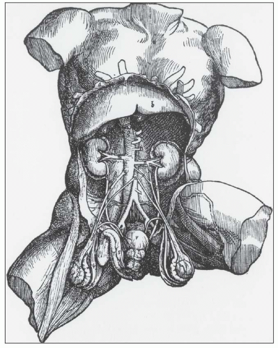
FIGURE 1.1 Andreas Versalius’ 1543 illustration of the male genitourinary tract. Although most anatomic details are correct, the left kidney is incorrectly placed lower than the right kidney. (From Book 5 De Humani Corpus Fabrica.)


GROSS ANATOMY AND MACROSCOPIC FEATURES OF THE KIDNEYS AND THEIR ENVIRONS


Retroperitoneum

The retroperitoneum is a large compartment containing fat and several organs bounded anteriorly by the peritoneum and posteriorly by the transversalis fascia (Fig. 1.3). It is divided into three fascia-invested compartments or spaces: the anterior, posterior, and perirenal spaces (13,14,15). The anterior pararenal space contains the pancreas, duodenal loop, ascending and descending colon, and the hepatic, splenic, and proximal superior mesenteric arteries. The posterior pararenal space contains fat but no organs. The perirenal space is home to the kidneys and adrenal glands. Its anterior and posterior (Zuckerkandl) fascial investments are known as Gerota fascia, a loose connective tissue envelope that provides surgical dissection planes during radical nephrectomy. The perirenal space is bounded medially by dense fat and the adventitial connective tissues of the aorta and vena cava that impede communication across the midline of perinephric processes such as urine leaks, hemorrhage, or infection (15).


Kidneys

The kidneys are paired retroperitoneal organs insulated by adipose tissue within Gerota fascia. They extend from the 12th thoracic vertebra to the 3rd lumbar vertebra. Their position is 2.5 cm lower in the erect position compared with their supine location, and they exhibit craniocaudal movement of 1.9 to 4.1 cm during respiration (14,16,17). The right kidney is slightly lower than the left kidney (in contrast to the drawing by Versalius, Fig. 1.1), and their upper poles are tilted slightly toward the midline. The kidneys are covered by a less than 1-mm-thick fibrous true capsule. It is adherent to the underlying renal cortical parenchyma but easily stripped off the normal kidney. This firm layer can be felt with introduction of the needle into the kidney during renal biopsy.

The newborn kidney is smaller than the adult kidney with the same reniform shape, but grossly, it is distinguished by prominent fetal lobations (Fig. 1.4). Renal weight in newborns ranges from 13 to 44 g; this increases by adulthood to 125 to 170 g in males and 115 to 155 g in females and has been shown to be proportional to body surface area (16,17).

The number of glomeruli is constant in an individual between birth and adulthood; the increase in renal volume reflects expansion of tubular mass. The reduced tubular mass of the newborn kidney limits the capacity for salt and water regulation and explains neonates’ susceptibility to dehydration. With age, there is a progressive decline in renal mass and weight, and therefore a decrease in renal reserve after the third decade, even in the absence of comorbid conditions (18,19,20). The loss of renal mass is primarily cortical and is proportional to the loss of (sclerosis of) functioning glomeruli. The average adult kidney is 11 to 12 cm long, 5 to 7 cm wide, and 2.5 to 3 cm thick.

The left kidney is slightly larger than the right kidney. Renal volume can increase or decrease by 15% to 40% with major fluctuations in blood pressure, intravascular volume, or interstitial expansion by edema. The combined mass of the kidneys correlates with body surface area and is reduced in children of low birth weight (19,20).

The posterior surfaces of both kidneys are flatter than the anterior surface. The medial surface is concave with a 3-cm slitlike space called the hilum. The hilum is the vestibule through which pass the ureter, branches of the renal arteries and veins, nerves, and lymphatics as they enter and exit the renal sinus. The renal sinus is the fat-containing compartment housed within the confines of the kidney that also invests the calyceal and pelvic portions of the collecting system (Fig. 1.5).

The renal connective capsule extends a short distance into the renal sinus where it terminates. Portions of the renal cortex, the columns of Bertin, have no renal capsular investment (Fig. 1.6), resulting in direct continuity between the cortical interstitium and sinus tissues. This appears important in renal neoplasia, representing preferential routes for cancer dissemination (21). The subcapsular surface of the renal cortex may be smooth and featureless or may show grooves corresponding to the outlines of some or all of the individual renal lobes (Fig. 1.7). Persistence of distinct fetal lobations is common and
is a normal anatomic variant without functional consequences (22,23,24). In some of the kidneys, three subtle zones are created by two shallow superficial grooves that radiate from the hilum to the lateral border. The three regions correspond roughly to the upper pole, middle zone, and lower pole and usually reflect regions drained by the segmental veins.


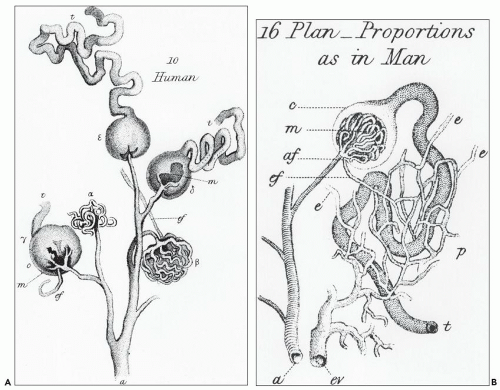
FIGURE 1.2 William Bowman’s 1842 dye study. A: Illustration of the glomerular vascular supply. B: Illustration of the relationship between the efferent arteriole and peritubular capillary plexus and the proximal tubule. (From Bowman W. Philos Trans R Soc Lond Biol 1842;132:57.)

The human kidney is a multipapillary type of mammalian kidney (Fig. 1.8). The cortex is continuous and undivided, and the medulla is discontinuous with individual pyramids that drain discrete regions of the associated cortex. Each lobe in a multipapillary kidney may be regarded as the counterpart of an entire unipapillary kidney, such as those in a rat or rabbit (22,23,24). Certain animals such as bears and whales have nonfused, multiple, unipapillary kidneys; each papilla is invested with the cortex that is autonomous in structure and function from its neighbors. The normal human kidney has a minimum of 11 to 14 lobes, each composed of a central conical medullary pyramid surrounded by a cap of cortex (22,23,24,25,26,27,28,29,30,31). Often, there are six lobes in the upper pole and four lobes each in the middle zone and lower pole. By the 28th week of gestation, the number of renal lobes is established; however, with the subsequent increase in renal mass, a process of lobar fusion ensues. In the adult kidney, this reduces the number of renal pyramids and their corresponding calyces to 9 to 11. The pyramids, within their minor calyces, angle toward the major calyces and renal pelvis from a slightly anterior or posterior direction (Fig. 1.9) (26,27,28).

The lobar fusion affects pyramids in the polar region more than the midportion of the kidney, creating a mixed population of simple and compound pyramids that characterizes most human kidneys. Simple pyramids (Fig. 1.10) occur predominately in the midpolar regions, drain a single lobe, and have a convex tip with slit-like openings of the ducts of Bellini, the area cribrosa. Compound pyramids are principally polar in location and drain two or more lobes (see Fig. 1.8). They have flattened to convex tips with rounded or gaping orifices of the
ducts of Bellini, creating the potential for intrarenal reflux during urinary tract obstruction and infection (30,31).


TABLE 1.1 Nomenclature of the kidney


Main divisions


Cortex


Cortical labyrinth


Medullary ray


Renal column (column of Bertin)


Medulla


Outer medulla


Outer stripe


Inner stripe


Inner medulla


Papilla


Renal pelvis


Pelvic cavity


Major calyx


Minor calyx


Fornix


Renal sinus


Renal hilum


Vessels, nerves, and interstitium


Renal vasculature


Main renal artery and vein


Anterior and posterior arterial divisions


Segmental arteries: apical, upper, middle, lower, and posterior


Interlobar artery and vein


Arcuate artery and vein


Interlobular artery (cortical radiating) and vein


Perforating radial artery


Stellate vein


Afferent arteriole


Efferent arteriole


Glomerular capillary


Peritubular capillary


Descending and ascending vasa recta


Vascular bundle


Interbundle capillary plexis


Renal lymphatics


Cortical


Capsular


Renal nerves


Renal interstitium


Cortical


Peritubular


Periarterial


Medullary


Nephron


Nephron types


Superficial nephron


Midcortical nephron


Juxtamedullary nephron


Short-looped nephron


Long-looped nephron


Nephron components


Renal corpuscle


Bowman capsule


Glomerulus or glomerular tuft


Parietal cell


Peripolar cell


Podocyte


Parietal podocyte


Endothelial cell


Mesangial cell


Mesangial matrix


GBM


Lamina rara interna


Lamina densa


Lamina rara externa


Urinary space (Bowman space)


Juxtaglomerular apparatus


Granular cell


Extraglomerular mesangium


Extraglomerular mesangial cells (lacis cell, Goormaghtigh cell)


Macula densa


Tubular portion of the nephron


Proximal tubule


Proximal convoluted tubule


Proximal convolutions (not synonymous with the PT)


Straight part


Loop of Henle


Thin descending limb


Thin ascending limb


Distal tubule


Thick ascending limb


MD epithelium


Distal convoluted tubule


Distal convolutions (not synonymous with the DT)


Connecting tubule


Collecting duct


Cortical CD


Outer medullary CD


Inner medullary CD (duct of Bellini)


The renal parenchyma consists of the granular brown cortex and the striated medulla (Fig. 1.10). The medullary pyramid is divided into an outer medulla, with an outer and inner stripe, and the inner medulla or papilla. In human beings, the relative volumes occupied by the cortex, outer medulla, and inner medulla are 70%, 37%, and 3%, respectively (32). The renal cortex forms a 1.0-cm layer beneath the renal capsule and extends down between the renal pyramids forming the columns of Bertin. The midplane of a column of Bertin is the line of fusion of two renal lobes, with each half draining into adjacent pyramids. The bases of the pyramids have faint perpendicular cortical extensions, the medullary rays that contain the straight portion of the proximal tubules (PTs), thick ascending limbs (TALs), and

collecting ducts (CDs). The papilla protrudes into a minor calyx. Its tip, the area cribrosa (with cribriform appearance grossly), has from 20 to 70 openings of the papillary CDs (Bellini ducts); the large variation reflects simple versus compound pyramid arrangement (24,26,28).


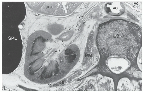
FIGURE 1.3 Cross section of a human cadaver at the level of the second lumbar vertebra viewed from above, demonstrating the relationships between the left kidney and adjacent organs. L2, second lumbar vertebra; AO, aorta; PM, psoas muscle; QL, quadratus lumborum muscle; SPL, spleen; JEJ, jejunum; C, calyx; P, pelvis; RV, renal vein; IVC, inferior vena cava.


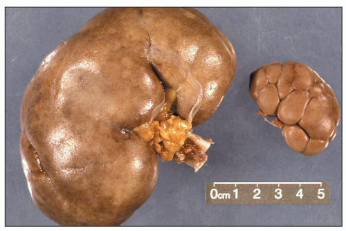
FIGURE 1.4 Composite adult and newborn human kidneys. Notice the smooth subcapsular surface of the adult kidney and the prominent lobations of the newborn kidney.


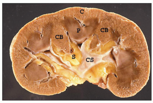
FIGURE 1.5 A hemisected human kidney shows the cortex (C) with columns of Bertin (CB) and renal pyramids (P). The renal papillae protrude into the minor calyces, which unite to form the major calyces. The collecting system (CS) is nestled within the fatty tissue of the renal sinus (S).


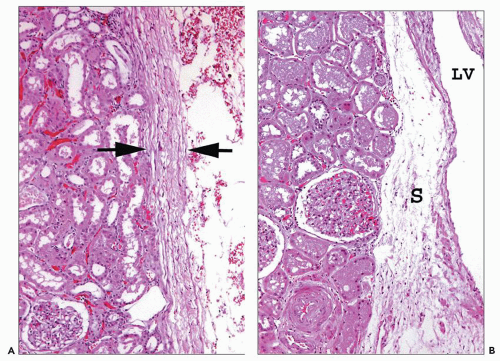
FIGURE 1.6 Human kidney. A: A dense connective tissue capsule (between arrows) separates the peripheral cortex from the perinephric fat (×100). B: The renal tubules and interstitium of the columns of Bertin have no connective tissue separation from the sinus (S) and its abundant lymphatics and vessels. (×120.) LV, lobar vein.


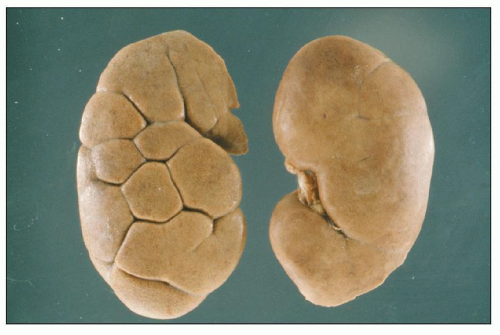
FIGURE 1.7 Two normal adult human kidneys, one with retained lobations.


Renal Vasculature

The arterial supply to the kidneys follows a general blueprint. Knowledge of its details is useful in recognition of lesions resulting from an arterial abnormality (32,33,34,35,36,37). In 1901, Brödel first appreciated the distinctive renovascular segmentation of the kidney (33). The nomenclature currently in use was established by Graves (Fig. 1.11) in 1954 (34).

The main renal arteries arise from the aorta; the right artery is slightly longer and often arises slightly higher from the aorta than the left artery (33,34,35). Each main artery gives off a suprarenal artery to supply the adrenal glands and a ureteric artery to each ureter. The most common main renal arterial division pattern is to divide into anterior and posterior branches (Figs. 1.11 and 1.12) that give rise to five segmental renal arteries. Most commonly, the anterior branch gives rise to four segmental arteries: the apical, upper, middle, and lower segmental arteries. Two segmental arteries supply the middle anterior portions of the kidney, and two polar segmental branches supply both the anterior and posterior polar aspects of the kidney. The posterior branch continues as a fifth segmental branch, the posterior segmental artery, to supply the middle posterior portions of the kidney. However, deviation from this pattern is common. This may reflect variation in the origin of a segmental artery from
either anterior or posterior branches or arising separately from the aorta as an aberrant, accessory, or polar artery (Figs. 1.13 and 1.14), which occurs in 25% of the kidneys (32). Another variation is supply of the upper or lower pole by the posterior segmental artery or a branch from the posterior segmental artery.


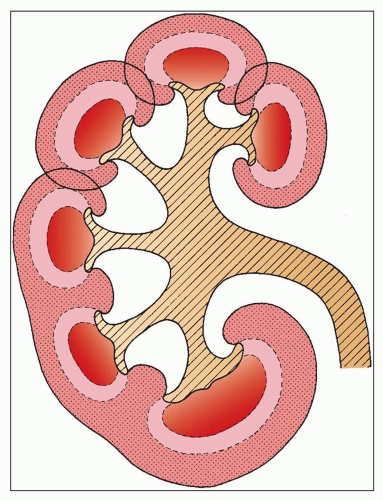
FIGURE 1.8 Diagram of the arrangement of the cortex and medulla in the human kidney and of its possible development. Unipapillary kidney anlagen fuse to form a multipapillary kidney (upper part of the drawing). The interpyramidal cortical intrusions (septa of Bertin) reach down to the renal sinus. In addition to the fusion of the cortical tissue, pyramidal fusion occurs and is generally found in the polar regions (compound papilla, shown in the lower part of the drawing).

From the five segmental arteries, the interlobar arteries, arcuate arteries, interlobular (cortical radiating) arteries, and arterioles are sequentially derived. All arteries are end arteries with no collateral blood flow. Thus, occlusion of a segmental artery or any of its subsequent branches results in infarction of the zone of parenchyma it supplies.

A segmental artery branches within the renal sinus, giving rise to several interlobar arteries. An interlobar artery pierces the parenchyma between the pyramid surface and a column of Bertin and forms a splay of six to eight arcuate arteries. The arcuate arteries curve along the corticomedullary junction and ascend up the lateral surface of the pyramid and over its basal surface to terminate at the midpoint of a renal lobe. Since the arcuate arteries do not anastomose, the central portion of the renal lobe is most susceptible to ischemic injury. At perpendicular or slightly oblique angles, the interlobular arteries (cortical radiating arteries) arise from an arcuate artery (Fig. 1.15) and may branch as they pass through the cortex toward the renal capsule. The interlobular arteries course between medullary rays and are encircled by tiers of five to six glomeruli, which they supply with an

afferent arteriole (Figs. 1.15 and 1.16). The efferent arterioles, on exiting the glomeruli, form a portal system of capillaries that supplies the adjacent cortical tubules, or provides the main arteriolar flow to the renal medulla in the case of juxtamedullary glomeruli (Figs. 1.2B and 1.17; see Cortical Microvascularization section below). A few interlobular arteries reach the renal capsule and anastomose with branches of the suprarenal and gonadal arteries.


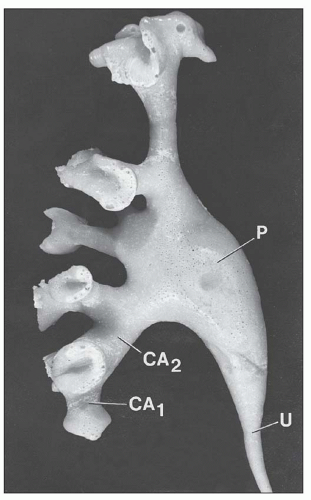
FIGURE 1.9 Minor calyces (CA1) fuse to form major calyces (CA2) that finally become the renal pelvis (P), which tapers to continue on as the ureter (U). Most calyces are angled toward the renal pelvis. Corrosion cast of a human renal pelvis. (From Sampaio FJB, Mandarim-De-Lacerda CA. 3-Dimensional and radiological pelvicaliceal anatomy for endourology. J Urol 1988;140:1352.)


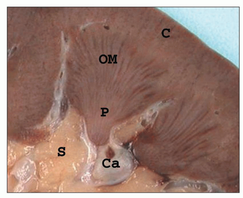
FIGURE 1.10 A simple human renal pyramid with a convex papilla (P) nestled within a minor calyx. S, sinus; OM, outer medulla; C, cortex; Ca, calyx.


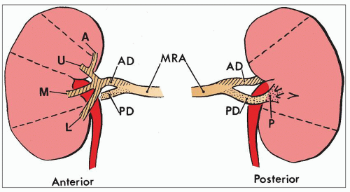
FIGURE 1.11 Diagram of the most common pattern of arterial supply to the kidneys demonstrating the main renal artery, anterior and posterior branches, and five segmental arteries. MRA, main renal artery; PD, posterior division; AD, anterior division. Segmental arteries are indicated by A (apical), U (upper), M (middle), L (lower), P (posterior). (Modified from Graves FT. The anatomy of the intrarenal arteries and its application to segmental resection of the kidney. Br J Surg 1954;42:132.)


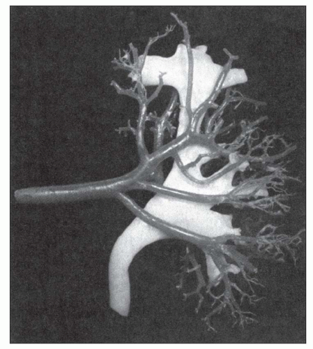
FIGURE 1.12 Cast of the arterial supply and collecting system of a human kidney. Notice the four anterior segmental arteries. (From Sampaio FJB, Aragao AHM. Anatomic relationship between the intrarenal arteries and the kidney collecting system. J Urol 1990;143:679.)


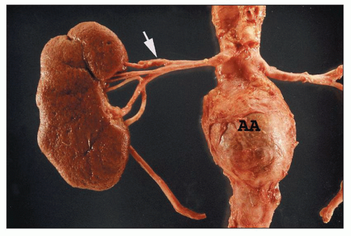
FIGURE 1.13 Human right kidney and aorta. The aorta has an aneurysm (AA). The main renal artery and its five segmental arteries are visible although not in the most common arrangement. The upper segmental artery branches first (arrow), and the posterior segmental artery appears to arise from the anterior group.


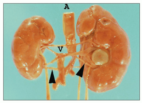
FIGURE 1.14 Newborn human kidneys. There are bilateral lower pole accessory (segmental) arteries (arrowheads). The crossing polar artery and segmental vein of the nonrotated duplex (two ureters) left kidney produced ureteropelvic junction obstruction. A, aorta; V, vena cava.


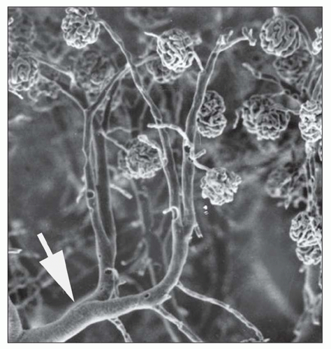
FIGURE 1.15 Arterial cast of the rat kidney. Two interlobular arteries give rise to arterioles that supply a single glomerulus. Arrow, arcuate artery. (From Gattone II VH, Evan AP, Willis LR, et al. Renal afferent arteriole in the spontaneously hypertensive rat. Hypertension 1983;5:8.)


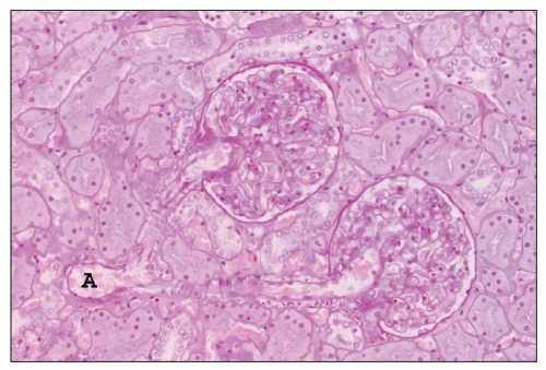
FIGURE 1.16 Normal human kidney. The interlobular artery (A) gives rise to two arterioles; each supplies one glomerulus. (PAS, ×200.)

The renal medulla has a dual blood supply, originating in part from its corticomedullary base and in part from the lateral distal papilla (38). The principal blood supply arises
from juxtamedullary glomeruli. Their efferent arterioles course directly into the medulla, forming the descending vasa recta (Fig. 1.17). The second blood supply originates from an interlobar artery as it courses along a minor calyx. Several spiral artery branches enter the papilla at the calyceal fornices, sending arterioles to the papillary tip. These arterioles anastomose freely with arterioles from the opposite side, forming a plexus around the distal ducts of Bellini.


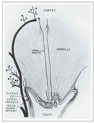
FIGURE 1.17 Diagram of the renal medulla showing its dual blood supply. (From Baker SB. The blood supply of the renal papilla. Br J Urol 1959;31:57. Reprinted from Wiley-Liss, Inc., Wiley Publishing Inc., a subsidiary of John Wiley & Sons, Inc., with permission.)

The major cortical venous return follows the arterial supply; the interlobular, arcuate, and interlobar veins run parallel to the arteries (Fig. 1.18) (39). A minority of veins originate as the stellate veins, draining the superficial cortex to join the arcuate veins. Unlike the arcuate and interlobar arteries, arcuate and interlobar veins are connected by abundant anastomoses and lateral tributaries that encircle the renal pyramids and calyces. Thus, the terms interlobar veins and intralobar veins are often used synonymously. The interlobar veins converge anterior to the pelvis and form two to three segmental veins that drain the three poles of the kidney and then unite to form the main renal vein. The convergence of the interlobar veins to form the main renal vein often occurs outside of the renal hilum. This is particularly common for the left main renal vein, which is substantially longer than the right main renal vein, because the vena cava lies to the right of the aorta (Fig. 1.19). No veins are located within the medulla.


Renal Lymphatic System

The kidneys have a dual lymphatic system (40,41). The major lymphatic drainage follows the vasculature. The lymphatics begin as small vessels in the adventitia of the peripheral interlobular (cortical radiating) arteries, enlarge, and become more numerous as they descend to the corticomedullary junction and enter the renal sinus (Fig. 1.20). In humans, there are no lymphatic vessels amid the glomeruli and renal tubules, although some animals (e.g., dogs) appear to have periglomerular and periarteriolar lymphatics (40,41). During inflammatory processes, lymphatic neovascularization within the cortical labyrinth has been documented (42). With the recent identification of immunohistochemical markers specific for lymphatic endothelium, more will be learned about the dynamics of cortical lymphatics (43). Lymph eventually exits through the hilum and terminates in hilar and lateral paraaortic lymph nodes.


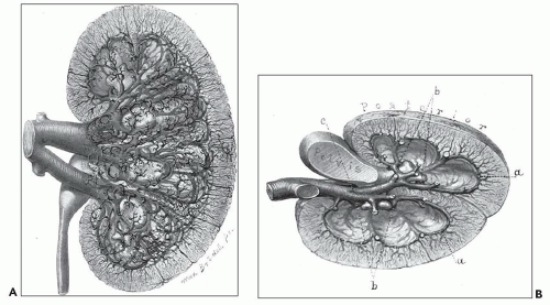
FIGURE 1.18 Brödel’s 1903 artist’s rendition of the human kidney following celloidin injection with tissue digestion, demonstrating details of the lush venous return. A: Anterior view of the left kidney. For the sake of clearness, the small veins of the cortex of the anterior portion have been omitted. B: The transverse section viewed from above. There is no collecting vein posterior to the renal pelvis; all of the veins of the posterior region cross over to the anterior portion between the necks of the minor calices (b) to join the veins of the anterior region at a point indicated by c. (Brödel M. The intrinsic blood vessels of the kidney and their significance in nephrotomy.Johns Hopkins Hosp Bull 1901;118:10.)


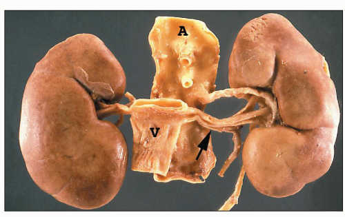
FIGURE 1.19 Human kidneys with aorta (A) and vena cava (V). The left main renal vein (arrow) is much longer than the right main renal vein.


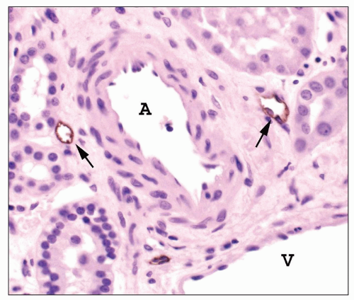
FIGURE 1.20 Human interlobular arteries and lymphatics. Adjacent to an interlobular artery (A) are several small lymphatics (arrows). These will converge and progressively enlarge as they drain toward the medulla. The lymphatic endothelium is stained for podoplanin, a lymphatic endothelial cell marker. V, vein (Immunoperoxidase stain for podoplanin, ×400.)

The second separate lymphatic system exists within the renal capsule. It receives drainage from the most superficial cortex. Lymph courses along the capsule and around to the hilum to join the major lymphatic flow exiting the renal sinus. Valves present in the capsular system prevent retrograde flow back to the cortex. The capsular lymphatic system normally contributes little to lymph flow. However, under certain conditions such as urinary tract obstruction, it becomes the principal pathway for lymphatic flow (41).


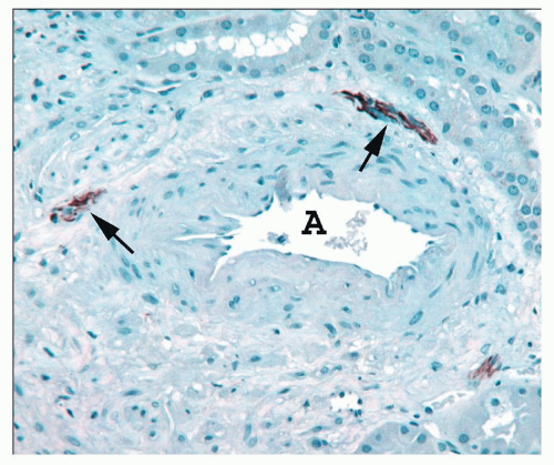
FIGURE 1.21 Human interlobular artery. The adventitia of this artery (A) contains three small nerves stained for neurofilament (arrows). (Immunoperoxidase stain for neurofilament.)


Renal Innervation

The celiac plexus sends sympathetic fibers via splanchnic nerves to synapse with ganglia in the renal plexus. Nerve fibers extend from the plexus to accompany the arterial system (Fig. 1.21) as it ramifies throughout the cortex (44,45,46,47). This system innervates the renal vasculature and extensively innervates the juxtaglomerular apparatus (JGA). Animal studies show that nerve fibers continue along with efferent arterioles and the descending vasa recta until they lose their smooth muscle layer. Nerve terminals also innervate cortical tubules, most concentrated in the pre-JGA thick ascending limb of Henle (Fig. 1.22). Sensory fibers from the kidney travel along the sympathetic pathways to T10-11, accounting for the flank location of renal-derived pain impulses.


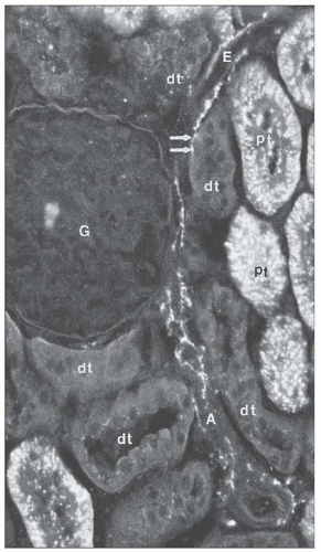
FIGURE 1.22 Nerve fibers viewed by catecholamine fluorescence histochemistry are seen along the afferent (A) and efferent (E) arterioles of the rat kidney. A spot of fluorescence (arrows) appears adjacent to a distal tubule (DT). G, glomerulus; PT, proximal tubule. (×306.) (From Barajas L. Innervation of the renal cortex. Fed Proc 1978;37:1192.)


Calyces and Renal Pelvis

The renal collecting system consists of the calyces, the sac-like renal pelvis, and the ureter (see Figs. 1.9 and 1.12) (48,49,50,51). The calyces and a portion of the renal pelvis are enveloped by the renal sinus fat. The most proximal portions of the collecting system are 9 to 11 funnel-shaped minor calyces that surround the individual papillary tips, both simple and compound (see Fig. 1.10). They have slender most proximal extensions termed fornices. The number of pyramids exceeds the number of calyces because of pyramid fusion. The major calyces represent the confluence of the minor calyces and unite to form the renal pelvis, which represents the expanded upper portion of the ureter. There is no distinct delineation between the pelvis and the ureter; rather, a gradual transition occurs.

The renal calyces and pelvis have a continuous muscular wall. Some smooth muscle fibers begin in the proximal fornices at the base of the papillae and extend along the calyces to the pelvis and ureter. In addition, there is a ring of smooth muscle that encircles the base of the pyramid. Pacemaker cells located in the most proximal fornix appear to initiate rhythmic peristaltic waves, 2 to 3 per minute, that aid urine movement toward the bladder (48,49,50,51). It has been proposed that during the rhythmic contractions, the ring of muscle fibers compress the papillae creating positive and negative pressures (40,41). With contraction, the papilla is elongated, and its diameter decreases by 20%. Fluid is forced along the ducts of Bellini to the papillary tip, and the vasa recta capillaries collapse. Fluid is also forced into the collapsed CDs cells. With relaxation, the negative pressure moves fluid from the CD cells into the interstitium and then into the descending and ascending vasa recta capillaries. Blood flow resumes first in the descending vasa recta capillaries and then in the ascending vasa recta capillaries, moving the water forward and thus contributing to the concentration mechanism of the renal papilla.


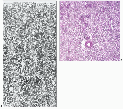
FIGURE 1.23 A: Longitudinal section through the cortex of a human kidney demonstrates the arrangement of the cortical labyrinth (CL) and the medullary rays (MRs). The cortical labyrinth contains the interlobular vessels and the glomeruli together with the convoluted tubules; the medullary rays contain the straight tubular portions and collecting ducts. (Paraffin section, ×75.) B: This section from a human kidney shows two medullary rays with their longitudinally oriented tubules. An artery and glomeruli are centrally located within the cortical labyrinth between the two medullary rays. Notice that the tubules of the outer stripe of the outer medulla located beneath the artery appear similar to those in the medullary rays. (Periodic acid-Schiff, × 20.)

The collecting system is lined by a unique epithelium known as transitional epithelium or urothelium. This epithelium is specialized to adapt to pelvic distension with major changes in volume of the collecting system. Urothelium is also impermeable to withstand the chemical environment of the urine, which fluctuates tremendously in chemical composition. The urothelium is thinner in its initial portions in the minor calyces but usually has five or six cell layers in the non-distended pelvis and ureter. It is covered by a superficial layer of large rounded cells, the umbrella cells. The urothelium rests on
a loose vascularized connective tissue layer, the lamina propria, with an underlying thin muscularis propria.


ARCHITECTURAL ORGANIZATION OF THE CORTEX AND MEDULLA


Cortical Labyrinth and Medullary Rays

The cortex of the human kidney is approximately 1 cm thick (excluding the column of Bertin) (see Figs. 1.5 and 1.10). It is organized into two architectural regions: the cortical labyrinth and the medullary rays (Figs. 1.23 and 1.24) (17,52,53,54,55,56,57,58,59,60,61). The cortical labyrinth contains the glomeruli, proximal, and distal convoluted tubules (DCTs), connecting tubules (CTs), and the initial portion of the CDs, as well as interlobular arteries and veins, arterioles, venules, capillaries, and lymphatics (Fig. 1.25). The principal components of the labyrinth, by volume, are the proximal convoluted tubules. In the normal cortex, the tubules are closely packed with their basement membranes (BMs) in close apposition. The interstitial space is scant and contains the peritubular capillary plexus and interstitial cells.


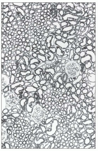
FIGURE 1.24 Cross-section (1-µm section of Epon-embedded tissue) through the cortex of the human kidney. In the cortex, the cortical labyrinth can clearly be delineated from the medullary rays (the cross section of one ray is marked by a dashed line). Within the labyrinth, the interlobular vessels (A, artery; V, vein), the glomeruli, and the convoluted tubular segments are found. The medullary rays contain the straight tubular segments and collecting ducts. (×140.)


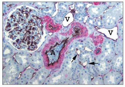
FIGURE 1.25 Human renal cortical labyrinth. The cortical labyrinth contains glomeruli, vessels, and tubules, mostly proximal tubules. The tubules have closely apposed basement membranes with little interstitial space, largely occupied by peritubular capillaries (arrows). The smooth muscle of the interlobular artery (A) and arterioles is stained red for smooth muscle actin. The veins (V) have very thin walls lacking smooth muscle. The endothelial cells of glomeruli, peritubular capillaries (arrows), and larger vessels are stained black for an endothelial marker, CD31. (×150.)

The medullary rays are elongated conical regions faintly visible in optimal planes of section of the renal cortex (see Fig. 1.10). Their name derives from the tubular segments they carry that are identical to those of the outer stripe of the outer medulla. They are, in effect, projections of medullary tissue into the cortex (see Fig. 1.23B). The medullary rays are aligned perpendicular to the corticomedullary junction.

They form from the confluence of parallel arrays of CDs and the proximal and distal straight tubules of the superficial and midcortical nephrons as they course down into, and back up from, the medulla. The straight tubules of the superficial nephrons are in the central portion of a medullary ray; straight tubules from the deeper nephron form the outermost layers.


Renal Lobule

There are two anatomic versions of the renal lobule (17,52,53,54,55,56). In one version, the nephrons that empty into the CDs of a single medullary ray constitute a lobular unit. A general sense of the lobular organization is apparent in well-oriented histologic sections perpendicular to the medullary rays and parallel to the capsular surface (see Fig. 1.24). The other concept of a renal lobule is based on vascularization relationships. In this viewpoint, the interlobular artery is the center of the lobule, which then includes the nephrons it supplies (see Fig. 1.16). Its borders would be the CDs within the medullary rays. In both concepts of a renal lobule, the limits of the lobule are indistinct because there is no connective tissue separation from an adjacent lobular unit.


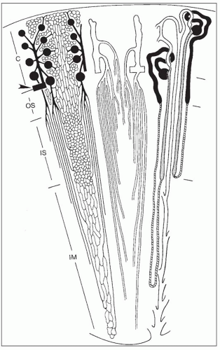
FIGURE 1.26 Schematic diagram of microvasculature and nephrons (not drawn to scale). C, cortex; OS, outer stripe; IS, inner stripe; IM, inner medulla. Left: Arterial vessels and capillaries. An arcuate artery (arrowhead) gives rise to an interlobular artery (cortical radial artery) from which afferent arterioles originate. Efferent arterioles from superficial and midcortical glomeruli split off into the cortical peritubular capillaries. Efferent arterioles of juxtamedullary glomeruli descend into the outer stripe and divide into the descending vasa recta, which supply the different capillary plexuses in the medulla. Middle: Venous vessels. Interlobular veins start in the superficial cortex. Stellate veins (characteristically found in the human kidneys), which begin on the renal surface, are not shown. The deep portions of interlobular veins and arcuate veins accept the ascending vasa recta, which drain the venous blood from the medulla. Ascending vasa recta and descending vasa recta together establish the vascular bundles. Note the dense pattern of ascending recta traversing the outer stripe as wide, tortuous channels. Right: A short- and a long-looped nephron together with a collecting duct. Glomeruli and proximal tubules are drawn black. Thin limbs are hatched; thick limbs are dotted. Distal convoluted tubules, connecting tubules (including an arcade), and collecting ducts are white. This drawing allows for the correlation of the location of tubules and vessels; the left, middle, and right views should be imagined as being superimposed on each other.


Cortical Microvascularization

The gross vasculature and general aspects of the initial portions of renal vascularization have been previously described. The microvascularization of the kidney (tubulovascular relationships) has been studied with various injection techniques, coupled with microscopic, ultrastructural, and radiographic techniques in many species. They show a similar pattern of organization (55,56,57,58,59,60,61).

The microvascularization of the cortex begins with the glomerular afferent arteriole (Figs. 1.26 and 1.27). The afferent arteriole enters the renal corpuscle at the hilum and immediately branches to form the capillary loops of the glomerular tuft; these loops ultimately converge to become the efferent arteriole. The efferent arterioles of the superficial and midcortical
nephrons are short and quickly transition into a peritubular capillary plexus that forms a uniformly distributed anastomosing vascular lattice amid the cortical tubules of the labyrinth. In the medullary rays the capillary plexus assumes a more longitudinal orientation, following the course of the straight tubules.


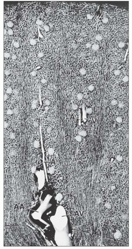
FIGURE 1.27 Longitudinal section through the cortex of a human kidney. The arteries have been injected with silicone rubber (Microfil). The different capillary patterns of the cortical labyrinth and of the medullary rays are visible. Within the cortical labyrinth, the interlobular (corticoradial) arteries (A) and veins (V) as well as the glomeruli are found. Within the medullary rays, the capillary plexus consists of elongated meshes. (×70.) AA, arcuate artery; AV, arcuate vein.

The efferent arterioles of the deep or juxtamedullary glomeruli descend into the medulla as discussed below.

The tubules of the superficial glomeruli are perfused by capillaries derived from their efferent arterioles, whereas in the midcortical nephrons, efferent arterioles perfuse both tubules of their originating nephron and tubules of adjacent nephrons. Since the efferent arterioles of the juxtamedullary nephrons enter the renal medulla, tubules of these nephrons are perfused by efferent arterioles of midcortical glomeruli. The peritubular capillary plexus of the cortex drains first from the medullary rays into the labyrinth and then enters small venules that converge and continue on as the major venous drainage previously discussed.


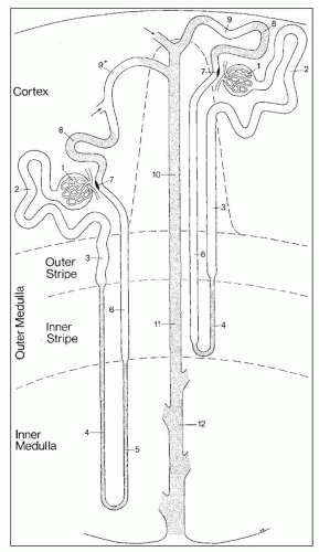
FIGURE 1.28 Schematic drawing of the nephron. This scheme depicts a short-looped and a long-looped nephron together with the collecting system. Not drawn to scale. Within the cortex, a medullary ray is delineated by a dashed line. 1: Renal corpuscle including Bowman capsule and the glomerulus (glomerular tuft). 2: Proximal convoluted tubule. 3: Proximal straight tubule. 4: Descending thin limb. 5: Ascending thin limb. 6: Distal straight tubule (thick ascending limb). 7: Macula densa located within the final portion of the thick ascending limb. 8: Distal convoluted tubule. 9: Connecting tubule. 9*: Connecting tubule of the juxtamedullary nephron that forms an arcade. 10: Cortical collecting duct. 11: Outer medullary collecting duct. 12: Inner medullary collecting duct. (From Kriz W, Bankir L, Bulger RE, et al. A standard nomenclature for structures of the kidney. The Renal Commission of the International Union of Physiological Sciences. Kidney Int 1988;33:1.)


Medulla

The medulla is divided into an outer medulla, composed of an outer stripe and an inner stripe, and the inner medulla or papilla (Figs. 1.26 and 1.28). The anatomic limits of each region are defined by their differing tubular composition (Table 1.2).

They also exhibit unique and physiologically important tubulovascular relationships (52,53,54,55,56,62,63,64,65,66).


Outer Medulla


OUTER STRIPE

The outer stripe is the thinnest portion of the renal medulla in humans but is thicker in other animals such as in the rat’s unipapillary kidney (Fig. 1.29). The outer stripe contains the continuation of the straight portion of the PTs, CDs, and thick ascending limbs of Henle. The straight portion of the PTs and ascending thick limbs of the juxtamedullary nephrons are closest to the vascular bundles (Figs. 1.30 and 1.31). These are surrounded by the straight portion of the PTs and ascending thick limbs of the mid- and superficial nephrons. Farthest from the vascular bundles are the CDs, which reside in the interbundle zone.


INNER STRIPE

The beginning of the inner stripe is defined by transition of the straight portion of the PT into the thin descending limb of Henle (Figs. 1.28 and 1.32). The short-looped nephrons of the superficial and midcortex loop around to return to the cortex at various levels in the inner stripe. The vascular bundles beginning to aggregate in the outer strip become well defined in the inner stripe (Fig. 1.33).

The tubulovascular organization of the inner stripe shows species variation that affects urine-concentrating ability. In humans and other species such as rabbits, pigs, and
monkeys, the vascular bundles are of the simple type, and the tubulovascular relationships of the outer stripe are maintained in the inner stripe. The descending thin limbs (DTLs) and ascending thick limbs of the long-looped juxtamedullary nephrons are situated close to the vascular bundle, whereas the DTLs and ascending thick limbs of the short-looped nephron and the CDs are more peripheral (Fig. 1.33). In other species, such as rat, mouse, and desert rodents, the vascular bundles are of the so-called complex type. In this type, DTLs of shortlooped nephrons leave the interbundle region and curve toward the vascular bundles, intermingling with ascending vasa recta. The thick limbs of both short- and long-looped nephrons and the thin limbs of long-looped nephrons remain in the interbundle region with CDs. These histotopographic differences in tubulovascular relationships generally correlate with urinary concentration ability. However, exceptions do occur such as in the hamster with its simple vascular bundles and high concentration capacity.


TABLE 1.2 Tubular segments within each zone of the medulla


Outer stripe


Straight part of the proximal tubule (pars recta)


Thick ascending limb of Henle


Collecting ducts


Inner stripe


Thin descending limb Henle


Thick ascending limb of Henle


Collecting ducts


Inner medulla


Thin descending limb of Henle


Thin ascending limbs of Henle


Large collecting ducts (ducts of Bellini)


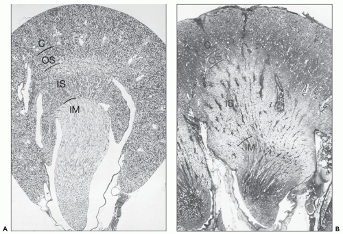
FIGURE 1.29 Longitudinal sections (with respect to the papilla) through a unipapillary kidney (rat) (A) and a single papilla of a multipapillary kidney (human) (B). Note the different proportions of the individual zones. The outer stripe in the human kidney is very narrow, and the inner stripe is thick. (Paraffin sections, A: ×10; B: ×3.) C, cortex; OS, outer stripe; IS, inner stripe; IM, inner medulla.


Inner Medulla (Papilla)

The inner medulla is defined as the point at which the thin ascending limb of Henle of the long-looped nephrons transforms into the thick ascending limbs of Henle. The inner medulla therefore contains the thin descending and thin ascending limbs of Henle derived from the long-looped nephrons of the deep cortex and the CDs (ducts of Bellini) (Figs. 1.28, 1.34, and 1.35). Since the short-looped and long-looped nephrons make their looping return back toward the cortex at various levels in the inner stripe of the outer medulla and the inner medulla, respectively, the absolute number of tubules decreases, which accounts for the tapering of the medulla toward the papillary tip. In parallel, the confluence of CDs with descent through the medulla results in a progressive increase in CD size from the outer medulla to the papillary tip. In animals with higher concentrating need, the inner medulla progressively increases in size relative to the rest of the kidney. At the papillary tip, the area cribrosa, the ducts of Bellini epithelium transforms into the urothelium of the collecting system.

The vascular bundles become attenuated in the inner medulla as they lose their muscular investments and progressively decrease in number. The CDs remain peripheral to the vascular elements, and the thin descending and thin ascending limbs are interspersed within the vasculature. Near the papillary tip, the vasa recta disappear. The interstitial space increases distally, and interstitial cells become increasingly prominent (Fig. 1.35).


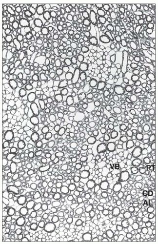
FIGURE 1.30 Cross section (1-µm section of Epon-embedded tissue) through the outer stripe in the outer medulla. The tubules are arranged around the vascular bundles (VB). In the vicinity of the bundles, the nearest segments are the straight proximal and distal tubules of the jux-tamedullary nephrons. More peripherally located to them are the straight proximal and distal tubules of midcortical and superficial nephrons; the collecting ducts are lying distant from the bundles. (×140.) PT, proximal tubule; AL, ascending limb (straight parts of distal tubule); CD, collecting duct.


Medullary Microvascularization

The medullary blood supply has two sources as previously mentioned: the efferent arterioles of the juxtamedullary nephrons and arteriolar branches from the spiral arteries that are derived from the interlobar arteries and supply the papillary tip (see Figs. 1.17 and 1.26). There are no arteries, veins, or lymphatics within the renal medulla (62,63,64,65,66).

The principal blood supply to the medulla derives from efferent arterioles of the juxtamedullary glomeruli. The efferent arterioles begin as a splay of arterioles that descend toward the medulla, where they branch in the outer stripe. A few branches provide a capillary plexus to the tubules of the outer stripe, although most continue on into the inner stripe. They converge as the descending arteriolae rectae, forming organized bundles in the inner stripe of the outer medulla (Figs. 1.33 and 1.36). At various points as they descend, additional branches supply the tubules within the interbundle regions of the inner stripe with a rich capillary plexus. The bundle size diminishes as it descends into the inner medulla.


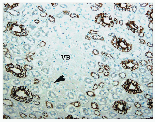
FIGURE 1.31 Human kidney, outer medulla. The central vascular bundles (VB) are surrounded by the descending straight tubules (arrowhead), which are unstained, and the smaller stained ascending thick limb tubules. The largest stained tubules are collecting ducts. (Immunoperoxidase stain for cytokeratin AE 1/3, ×250.)

The descending arteriolae rectae destined for the inner medulla supply no branches to the tubules of the inner stripe. Therefore, there is complete separation of the blood supply to the inner stripe from that of the inner medulla. The capillary plexus of the inner medulla is sparse but richer at the papillary tip than in the more superficial portions of the inner medulla where almost all the vessels are vasa recta arterioles. Arterioles from spiral arterial branches of the intralobar arteries contribute to the blood supply of the papillary tip (see Fig. 1.17). These compartmentalized vascular patterns provide a basis for the differing zones of medullary injury associated with the various causes of papillary necrosis.

As the arterioles enter the interbundle regions, they gradually lose their smooth muscle and pericytic coats to enter a fenestrated venous system that ascends back toward the cortex to form the ascending venae rectae (Figs. 1.26, 1.34, and 1.37). The inner medullary venous outflow occurs exclusively via vascular bundles. The inner medullary ascending venae rectae enter the vascular bundles at the junction of the inner and outer medulla and then leave the vascular bundles as they approach the outer stripe and course outward. The capillary plexus of the outer stripe also join the ascending venae rectae. These two venous outflows remain separate within the inner stripe, but both receive capillary venous outflow as they enter the outer stripe. This arrangement is important in urine concentration.

Within the outer stripe, the venous ascending venae rectae have a thin attenuated and fenestrated endothelium and represent a large fraction of the vascular plexus of the outer stripe,
which is sparse in true capillaries. They also travel outward from the bundle as they enter the outer stripe. The ascending venules finally empty into interlobular or arcuate veins. The venae rectae and the arteriolae rectae are arranged in close proximity throughout their course as they travel down to the papillary tip and back up. The intermingled venous and arterial limbs compose the countercurrent exchange system that maintains the medullary osmotic gradient.


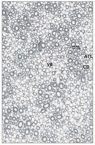
FIGURE 1.32 Cross-section (1-µm section of Epon-embedded tissue) through the inner stripe of the outer medulla. The vascular bundles (VB) are well developed and surrounded by the limbs of short and long loops of Henle in an arrangement similar to that in the outer stripe. (×140.) DTL, descending thin limb; ATL, ascending thick limb; CD, collecting duct.


NEPHRONS

The nephron consists of the metanephric-derived structures, the renal corpuscle, and a lengthy cylindrical epithelial-lined tubular component. The nephron does not, strictly speaking, include the CD, which is ureteric bud derived (28,32).

However, it is not uncommon for the term nephron to be used in reference to both the true nephron components as well as the CD. The status of the CTs is unclear. Ultrastructural studies implicate ureteric bud origin, whereas microdissection studies support metanephric blastema derivation (28,32).
The tubular portion of the nephron has complex spatial and topographic relationships with its microvasculature and demonstrates sequential variation in its cellular constitution tightly linked to function.


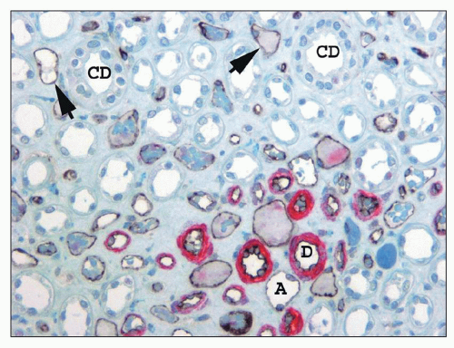
FIGURE 1.33 Human kidney, outer medulla. The descending arteriolae rectae (D) in the vascular bundle have a prominent smooth muscle media (stained red for smooth muscle actin), whereas the thin ascending venae rectae (A) and the interbundle capillary plexus (arrows) have no smooth muscle but can be distinguished from the thin limbs of Henle because their endothelial cell lining is stained black with CD 31. (×350.) CD, collecting duct.


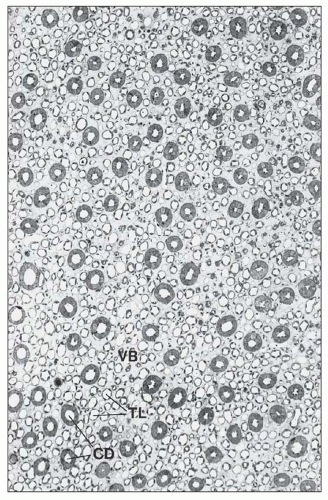
FIGURE 1.34 Cross-section (1-µm section of Epon-embedded tissue) through the inner medulla. The vascular bundles (VB) are small and cannot clearly be separated from the surrounding thin limbs (TLs). (×112.) CD, collecting duct.


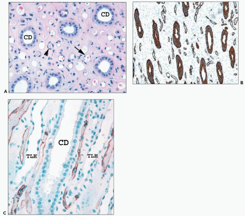
FIGURE 1.35 Human kidney inner medulla. A: The collecting ducts (CDs) are easily identified. However, the thin limbs (arrowhead) and vessels (arrow) appear similar. (×300.) B: The interstitium of the inner medulla is prominent compared with the density of tubules. The thin limbs of Henle and collecting ducts are highlighted by cytokeratin 7 stain. (×300.) C: CD31 labels vascular endothelium of the vasa recta, which are positioned between the thin limb (TLH) and collecting ducts (CDs). The descending vasa recta at the lower right are shown looping back as ascending vasa recta. (×350.)


Nephron Number

The number of nephrons in the normal kidney is important because nephron number may play a role in the development of hypertension and progression of chronic renal disease (67,68,69,70,71). Nephron number is influenced by many factors that affect renal development. Low birth weight, preterm birth, reduced kidney mass and size, short stature, maternal hyperglycemia, maternal protein malnutrition, and gene polymorphism for PAX2 and RET are associated with reduced nephron numbers (68). Low nephron numbers are associated with increased glomerular volume and an increased risk of hypertension. Older patients also have fewer nephrons because of age-related obsolescence (67,68,69,70,71). Recent studies have shown a broad range in nephron numbers, with some patients having substantially fewer nephrons than others. Neugarten et al. (70) found that nephron numbers per kidney ranged from 400,000 to 800,000. Hughson et al. (71) found similar average numbers but an even broader range, between 227,000 and 1,825,000 nephrons per kidney, an eightfold range that varied with birth weight. In rats and rabbits, nephron numbers are much lower, estimated at 30,000 per kidney.


Types of Nephrons

Classification of nephrons is based on either topographic location in the cortex or on function (52,53,54). The three types of nephrons as classified according to their topographic location in the cortex are the superficial, midcortical, and juxtamedullary. As their name indicates, the superficial nephrons are located within the outer cortex. They send arterioles to the subcapsular regions. The juxtamedullary nephrons are deeply situated at the corticomedullary junction and send arterioles into the medulla, which converge to form the descending vascular bundles. The midcortical nephrons are situated between
the other two, but there is no distinct boundary between these three regions.


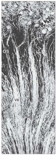
FIGURE 1.36 Arterial vessels of the medulla of a mouse injected with silicone rubber (Microfil). The juxtamedullary glomeruli give rise to efferent arterioles (arrows), which split into the descending vasa recta, establishing the arterial part of the vascular bundle (VB). The bundles are best developed in the inner stripe (IS) and decrease after transition into the inner medulla (IM). C, cortex; OS, outer strip. (×85.)


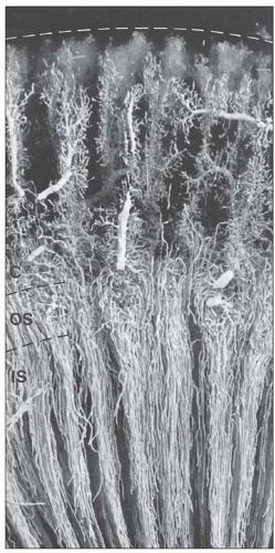
FIGURE 1.37 Venous vessels of rabbit kidney injected with silicone rubber (Microfil). The interlobular veins of the cortex accept the blood from the cortical plexuses and descend to the corticomedullary border. The venous vessels of the medulla are the ascending vasa recta, which ascend within and between the vascular bundles toward the corticomedullary border, where they empty into arcuate veins or the basal portions of the interlobular veins. C, cortex; OS, outer stripe; IS, inner stripe. (×20.)

Nephrons are more commonly classified functionally by the length of their loop of Henle, which varies in the point at which it bends to return to the cortex (72). Short-looped nephrons arise in the superficial and midcortical regions. They send the loops of Henle into the outer medulla where they bend and return toward the cortex. Juxtamedullary and some deep midcortical nephrons are the long-looped nephrons. They send the loops of Henle into the inner medulla before bending at various levels to return to the cortex. The percentage of longlooped nephrons shows great species variation; to some degree,
this is related to the needs for urinary concentration and water conservation. However, urinary concentration correlates more with the complexity of medullary development rather than simply the number of long-looped nephrons (62,63,64,65,66). In the human kidney, approximately 15% of nephrons are long looped. In the rodent kidney, this is almost doubled to 28%, whereas in cats and dogs, all nephrons are long looped (72).


RENAL CORPUSCLE


General Structure and Histology

The renal corpuscle or glomerulus is a spherical to ellipsoid cluster of capillaries and matrix housed within a connective tissue structure, the Bowman capsule (BC). The BC is the dilated proximal-most extension of the PT (17,32,52,73,74,75). The glomerular tuft floats within a fluid-filled cavity, the Bowman space, and is tethered to the BC at its vascular pole. The glomerular tuft consists of the glomerular capillaries, and the glomerular mesangium, which is contiguous with the extraglomerular mesangium that forms the central portion of the JGA. In rarely observed, optimally oriented sections, the glomerular vascular pole pedicle and the urinary pole outflow are usually positioned on opposite sides of the glomerulus (Fig. 1.38). The glomerulus has a mean diameter of approximately 200 µm in the adult human; juxtamedullary glomeruli and glomeruli in patients with a solitary kidney are larger (17,73).


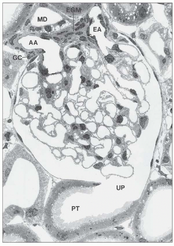
FIGURE 1.38 Glomerulus with juxtaglomerular apparatus of the rat kidney. The afferent arteriole (AA), the efferent (EA), and the extraglomerular mesangium (EGM) are in close contact with the macula densa (MD) of the thick ascending limb. UP, urinary pole; PT, proximal tubule; GC, granular cell. (Electron micrograph, ×900.)

The glomerular tuft consists of a central axial branching mesangium invested by a profusion of capillaries (17,32,52,73,74,75,76,77) (Figs. 1.38, 1.39, 1.40, 1.41). The glomerular capillaries have a unique status within the vascular system; they are situated between two arteriolar systems. This arrangement is necessary to maintain, and to allow, modulation of the intravascular pressure required for glomerular filtration. The glomerular tuft is formed by an afferent arteriole that divides immediately on entering the BC to form lobules of glomerular capillaries. Two to five lobules are observed in reconstructions of rat glomeruli, although this lobular arrangement is difficult to appreciate in histologic sections.
There are afferent, more peripherally arrayed glomerular capillary domains and efferent, more centrally located domains, with the former constituting most of the capillaries (73,74,75,76,77). There are multiple anastomoses between capillaries within a lobule, and between capillaries in separate lobules, creating a capillary network (Fig. 1.41).


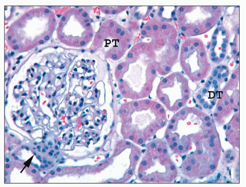
FIGURE 1.39 Human cortex. The macula densa (arrow) and the adjacent extraglomerular lacis cells are on the left; the ostium of the proximal tubule (PT) is on the right. The parietal epithelium lining Bowman capsule is flattened and abruptly transitions to the columnar proximal tubular epithelium at the urinary pole. Most tubules are proximal, but two distal tubules (DTs) are present. (H&E, ×200.)

The capillaries of the efferent domain converge on the vascular pole to become the efferent arteriole (77) (see Figs. 1.38 and 1.40). This convergence is established within the glomerular tuft and splits the two initial branches of the entering afferent arteriole as the efferent arteriole exits the glomerulus. The afferent arteriole has a larger diameter than the efferent arteriole due to a more substantial media and a larger lumen, but these features are often difficult to appreciate since it is rare to capture both in an identical plane of section. The afferent arteriole in Figure 1.40 from a patient with hypertension is easy to recognize because of hyalinosis, which affects only the afferent arteriole in nondiabetics.


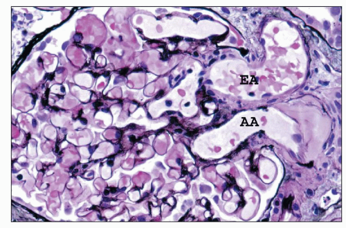
FIGURE 1.40 Human glomerulus with vascular pole. The thin delicate capillary loop basement membranes and slender profiles of mesangial matrix are stained black. The afferent arteriole is identified by the presence of hyalinosis. (Jones methenamine silver, (×400.) AA, afferent arteriole; EA, efferent arteriole.


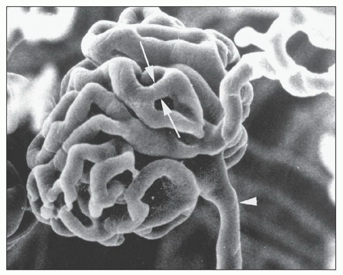
FIGURE 1.41 Scanning electron micrograph of a vascular cast of a rat glomerulus. The afferent arteriole (arrowhead) and efferent arteriole are shown. A capillary loop anastomosis is shown (between arrows). (×450.) (From Gattone VH II, Evan AP, Willis LR, et al. Renal afferent arteriole in the spontaneously hypertensive rat. Hypertension 1983;5:8.)

The glomerular tuft is supported by two forms of the extracellular matrix: the mesangial matrix and the glomerular capillary loop basal lamina (Fig. 1.42). A central scaffolding function is provided by the axial branching mesangial matrix.

The encircling capillary loops are principally supported by their basal lamina, which forms an incomplete vascular cylinder since the mesangium constitutes a portion of the capillary loop wall. Although basal lamina is the correct anatomic term for the extracellular matrix layer demonstrable by electron microscopy that separates podocytes and endothelial cells, the basal lamina is commonly referred to as the glomerular basement membrane (GBM). The glomerular capillary loop component demonstrable by light microscopy (LM) includes both the cellular elements—podocytes and endothelial cells—and the central matrix component, the GBM.

The glomerulus consists of three cell types: mesangial cells, endothelial cells, and epithelial cells (17,52) (Fig. 1.42). Mesangial cells reside within the mesangial matrix. In the normal human glomerulus, mesangial cells should number no more than two per mesangial area. Endothelial cells completely line the inner surface of the glomerular capillaries with an attenuated layer of cytoplasm, and they have an oval nucleus that projects into the capillary lumen usually near the mesangial interface. There are two types of epithelial cells: podocytes, also known as the visceral epithelial cells, and parietal epithelial cells (PECs). Podocytes are arrayed along the exterior surface of the capillary loops and over the mesangial waist region (a region not covered by capillaries). They have prominent protruding nuclei and lightly stained cytoplasm. At the vascular pole, the podocytes transition into the PECs that line the BC. PECs are thin squamous-like epithelial cells that line the entire inner surface
of the BC. Parietal cells abruptly transition into columnar proximal tubular cells at the urinary pole (see Figs. 1.38 and 1.39).


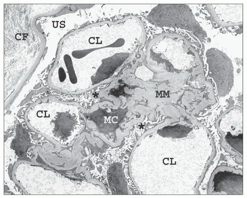
FIGURE 1.42 Transmission electron micrograph of a relatively normal human glomerulus. The mesangial matrix (MM) contains a mesangial cell (MC), and the capillary loops (CLs) are lined by thin fenestrated endothelium. The endothelial cell nuclei are located near the mesangial interface. Podocytes invest both the outer aspects of the capillary loops and the mesangial waist regions (asterisk). The urinary space (US) separates the glomerulus from the Bowman capsule, which is lined by flat parietal epithelial cells. Interstitial collagen fibrils (CF) are visible on the outer aspect of the basal lamina of the Bowman capsule. (×2200.)

In some glomeruli, two distinct cells may be interposed between the visceral and PECs, parietal podocytes, and periportal cells (78,79,80). The parietal podocyte is present in most glomeruli. It is a podocyte that sends cell processes to cover capillary loops as a typical podocyte but also sends cell processes to the parietal BM of the BC (79). Cell processes in both areas have foot processes (FPs) with slit diaphragms (SDs), suggesting some filtration function across the BC either into the JGA or the cortical interstitium.

Periportal cells are granulated glomerular epithelial cells that form around the vascular pole. Peripolar cells are present in only a minority of human glomeruli (3% to 28%), primarily in outer cortical glomeruli (79,80). These cells form junctional complexes with podocytes and PECs. Peripolar cells are closely associated with renin-containing cells in the afferent arteriolar wall. Periportal cells contain dense granules, morphologically similar to renin granules; but the granules do not appear to contain renin. Their function is not known.

The three glomerular cells—podocytes, endothelial cells, and mesangial cells—can often be recognized in hematoxylin and eosin (H&E)-stained sections, most readily identified in the peripheral portions of the glomerular tuft where structural relationships are best appreciated. The glomerular cells are difficult to identify separately in the central regions of the glomerulus and in the abnormal glomerulus. Histochemical stains that permit precise resolution of the relationship between cells and matrix components facilitate more confident cellular identification. Periodic acid-Schiff (PAS) stain and a silver-based stain, two commonly used stains in renal biopsy evaluation, are most suitable for this purpose. The mesangial matrix is well delineated by both stains. However, the silver stain reveals a more delicate detail of the GBM since it stains only the lamina densa (Figs. 1.40 and 1.43). The PAS stain provides better cellular detail but stains all cytoplasmic and matrix components of the capillary loop. Appreciation of general structural features of the glomerulus and tubules is also possible by
viewing an H&E-stained section under fluorescence (Fig. 1.44). This is useful in kidney biopsies for correlation with immunofluorescence findings (81).


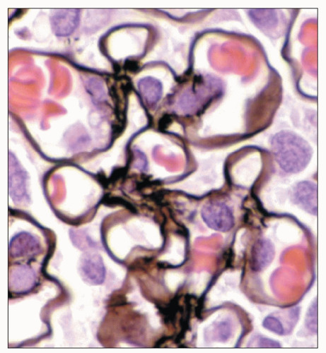
FIGURE 1.43 Normal human glomerulus. The thin delicate detail of the capillary loops and scant mesangium is nicely delineated by the Jones methenamine silver stain. (×650.)


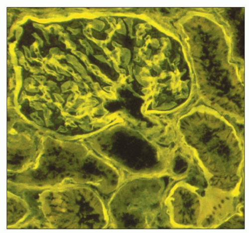
FIGURE 1.44 Normal human cortex. This H&E-stained section is viewed under fluorescent microscopy. The general architectural features of the glomerulus and tubules are easily recognized. (H&E, ×250.)

In addition to routine histochemical stains mentioned above that rely on relationships between cells and the matrix for cell identification, more precise analysis is possible. Many molecules can be targeted with modern immunohistochemical reagents and other techniques to identify specific matrix components, clarify cell lineage, assess cell cycling status, or identify other physiologically important molecules, such as adhesion molecules (integrins, selectins, cadherins), cytokines and their inhibitors, complement inhibitors, other protein inhibitors, antiinflammatory eicosanoids, antithrombotic molecules, antioxidants, heat shock proteins, and protein phosphatases.


Cellular Components of the Glomerulus


Podocyte

Podocytes are the largest cells in the glomerulus. They have a highly specialized three-dimensional structure and unique molecular profile closely linked to the critical functions they perform (82,83,84,85,86,87,88,89,90,91,92,93,94). Podocytes participate in GBM synthesis and repair, provide structural support for the capillary loop, and are the principal component governing the permselectivity of the capillary loop. Podocytes also participate in cross talk with endothelial cells and mesangial cells and may have contractile properties that affect capillary loop diameter (83,85,95,96,97,98,99).

The normal adult podocyte is a terminally differentiated cell that relinquishes mitotic capability during maturation once formation of FPs occurs (83,85,95,96,97,98,99). Nuclear division without cell division does on occasion occur, resulting in binucleated and multinucleated podocytes, a phenomenon that is most marked in nephropathic cystinosis (100). The state of fixed quiescence appears necessary for proper podocyte cell function. Cell division requires reorganization of the cytoskeleton, which would disrupt the highly ordered arrangement of the FPs, affecting cell attachment to the GBM and the filtration selectivity of the SD. The quiescent state is maintained by a tightly regulated balance between several families of molecules that control cell cycle: cyclins, cyclin-dependent kinases, and the cyclin-dependent kinase inhibitors such as p21, p27, and p57. The mature podocyte expresses p27, p57, and cyclin D3 (95,101,102,103,104). Although podocytes were previously regarded as fixed in a terminally differentiated state, it is now clear that reentry into the cell cycle occurs in a few glomerular diseases, such as HIV nephropathy and other collapsing forms of focal segmental glomerulosclerosis, leading to the concept of the dysregulated podocyte. In these conditions, p21 is reexpressed, cyclin D1 increases, and p27, p57, and cyclin D3 decrease with resultant podocyte proliferation confirmed by PCNA and Ki-67 staining (95,101,102,103,104).

The molecular signature of podocytes is unique among epithelial cells of the kidney. Podocytes are the only cells in the adult kidney to express intranuclear WT-1 and are the only glomerular cells to express C3b receptor and at least in cell culture smooth muscle proteins smoothelin, calponin, and myocardin (Fig. 1.45) (94). Podocytes also have a paradoxical intermediate filament profile (105). Podocytes express vimentin, a ubiquitous intermediate filament expressed by mesenchymal cells but not usually by epithelial cells (Fig. 1.46A). Vimentin expression shows a restricted pattern, limited to the cell body and cell processes but absent in the FPs (Fig. 1.47). Cytokeratin, the intermediate filament of most epithelial cells, is not expressed in podocytes but is expressed in PECs and all other renal epithelial cells (Fig. 1.46B).


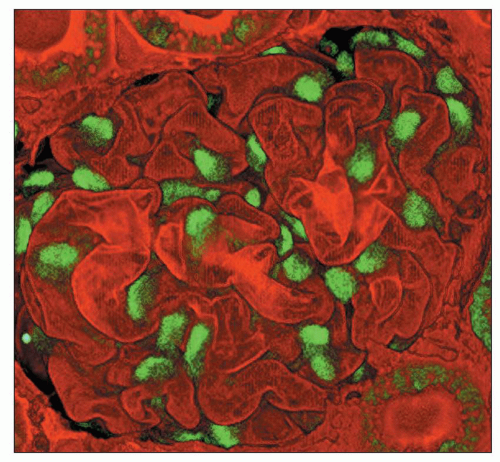
FIGURE 1.45 Two-photon microscopy of a rat glomerulus. Podocytes are the only renal cells of the mature kidney that express WT-1; note the intense nuclear stain. The GBM is stained with Lens culinaris. (×550.) (Photograph courtesy of Carrie Phillips, MD.)


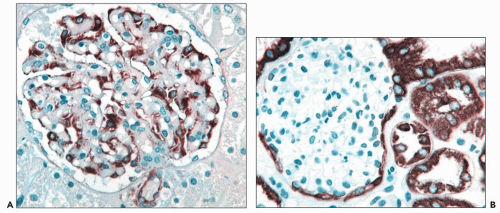
FIGURE 1.46 Human kidney. A: Podocytes express vimentin intermediate filaments while parietal epithelial cells are negative. (Immunoperoxidase stain for vimentin, ×350.) B: Parietal epithelial cells and proximal and distal tubular cells express cytokeratin while podocytes are negative. (Immunoperoxidase stain for cytokeratin 8, ×300.)


PODOCYTE ULTRASTRUCTURE

Podocytes are large octopus-like cells polarized relative to the GBM (86,87,88,92,93,94). Transmission electron microscopy and scanning electron microscopy (SEM) have unveiled their unique and complex structure (Figs. 1.48 and 1.49). Three major structural regions have been identified: the cell body, cell processes, and FPs. The cell body is usually located within a valley created by the reflection of adjacent capillary loops. The cell body contains the nucleus and other major organelles such as abundant rough endoplasmic reticulum (RER), an extensive Golgi apparatus, mitochondria, and frequent lysosomes. Occasionally, rudimentary cilia are present projecting from the surface. The cell body does not rest directly on the GBM. It is separated from the GBM by the subpodocyte space and a layer of FPs (Fig. 1.49). Based on serial three-dimensional transmission electron microscope (TEM) reconstructions, the subpodocyte space covers approximately 60% of the GBM. It may be a dynamic compartment that can fluctuate in volume, restrict movement of the ultrafiltrate into the urinary space, provide a mechanism for podocyte participation in modulating capillary permeability, and chemically communicate with endothelial and mesangial cells possibly by reverse flux across the GBM (91).


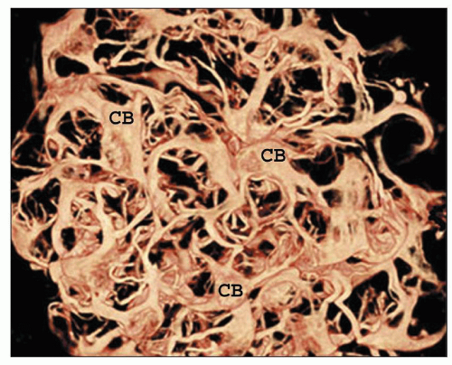
FIGURE 1.47 Two-photon image of a rat glomerulus. Vimentin intermediate filaments are restricted by the cell body (CB) and cell processes. The intervening foot processes are not visible since they lack vimentin. (Immunofluorescent stain for vimentin, ×600.) (Photograph courtesy of Carrie Phillips, MD, and Indiana Center for Biological Microscopy.)

Long, slender, arborizing cell processes extend from the cell bodies. These processes contain few organelles but are rich in microtubules and intermediate vimentin filaments that run parallel to the long axis (88,89,90,91,92,93,94,95,96). The cell processes participate in cell trafficking and maintain cell shape and rigidity.

They may branch into secondary or even tertiary cell processes before investing multiple capillary loops (Figs. 1.48 and 1.49). The cell processes ultimately give rise to terminal structures called FPs or pedicels.

The name foot processes is derived from the original transmission electron microscopic studies of normal kidneys that revealed slender processes arranged perpendicular to the capillary loop BM (Fig. 1.50). The three-dimensional architecture of the podocyte and its FPs was first understood following the beautiful panoramic SEM studies published in the 1970s and 1980s (86,87,88) (see Fig. 1.49). These investigators demonstrated that the podocyte FPs are slender pectinate cytoplasmic finger-like extensions. The FPs have a smooth luminal surface and interdigitate with the FPs of adjacent podocytes. FPs are attached to the BM by integrins, the principal ones are α3β1 integrin and α and β dystroglycans (83,95,96,97,98,99).

Only gold members can continue reading. Log In or Register to continue

Stay updated, free articles. Join our Telegram channel

Jun 21, 2016 | Posted by in UROLOGY | Comments Off on Renal Anatomy and Histology

Full access? Get Clinical Tree

Get Clinical Tree app for offline access