Pauci-Immune and Antineutrophil Cytoplasmic Autoantibody-Mediated Crescentic Glomerulonephritis and Vasculitis


Pauci-Immune and Antineutrophil Cytoplasmic Autoantibody-Mediated Crescentic Glomerulonephritis and Vasculitis


J. Charles Jennette

David B. Thomas


A landmark publication in 1979 by Stilmant et al. (1) reported that many if not most patients with crescentic glomerulonephritis did not have evidence for anti-glomerular basement membrane (anti-GBM) antibodies or immune complexes in glomeruli. Of 46 patients with crescentic glomerulonephritis, 16 (35%) had no substantial glomerular immunoglobulin deposits, 10 (22%) had linear IgG deposits indicative of anti-GBM disease, 19 (41%) had immune complex disease, and 1 had malignant hypertension (1). This category of crescentic glomerulonephritis with no evidence for mediation by anti-GBM antibodies or deposited immune complexes has been called idiopathic crescentic glomerulonephritis or primary crescentic glomerulonephritis; however, the most widely used term currently is pauci-immune crescentic glomerulonephritis, which is based on the paucity of glomerular staining for immunoglobulins. Pauci-immune crescentic glomerulonephritis occurs as a renal-limited disease and as a component of systemic necrotizing small-vessel vasculitis (2,3,4,5,6,7,8). In fact, most patients with pauci-immune crescentic glomerulonephritis have at least some constitutional signs and symptoms that raise the possibility of systemic vasculitis, and approximately 75% have overt evidence for systemic vasculitis. Some have advocated calling it renal-limited vasculitis (RLV) because the glomerular lesion in patients with necrotizing small-vessel vasculitis, such as microscopic polyangiitis (MPA) and granulomatosis with polyangiitis (GPA; Wegener granulomatosis), is indistinguishable from that seen in patients with pauci-immune crescentic glomerulonephritis in the absence of systemic vasculitis (4,5).

In 1988, Falk and Jennette (6) demonstrated that most patients with pauci-immune crescentic glomerulonephritis have antineutrophil cytoplasmic autoantibodies (ANCA), including patients with and without systemic vasculitis (6). This has been confirmed by many investigators (2,5,7,8,9,10,11,12,13). Approximately 85% to 95% of patients with active untreated pauci-immune crescentic glomerulonephritis are found to be ANCA positive (11,13,14,15,16,17,18,19,20). Crescentic glomerulonephritis that is not associated with ANCA and does not have evidence for immune complex or anti-GBM mediation is rare and accounts for no more than 5% of all crescentic glomerulonephritis (5,21).

Pauci-immune crescentic glomerulonephritis (i.e., ANCA crescentic glomerulonephritis) is the most common cause of crescentic glomerulonephritis (i.e., with crescents in 50% or more of glomeruli) (Fig. 16.1) (7,21,22). Pauci-immune crescentic glomerulonephritis is most prevalent in older patients. For example, pauci-immune crescentic glomerulonephritis was the category of crescentic glomerulonephritis found in 79% (201 of 256) of patients older than 60 years of age, compared with 48% (145 of 303) of patients 21 to 60 years old and 42% (31 of 73) of patients younger than 21 years of age (21). Most patients with pauci-immune crescentic glomerulonephritis are ANCA positive, and approximately 90% of ANCA-positive patients who have glomerulonephritis have crescent formation (21). The pathologic and clinical features of disease, including the association with systemic vasculitis, are similar in patients with pauci-immune crescentic glomerulonephritis who are
ANCA positive compared with those who are ANCA negative (23). ANCA-associated pauci-immune crescentic glomerulonephritis is the most common cause for the renal component of pulmonary-renal vasculitic syndrome (24,25). For example, in a study of 88 patients with pulmonary-renal syndrome by Niles et al. (24), 48 (55%) had ANCA, 6 (7%) had anti-GBM antibodies, and 7 (8%) had both. Thus, 63% of patients with pulmonary-renal syndrome had ANCA.


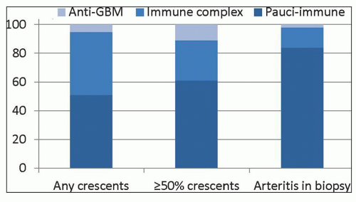
FIGURE 16.1 Frequency of immunopathologic categories of glomerulonephritis in native kidney biopsies evaluated by immunofluorescence microscopy in the University of North Carolina Nephropathology Laboratory. Data are derived from 540 patients with any crescents, 195 patients with ≥50% crescents, and 37 patients with arteritis in the biopsy (137). The anti-GBM patient with arteritis was ANCA positive.

The systemic vasculitides that may be accompanied by pauci-immune crescentic glomerulonephritis include granulomatosis with polyangiitis (GPA, Wegener granulomatosis), microscopic polyangiitis (MPA), and eosinophilic granulomatosis with polyangiitis (EGPA, Churg-Strauss syndrome) (3,22,26,27). In a patient with pauci-immune crescentic glomerulonephritis, a diagnosis of GPA is appropriate when there is evidence for necrotizing granulomatous inflammation but no asthma. A diagnosis of EGPA is appropriate when there is a history of asthma and eosinophilia and there is granulomatous pulmonary disease. And a diagnosis of MPA is appropriate when there is systemic necrotizing small-vessel vasculitis with no evidence for granulomatous inflammation or asthma (Table 16.1) (26). Clinical or pathologic evidence of renal disease is seen in approximately 90% of patients with MPA, 80% of patients with GPA, and 45% of patients with EGPA (Table 16.2) (28,29,30,31,32,33,34,35,36). EGPA syndrome has the lowest incidence of renal disease, but, when present, it is pathologically indistinguishable from the renal injury caused by MPA and GPA (22,37,38). The proportion of patients with renal involvement varies among cohorts in part based on the subspecialty interests of the investigators, with cohorts studied by nephrologists having a higher frequency of renal disease than those studied by pulmonologists and otolaryngologists.


TABLE 16.1 Definitions of ANCA-associated vasculitis, microscopic polyangiitis, granulomatosis with polyangiitis, and eosinophilic granulomatosis with polyangiitis based on the 2012 Chapel Hill Consensus Conference on the Nomenclature of Systemic Vasculitis


ANCA-associated vasculitis (AAV)


Necrotizing vasculitis, with few or no immune deposits, predominantly affecting small vessels (i.e., capillaries, venules, arterioles, and small arteries), associated with MPO-ANCA or PR3-ANCA. Not all patients have ANCA. Add a prefix indicating ANCA reactivity, for example, PR3-ANCA, MPO-ANCA, ANCA-negative.


Microscopic polyangiitis (MPA)


Necrotizing vasculitis, with few or no immune deposits, predominantly affecting small vessels (i.e., capillaries, venules, or arterioles). Necrotizing arteritis involving small and medium arteries may be present. Necrotizing glomerulonephritis is very common. Pulmonary capillaritis often occurs. Granulomatous inflammation is absent.


Granulomatosis with polyangiitis (GPA)


Necrotizing granulomatous inflammation usually involving the upper and lower respiratory tract and necrotizing vasculitis affecting predominantly small to small vessels (e.g., capillaries, venules, arterioles, arteries, and veins). Necrotizing glomerulonephritis is common.


Eosinophilic granulomatosis with polyangiitis (EGPA)


Eosinophil-rich and necrotizing granulomatous inflammation often involving the respiratory tract and necrotizing vasculitis predominantly affecting small to medium vessels and associated with asthma and eosinophilia. ANCA is more frequent when glomerulonephritis is present.


Modified from Jennette JC, Falk RJ, Bacon PA, et al. 2012 Revised International Chapel Hill Consensus Conference Nomenclature of Vasculitides. Arthritis Rheum 2013;65:1-11.


Godman and Churg (39) recognized the relatedness of MPA, GPA, and EGPA in 1954, and this conclusion is supported by the association of these three disorders with ANCA (13,14,15,16,17,18,19,20). MPA, GPA, and EGPA also share pathologically indistinguishable pauci-immune systemic vasculitis that involves many different vessels, such as alveolar capillaries, dermal venules, sinus mucosal venules, and arterioles and arteries in peripheral nerves, skeletal muscles, and most viscera (22,26,27). The major pathologic difference in the kidneys is the occurrence of interstitial granulomatous inflammation in GPA and EGPA; however, identification of interstitial granulomatous inflammation in renal biopsy specimens from patients with GPA or EGPA is rare.


CLINICAL PRESENTATION


ANCA Serology

Pauci-immune crescentic glomerulonephritis is almost synonymous with ANCA crescentic glomerulonephritis, because 85% to 90% of pauci-immune crescentic glomerulonephritis occurs in ANCA-positive patients and 80% to 90% of the crescentic glomerulonephritis that occurs in ANCA-positive patients is pauci-immune (5,7,8,9,10,11,13,21,75,76). Serologic detection of ANCA is a useful diagnostic marker not only for renal-limited pauci-immune crescentic glomerulonephritis but also for MPA and GPA, and to a lesser extent EGPA. As with all serologic markers, however, ANCA are not absolutely specific or sensitive and thus must be interpreted in light of the other available data. Approximately 90% to 95% of patients with active untreated GPA or MPA, and approximately 45% of patients with EGPA, are ANCA positive (30). Thus, ANCA serology is least sensitive for EGPA; however, three quarters or more of EGPA patients with evidence for glomerulonephritis have ANCA (77).

ANCA are specific for proteins in the cytoplasmic granules of neutrophils and the lysosomes of monocytes (6,14,17,18,78). Davies et al. (79) first reported ANCA in the serum of patients with systemic small-vessel vasculitis and pauci-immune necrotizing glomerulonephritis. This association was confirmed by Hall et al. (80) in 1984. ANCA did not receive much attention until 1985 when a collaborative European group reported a strong association between ANCA and active GPA (81). Subsequent investigations revealed that ANCA also are associated with MPA, EGPA, renal-limited pauci-immune crescentic glomerulonephritis, and certain forms of drug-induced vasculitis (2,6,9,63,82,83,84).

Although ANCA are far more frequent in pauci-immune small-vessel vasculitis and glomerulonephritis, ANCA are identified in the serum of approximately one third of patients with anti-GBM disease (21,85,86,87,88,89,90). Compared with patients with anti-GBM alone, patients with both autoantibodies are older, often have features of systemic small-vessel vasculitis, and have a better prognosis for renal survival with treatment (85). The concurrent anti-GBM and ANCA specificity of serum is the result of two different antibody populations rather than one cross-reacting population (88,91), and the antigen specificities of anti-GBM antibodies are the same in patients with anti-GBM antibodies alone compared to those with ANCA and anti-GBM antibodies (91).

Two major antigen specificities for ANCA are seen in patients with pauci-immune crescentic glomerulonephritis and small-vessel vasculitis. These two specificities cause two staining patterns, cytoplasmic (c-ANCA) and perinuclear (p-ANCA), when ANCA are detected by indirect immunofluorescence microscopy using alcohol-fixed neutrophils as substrate. The perinuclear distribution of antigen is an artifact of substrate preparation that results from redistribution of the antigen from the cytoplasm to the nucleus during substrate preparation (92).
More precise determination of antigen specificity is accomplished by enzyme immunoassay. The most common cANCA specificity is for proteinase 3 (PR3-ANCA) (93,94,95), and the most common p-ANCA specificity is for myeloperoxidase (MPO-ANCA) (6). p-ANCA that do not have specificity for MPO are associated with various inflammatory diseases, such as ulcerative colitis, sclerosing cholangitis, autoimmune hepatitis, rheumatoid arthritis, and Felty syndrome (18). Thus, a positive p-ANCA result in this context is a “false-positive” result with respect to pauci-immune crescentic glomerulonephritis even though it is an analytically true positive result and is a true positive result for inflammatory bowel disease. Because of this and other instances in which a positive indirect immunofluorescence microscopy assay (IFA) result does not correspond to a positive MPO-ANCA or PR3-ANCA, an international consensus conference recommendation is to perform both IFA for p-ANCA and c-ANCA and enzyme-linked immunosorbent assay (ELISA) for MPO-ANCA and PR3-ANCA when testing for the presence of pauci-immune crescentic glomerulonephritis or small-vessel vasculitis (96). When both a positive IFA and positive ELISA are required to conclude that the ANCA serology is positive, this positive result has a sensitivity of approximately 81% and a specificity of approximately 96% for pauci-immune crescentic glomerulonephritis (with or without systemic vasculitis) (16). Figure 16.2 shows the calculated predictive value of an ANCA serology result with this sensitivity and specificity. As with all diagnostic testing, the positive and negative predictive values of the test are dependent on the pretest likelihood of the disease, which in turn is determined by the prevalence of the disease in patients with the signs and symptoms of the patient being tested. As noted in Figure 16.1, pauci-immune crescentic glomerulonephritis has a prevalence of approximately 50% in all patients with crescents and an even higher prevalence when there are more than 50% of glomeruli with crescents. Assuming a prevalence (pretest likelihood) of 50%, a positive ANCA result in a patient with signs and symptoms of rapidly progressive glomerulonephritis would yield a posttest likelihood of the disease (positive predictive value) of 95%, and a negative result would yield a posttest likelihood of no disease (negative predictive value) of 85%. The negative predictive value in this instance is very close to the percentage of all patients with pauci-immune crescentic glomerulonephritis who in fact are ANCA negative using first-generation ANCA immunoassays. When there is low pretest likelihood of pauci-immune crescentic glomerulonephritis, for example, in patients with hematuria and proteinuria but no renal insufficiency, the major value of ANCA testing is to rule out ANCA disease or to increase the suspicion for ANCA disease to a level that will prompt more extensive and rapid diagnostic evaluation (e.g., renal biopsy). In this setting, a negative result provides a negative predictive value mathematically at essentially 100%. Although a positive result in this setting has a very low positive predictive value that certainly would not warrant immediate institution of immunosuppressive therapy, it does increase the likelihood of this life-threatening disease to a level that would warrant more careful and timely diagnostic evaluation.

The antigen specificity of ANCA correlates to a degree with the category of pauci-immune small-vessel vasculitis, although overlap is so great that antigen specificity alone cannot be used to differentiate between them. Patients with active GPA usually have PR3-ANCA. In North America and Europe, patients with MPA have slightly more MPO-ANCA than PR3-ANCA, and patients with EGPA and renal-limited pauci-immune crescentic glomerulonephritis have predominantly MPO-ANCA (Fig. 16.3). ANCA specificity correlates with clinical symptoms independent of the clinicopathologic variants, for example, 90% of patients with ANCA who have destructive upper respiratory tract disease (e.g., saddle nose deformity) have PR3-ANCA, whereas most patients with exclusively or predominantly renal involvement have MPO-ANCA (50). Dual positivity for both MPO-ANCA and PR3-ANCA is rare (13) except in the setting of drug-induced ANCA (71). It is extremely important to note that some patients with otherwise absolutely classic disease have pauci-immune crescentic glomerulonephritis, MPA, GPA, or EGPA, but are ANCA negative. Thus, a negative ANCA result by no means rules out these diseases, and patients who are ANCA negative have the same prognosis and should receive the same treatment as ANCA-positive patients (23).


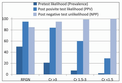
FIGURE 16.2 Predictive value of ANCA serology for pauci-immune crescentic glomerulonephritis in adults with clinical evidence for glomerulonephritis (e.g., dysmorphic hematuria and proteinuria) and different degrees of renal insufficiency. In a patient with RPGN, the pretest likelihood of pauci-immune crescentic glomerulonephritis is approximately 50%, increases to 95% with a positive result, and falls to 15% with a negative result. By comparison, in a nephritic adult with normal renal function, the pretest likelihood of ANCA disease of less than 5% increases to only approximately 30% with a positive result but is essentially 0% with a negative result; however, the positive result increases the likelihood enough to warrant expeditious further evaluation including renal biopsy. RPGN, rapidly progressive glomerulonephritis with rapid loss of GFR and overt active urinary finds of glomerulonephritis; PPV, positive predictive value; NPV, negative predictive value. (Data derived from Lim LC, Taylor JG III, Schmitz JL, et al. Diagnostic usefulness of antineutrophil cytoplasmic autoantibody serology: Comparative evaluation of commercial indirect fluorescent antibody kits and enzyme immunoassay kits. Am J Clin Pathol 1999;111:363.)

Roth et al. (97) studied the epitope specificity of MPO-ANCA IgG using a highly sensitive assay. They detected low titer MPO-ANCA natural autoantibodies in healthy controls directed against a limited set of epitopes. In patients with MPO-ANCA disease, autoantibodies against greater than 20 MPO epitopes were identified. MPO-ANCA IgG in patients
with active disease reacted with a restricted set of epitopes that were not recognized by IgG from patients in remission. Interestingly, this assay method detected MPO-ANCA in ANCA-negative disease that reacted against a sole linear MPO epitope. This reactivity could only be detected using IgG rather than serum because serum contains a ceruloplasmin fragment that binds to MPO and blocks detection of these autoantibodies by routine serologic methods (97). Thus, some ANCA-negative patients appear to in fact have MPO-ANCA that is not detected by first-generation clinical ANCA assays; however, this experimental observation has not been confirmed, and an assay to detect these masked ANCA is not commercially available at this time.


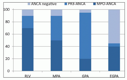
FIGURE 16.3 Approximate frequency of MPO-ANCA and PR3-ANCA in patients with active untreated GPA, MPA, EGPA, or renal-limited pauci-immune crescentic glomerulonephritis (RLV). GPA patients have a lower frequency of ANCA-positivity if there is no renal involvement. EGPA patients are greater than 75% ANCA-positive when glomerulonephritic is present. These data are for patients in North America and Europe. Patients in Asia have a marked predominance of MPO-ANCA in all clinicopathologic variants. MPA, microscopic polyangiitis; GPA, granulomatosis with polyangiitis; EGPA, eosinophilic granulomatosis with polyangiitis.

Kain et al. (98,99) have reported that lysosomal membrane protein 2 (LAMP2) is another major ANCA autoantigen in addition to MPO and PR3 in patients with pauci-immune small-vessel vasculitis and glomerulonephritis. LAMP2-ANCA were detected in patients with either MPO-ANCA, PR3-ANCA, or ANCA-negative pauci-immune crescentic glomerulonephritis. LAMP2 is homologous to a bacterial adhesin called FimH, and LAMP2-ANCA have been hypothesized to arise through molecular mimicry caused by infection with fimbriated Gram-negative bacteria. To support this hypothesis, Kain et al. reported induction of crescentic glomerulonephritis in rabbits immunized with FimH. However, our research group has not been able to reproduce these results (100).


PATHOLOGIC FINDINGS


Gross Pathology

In patients with severe disease, the kidneys are normal or slightly increased in size. When acute pauci-immune crescentic glomerulonephritis is extensive and severe, the subcapsular surface and cut surfaces have scattered small red dots caused by blood within the Bowman spaces and in tubular lumens. Slightly larger irregular petechial lesions may be present and represent hemorrhage from necrotic glomeruli and small interlobular arteries. Occasionally, pale or hemorrhagic nodular inflammatory lesions can be discerned in arcuate arteries and interlobar arteries. However, grossly discernible nodular and aneurysmal lesions in interlobar and larger arteries are rare in the pauci-immune small-vessel vasculitis, unlike the higher frequency of such lesions in polyarteritis nodosa and Kawasaki disease (see Chapter 17). When grossly identifiable arterial lesions are present, they are usually in the arcuate and interlobar arteries and may manifest as pale nodules or foci of hemorrhage. One may see cortical infarcts, which are usually small. In Heptinstall’ series (101) of 18 patients with systemic arteritis and glomerulonephritis, which was consistent with MPA, 16 patients had arteritis in the kidney at postmortem examination, 4 had grossly identifiable aneurysms, and 7 had infarcts. Therefore, the presence of grossly identifiable aneurysms does not differentiate between the pauci-immune small-vessel vasculitides and polyarteritis nodosa, although such aneurysms are much more frequent in the latter.

A few patients, especially those with GPA, have focal hemorrhagic papillary necrosis that is caused by leukocytoclastic angiitis affecting the medullary vasa recta (3,102,103). The lesions usually appear as fan-shaped hemorrhagic areas in the inner medulla. Watanabe et al. (102) observed papillary necrosis in 5 of 23 autopsies of patients with GPA.

Grossly apparent granulomatous inflammation is rare in the kidneys of patients with GPA. It causes irregular pale to hemorrhagic zones that may involve the cortex or medulla.

Hydronephrosis caused by ureteral obstruction from vasculitic and granulomatous lesions is a rare complication of GPA (104,105). Ureteral involvement by granulomatous inflammation has been a rare problem in renal transplants in patients with GPA (106). Even when ureteral obstruction is not present, careful examination of the ureter may reveal areas of hemorrhage or nodularity that prove to be vasculitic or granulomatous lesions when they are examined microscopically.

Pauci-immune small-vessel vasculitis can affect any organ of the body and may produce gross lesions in these organs. Favored sites, in addition to the kidney, include the skin, respiratory tract, gastrointestinal tract, skeletal muscle, and peripheral nerves. Small foci of hemorrhage at sites of vascular rupture caused by necrotizing inflammation are the most frequent manifestation of vasculitis. A variation on this theme is the more extensive pulmonary hemorrhage that can result from alveolar capillaritis. Grossly, the granulomatous inflammation of GPA and EGPA, which most often affects the respiratory tract, typically produces irregular pale zones with varying degrees of hemorrhage. Granulomatous inflammation in the lungs may produce cavities, and granulomatous inflammation in the nose may cause perforation of the nasal septum or collapse of the bridge of the nose.


Light Microscopy


Glomeruli

The hallmark histologic lesions of acute pauci-immune ANCA glomerulonephritis are crescents and fibrinoid necrosis (Figs. 16.4, 16.5, 16.6, 16.7), which occur at the same frequency irrespective
of the presence or absence of associated systemic vasculitis (2,3,11,12,21,22,36,70,71,107). The glomerular inflammation is accompanied by proportional nonspecific tubulointerstitial inflammatory lesions (107). In an analysis of 45 ANCA-positive patients with glomerulonephritis and systemic small-vessel vasculitis, renal biopsies demonstrated crescents in 93% of patients and glomerular necrosis in 98% (2). In a study of 32 renal biopsies from patients with MPA, Savage et al. (36) identified glomerular segmental necrosis in 100% and crescent formation in 88%. Likewise, in a study of 20 renal biopsies in MPA by D’Agati et al. (108), 80% of 20 specimens had segmental glomerular necrosis, and 85% had crescents. The severity of acute lesions ranges from focal segmental fibrinoid necrosis and crescent formation affecting less than 10% of glomeruli to severe diffuse necrotizing and crescentic glomerulonephritis with global necrosis of virtually all glomeruli. On average in a given renal biopsy specimen, 45% to 55% of glomeruli have crescents, and 20% to 25% of glomeruli have fibrinoid necrosis (12). The fibrinoid necrosis usually is accompanied by crescent formation. For example, Hauer et al. (109) identified fibrinoid necrosis in the absence of extracapillary hypercellularity in only 1 of 87 glomeruli from renal biopsy specimens of pauci-immune crescentic glomerulonephritis that were examined by serial section. Eisenberger et al. (23) compared glomerular crescents, necrosis, and sclerosis in patients with ANCA-negative pauci-immune glomerulonephritis and patients with ANCA-positive pauci-immune glomerulonephritis and found no significant difference, which corresponds to our experience.


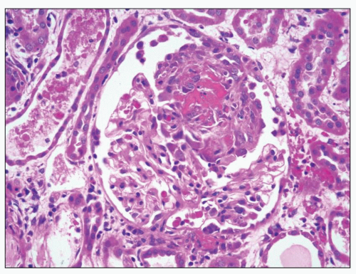
FIGURE 16.4 Segmental fibrinoid necrosis that is deeply acidophilic surrounded by early crescent formation. Note that the nonnecrotic segments are relatively unremarkable with thin capillary walls and no hypercellularity. Also note the acute tubulointerstitial changes including interstitial edema and tubular epithelial cell simplification (e.g., of the tubule in the upper left that has flat epithelium and cellular debris in the lumen). (H&E.)


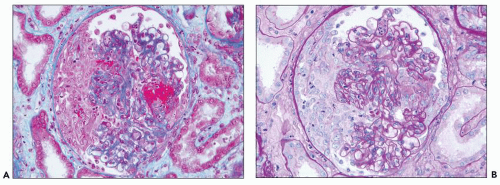
FIGURE 16.5 Glomerulus from a patient with MPO-ANCA renal-limited pauci-immune crescentic glomerulonephritis stained with Masson trichrome (A) and PAS (B) showing a well-formed cellular crescent on the left and a well-defined irregular focus of segmental fibrinoid necrosis on the right that stains red with the Masson trichrome stain (A). Most capillary lumens are patent, but there are a few neutrophils in some capillaries, for example, at the top of the tuft in (B). Note also the edema in the periglomerular interstitium.

The extent of crescent formation is not different between patients with PR3-ANCA versus MPO-ANCA. Jennette (21) observed a mean of 48% glomerular involvement with crescents in patients with MPO-ANCA (n = 102) versus 46% with PR3-ANCA (n = 52), Hauer et al. (12) observed 46% with MPO-ANCA (n = 58) versus 50% with PR3-ANCA (n = 63), and Vizjak et al. (11) observed 44% with MPO-ANCA (n = 74) versus 39% with PR3-ANCA (n = 55).

Fibrinoid necrosis is fuchsinophilic (red) with trichrome staining (Figs. 16.5A and 16.6A), a feature that distinguishes it from the sclerosis (blue or green) that develops at sites of scarring caused by fibrinoid necrosis. Special stains for basement membranes, such as Jones silver methenamine (Fig. 16.6B) and periodic acid-Schiff (PAS) (Fig. 16.6C), reveal disruption of the GBM, and often the Bowman capsule as well, in the areas of fibrinoid necrosis. Fibrin thrombi often are present in glomerular capillaries adjacent to necrotic zones (110) and have the same tinctorial properties as the fibrinoid material in necrotic segments and the fibrin between crescent cells. Foci

of fibrinoid necrosis in pauci-immune crescentic glomerulonephritis often contain neutrophil granule constituents indicating that there has been extensive neutrophil activation and degranulation at these sites. For example, using double immunohistochemical staining procedures, Bajema et al. (111) identified PR3, MPO, elastase, and lactoferrin at sites of glomerular fibrinoid necrosis.


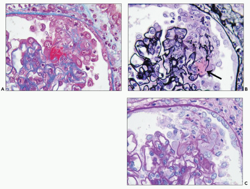
FIGURE 16.6 Glomerulus from the same patient as in Figure 16.5 stained with Masson trichrome (A), Jones silver methenamine (B), and PAS (C) showing a small focus of fibrinoid necrosis with adjacent early crescent formation. The fibrinoid material is bright red (fuchsinophilic) with the trichrome stain (A) and is associated with breaks in the glomerular basement membrane that can be seen best with the Jones stain (arrow in B).


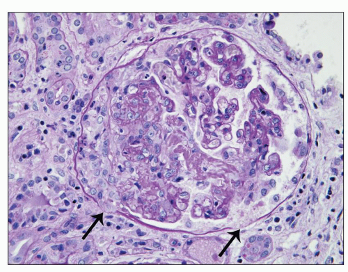
FIGURE 16.7 Glomerulus from a patient with microscopic polyangiitis showing segmental fibrinoid necrosis with an adjacent relatively small cellular crescent. The PAS stain allows demarcation of focal dissolution of Bowman capsule basement membrane (arrows). Glomerular basement membranes and mesangial matrix have been destroyed in the area of fibrinoid necrosis. Note the periglomerular tubulointerstitial inflammation and edema.


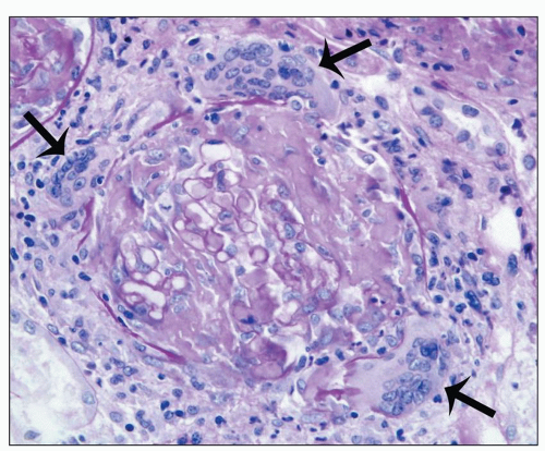
FIGURE 16.8 Glomerulus from a child with cANCA GPA showing severe fibrinoid necrosis of the glomerular tuft, extensive disruption of Bowman capsule basement membrane, and prominent periglomerular granulomatous inflammation with multinucleated giant cells (arrows). (PAS stain.)

Crescent formation appears to begin adjacent to foci of segmental necrosis (Figs. 16.4, 16.6, and 16.7) (109), but it can extend to surround most, if not all, of the tuft forming a circumferential crescent. Breaks in the Bowman capsule, which are identified best with special stains such as PAS or Jones silver methenamine, are common in pauci-immune crescentic glomerulonephritis (75,76) (see Fig. 16.7). There may be extensive dissolution of the Bowman capsule with severe necrotizing glomerular injury, resulting in continuity between the glomerular inflammation and pronounced periglomerular inflammation (Fig. 16.8). This extremely lytic necrosis is similar to focal lytic lesions in many other small vessels in ANCA disease (112). Periglomerular interstitial infiltrates of leukocytes sometimes have a granulomatous appearance (11,113). Vizjak et al. (11) observed periglomerular granulomatous inflammation more often in MPO-ANCA disease than in PR3-ANCA disease, which indicates that this reaction is not related to the type of granulomatous inflammation that corresponds to GPA. Hauer et al. (12) observed no difference in the extent of periglomerular granulomatous inflammation in patients with GPA versus MPA or in patients with PR3-ANCA versus MPO-ANCA. Occasional specimens have multinucleated giant cells in the glomerular or periglomerular infiltrates. The multinucleated giant cells seem to localize preferentially to sites of fragmentation of the Bowman capsule. Which is cause and which is effect, if either, is unknown. When there are only a few residual fragments of glomerular structure remaining, care must be taken not to mistake this destructive glomerular inflammation for purely interstitial necrotizing granulomatous inflammation. This may require examination of the specimen at multiple levels of section with special stains that highlight residual fragments of GBM and the Bowman capsule.

Only gold members can continue reading. Log In or Register to continue

Stay updated, free articles. Join our Telegram channel

Jun 21, 2016 | Posted by in UROLOGY | Comments Off on Pauci-Immune and Antineutrophil Cytoplasmic Autoantibody-Mediated Crescentic Glomerulonephritis and Vasculitis

Full access? Get Clinical Tree

Get Clinical Tree app for offline access