Membranoproliferative Glomerulonephritis


Membranoproliferative Glomerulonephritis


Xin J. (Joseph) Zhou

Fred G. Silva


Membranoproliferative glomerulonephritis (MPGN) refers to a morphologic pattern that includes many etiologically distinct forms of glomerulonephritis in which, as the name implies, there is thickening of the glomerular capillary wall (membrano-) as well as an increase in the number of cells in the glomerular tuft (-proliferative). Since the hypercellularity, which often is most prominent in the mesangium, and capillary wall thickening cause consolidation and expansion of the segments (lobules), there is often accentuation of the lobules of the glomeruli (hypersegmentation). An old term for this pattern that is rarely used today is lobular glomerulonephritis. This pattern is also referred to as mesangiocapillary glomerulonephritis because of the extensive involvement of the mesangium and the extension of the mesangial cells into the subendothelial portions of glomerular capillary walls. The MPGN pattern of glomerular injury may be either idiopathic (primary) or secondary to a wide variety of disease states (Tables 8.1 and 8.2). The idiopathic category is diminishing as more recognizable etiologies are identified. For example, hepatitis C is associated with a majority of cases that were previously identified as idiopathic MPGN type I and essential mixed cryoglobulinemia (52,53). An additional subset of previously idiopathic type I MPGN is now known to be caused by genetically determined or acquired defects in regulation of the alternative complement pathway (C3 glomerulopathy with MPGN type I pattern) (Fig. 8.1). Hence, MPGN is a morphologic pattern that should be interpreted in the context of etiologies and underlying diseases/conditions. The term idiopathic should be avoided in the pathologic diagnosis because it may impede the search for a recognizable cause.

Historically, the MPGN is subclassified into MPGN type I (the most common form), type II (dense deposit disease [DDD]), and type III based on the combined features of light, immunofluorescence (IF), and electron microscopy (EM). In recent years, there have been great advances in our understanding of the pathogenesis of MPGN, particularly in the area of complement-mediated C3 glomerulopathies, including DDD and C3 glomerulonephritis (52,53,117,118). Thus, the
traditional classification of MPGN requires modification. A useful classification system should meet the following criteria: (a) define the entity clearly so that the precise connotation of the term can be immediately apparent; (b) be clinically significant, useful, and therapeutically relevant; (c) be based on pathogenesis within the limitation of our current knowledge; and (d) be relatively easy for all to use and morphologically reproducible (119). The traditional pathologic classification of MPGN was based primarily on ultrastructural features and included MPGN type I, MPGN type II, and two variants of MPGN type III. The current classification recognizes the importance of IF (or immunohistochemistry) microscopy in further dividing MPGN into immune complex-mediated MPGN with glomerular immunoglobulins and complement deposition and MPGN with abnormalities in alternative complement pathway regulation resulting in isolated C3 deposits with little or no immunoglobulins by IF. MPGN type II is currently designated DDD and is recognized as a variant of C3 glomerulopathy (see Table 8.1 and Chapter 9).


TABLE 8.1 Pathologic variants of membranoproliferative glomerulonephritis


Membranoproliferative glomerulonephritis (MPGN) type I


Immune complex MPGN type I (Ig and C3 by IF)


Infection (e.g., bacterial endocarditis, hepatitis C)


Autoimmunity (e.g., mixed cryoglobulinemia)


Neoplasia (e.g., carcinoma, lymphoma)


C3 glomerulopathy variant of MPGN type I (C3 with little or no Ig by IF)


Genetic abnormality (e.g., mutations in complement factor H)


Autoimmunity (e.g., anti-complement factor H)


Dense deposit disease (MPGN type II) (C3 with little or no Ig by IF)


Genetic abnormality (e.g., mutations in complement factor H)


Autoimmunity (e.g., anti-complement factor H)


Infection (e.g., streptococcal)


MPGN type III


MPGN type III of Burkholder


Immune complex MPGN type IIIB (Ig and C3 by IF)


C3 glomerulopathy variant of MPGN type IIIB (C3 with little or no Ig by IF)


MPGN type III of Strife/Anders


Immune complex MPGN type IIIS/A (Ig and C3 by IF)


C3 glomerulopathy variant of MPGN type IIIS/A (C3 with little or no Ig by IF)


The less well-understood form of MPGN that is called MPGN type III is further divided into two variants. Burkholder et al. (120) identified a variant that has many of the features of MPGN type I but with the added presence of numerous electron-dense deposits on the subepithelial side of the glomerular capillary basement membranes. This pattern has some glomerular capillaries that are indistinguishable from type I, yet other segments of the glomerular tuft show changes similar to membranous glomerulopathy. This form of MPGN is now termed MPGN type III of Burkholder. The issue is complicated by the use of the term type III to describe a different form of MPGN described by Strife, Anders and coworkers (121,122). This form has intramembranous deposits of moderate to low electron density that bridge the glomerular basement membrane (GBM) and may connect to irregular subendothelial and the subepithelial deposits. EM of silver-stained sections accentuates the GBM abnormalities by demonstrating irregular silver-negative zones. This form of MPGN is now termed MPGN type III of Strife/Anders.

MPGN was described as a mixed proliferative and membranous glomerulonephritis in early reports prior to the recognition of the characteristic features by EM and IF. Fahr (123) probably included MPGN in the “intracapillary form” of subchronic glomerulonephritis. Cameron (124) concluded that at least two cases originally studied by Richard Bright at Guy’s Hospital in the 1820s represented a form of MPGN. Three of the original kidneys in the Gordon Museum at Guy’s Hospital were sectioned, and two of the kidneys had the histologic features of MPGN. As Cameron (124) states, it is remarkable that this pattern can still be recognized 150 years later, after preservation first with brandy and then with formalin. MacCallum in 1934 (125) and Ellis in 1942 (126) described patients with chronic progressive glomerulonephritis showing membranoproliferative and lobular features. Bell (127) described a series of patients with “latent chronic glomerulonephritis” or “chronic azotemic glomerulonephritis,” which probably represents what we now call MPGN. Allen (128) used the term chronic lobular glomerulonephritis and noted that there was a tendency for the “periphery of each lobule to be laminated by two or three layers of endothelial cells” and for splitting of the basement membranes to be present. Churg and Grishman (129) described 28 patients with subacute glomerulonephritis with mesangial cell interposition (i.e., between the glomerular endothelium and the GBM). They also commented that with time, the cellularity of the glomerular lesions decreased and central nodular scars developed. Habib et al. (130), in 1961, presented 108 nephrotic patients at a Ciba Foundation symposium. Fifteen patients had “endocapillary proliferative glomerulitis associated with hyaline nodules” (“lobular glomerulitis”). Seven additional patients had complex or unclassified forms of glomerular disease with thick glomerular capillary walls and endocapillary hypercellularity. The light and electron microscopic findings described at this benchmark symposium (that launched the modern era of renal biopsy) conform to what we now accept as MPGN type I.

David Jones (131), using an elegant staining method (periodic acid-methenamine silver stain) on thin paraffin-embedded and plastic-embedded sections, defined the characteristic glomerular capillary wall lesions in MPGN type I. He demonstrated the formation of a new basement membrane internal to the original GBM and described the continuity of the mesangial region with the peripheral capillary wall lesion. Arakawa and Kimmelstiel (132) described the histologic and ultrastructural appearance of circumferential mesangial cell interposition in a series of patients with diffuse (and often lobular) glomerulonephritis. They concluded that circumferential mesangial cell interposition can be regarded as a distinct form of mesangial proliferation in glomerulonephritis.

West et al. (133) and Gotoff et al. (134) almost simultaneously noted depletion of serum complement in children with chronic renal disease and named it hypocomplementemic persistent or chronic glomerulonephritis. The glomerular pathologic characteristics in these patients were increased cellularity,
thickening of the glomerular capillary walls, argyrophilic “splitting” of the basement membranes by nonargyrophilic material, and prominent lobulation of the tuft with some central hyaline zones (133,134). Subsequent studies showed similar glomerular lesions in other patients including patients who did not have hypocomplementemia.


TABLE 8.2 MPGN type I associated with known conditions


Autoimmune diseases


Mixed cryoglobulinemia (1,2,3,4)


Systemic lupus erythematosus (5,6)


Sjögren syndrome (7,8)


Henoch-Schönlein purpura (9)


Rheumatoid arthritis (10)


Infectious diseases


Bacterial


Infected ventriculoatrial shunts (11)


Endocarditis (12)


Visceral abscesses (13)


Brucellosis (14)


Tuberculosis (15)


Leprosy (16)


Lyme disease (17,18)


Mycoplasma (19)


Meningococcal meningitis (20)


Viral


Hepatitis B (21,22,23,24,25,26,27,28)


Hepatitis C (29,30,31,32,33)


HIV (7,34,35)


Hantavirus (36,37)


BK virus (38)


EBV (39)


Fungal


Candida endocrinopathy (40)


Protozoal


Filariasis (41)


Malaria


Schistosomiasis


Hydatid disease (42)


Dysproteinemia


Cryoglobulinemia


Monoclonal immunoglobulin deposition disease


Monoclonal gammopathy of undetermined significance (43)


Waldenström macroglobulinemia


Fibrillary glomerulonephritis


Immunotactoid glomerulonephritis


Neoplasms


Leukemias and lymphomas (44)


Epithelial tumors (45,46,47,48,49)


Abdominal desmoplastic round cell tumor (50)


Mixed-cell germinal ovary tumor (51)


Hereditary or genetic


Hereditary deficiencies of complement components including regulatory factors (52,53,54,55,56,57,58,59,60,61,62,63,64,65,66)


α1-Antitrypsin deficiency (67,68)


X-linked (69,70)


Autosomal dominant (70,71)


Autosomal recessive MPGN type I (66)


Down syndrome (72)


Gaucher disease (73)


Kartagener syndrome (74)


Nephropathy-gonadal dysgenesis type II (75)


Prader-Willi syndrome (76)


Turner syndrome (77)


Hereditary angioedema (78)


Familial Mediterranean fever (79)


Miscellaneous


Sarcoidosis (80)


Addison disease (81)


Castleman disease (82)


Celiac disease and sprue (83)


Coexisting glomerulonephropathies


Amyloidosis (84)


Diabetes mellitus (85)


Alport syndrome (86)


Polycystic kidney disease (87)


Cushing disease (88)


Drug abuse (89)


Hemolytic uremic syndrome (90)


Immunoglobulin and IgG subclass deficiency (91)


Pregnancy related (92)


Psoriasis vulgaris (93)


Renal artery dysplasia (94)


Renal vein thrombosis (95)


Takayasu arteritis (96)


Toxic oil epidemic syndrome (97)


Cryptogenic organizing pneumonia (98)


Ulcerative colitis (99)


Hypocomplementemic urticarial vasculitis syndrome (100)


Bone marrow transplantation (101)


Renal allografts


Recurrent glomerulonephritis (102,103,104,105,106,107,108,109,110,111,112,113,114,115)


De novo glomerulonephritis (112,116)


Confusion over the diagnosis of MPGN has been caused by the lack of clear understanding of the pathogenesis of the disease and the inability of authors to distinguish between the different types. The term MPGN has been used loosely by both pathologists and clinicians, and its precise meaning is not always apparent. This chapter deals with types I and III MPGN and refers to DDD (type II MPGN) only for comparison. Chapter 9 focuses on DDD and other variants of C3 glomerulopathy and thus also will refer back to the MPGN variants of C3 glomerulopathy. Whenever possible, the more specific term (i.e., MPGN type I, MPGN III of Burkholder, and MPGN type III of Strife/Anders) will be used. Unfortunately, many situations where it is not possible to know what specific MPGN variant is being referenced, the generic term MPGN will be applied.


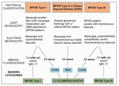
FIGURE 8.1 The evolving classification of membranoproliferative glomerulonephritis. Until recently, the classification of primary MPGN into types I, II, and III was based primarily on histologic features (light microscopy) and the ultrastructural location and electron density of the deposits (electron microscopy). With our increased understanding of the role of complement in the pathogenesis of these conditions, the IF findings now play a crucial role in categorizing MPGN as immunoglobulin-mediated versus non-immunoglobulin-mediated disease; the latter grouping, which is distinguished by isolated C3 staining on IF, has been termed “C3 glomerulopathy.” C3 glomerulopathy encompasses C3 glomerulonephritis (C3GN) and DDD. GBM, glomerular basement membrane; IgG, immunoglobulin G; IgM, immunoglobulin M. (Reproduced from D’Agati VD, Bomback AS. C3 glomerulopathy: what’s in a name? Kidney Int 2012;82:379-381,with permission.)


MEMBRANOPROLIFERATIVE GLOMERULONEPHRITIS TYPE I


Clinical Presentation and Epidemiology

The incidence and prevalence of MPGN vary in different parts of the world and have been declining in most developed countries (135,136) probably because of the decline in persistent infectious diseases. MPGN type I has been recorded in patients of all ages, although it is described most commonly in children (133,137,138,139,140,141,142,143,144). In a study of 79 patients with predominantly MPGN type I, the mean age at diagnosis was 34.6 years old, with a range of 6 to 79 years old, and 20 patients less than 20 years old (135). It has been diagnosed in patients younger than 2 years old (69,137,140,145), but most patients show signs of the disease after the age of 8 years (140). It can appear in adults of all ages, including patients beyond the seventh decade (141,146,147,148,149). In a retrospective study of renal biopsies in 150 patients aged 70 years or older during the period of 2000 to 2007 in Western France (150), 45 presented with nephrotic syndrome. Nine (20%) of the 45 patients had MPGN type I. None of these patients had detectable underlying infectious etiology although some had monoclonal gammopathy of undetermined significance (MGUS). This disease may favor Caucasians. There does not appear to be a substantial male or female predominance, although a few series suggest a slight male predominance.

The clinical characteristics are varied depending on the timing of the diagnostic renal biopsy relative to the clinical course. In about half of the patients, the clinical onset is preceded by a history of a respiratory infection (139). Although acute group A streptococcal infections are not thought to play a role in the genesis of this pattern of renal disease, two studies have shown elevated antistreptolysin O (ASO) titers in 38% (151) and 25% (140) of patients, respectively. These figures differ little from the prevalence rate in the general population (139). However, it may be difficult to distinguish MPGN from acute postinfectious glomerulonephritis on clinical and morphologic grounds in some patients. Patients may present with clinical symptoms of a nephritic or a nephrotic syndrome or both. Some patients have a clinical picture resembling acute glomerulonephritis with macroscopic hematuria and red blood cell casts (140). Dysmorphic or distorted red blood cells may be found in the urine. On average, approximately 10% to 20% of patients have an acute nephritic syndrome (140), with oliguria, edema, hematuria, hypertension, and renal insufficiency. Most patients have microscopic hematuria. Attacks of gross hematuria recur in a minority of patients and are more common in children than in adults (148). Persistent microscopic
hematuria is frequently a finding. MPGN may be asymptomatic and detected only, for example, in school urinary screening of children (152). It has been noted that patients diagnosed with MPGN type I on routine urinary screening usually have lower blood pressure, less proteinuria, and less chronic renal disease when compared to those who are diagnosed when symptomatic, indicating that early identification of the disease by urinary screening may allow for early therapy and improve the prognosis of this disease (152).

Mild hypertension is commonly present at the clinical onset (noted in about a third of patients). Some series note it more commonly (141); occasionally, it may be severe (141,147,153,154,155), and malignant hypertension may even be the presenting sign (153). Hypertension is typically observed as the renal disease progresses and is more common in adults than in children (148). Encephalopathy owing to hypertension is rare at presentation, but has been reported during follow-up in both treated and untreated patients (155).

Proteinuria is almost uniformly present. The nephrotic syndrome is a typical mode of presentation and has been noted in over 1/2 to 2/3 of patients (140,141,144). The nephrotic syndrome was found in more than 80% of patients in one series in children with MPGN type I (137). If the nephrotic syndrome is not evident at clinical presentation of a patient with MPGN, it often develops during the course of the disease. Heavy proteinuria is very common and, when studied, is generally of a moderately or poorly selective type (151). Patients with proteinuria generally also have accompanying microscopic hematuria. Some patients may not have overt clinical symptomatology, and the proteinuria is simply discovered on routine urinalysis. This was true of almost half the children in one study (140). MPGN is one of the major histopathologic patterns found in children with idiopathic nephrotic syndrome. It accounted for approximately 5.8% of cases of idiopathic nephrotic syndrome among pediatric patients in the report of the International Study of Kidney Disease in Children (156). In adults, the relative frequency of type I MPGN as a cause of nephrotic syndrome has declined in the United States from 6% for the period of 1976 to 1979 to 2% for the period of 1995 to 1997 (157). However, it should be noted that MPGN type I is much more common in underserved countries where there is a higher frequency of MPGN type I secondary to persistent infectious disease. For instance, in Asia (Saudi Arabia), South America (Peru), and Africa (Nigeria), MPGN type I is one of the most common causes of nephrotic syndrome and accounts for approximately 30% to 40% of all cases (158).

Blood urea nitrogen (BUN) and serum creatinine are elevated at clinical onset in about a fourth of patients. These values may stay elevated (presaging the onset of permanent renal failure) or return to normal over a few weeks in about half the patients. Depression of the glomerular filtration rate (GFR) is more often noted in adults than in children (148). Renal tubular dysfunction or injury has been described in patients with MPGN type I, as evidenced by increased urinary excretion of N-acetyl-beta-glucosaminidase (159), defects in maximal urinary concentration and urinary acidification, and elevated levels of the fractional excretion of sodium (160). Potassiumlosing nephropathy, generalized aminoaciduria, and glycosuria (reversible with steroid therapy) have also been described (161).

The commonly encountered feature of hypocomplementemia was recognized (and was the impetus for the discovery of this type of glomerulonephritis) by West et al. (133) and Gotoff et al. (134). There is often a decline in C3 levels in the serum (141,144,146,147,162,163), although the levels tend to fluctuate. Depressed levels of serum C3 have been found at the time of diagnosis in approximately a third to half of the patients (137,139,144,164). In most patients, serial determinations usually reveal hypocomplementemia sometime during the course of renal disease. Some patients do not appear to have depression of the serum complement level. A normal level may persist throughout the course of the illness; alternatively, as mentioned earlier, the level may drop at a later time (146,163). In the series of Habib et al. (130) and Servais et al. (144), as many as 40% and 54% of patients with MPGN did not have depressed serum complement, respectively. Serum concentrations of the early (i.e., C1q, C4, and C2) and terminal (C5, C6, C7, or C9) components of the classic pathway as well as components of the alternative pathway (i.e., factor B, properdin) are also frequently low (163,164,165). The complement profile (either classical or alternative complement pathway activation) depends on whether the cause is an immune complex disease or C3 glomerulopathy (52,53).

Immune complex MPGN type I but not the C3 glomerulopathy variant of MPGN type I might have detectable circulating immune complexes (CICs). CICs have been searched for using a number of different techniques and have been found in approximately 20% or more of patients with MPGN; this percentage depends on the sensitivity of the technique used for identifying the complexes (166,167,168). Davis et al. (167) demonstrated the presence of CICs when renal disease was mild or silent, but CICs were almost always absent by the time renal impairment developed. This finding suggested to the authors that either the CICs detected were not nephritogenic or that they programmed subsequent renal events that augment renal parenchymal injury in the absence of CICs. These workers also suggested that the measurement of CICs was of minimal value in the diagnosis or prognosis of patients with MPGN. Some groups have failed to find evidence of CICs altogether (168). IgM rheumatoid factors (i.e., autoantibodies to IgG) have been noted (169), as has cryoglobulinemia (1), in patients with MPGN type I.

Renal vein thrombosis can occur with MPGN. In one series, MPGN was the most common form of nephropathy associated with renal vein thrombosis (95). Successful pregnancies are the norm in affected patients (170), although Surian et al. (92) noted a high incidence of complications. Abrupt deterioration of renal function during pregnancy in a patient with preexisting MPGN has been reported (171). Plasmapheresis, albumin replacement, and antihypertensive therapy allowed for continuation of the pregnancy until a healthy infant could be delivered (171).

Although several formulas have been proposed (based on clinical and laboratory findings) to establish the diagnosis of MPGN without renal biopsy (156,172), biopsy is still the only way to determine with certainty the exact pattern of glomerular disease in the individual patient. Despite recent attempts to identify new biomarkers for MPGN (173), none has been identified.


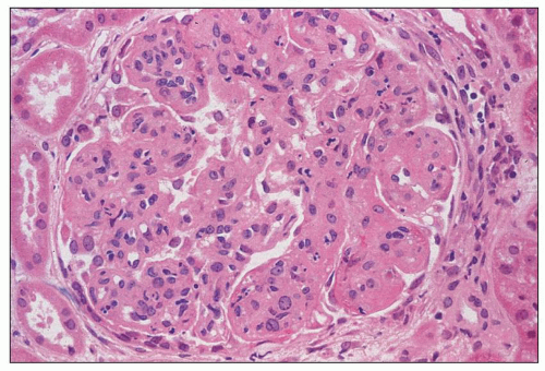
FIGURE 8.2 MPGN type I. There is increased lobulation, intracapillary hypercellularity (including mild neutrophil infiltration), and thickening of the capillary walls. (H&E, ×400.)


Pathologic Findings


Gross Pathology

By the time the gross appearance of the kidneys is studied, either at nephrectomy before transplantation or at autopsy, the kidneys are usually pale. Yellow flecking may be seen in the cortex that is caused by the accumulation of lipid in tubular epithelial cells and interstitial foam cells. With advancing disease, the kidneys become small and have a granular surface. There is a firm consistency to the renal parenchyma, and the arteries may be prominent.


Light Microscopy


GLOMERULI

The glomeruli have characteristic and uniform changes. Glomeruli typically are enlarged, with a diffuse increase in glomerular tuft cellularity (Figs. 8.2 and 8.3). The intracapillary hypercellularity is usually global (i.e., involving all portions of each glomerular tuft to about the same degree). The increase in cellularity within each glomerular lobule creates an accentuation of the normal lobularity of glomerular tufts (Fig. 8.4). The term “lobular glomerulonephritis” is purely descriptive and nonspecific and should not be used as a diagnostic term.


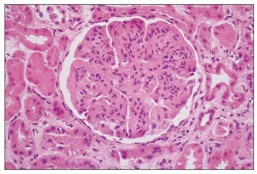
FIGURE 8.3 MPGN type I. There are capillary wall thickenings, increased cellularity, and pronounced lobulation. (H&E, ×360.)


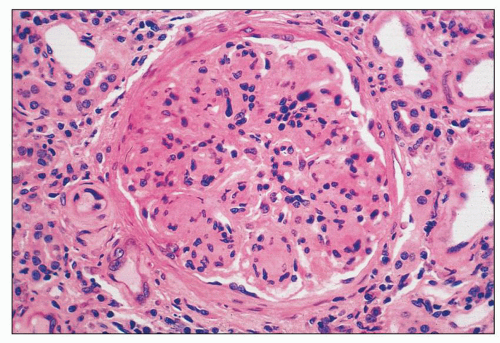
FIGURE 8.4 MPGN type I. Accentuation of the lobular pattern with sclerotic mesangial nodules. (H&E, ×400.)

The increase in cells in the mesangial regions and the increase in the amount of mesangial matrix create a much larger mesangial (centrilobular) area with the lobules sometimes assuming a club shape. In some patients, however, there is widespread glomerular hypercellularity with little accentuation of the lobular pattern. It has been suggested that the severity of the mesangial lesions, especially sclerosis, relates to the duration of renal disease. In some repeat biopsies, as the lobular lesion progresses, the cellularity tends to diminish and is replaced by mesangial matrix (sclerosis). In approximately one fourth of the cases, there is a marked polymorphonuclear leukocytic infiltration (exudative form) (Fig. 8.5) (174). Laohapand et al. (175), using α1-antitrypsin as a marker for mononuclear leukocytes, noted an abundance of monocytes in
severe cases of MPGN. The greatest numbers of monocytes were noted in renal biopsies with the most glomerular hypercellularity and the largest number of glomerular subendothelial and subepithelial deposits. Soma et al. (176) examined the nature of the intraglomerular immune cell infiltration and its relationship to C3 deposits over time. These investigators found monocytes/macrophages and leukocytes to be the predominant cell type at first biopsy (with many fewer T cells). Second biopsy showed either less complement deposition with fewer leukocytes of all types or greater complement deposition with a positive correlation between the number of intraglomerular T cells and monocytes/macrophages (176). Yang et al. (177) and Lan et al. (178), using double immunostaining for CD68 and the proliferating cell nuclear antigen (PCNA), demonstrated that MPGN (presumably type I) is associated with marked macrophage infiltration, with proliferating macrophages (CD68+PCNA+ cells) accounting for up to 42% of total macrophage population. Macrophage proliferation was largely restricted to areas of severe tissue damage (i.e., glomerular hypercellular lesion and foci of tubulointerstitial damage), suggesting that local proliferation is a mechanism for amplifying macrophage-mediated tissue injury. Macrophage accumulation may be partially related to the marked up-regulation of renal expression of macrophage migration inhibitory factor (MIF). In addition, the glomerular and interstitial macrophage proliferation correlated with loss of renal function and histologic lesions but not with proteinuria. In a recent study, Wu et al. (179) characterized and quantified the proliferating cells in MPGN (presumably type I) using monoclonal antibodies for various cell markers. They demonstrated marked mesangial proliferation/activation coupled with increased neutrophils, macrophages, and T cells. However, endothelial cell proliferation was not obvious. Cases of primary mixed or essential cryoglobulinemic glomerulonephritis with a membranoproliferative glomerular pattern generally have a large number of infiltrating monocytes and macrophages (180,181).


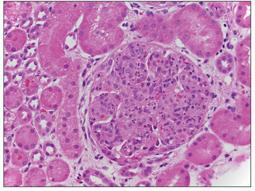
FIGURE 8.5 MPGN type I. There are numerous infiltrating neutrophils (exudative form) resembling acute postinfectious glomerulonephritis by light microscopy. (H&E, ×400.)

Podocyte changes also develop, especially in patients with nephrotic proteinuria. The early events are characterized by molecular alterations of the slit diaphragm followed by podocyte detachment, hypertrophy, and death if early damage is not reversed. Patrakka et al. (182) investigated the nephrin expression by immunohistochemistry in pediatric kidney diseases including six cases of MPGN type I. The findings did not reveal major alterations of nephrin in MPGN type I when compared with normal controls. In another study, Wang et al. (183) demonstrated a down-regulation of nephrin in patients with MPGN (types not specified) in the glomeruli.

There is marked diffuse thickening of the glomerular capillary walls. The thickening can be more prominent in some glomeruli and in some capillary loops than in others. Periodic acid-Schiff (PAS) and methenamine silver stains show that the thickened glomerular capillary walls often have two basement membranes with a clear or nonargyrophilic region between them. This double contour is sometimes termed tram tracking, splitting, or reduplication of the GBM (Fig. 8.6). In some capillaries, the production of basement membrane-like structures is very complex, resulting in multiple laminations. The double contour is brought about by mesangial interposition, which refers to the outward migration of mesangial cells, infiltrating mononuclear cells, or even margination of portions of endothelial cells along the inside of the capillary walls, interposing themselves between the endothelium and GBM. Mesangial interposition can be circumferential or partial depending on whether the entire circumference or only a segment of the peripheral capillary wall is involved. Because mesangial or endothelial cells can produce basement membrane-like material, the cytoplasm of these cells and immune complexes are covered on the outside (the Bowman space side) by the original basement membrane and on the inside by the newly formed GBM-like “membrane.” Both the membranes stain positively with silver and thus give rise to the double contour.


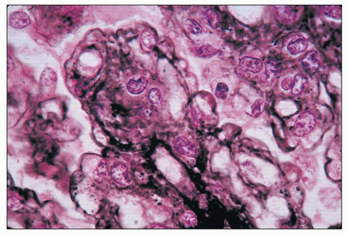
FIGURE 8.6 MPGN type I. Glomerulus with silver stain shows tram tracking or reduplication of the GBM. (Jones silver methenamine, ×600.)

The newly formed basement membrane-like material (the inner contour) may be thin, incomplete, and at times difficult to discern because of extreme glomerular hypercellularity. Although circumferential mesangial interposition can be seen in a wide variety of glomerular lesions, it is most common, marked, and diffuse in MPGN type I. Nakamoto et al. (184) theorize that this interposition is related to low-grade mesangiolysis and subsequent passive dislocation of the mesangial cells toward the peripheral glomerular capillary walls because of a high hydraulic pressure of blood flow penetrating the lysed mesangium. A widely accepted alternative theory proposes that an active movement by the muscle-like mesangial cells from the contiguous mesangial regions along the lamina rara interna of the GBM, probably in response to subendothelial immune deposits. In certain cases, capillary loops show the spiking phenomenon noted in membranous nephropathy as a result of subepithelial deposits. This finding is more pronounced in the so-called type III MPGN of Burkholder (120,137,138,140).

The glomerular capillary lumens are often diffusely and globally diminished by the increase in matrix and cellularity in the mesangial regions as well as by the thickening of the capillary walls. Generally, the subendothelial deposits contribute little to this luminal narrowing. Infiltrating inflammatory cells also may contribute to this endocapillary hypercellularity and capillary lumen closure. Intraglomerular lipid deposition (mainly apolipoprotein B) can be noted (185). Discrete glomerular subendothelial deposits may be identified with the use of trichrome stains, although they are better demonstrated using EM. Mesangial deposits are generally small and difficult to identify by light microscopy. In some cases,
scattered glomerular fuchsinophilic subepithelial humps may be detected with trichrome stains and oil immersion. Bohle et al. (186) have suggested that “hyperperfusion injury” can be seen with great frequency in MPGN. This is defined as the presence of glomerular adhesions (synechiae), glomerular subendothelial hyalinosis, and fat droplets in the hyaline material.


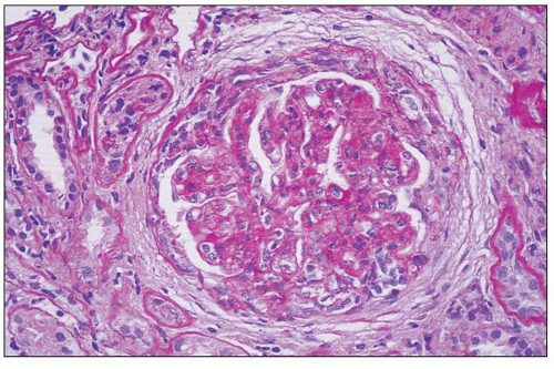
FIGURE 8.7 MPGN type I. Glomerulus from a case of MPGN type I with crescent. (PAS, ×400.)

Crescents occur in approximately 10% of patients (139) (Fig. 8.7). These crescents may be small and focal (147) or large, affecting most of the glomeruli (138). They are often indicative of a poor prognosis (138,139,140,148,187). In several studies of all types of crescentic glomerulonephritis in children, up to approximately one fourth of the cases are MPGN type I (188). Crescent formation has been noted to develop within weeks following an initial biopsy showing only an MPGN type I pattern with no crescents (189). Parietal epithelial cells may be prominent without the presence of obvious crescents (190).

Serial biopsies are not commonly performed, but in those few patients in whom they have been reported, the glomerular tuft hypercellularity may become less pronounced with an increase in the amount of mesangial matrix (sclerosis) (178). Taguchi and Bohle (191) have described sequential biopsies (separated by a mean of 39 months from initial biopsies) from 33 patients with MPGN type I and DDD. Twenty-four of twenty five patients with diffuse forms of MPGN maintained that pattern on subsequent biopsies, whereas 4 of 6 patients with a focal MPGN pattern showed signs of a diffuse form on the second biopsy. Two patients who had no histologic findings of MPGN on the initial biopsy (one had focal MPGN and another had mild mesangial proliferative glomerulonephritis with small crescents) later showed evidence of a diffuse form of MPGN on subsequent biopsy. End-stage sclerotic glomeruli can develop later.

Striker et al. (192) studied the extracellular components of several renal diseases with progressive glomerular sclerosis (including MPGN) using a variety of immunohistochemical analyses. In advanced stages, the amount of types IV and V collagens, laminin, and fibronectin was increased in the mesangial and sclerotic lesions; however, the staining intensity for type IV collagen, laminin, and fibronectin gradually declined during the progression of glomerular sclerosis. The authors have found types I and III (interstitial) collagens in the glomeruli of those patients with severe damage to the Bowman capsules (as with crescent formation).


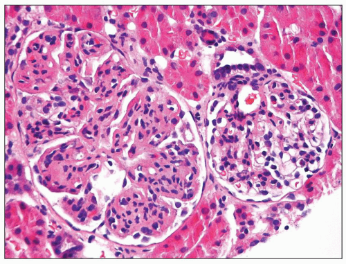
FIGURE 8.8 Focal MPGN type I. One glomerulus shows typical changes of MPGN type I, while the other glomerulus reveals only mild mesangial hypercellularity. (H&E, ×400.)

There are reports of focal or segmental MPGN (Fig. 8.8) (138,154,193). In these reports, in which only some glomeruli show lesions while others do not, it is not always clear whether the glomeruli that are normal by light microscopy contain deposits when viewed by electron microscopic and immunofluorescence methods. Focal MPGN may progress to typical MPGN, and typical MPGN may regress to focal MPGN. Therefore, focal MPGN is considered to be an early type of typical MPGN or a stage of recovery from typical MPGN. In a case report, Kano et al. (194) described a girl with MPGN type I diagnosed by the third biopsy. The first biopsy revealed endocapillary proliferative glomerulonephritis, and the second biopsy showed focal MPGN. D’Amico and Ferrario (195), in a review of a large number of patients with MPGN type I and DDD, suggest that there are six characterized morphologic variants: classic, nodular, exudative, focal segmental, with massive subendothelial deposits, and crescentic. They believe that these different forms involve different etiologic and pathogenetic factors and that the clinical outcome correlates with the histopathologic patterns. Other researchers believe that these different patterns in MPGN do not relate well to specific etiologic or pathogenetic factors or clinical findings, but rather, represent different points in a continuum of morphologic manifestations of MPGN. In renal biopsies of MPGN, it is important for the pathologist to look for refractile eosinophilic hyaline globules in the glomerular capillary lumens (“hyaline thrombi”) suggestive of cryoglobulin deposits. More is discussed about this in the section dealing with cryoglobulinemia later in this in chapter.

Quantitative studies have been conducted on renal biopsies of patients with MPGN type I (196). Glomerular and mesangial volume fractions were increased and related to a diminished GFR, enhanced glomerular permeability to protein, hypertension, and volume fraction of the cortical interstitium. The percentage of the glomerular capillary endothelial circumference (filtration surface) was also smaller. Thus, quantitative measures of glomerular structure were highly correlated with glomerular function.


TUBULES

Morphologic changes in the tubules and interstitium generally reflect the changes noted in the glomeruli (139). The tubules may contain hyaline droplets that are protein and lipid resorption droplets (phagolysosomes). These droplets are directly related to the glomerular permeability to proteins and lipids. Tubular lumens may also contain red blood cells. With evolution of the disease toward more severe renal parenchymal damage, interstitial inflammation and edema as well as tubular atrophy and fibrosis develop. However, the study by Schmitt et al. (197) suggests that the tubulointerstitial findings are unrelated to the severity of the glomerular alterations. Severe glomerular lesions can arise in the absence of tubulointerstitial disease; conversely, severe tubulointerstitial disease can be seen with mild glomerular disease. These authors suggest that the tubulointerstitial changes result in many of the renal functional disturbances.


INTERSTITIUM

Clusters of interstitial foam cells are observed quite often in MPGN type I and, to an even greater extent, in Alport syndrome. Clefts—sometimes noted in the lumina of tubules— probably are caused by cholesterol ester. Cholesterol granulomas are rare. There is a good correlation between interstitial fibrosis and the level of serum creatinine (198) and other functional abnormalities (196,199). Various cells have been found in the renal interstitium of patients with MPGN type I. Segerer et al. (200) found a predominance of T cells that are positive for CXCR3 (a receptor for the CXC chemokines IP-10 and Mig) and CCR5 (a receptor for the C-C chemokine RANTES). In addition, the number of CXCR3- as well as CCR5-positive T cells correlates with renal function, proteinuria, and percentage of globally sclerotic glomeruli, suggesting an important role during progressive loss of renal function, and as such may represent a potential therapeutic target.


BLOOD VESSELS

Arteries and arterioles are affected in those patients in whom renal failure and hypertension develop. There is severe arterial intimal thickening in patients with long-standing renal disease and in those in whom dialysis has been instituted. If vasculitis is identified, cryoglobulinemia, hepatitis B- or hepatitis C-related MPGN, as well as antineutrophil cytoplasmic antibody (ANCA)-associated vasculitis should be ruled out.


Immunofluorescence Microscopy

As noted earlier, findings by IF microscopy (or immunohistochemistry) are used to classify MPGN into an immune complex variant and a complement dysregulation variant (see Fig. 8.1 and Table 8.1). IF staining patterns are generally quite characteristic (137,140,141,201,202,203,204). In the immune complex variant of MPGN, the most consistent finding is positive staining for IgG in a fine to coarse granular pattern along the glomerular capillaries (Fig. 8.9) (201,204). A characteristic picture is produced, with the glomerular capillaries around the periphery of the expanded lobules predominantly (and often solely) affected, so that the lobules stand out quite clearly as a negative zone, cloaked by strongly positive IF. In some patients, staining for IgG decreases as the disease progresses and the deposits become replaced or obscured by the increase in mesangial matrix. Serial renal biopsy studies have shown that some patients have IgG deposits during the early stages of the disease, with subsequent biopsies showing only deposits of complement (163). This may prove to be a confounding problem for classification based on relative predominance of immunoglobulin versus complement. Doi et al. (205) demonstrated all four IgG subclasses in renal biopsies of MPGN, but in contrast, other studies (206,207) noted an excess of IgG3 compared with the other classes. IgM is less commonly evident than IgG, but it was present in 86% of patients in one series (204), 73% in another (202), and 60% in a third (147) (Fig. 8.10). A predominance of IgM is typically observed in MPGN type I stemming from chronic bacterial infection, such as osteomyelitis or infected ventriculoatrial shunt. IgA is found even less often but has been observed in one third of the patients in some studies (147,204). It is important to ascertain whether the IgA is the predominant or codominant immunoreactant to diagnose a membranoproliferative form of an IgA nephropathy or an IgA-dominant postinfectious glomerulonephritis, both of which are rare. Orfila et al. (208) noted the presence of both kappa and lambda light chains in MPGN; however, MPGN type I with typical features by light microscopy and electron microcopy can be caused by monoclonal
immunoglobulin deposition (52). Conspicuous IgM or IgA even in the absence of IgG is indicative of an immune complex MPGN rather than a C3 glomerulopathy variant of MPGN.


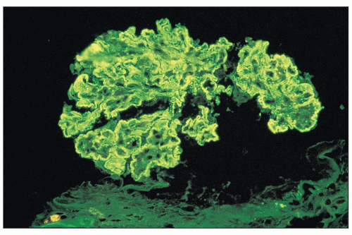
FIGURE 8.9 Immunofluorescence of MPGN type I. There is intense glomerular mesangial and capillary wall staining with anti-IgG antiserum. (×400.)


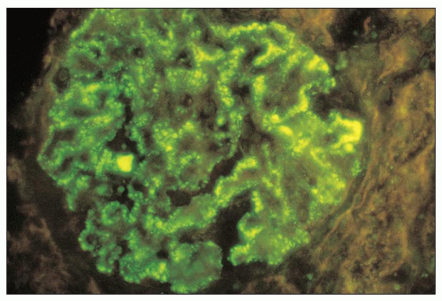
FIGURE 8.10 Immunofluorescence of MPGN type I. There is moderately intense staining for IgM along the glomerular capillary walls in a granular pattern. (×400.) (Courtesy of Dr. Zoltan Laszik.)

In immune complex MPGN type I, C3 is noted in a similar pattern to IgG in all patients and may stain more intensely (Figs. 8.11). In the C3 glomerulopathy variant of MPGN type I, staining for C3 is intense with little or no staining for immunoglobulin (52,53,118). Staining for immunoglobulin and/or C3 typically produces a granular to semilinear staining along the capillary walls. Smooth outer contour of the deposits (due to their conformation by the delimiting outer GBM) provides a useful clue indicating that the deposits are subendothelial (rather than subepithelial). In some instances, the peripheral glomerular capillaries show coarse, somewhat elongated areas of IF that may impart a broken, wide, or band-like pattern.

Immunoglobulin and/or C3 may also be noted in the glomerular mesangium. When there is a great increase in the amount of mesangial matrix, mesangial deposits may be obscured or absent. Early components of the classic pathway of complement activation (especially C1q) are sometimes visible and are present in about one half to two thirds of the patients (201,204). Yamashina et al. (209), using an avidin-biotin-peroxidase complex method and proteolytic digestion of formalin-fixed paraffin-embedded sections, verified the presence of all complement components. Properdin is virtually always present (139). Properdin deposits were found in 90% of cases in one series (147) and in 100% in another (203). Kazatchkine et al. (210) found normal staining of the glomerular visceral epithelial cells (podocytes) with antibodies to C3b receptor in renal biopsies of MPGN type I. Although IF findings along the tubular basement membranes (TBMs) are usually negative, a minority of patients have immune complexes in this area (211).

Early studies by Levy et al. (140,163) of the IF patterns in MPGN type I are consistent with the current concept of two immunopathologic variants, one medicated by immune complexes and one by complement dysregulation. In their study of 36 children, group 1 (26 patients), which appeared to be immune complex mediated, exhibited immunoglobulins and C3; 15 of them had immunofluorescent staining along the glomerular capillary walls but not in the mesangial regions (163). In these 15 patients, IgG and IgM were the most common immunoglobulins, and C1q and C4 were also present. The other 11 patients in group 1 had C3 and immunoglobulins (mainly IgG) along the glomerular capillaries but also mesangial deposits of C3. C1q and C4 were noted in a location similar to that of the immunoglobulins in all patients studied. Group 2 (10 patients) displayed the presence of C3 only, without any immunoglobulin, and thus are consistent with C3 glomerulopathy. C3 was noted along the glomerular capillary walls and in the mesangial regions. No early complement components of the classic pathway were found in group 2.


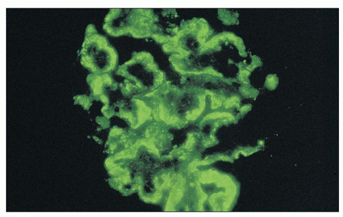
FIGURE 8.11 Immunofluorescence of MPGN type I. There is intense staining aong all glomerular capillary walls for C3. The granular staining is quite broad in this instance. (×400.)

IF techniques also have been used to search for antigens other than immunoglobulins and complement. Murphy and d’Apice (212) and Nakamura et al. (213) studied the kidneys with MPGN with a variety of monoclonal antibodies to glomerular proteins and demonstrated fibronectin in both the GBM and mesangium. Hara et al. (214), using antisera to the human GBM, type IV collagen, and P3 antigen (which is a nephritogen in rats), noted that in MPGN, the expanded mesangium and expanded glomerular capillary walls reacted positively with anti-GBM and anti-type IV collagen antibodies; only the outer portion of the capillary walls was positive with anti-P3. Büyükbabani and Droz (215) further studied alterations of the matrix compartment. They observed strong accumulation of fibronectin in the expanded mesangial regions (as well as along the migration track of the proliferating mesangial cells), with accumulation of laminin, α12 chains of type IV collagen, and heparan sulfate proteoglycan. Type I collagen was also present in the central part of the mesangial regions. The distribution of these matrix components was different in cases of MPGN than in those of other glomerular lesions, such as membranous glomerulopathy, focal segmental glomerulosclerosis (FSGS), or crescentic glomerulonephritis.


Electron Microscopy

EM has helped to distinguish classic MPGN type I from other glomerular lesions with membranoproliferative features and clarify the findings of light microscopy (120,137,140,141,147,151,204,216,217,218) (Figs. 8.12, 8.13, 8.14, 8.15, 8.16). Ultrastructural studies have shown subendothelial dense deposits (see Figs. 8.12, 8.13, 8.14) with varying amounts of mesangial, intramembranous, and subepithelial deposits. There are mesangial hypercellularity and increased mesangial matrix, both of which are often present between the normal-appearing GBM and the glomerular endothelium (mesangial interposition) (see Fig. 8.15). As described earlier, the new mesangial matrix-like material produced by the migrating mesangial cells and endothelial cells creates an inner “basement membrane.” The thickened capillary wall is therefore composed of two or more layers of basement membrane-like material, interposed mesangial cells, and electron-dense immune-type deposits (see Figs. 8.12, 8.13, 8.14, 8.15). In some cases, the double contour of the peripheral glomerular capillary wall is caused by the interposition not of mesangial cells but of monocytes instead (2); this finding is especially common in patients with cryoglobulinemia and MPGN. Some have suggested that cells in these positions are portions of invaginated endothelial cells.

The electron-dense deposits are generally described as subendothelial but are really nearer the inner aspect of the original basement membrane. Their exact position is somewhat obscured because of the migrating mesangial cells and the new layers of basement membrane-like material. The deposits
range from small and discrete to large and elongated; they are found both along the periphery of the filtering glomerular capillary walls and just under the paramesangial GBM as it overlies the mesangium. Discrete electron-dense deposits also may be found within the mesangium; at this site, they are usually small, but sometimes, they are more bulky. They are associated with an increase in mesangial or endocapillary hypercellularity (see Fig. 8.16). Sometimes, the IF discloses intense and widespread positive staining along the capillary walls, whereas only scant deposits are present on the EM.


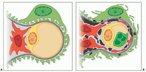
FIGURE 8.12 Drawing depicting a normal glomerular capillary and adjacent mesangium (A) compared to the ultrastructural changes of MPGN type I (B). Note the subendothelial, mesangial and few subepithelial dense deposits (black), capillary wall mesangial interposition (red), new layers of subendothelial matrix material (gray), and increase in mesangial cell numbers.

Some patients have scattered, small glomerular subepithelial deposits quite similar to the humps noted in classic acute postinfectious (poststreptococcal) glomerulonephritis (137,141,149,204). They are found in as many as 30% (141) to 50% (140) of cases studied. Small subepithelial deposits may have accompanying protruding spikes of GBM-like material similar to that noted in membranous nephropathy. Sato (219) evaluated the glomerular subepithelial deposits in patients with various forms of MPGN. Most patients also had undergone serial biopsies. Despite diminished glomerular cell proliferation in most second biopsies, the glomerular subepithelial deposits were increased along with thickening and irregular structuring of the GBM (219). As described later, the presence of a large number of glomerular subepithelial deposits forms part of the pattern of MPGN type III of Burkholder. Occasionally, intramembranous deposits are noted in MPGN type I, and fragmentation of the GBM can occur (149). The application of silver impregnation to renal EM may help resolve the various patterns of MPGN (220). Ultrastructural studies at high magnification (e.g., ×30,000) are important to determine if the deposits have a microtubular substructure such as seen in immunotactoid glomerulopathy or cryoglobulinemia (1).

There is an increase in mesangial cellularity and matrix formation. Hypercellularity, production of matrix (sclerosis), and movement or extension of the mesangial cells around the glomerular capillary wall are all manifestations of the great activity on the part of the mesangial cells. Platelets or fibrin tactoids may be seen in one fourth of the biopsies studied by EM. Glomerular visceral epithelial cell foot process effacement is common. Using immunoelectron microscopy, Huh et al. (221) observed that the expression of nephrin in human glomerular disease (including MPGN type I) was lower in regions where the foot processes were effaced and comparable to normal controls where the foot process interspaces were preserved. Bonsib (222) used enzymatic digestion to remove cellular elements and deposits, coupled with scanning electron microscopy, to study the three-dimensional aspects of the glomerulus. The picture of MPGN type I is illustrated in Figure 8.17.


Clinical Course, Prognosis, Therapy, and Clinicopathologic Correlations

MPGN type I is generally progressive, and overall, renal prognosis is poor. However, the clinical course of patients can be quite variable (52,53,117,137,140,144,147,223). The nephrotic syndrome persists in some, whereas others have intermittent nephrotic or nephritic episodes with abnormal findings on urinalysis between the episodes. Rarely, patients may become entirely asymptomatic, with normal renal function and urinary findings (151,224). This clinically silent phase may last for many years despite persistence of the renal morphologic abnormalities.

Clinical remission has been noted in 5% to 20% of patients (138,175,225). Complete remission occurred in only
a few patients for whom there has been long-term follow-up (124,140,148). The series of Levy et al. (140) showed complete remission in only 4 of 84 children. In this study, 17 children went into remission, but 13 relapsed; in only 4 patients was the remission maintained for periods of up to 4 years (140). Kim et al. (139) noted complete clinical remission in 5 of 63 patients.

Only gold members can continue reading. Log In or Register to continue

Stay updated, free articles. Join our Telegram channel

Jun 21, 2016 | Posted by in UROLOGY | Comments Off on Membranoproliferative Glomerulonephritis

Full access? Get Clinical Tree

Get Clinical Tree app for offline access