Ischemic and Toxic Acute Tubular Injury and Other Ischemic Renal Injuries


Ischemic and Toxic Acute Tubular Injury and Other Ischemic Renal Injuries


Gilbert W. Moeckel

Michael Kashgarian

Lorraine C. Racusen


ACUTE TUBULAR INJURY

Acute tubular injury (ATI) is a major cause of acute renal failure (ARF), a clinical syndrome characterized by rapid deterioration in renal function and glomerular filtration rate (GFR) over a relatively short period of time, ranging from hours to days. The result is a sudden inability to maintain normal fluid and electrolyte homeostasis. The acute reduction in renal function can be the result of the impairment of blood flow (so-called prerenal failure), obstruction of the urinary collecting system (so-called postrenal failure), or a variety of intrinsic renal diseases ranging from glomerulonephritis to interstitial nephritis to ATI (Fig. 26.1), which is the primary topic of discussion in this chapter. Based on the available literature, which uses somewhat variable definitions, ARF is commonly encountered in hospitalized patients, has a variety of risk factors and etiologies, and is associated with increased mortality (1). Its frequency ranges from 1% at admission to the hospital to as high as 31% in patients undergoing cardiopulmonary bypass or with other high-risk conditions. Clinical manifestations range from mild increase in serum creatinine (sCr) to anuric renal failure (2). A consensus panel of the American Society of Nephrology recommended that “acute renal failure” be replaced by the term acute kidney injury (AKI). The term AKI could be used to distinguish early from more advanced stages of kidney disease, in which there is more overt “failure” of clearance by the kidney. Use of the more general term AKI highlights the predictive value of acute or small changes in sCr and facilitates recognition of renal injury and dysfunction at earlier stages of disease, since even transient rise in creatinine is correlated with increased risk of death (3).

In 2004, a consensus definition was published, which included both GFR and urine output criteria (Table 26.1). The earliest phase, risk of renal dysfunction, was defined by an increase in sCr by 1.5 times, GFR decrease of more than 25%, or urine output below 0.5 mL/kg/h for 6 hours. Renal injury was defined by sCr increase by 2 times, GFR decrease of more than 50%, or urine output less than 0.5 mL/kg/h for 12 hours. Renal failure was defined as sCr increase by three times (or over 4 mg/dL), GFR decrease by 75%, urine output below 0.3 mL/kg/h for 24 hours, or anuria for 12 hours. Added to loss of function and end-stage renal disease, these comprise the “RIFLE” criteria (4). These criteria appear to be clinically relevant and have been widely used (5). They were modified in 2007 as the AKIN (acute kidney injury) criteria, eliminating the loss-of-function and ESRD categories (6). AKIN criteria defined AKI as “functional or structural abnormalities or markers of kidney damage including abnormalities in blood, urine or tissue tests or imaging studies present for less than 3 months.” However,
both classifications use diagnostic criteria of renal impairment (urine output, rise in sCr) that manifest at a late stage of injury and rely on knowledge of baseline creatinine (7). Inclusion of other more sensitive criteria, including biomarkers, may enhance definition criteria (8). There has also been lack of precision in the use of the term acute tubular injury/necrosis. The term should be reserved for the clinical pathologic entity of intrinsic renal failure that is the result of either an ischemic or toxic insult to the kidney, with evidence of tubular injury/dysfunction such as altered fractional excretion of sodium (8) and potentially other more specific biomarkers, when other causes have been excluded. As a result of the lack of uniformity in terminology, the percentage of cases of ARF that can be attributed to “acute tubular necrosis/injury” are difficult to accurately ascertain, but the condition is likely responsible for the majority of cases of ARF that require acute renal replacement therapy. The term acute tubular necrosis itself is a misnomer, since necrosis, while classically a feature of animal models, is only one morphologic manifestation of clinical ATI. It should also be noted that morphologic evidence of frank tubular necrosis is not a frequent feature in kidney biopsies obtained in the context of clinical ARF; however, morphologic changes of sublethal tubular injury are usually present. Just as in the clinical classifications, however, morphologic signs of injury appear in a later stage of injury; more sensitive markers are required to identify early tubular cell injury. The term acute tubular injury is more accurate and will be used throughout this chapter.


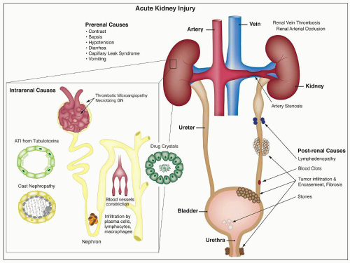
FIGURE 26.1 Causes of acute renal failure. This figure depicts several etiologies that may lead to AKI. Intrarenal causes of tubular injury include tubulotoxins, light chain casts, ischemic injury, and lesions that indirectly affect tubular cell viability through impaired glomerular blood flow (i.e., TMA and necrotizing GN). Other common causes of ATI include drug crystals with associated tubular obstruction, interstitial inflammatory infiltrate by lymphocytes, plasma cells, and macrophages. Prerenal causes include hypotension, diarrhea, vomiting, and contrast. Postrenal causes include lymphadenopathy, tumor infiltration, and urinary outflow obstruction by fibrosis and blood clots and stones.


Historical Background

It was not until World War I that acute renal dysfunction was recognized as a distinctive clinical and pathologic entity. Hackradt (9) described what he called “vasomotor nephrosis” following crushing injuries. In a review of autopsy material, Minami (10) described the presence of pigment casts in medullary tubules associated with tubular changes and an interstitial infiltrate and suggested that myohemoglobinuria and subsequent precipitation in the tubules were factors involved in producing the observed anatomic and functional renal abnormalities. Shortly
thereafter, Baker and Dodds (11) studied a rabbit model of ARF and emphasized tubular obstruction as important in the pathogenesis. Progress in the field was relatively dormant until the advent of World War II, when Bywaters and Beall (12) revived interest in ARF as a result of their study of London air casualties. Bywaters and Dible (13) and Dunn et al. (14) described intratubular hemoglobin and myoglobin casts associated with focal necrosis of tubules, interstitial edema, and mild interstitial inflammation localized to specific portions of the nephron in the “crush syndrome or traumatic anuria.” This finding led to the hypothesis that tubular obstruction by necrotic debris and precipitated pigment was the prime cause of the observed oliguria.


TABLE 26.1 Clinical phases of AKI


1. Risk of renal dysfunction


Increase in sCr by 1.5 times OR


Decrease in GFR >25% for 6 h OR


Urine output <0.5 mL/kg/h for 6 h


2. Renal injury


Increase in sCr by 2 times OR


Decrease in GFR >50% for 12 h OR


Urine output <0.5 mL/kg/h for 12 h


3. Renal failure


Increase in sCr by 3 times or >4 mg/dL OR


Decrease in GFR by 75% or more for 24 h OR


Urine output <0.3 mL/kg/hr for 24 h OR


Anuria for 12 h


sCr, serum creatinine; GFR, glomerular filtration rate.


Modified from Bellomo R. Defining, quantifying, and classifying acute renal failure. Crit Care Clin 2005;21:223.


Bywaters and Dible (13), however, believed that obstruction alone could not explain all the clinical findings or the abnormal character of the urine that was produced by such patients. Because patients with ARF had urine that was quite abnormal and resembled an unaltered glomerular filtrate, and because there was a marked discrepancy between the degree of anatomic change and the severity of the oliguria, they postulated that other factors must play a part. Lucke (15) emphasized that the discrepancy between structure and function could be related to the observed localization of histologic changes to the distal nephrons, and he coined the term lower nephron nephrosis.

Oliver et al. (16) used nephron dissections to study cases of ARF, and they were able to show two distinct types of renal tubular injury. In the first, as a result of the direct cytotoxic effect of a specific nephrotoxin, there was segmental necrosis of the proximal tubular epithelium with denudation of the basement membrane, which remained intact. In the second type, which they termed the tubulorrhexic lesion, there was focal necrosis of tubular cells associated with rupture of the adjacent basement membrane, allowing communication of the tubular lumen with the interstitial tissue, a lesion most commonly seen in the distal portions of the proximal tubule, most likely ischemic in origin. The focal and patchy nature of the necrosis, which was usually not associated with any interstitial reaction, was thought to be the reason that random histologic sections of such kidneys frequently did not demonstrate significant pathologic change. Oliver et al. suggested that these lesions could lead to the leakage of tubular fluid into the interstitial tissue, diminishing the amount of urine produced. Such leakage could cause a rise in intrarenal pressure, which could further potentiate oliguria by compression of capillaries, resulting in diminished glomerular filtration.

While these investigators were concentrating on the tubular changes, Goormaghtigh (17,18) called attention to the renal arteriolar changes in kidneys of patients with the crush syndrome. He observed considerable hypertrophy and an increase in granularity of the juxtaglomerular cells in the kidneys of patients with ARF and theorized that these cells produced a vasoactive or prevasoactive substance that could act on the renal vasculature. He suggested that the anuria observed in the crush syndrome was the result of vasoconstriction and postulated that glomerular hemodynamic changes would result in the diminution of glomerular filtration, emphasizing decreased glomerular function as a mechanism of oliguria.

Based on direct observation of the kidney as well as angiographic and morphologic studies of intrarenal vascular patterns, Trueta et al. (19) proposed that the fundamental defect in the crush syndrome was a reduction of glomerular filtration as a result of the diversion of blood away from the outer cortical glomeruli through a juxtamedullary shunt. Brun et al. (20,21) produced even more convincing evidence by measuring renal hemodynamics in vivo. Sheehan and Davis (22) and Sevitt (23) also believed that ischemia was important, but they suggested that the mechanisms of action were related to vascular damage following an initial period of ischemia, which prevented adequate reperfusion once blood flow had been established. Hollenberg et al. (24,25) studied patients with ARF following a variety of initiating injuries and noted that it was impossible to see the cortical arteries in such patients; they also documented disappearance of the cortical flow component of xenon washout from the kidney. Munck (26) verified that such a decrease in blood flow was sufficient to result in renal hypoxia. These early studies suggested several physiologic mechanisms of action for the resultant oligoanuria, including tubular obstruction, back leakage of tubular fluid, and changes in hemodynamics resulting in decreasing glomerular filtration.

The advent of micropuncture techniques led to the investigation of several animal models of ARF to identify the pathophysiologic mechanisms of action in greater detail. Oken et al. (27) studied experimental mercury- or glycerol-induced ARF, demonstrating that glomerular filtration progressively diminished as oliguria developed and suggesting that suppressed filtration was the key pathophysiologic factor. Early work in our own laboratory (M.K.) studied two different models of ARF (28), potassium dichromate-induced toxic cellular damage to the early (S1-2) part of the proximal tubule and administration of purified human globin, producing an intrarenal obstructive lesion of the distal nephron. Decrease in urine flow was accompanied by a diminution of the total and individual nephron GFR associated with a decrease in the tubular reabsorptive capacity, and there was evidence of mechanical tubular obstruction, reflected by an elevation of free-flow intratubular pressure. In addition, the glomerular filtration pressure appeared to be diminished, suggesting that the decreased glomerular blood flow and glomerular filtration were the result of preglomerular arteriolar constriction, mediated by activation of the local renin-angiotensin system. Studies of renal blood
flow distribution demonstrated that a diminution of outer cortical flow correlated best with decreased glomerular filtration (29,30). These and other studies led to a proposal that tubular epithelial injury induced either by ischemia or by a toxin could be sublethal but had to be severe enough to result in decreased epithelial transport activity, which then would result in decreased tubular sodium reabsorption and local activation of the renin-angiotensin system. This, in turn, would alter glomerular hemodynamics and result in decreased glomerular filtration. Decreased tubular urine flow associated with the shedding of cellular debris and the presence of Tamm-Horsfall protein would result in tubular obstruction. When combined with focal areas of necrosis, as demonstrated by the microdissection studies of Oliver, this could lead to a back leakage of fluid, all of which contribute to the end result of oliguria. The term acute renal success was suggested by Thurau and Boylan (31), interpreting the pathophysiologic changes of decreased glomerular filtration as a defense against loss of intravascular volume caused by the inability of the damaged tubules to reabsorb the glomerular filtrate.

Although these studies primarily focused on pathophysiology and did not address the cellular pathologic characteristics associated with the oliguria of ARF, they did note the discrepancy between structure and function and emphasized the central role of alteration of renal tubular epithelial transport function by ischemic or toxic injury. Studies by Rosen and colleagues on renal pathology in experimental and human ATI have also emphasized the importance of sampling the nephron segments that are most vulnerable to a particular type of injury (32). This section of the chapter will focus on ATI caused by ischemia and/or nephrotoxins. Clinical features, pathology, and pathogenesis will be discussed for ischemia and toxic injury in general and for the major nephrotoxic agents.


Clinical Presentation

Patients with injury to the tubular epithelium have clinical and laboratory evidence of tubular dysfunction that is sometimes quite subtle. Loss of normal resorptive function may lead to polyuria, glucosuria, phosphaturia, or aminoaciduria; Fanconi syndrome has occasionally been reported. With more severe injury, intact and necrotic tubular cells appear in the urine sediment, individually or in cast form. Patients may become oliguric. In some cases, crystals, leukocyturia, and hematuria may also be detected on urinalysis.

Enzymuria is a useful marker for tubular cell injury; it is more sensitive than a rise in sCr and may be used to some extent to gauge the severity of cell injury. Elevated levels of β2-microglobulin or enzymes may be detected in the urine and have been used in many clinical studies as markers of tubular toxicity. The presence in the urine of brush border enzymes, such as alkaline phosphatase and gamma-glutamyl transpeptidase, may reflect mild cellular injury. The appearance of lysosomal enzymes, such as N-acetyl glucosaminidase, and of cytoskeletal elements reflects more severe injury and cell loss from the tubular epithelium. Measurement of these factors has been used as a noninvasive marker of injury to the renal tubule in both ischemic and toxic tubular injury (33,34,35). New unbiased genomic and proteomic techniques are leading to the discovery of many potential biomarkers that may be useful in detecting early tubular injury. Biomarkers proposed for early detection of AKI include proteins present in urine (kidney injury molecule-1 (KIM-1), neutrophil gelatinase-associated ligand (NGAL), IL-18, cysteine-rich-61 (cyr61), Na(+)—H(+) antiporter isoform 3 (NHE3), lipocalin, actin), and, serum (cystin C, tumor necrosis factor-α (TNF-α) receptor, and NGAL) (33,34,36,37). Many of these markers can be found in prerenal azotemia as well as in renal injury, reflecting a continuum of ischemic injury. Differentiation of the two conditions relies on response of creatinine to expansion of circulating volume. Use of biomarkers for this purpose will likely require assay of a panel of candidate markers rather than a single marker.

ATI often results in reduction of GFR, with development of acute renal insufficiency and ARF. The acute reduction in renal function results in both biochemical and clinical abnormalities, related to the inability of the kidney to eliminate water, metabolic by-products, and acids and to regulate electrolyte balance. Oliguria is classically seen as an initial feature of many cases of ATI, but nonoliguric ARF is commonly recognized as well. While altered urine output is part of the definition of AKI, urine output can only be accurately measured in patients with a urinary catheter and is affected by blood volume status and diuretic use (8). Significant laboratory findings include elevations in blood urea nitrogen, sCr, and serum potassium; as noted above, rise in creatinine/fall in creatinine clearance (combined with urine output and other factors) defines AKI. Urinary sodium excretion is markedly amplified, with increased fractional excretion of sodium consistent with a decreased resorptive capacity of the damaged tubules.

Patients with toxic injury to the tubular epithelium also often show signs of renal failure. There is active research on biomarkers of nephrotoxic AKI (34). Hemoglobin and myoglobin are endogenous proteins that can function as nephrotoxins when they are present in large concentrations in the urine. ARF is associated with hemoglobinuria following acute hemolysis in patients with transfusion reactions and in patients with Plasmodium falciparum malaria. While the toxicity of hemoglobin may contribute to the pathogenesis of ARF in these instances, ischemia and microcirculatory disturbances probably play a greater role in its development. Myoglobinuria stemming from rhabdomyolysis as a result of trauma, viral infection, or heat stress produces a similar clinical picture. Rhabdomyolysis associated with cocaine abuse has also been demonstrated to result in ATI, as discussed in this chapter in the section on nephrotoxins. High concentrations of filtered light chains may also produce ATI, often with crystalline deposits in tubular cells (see Chapter 22).

A variety of exogenous agents can also cause ATI (Table 26.2). Between 15% and 30% of AKI is caused, at least in part, by exposure to drugs (1,38,39). The use of potentially nephrotoxic drugs such as aminoglycoside antibiotics or nonsteroidal anti-inflammatory drugs, which can accentuate renal ischemia, may predispose seriously ill patients to develop overt ARF. It must be noted that association of a particular drug or toxin with renal injury and dysfunction may be missed or may be difficult to establish as causative, especially in complex clinical settings. Repeated correlation of exposure and injury help to establish nephrotoxicity, and experimental models are often useful in defining the mechanism of injury.

Depending on the specific drug or toxin causing injury, renal dysfunction may occur soon after exposure or after a predictable interval, as is seen with aminoglycosides. Most patients experience a fall in GFR that is detectable on clearance
studies, but only a minority progress to overt renal failure. A few tubular toxins cause injury only at high doses; with other agents, some level of injury can be detected at the usual therapeutic doses in most patients. Pigmented casts or crystals may appear in the urine, providing a clue to the diagnosis; in such cases, oliguria and even anuria may be the presenting feature. Hydration and maintenance of diuresis help prevent renal dysfunction or hasten recovery in cases with intratubular crystals or cast formation. Radiographic studies generally reveal normal-sized kidneys with increased echogenicity. Clinical features of major nephrotoxins are described in the text following; the focus of this chapter is on toxic effects of therapeutic agents. Toxic nephropathies caused by heavy metals and other environmental and food toxins have been reviewed (40).


TABLE 26.2 Drugs that are injurious to renal tubular epithelium


Antiviral agents


Nucleoside analogs


Antiretroviral agents


Antibiotics


Aminoglycosides


Amphotericin B


Cephalosporins


Colistin/polymyxin B


Rifampicina


Sulfonamidesa


Vancomycin


Immunomodulatory agents


Calcineurin inhibitors


IVIG


Sirolimus


Antineoplastic agents


Cis-platinum


Other


Radiologic contrast media


Narcotics


Anesthetics


Herbal medications


a Discussed in Chapter 25.


New imaging techniques are emerging to diagnose and to study AKI, even at early stages. Rapid and accurate measurement of GFR using radioactive and nonradioactive clearance techniques is now possible, even when GFR is not stable. New imaging techniques enable multidimensional kinetic analyses, enhance the study of intrarenal perfusion and oxygenation, and can detect metabolic perturbations and molecular alterations in the kidney in vivo (41,42,43).


Clinical Features Associated With Specific Toxic Agents


ANTIBIOTICS

Antiviral Agents The nephrotoxicity of antiviral drugs has been reviewed (44,45,46,47,48). Acyclovir, an early nucleoside analog, was reported to be associated with renal dysfunction, and there is a significant incidence of renal dysfunction with newer agents as well. Tubular injury may lead to proximal tubulopathy and even Fanconi-like syndrome (cidofovir, tenofovir, foscarnet), distal tubular acidosis (foscarnet) and nephrogenic diabetes insipidus (foscarnet, tenofovir). The nucleoside reverse transcriptase inhibitors didanosine, stavudine, and lamivudine have rarely been associated with renal tubular dysfunction with acidosis and hypophosphatemia. Adefovir has been reported to produce proximal tubulopathy in up to 50% of patients at high doses (49). A variety of these agents have been associated with renal failure, including acyclovir (50,51), foscarnet (52), ganciclovir (53), cidofovir (54), indinavir (55,56), tenofovir (57,58), ritonavir (59), and adefovir (49). The widely used agent tenofovir has a rate of renal failure of less than 1% in those without preexisting disease (58), though renal failure is more frequent in those with preexisting disease. Risk factors for toxicity include baseline renal dysfunction, low CD4 counts, low body weight, and concomitant use of lopinavir or didanosine (47). Some agents are associated with crystalluria and/or nephrolithiasis (acyclovir, ganciclovir, indinavir, and much less commonly with the newer protease inhibitors atazanavir, saquinavir, nelfinavir, and lopinavir-ritonavir), and flank or abdominal pain may occur. Incidence may be up to 20% with antiretroviral regimens including indinavir, and more frequent when indinavir is pharmacologically boosted with ritonavir. Crystals are needle shaped and birefringent under polarized light and can be seen in voided urine. Proteinuria has also been described with cidofovir, more commonly than ARF.

Toxicity is dose dependent, and volume depletion may predispose to toxicity. For some agents such as tenofovir, toxicity depends on accumulation of drug in proximal tubular cells and may take weeks to months for injury to be detected (44). Renal failure often resolves rapidly when these drugs are discontinued, and the patient may be rechallenged with a lower dose of the drug without development of renal dysfunction. However, occasional cases of chronic renal failure have been reported in patients receiving cidofovir (60), indinavir (48), and tenofovir (57,61). Renal tubular functional defects may persist as well. Hydration to maintain diuresis may prevent renal toxicity, especially in those agents causing crystalluria. With acyclovir, toxicity has been described frequently with intravenous administration, but cases have been reported with oral administration as well. Combination with other nephrotoxic agents may enhance toxicity (57). Older age and preexisting renal failure are risk factors for ARF in patients receiving acyclovir. Preexisting renal impairment, common in the HIV population, is also a risk factor for ARF induced by tenofovir, cidofovir, foscarnet, indinavir, interferon, and ritonavir, and dosage adjustment is required. Because of renal toxicity, adefovir, cidofovir, and indinavir are not approved/recommended for primary antiretroviral therapy.

Aminoglycosides Aminoglycoside antibiotics have long been recognized as nephrotoxic and ototoxic. They continue to be used, however, because of their efficacy in treating gram-negative infections. The incidence of ARF in patients treated with gentamicin is about 20% (62). The nephrotoxicity of the various aminoglycosides is greatest in those with the largest number of free amino groups (63). Streptomycin, the least toxic, has two amino groups: those with intermediate toxicity, such as gentamicin, tobramycin, and kanamycin, have four to five groups, and neomycin, which is the most toxic, has six free amino groups. Changes in dosing to once-daily administration have evolved to avoid nephrotoxicity. Once-daily dosing
with monitoring of trough levels may enable avoidance of significant renal toxicity, even in elderly patients (64). However, with prolonged treatment, differences between once-daily and twice-daily dosing diminish (65). The toxicity of aminoglycosides may be potentiated by ischemia or other drugs, including thalidomide (66).

Gentamicin is a broad-spectrum antibiotic that has intermediate nephrotoxicity, and kanamycin also has an intermediate potential for nephrotoxicity. In humans, gentamicin alone may cause elevation of serum urea nitrogen (SUN) and sCr, although the incidence is difficult to assess because of a variety of concomitant clinical variables, including advanced age, presence of preexisting renal damage, or administration of other drugs that are potentially nephrotoxic. The frequency with which renal toxicity is reported varies from study to study, in part because of variable criteria for defining significant elevations in SUN and creatinine. Incidences ranging from 8% to 37% have been reported. Identified risk factors include advanced age, poor nutritional status, severe systemic illness, and administration of other drugs, including amphotericin B, vancomycin, methicillin, or cephalosporins, which are themselves potentially nephrotoxic.

Onset of a detectable rise in sCr is typically delayed for 8 to 10 days from initiation of therapy. Renal failure is usually mild, and most patients recover. Enzymuria may be detected in cases without elevations in sCr, suggesting the presence of subclinical injury in many patients. Occasional cases have been reported in which proximal tubular dysfunction is severe enough to produce Fanconi syndrome (67). Neomycin is the most nephrotoxic of the aminoglycoside antibiotics. Because it is poorly absorbed from the gastrointestinal tract, it is used largely as a bowel-sterilizing drug. Neomycin also has been used parenterally and has caused deafness and renal damage. ARF, usually of an oliguric type, has been reported; recovery has been reported in most patients (68,69). ARF occurs most commonly after intravenous or intramuscular administration of the drug, although it has been recorded after oral administration as well (69).

Amphotericin B Nephrotoxicity is the side effcect that most commonly limits the use of this important antifungal agent. Renal insufficiency is frequently observed, with a fall in the GFR and renal blood flow. In one large prospective series of patients being treated for cryptococcal meningitis, 26% had an increase in sCr level of more than 2 mg/dL (70). Such renal failure is usually reversible, but renal function may be permanently impaired in 40% of patients who receive more than 5 g of amphotericin (71). In addition, there is a defect in acid excretion by the tubules, resulting in renal tubular acidosis (72), which may precede a significant fall in the GFR and is generally reversible. A common side effect is an impaired ability to concentrate urine (73); this may be present without azotemia. Liposomal and lipid complex formulations may reduce nephrotoxicity (74). The different formulations are probably comparable (75).

Cephalosporins While acute (proximal) tubular injury is rare with the penicillins and uncommon with the current generation of cephalosporins, it is a greater risk with the penems. The cephalosporin group of antibiotics comprises several “generations” of these useful agents, defined on the basis of antimicrobial activity. The first generation includes cefazolin, cephalothin, and cephalexin. Cefamandole, cefonicid, cefuroxime, cefaclor, cefoxitin, and cefotetan are second generation, while the third generation includes ceftazidime, cefotaxime, and ceftriaxone. Cefepime is a fourth-generation cephalosporin that is more resistant to β-lactamase than the previous agents. Many of these drugs may be nephrotoxic (76). Cephaloridine and cephaloglycin are the most toxic of the group and are no longer used clinically in the United States but are used experimentally for toxicity studies. On the other hand, ceftazidime and cefepime are not nephrotoxic.

The toxic cephalosporins are most likely to produce renal failure in patients with preexisting renal insufficiency, in those with drug overdose, and in those receiving other antibiotics or furosemide, probably related to the ability of furosemide to prolong the half-life of the cephalosporins. Many of the patients reported to have nephrotoxicity owing to cephalosporins are acutely ill with severe infections, and many are elderly. Cephalothin given alone or with gentamicin, tobramycin, or other agents can cause ARF in humans or can worsen preexisting renal insufficiency. The ARF is usually reversible. Cephalexin is less likely to cause nephrotoxicity, but acute renal dysfunction has been reported, with “acute tubular necrosis” (77,78). ARF with tubular proteinuria has been described with a combination of ceftriaxone and acyclovir (79) and cefodizime and vancomycin (80).

Polymyxin B and Colistin Polymyxin B and colistin (polymyxin E) are older antibiotics that are reemerging for treatment of multiple-drug-resistant gram-negative bacteria and are used for “salvage” therapy in critically ill patients. These antibiotics have well-recognized nephrotoxicity. At lower doses, proteinuria, casts, and hematuria may be seen, and at higher doses, renal failure occurs. Reduction in dosing, avoidance of coadministration of other nephrotoxic agents, and other supportive measures likely explain a lower incidence of nephrotoxicity in more recent clinical series compared to older reports (81,82). Incidence of nephrotoxicity in recent studies has been 10% to 24%, with comparable toxicity for colistin and polymyxin B regimens (83,84,85,86). When there is preexisting impaired renal function, smaller doses can produce renal symptoms. Renal failure may occur with oliguria. Recovery is usual after withdrawal of the drug.

Vancomycin Vancomycin is a glycopeptide antibiotic that has been associated with nephrotoxicity and ARF since its introduction (87), limiting clinical use of the drug until the advent of methicillin-resistant Staphylococcus aureus (MRSA) and other drug-resistant organisms. Nephrotoxicity was initially reported at low rates of ≤5% with standard dosing (88), though higher rates were reported with use of concomitant nephrotoxic agents (89). With newer recommendations for use of higher doses for MRSA and hospital-acquired infections, increased rates of nephrotoxicity have been reported over the past decade (90,91,92). Risk factors include African American race, initial trough level, duration of treatment, and concomitant aminoglycoside use.


IMMUNOSUPPRESSIVE/IMMUNOMODULATORY AGENTS

Cyclosporine Cyclosporine (CsA) is widely used in the prevention and treatment of transplant rejection and to treat autoimmune disease. The major side effect is nephrotoxicity, which is to some extent dose dependent. Both acute and chronic toxic effects have been described (93). With nephrotoxicity broadly
defined to include an asymptomatic mild decline in the GFR, it is likely that many patients treated with immunosuppressive doses of CsA experience nephrotoxicity. When more overt CsA-induced renal failure is superimposed on mild functional toxicity, it may manifest in the form of one or more clinical syndromes: acute reversible renal functional impairment, delayed renal allograft function, tubular cell effects, acute vasculopathy (thrombotic microangiopathy), and chronic nephropathy with interstitial fibrosis.

The occurrence of acute reversible renal failure, while not absolutely related to circulating drug levels, is generally seen with serum levels rising above 200 ng/mL and is common at drug levels above 400 ng/mL. Other features may include hyperuricemia, hyperkalemia, hypomagnesemia, sodium retention, and concentrating defects (94,95,96). These relatively high levels are seen more commonly in heart and liver allograft patients than in patients with renal allografts. ARF may be severe, with polyuria or oliguria (and even rarely anuria). In some cases, renal functional impairment can be rapidly reversed when CsA dosing is reduced (97). This rapid return of function is evidence that there is no direct tubular toxicity, as is the low fractional excretion of sodium, which indicates intact tubular reabsorption. In early phases, the underlying vasoconstriction can be reversed by dopamine (98). Cyclosporine can also produce significant injury to proximal tubule epithelium, potentially related to direct effects as well as to ischemic injury due to prolonged vasoconstriction. In this setting, renal dysfunction is not rapidly reversible on reducing dosage of the drug.

Cyclosporine also has a propensity for producing endothelial cell damage, which can lead to thrombotic microangiopathy. Glomerular thrombi and thromboembolic complications have been described in several series, and a hemolytic uremic type of syndrome (HUS) has been reported, initially in bone marrow transplant recipients and subsequently in other contexts as well (99,100). There may be ischemic tubulointerstitial changes downstream from involved vessels.

Myers et al. (101) were the first to show fibrosis with cyclosporine treatment, and many others have drawn attention to the fact that chronic nephropathy with striped interstitial fibrosis may occur following long-term CsA therapy, particularly in cardiac and other solid organ allograft recipients, as well as in patients receiving chronic CsA for autoimmune disease (102). Proposed risk factors include episodes of clinical toxicity, high CsA trough levels, concurrent administration of other nephrotoxic drugs, acute rejection episodes and therapy, and high variability in CsA levels (103,104). Myers et al. showed significant reductions in the GFR in cyclosporine-treated patients to approximately 50% of that in azathioprine-treated patients (101,105). Patients may also have severe hypertension, mild proteinuria, and evidence of tubular dysfunction. A similar long-term reduction in the GFR has also been reported in liver allograft recipients (106), and comparable changes have been reported in the kidneys of pancreas transplant recipients as well (107). Even low-dose CsA therapy for psoriasis may effect long-term changes (108,109). This type of chronic cyclosporine toxicity may not be reversible. Risk factors for the development of chronic cyclosporine nephrotoxicity include previous episodes of ARF, high-dose treatment, and (for heart transplant patients) increasing age (110,111).

Tacrolimus Tacrolimus (FK506) produces a spectrum of nephrotoxicity very similar to that of CsA (93,94) and is generally dose dependent; toxic reactions are common at or above 20 ng/mL (112) but can occur even when trough levels have been in a lower range (103). Reversible renal dysfunction has been reported with the use of FK506 for prevention of graft versus host disease in bone marrow transplantation and in renal and nonrenal solid organ allografts. Tacrolimus may have a lower nephrotoxic potential than cyclosporine in renal allografts, with less reduction in blood flow (113), and lower sCr and/or higher GFR at doses with comparable efficacy (114,115,116). Better graft survival has been reported in renal allografts (116,117), and less CRF may occur in other solid organ allografts with tacrolimus versus cyclosporine (118,119,120). In addition to induction of posttransplant diabetes, patients may develop hypertension (121). Higher incidences of urinary tract infection, of pyelonephritis, and of polyoma virus infection (122) have been reported as well, perhaps owing to the more potent immunosuppressive activity of the drug.

Intravenous Immunoglobulin Intravenous immunoglobulin (IVIG) may produce ARF (123,124). Addition of sugar excipients, and especially sucrose, to IVIG formulations has reduced side effects of pain, fever, chills, and fatigue but may increase the frequency of ARF. Renal failure may be attenuated by slowing the rate of infusion. Renal function generally returns to normal with discontinuation of the drug. Switching to a D-sorbitol-stabilized formulation may prevent toxicity (124). Avoidance of sucrose-stabilized formulations is recommended in patients receiving other nephrotoxins, in the elderly, in those with preexisting dysfunction, and in diabetics.

Sirolimus Nephrotoxic effects of sirolimus have been reported in transplant and native kidneys. Delayed graft function is more common in series of patients treated with sirolimus peritransplant (125,126); another study demonstrated that sirolimus-treated patients were half as likely to resolve delayed graft function (127). A few cases of acute oliguric renal allograft failure associated with combined use of FK506 and sirolimus have been described, apparently owing to ATI (128). In one study of high-risk renal allograft recipients, FK506-treated patients on reduced sirolimus (5 to 10 ng/mL) had a significantly higher incidence of biopsy-proven FK506 toxicity (129). Severe acute renal dysfunction with tubular injury with myoglobin casts has been reported in renal allograft recipients, with ATI, with myoglobin casts noted only in the cohort treated with rapamycin (130), some with elevated creatine phosphokinase and/or serum myoglobin levels. ARF/AKI has also been reported in series of patients treated with rapamycin for chronic glomerulopathy (131); renal biopsies were not performed, but most recovered function after discontinuation of the drug. Some cases of acute renal dysfunction caused by sirolimus have been associated with thrombotic microangiopathy (132).


ANTINEOPLASTIC AGENTS

Several antineoplastic agents produce toxic effects in the kidney. Immunotherapeutic agents, discussed earlier, are among them. In addition, antineoplastic agents that lead to rapid tumor lysis may cause hyperuricemia, with precipitation of uric acid in renal tubules; this syndrome may be largely avoided by hydration and careful monitoring of the patient. Specific agents that are toxic to the kidney are discussed here.


Cis-Platinum Cis-platinum is a chemotherapeutic agent that frequently produces nephrotoxicity and is widely used in animal models of toxic tubular injury. Cis-platinum nephrotoxicity is dose related. In early studies, it was reported in 25% to 30% of patients on single-course therapy and 50% to 75% of patients on multiple courses (133,134) and remains high, affecting about one third of treated patients (135). Patients show gradual signs of elevations in SUN and sCr. Polyuria is a prominent early clinical feature, but even oliguric ARF may be seen. Other presenting symptoms include proteinuria, hyperuricemia, enzymuria, glycosuria, and electrolyte disturbances reflecting tubular dysfunction (136,137). Aggressive hydration, administration of diuretics, or coadministration of thiosulfate or thiophosphate reduces renal toxicity, and novel cytoprotective strategies based on understanding of pathophysiology are being tested in animal models (136,137,138). Delay of dosing is recommended if renal toxicity occurs. Recovery of renal function following cessation of therapy is the rule, but it may be delayed and incomplete, and subclinical dysfunction may persist (139). Chronic renal dysfunction is best predicted by the cumulative dose administered. Newer platinum derivatives, including carboplatin, spiroplatin, iproplatin, and oxaplatin, and liposome-entrapped platinum compounds appear to have limited nephrotoxicity. However, there is still a degree of nephrotoxicity with some of these formulations (138). Nephrotoxicity may be exacerbated by combination therapy with other agents such as Taxol (140).

Other Chemotherapeutic Agents Nitrosoureas also produce nephrotoxicity. Streptozotocin, a nitrosourea compound, is toxic to pancreatic beta cells and is used to treat metastatic islet cell carcinoma, carcinoid tumors, and lymphoma. Up to 75% of patients experience some degree of nephrotoxicity with prolonged administration (141,142). The alkylating agent cyclophosphamide has only transient effects on water excretion, increasing urine osmolarity and decreasing plasma osmolarity. However, its analog, ifosfamide, has significant renal toxicity. Renal proximal tubular dysfunction is the most common effect, and features of Fanconi syndrome and related electrolyte abnormalities, which may be severe, have been reported (137,138). Distal renal tubular acidosis occurs rarely. Mild decreases in the GFR are common, but severe ARF may occur as well, and irreversible chronic renal failure or continued deterioration after therapy has also been described (143,144). The major risk factor for toxicity is total dose of the drug (143). Other risk factors include age less than 5 years, previous exposure to cisplatin, underlying renal impairment, or tumor infiltrates in the kidney (145). Use of thiophosphates may reduce toxicity (146).

Other chemotherapeutic agents may also be nephrotoxic (137,138). High-dose therapy with mithramycin or methotrexate can result in renal failure, the latter via precipitation of methotrexate and 7-hydroxymethotrexate crystals in tubules. Azacitidine can produce renal symptoms and Fanconi syndrome or mild subclinical tubular dysfunction, which may necessitate bicarbonate and electrolyte supplementation. Imatinib and diaziquone can also produce Fanconi syndrome and AKI. Zolendronate, a bisphosphonate used in conjunction with chemotherapeutic agents, has also been associated with ARF and should be avoided in patients with severe underlying renal disease.


RADIOLOGIC CONTRAST MEDIA

Renal failure is an important complication of contrast media administration; the reported incidence of radiocontrast nephrotoxicity (RN) varies between 2% and 70%, averaging 5% to 10%. In the United States and Europe, RN has been reported to be the cause of 10% of hospital-acquired ARF (147). Differences in reported incidence are in part the result of issues with the definition of RN, optimally defined as “acute impairment in renal function following exposure to radiographic contrast materials.” This impairment is measured by a rise in sCr by most investigators. However, the degree of change in sCr that is considered to be diagnostic of RN shows great variation. Some prospective studies, which measured the sCr levels at regular intervals, diagnosed RN even after relatively small increases. Thus, some of these studies may overestimate the incidence of clinically significant RN. Urinary levels of tubular cell enzymes and markers of oxidative stress rise in the urine of patients with RN (148,149).

Certain underlying conditions predispose to the development of RN; the most important of them is preexisting renal insufficiency (150,151). Moore et al. (152) demonstrated that the incidence of RN in patients with baseline sCr levels between 1.5 and 1.9 mg/dL was 4.7%, the incidence for those with sCr levels between 2.0 and 2.4 mg/dL was 14.3%, and for levels between 2.5 and 2.9 mg/dL, it was 20%. Analogous findings were reported in a large study, with incidence of RN (rise in sCr of more than 0.5 mg/dL) ranging from 2.4% with sCr of 0.1 to 1 mg/dL to 30.6% for sCr above 3 mg/dL (150). A recent study in general ICU patients (153) using only iodinated nonionic low-osmolar or iso-osmolar contrast found an incidence of AKI of 16.4% using standard criteria (22.2% using KDIGO criteria); AKI was stage 3 (severe) in 25% of those who developed AKI. In contrast, with low-risk nonemergent CT, RN is uncommon among outpatients with mild baseline kidney disease (154). Dehydration is a risk factor for RN, which is not surprising because dehydration, and thus hypovolemia, may potentiate the development of renal failure owing to any insult. Effective prophylactic measures, such as rehydration, alleviate this problem. The efficacy of prophylactic hemodialysis and hemofiltration to reduce the incidence of RN in high-risk groups is controversial (151). A variety of protective measures have been proposed, including hydration and sodium bicarbonate, N-acetyl cysteine, combination therapy, and statin therapy (155).

Diabetes and multiple myeloma are also risk factors for RN. However, it appears that neither condition represents a higher risk if renal function is normal. Identified risk factors for contrast-induced nephropathy after coronary intervention studies include CHF, hypotension, intra-aortic balloon pump, age greater than 75 or 80 years, anemia, diabetes mellitus, contrast volume, and high preprocedural sCr (156). Whether dosage and route of administration are independent risk factors is a matter of debate. Some studies found a significant correlation between the volume of administered contrast media and the degree of nephrotoxicity, particularly in patients with underlying renal disease such as diabetes mellitus and in the setting of reduced renal function. Other studies did not confirm this relationship. These differences probably reflect biases in selection of patients. Dose may not be a significant risk factor in patients with normal renal function, but dosage as an independent risk factor has not been investigated in most published studies. An equation, to determine maximum acceptable
contrast dose, 5-mL contrast volume × body wgt (kg)/baseline sCr (mg/dL), has been developed (157). Several recent studies confirm that exceeding this threshold increases the risk for contrast-induced nephropathy (158,159), at least in percutaneous coronary intervention studies. Patients who develop RN reportedly have more frequent adverse events, including myocardial infarction, prolonged hospital stay, worse kidney function at discharge, and higher mortality (153,160).

Following the introduction of nonionic (low-osmolality) contrast media, some studies suggested that such media are less nephrotoxic than the conventional ionic (high-osmolality) contrast media. However, several prospective clinical studies comparing the nephrotoxic effect of low- and high-osmolality contrast media did not find differences in the incidence of nephrotoxicity (152). The randomized prospective multicenter Iohexol Cooperative Study group (161) found that in patients with normal renal function, the incidence rates of RN were not different between patients undergoing cardiac angiography using ionic (sodium diatrizoate) and those patients receiving nonionic (iohexol) contrast media. In contrast, patients with preexisting renal impairment receiving ionic contrast material were 3.3 times more likely to develop RN than patients receiving nonionic contrast material. Thus, it appears that the considerably cheaper conventional ionic contrast media can be safely used in patients with normal renal function, but ionic contrast media should be used with caution in those with preexisting renal insufficiency. Iso-osmolar (e.g., iodixanol) and (less expensive) low-osmolar (iomeprol, iopamidol, ioversol) contrast agents have also been compared in randomized trials. Most show some trend to lower rates of contrast-induced AKI with the iso-osmolar agent, but generally, the difference was not significant (162,163,164). However, in those receiving high contrast volume (163) or in diabetic patients (164), there may be a lower risk with the iso-osmotic agent.


NARCOTICS

Cocaine has been implicated in both acute and chronic renal failure (165). The clinical symptoms of myoglobinuric ARF associated with narcotic abuse are not different from myoglobinuric ARF of other origins. Patients show signs of ARF, with muscle pain and elevated serum levels of creatine phosphokinase, serum glutamic-oxaloacetic transaminase, serum glutamicpyruvic transaminase, and lactate dehydrogenase. Hypotension, hypoxia, hypovolemia, and acidosis are common findings. ARF may be polyuric or oliguric and of varying severity and duration (166,167). Only about a third of patients with cocaine-induced rhabdomyolysis develop renal failure; risk factors for ARF include higher creatine phosphokinase levels, hyperthermia, and hypotension (166). With appropriate supportive therapy, the majority of patients recover, but the mortality rate in some cohorts approaches 15% (166). Renal infarction is a rare complication; patients present with flank pain, fever, leukocytosis, elevated lactate dehydrogenase, and hematuria.


ANGIOTENSIN-CONVERTING ENZYME INHIBITORS

Angiotensin-converting enzyme (ACE) inhibitors have become widely used because of their proven beneficial effect on cardiovascular and renal disease. They decrease the GFR through reducing efferent arteriolar vascular tone by antagonizing the angiotensin II effect. There is ample evidence that by reducing glomerular transcapillary hydraulic pressure, ACE inhibitors slow the progression of chronic renal disease, particularly if it is attributable to glomerular hyperperfusion/hyperfiltration. However, data indicate that ACE inhibitors may induce ARF in some individuals (for review, see Textor (168). Risk factors include preexisting renal impairment, particularly if it is caused by compromise of the afferent arteriolar blood supply, such as renal artery stenosis. Another unwanted effect of ACE inhibitors is hyperkalemia. Fortunately, in the overwhelming majority of cases, renal failure is reversible, and the benefits of ACE inhibitors appear to far outweigh the risks. These agents may also produce interstitial nephritis and are discussed in detail in Chapter 25.


HERBAL MEDICATIONS

The use of herbal therapies has increased over the past decade in the Western world, and much of the world depends on botanical medicines to treat a variety of health problems (169,170,171,172). A number of renal manifestations have been reported with these preparations. These include ARF, Fanconi syndrome, and hypokalemia or hyperkalemia; the focus here will be on tubular injury in this context. Botanical/herbal preparations are inconsistent in composition and effect and, in general, are poorly regulated (173). The use of traditional herbal remedies may underlie about 35% of all cases of ARF in Africa. ARF produced by herbal medications may be a result of direct tubular injury, but may also be part of a systemic reaction or due to interstitial nephritis or urolithiasis. Herbs known to cause ATI/necrosis include Securida longepedunculata, Euphoria matabelensis, Callilepis laureola, Cape aloes, Taxus celebica, and Takaout roumia. Adulteration of herbal preparations by dichromate may underlie toxicity in some cases. Fanconi syndrome has been described with Chinese herbs containing aristocholic acids or adulterated with cadmium (169). Urinary excretion of β2-microglobulin and other low molecular weight proteins is increased, and proximal tubular enzymuria has been described with aristocholate exposure (173,174,175). Cases caused by Takaout roumia (paraphenylenediamine) are often associated with rhabdomyolysis (176).


Pathology of Acute Renal Failure/Acute Tubular Injury


Gross Pathology

At a gross level, as a result of extensive interstitial edema, the kidneys become enlarged and swollen. The combined weight of both kidneys is usually increased by about 25% to 30%. On cut section, the tissue bulges above the cut surface and has a flabby consistency. The cortex is widened and pale. The outer medulla may appear as a deep red band, in contrast to the more pale cortex and papillary tip, the result of congestion of the vasa recta. Glomeruli appear as distinct red dots in the pale cortex.


Light Microscopy


ACUTE TUBULAR INJURY

Our understanding of the pathology of human ARF is incomplete, since many cases occur without renal biopsy (177). Autopsy kidneys, even if optimally harvested and processed, often have major preservation artifacts, especially in tubules. Also, premortem ischemia induces tubular injury that is difficult to discern from a previous in vivo insult. Biopsy with rapid processing provides the best histologic preparation, although it has inherent sampling errors. However, while less than ideal, many useful observations have been made with available tissue from cases of ATI.


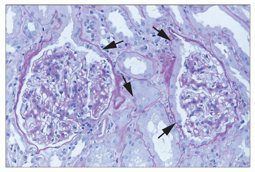
FIGURE 26.2 “Tubularization” of parietal epithelial cells lining the Bowman capsule (arrows). Reactive changes in the proximal tubule extend from the tubular takeoff to involve these epithelial cells, which have marked increase in cytoplasm compared to normal quiescent cells. (H&E; ×640.)

The lesions in both ischemic and toxic ATI primarily involve the tubules; the glomeruli are spared, as indicated by the nomenclature (178). Although no significant changes occur in the glomeruli, the parietal epithelium of the Bowman capsule is often prominent (Fig. 26.2), apparently reflecting reactive changes in the proximal tubule. Herniation of proximal tubular epithelium into the Bowman’s space is sometimes seen and may be the sole indicator of ATI when tubular epithelial changes are minimal. While these changes may be prominent, they are not specific. Glomeruli may show ischemic collapse, and the Bowman space may appear dilated.


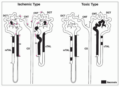
FIGURE 26.3 Cartoon demonstrating the difference in the distribution of lesions between ischemic and classic nephrotoxic tubular injury. In addition to differences in localization along nephron segments, different degrees of damage are visible between cortical and juxtamedullary nephrons. In the ischemic form, the S3 segments are most severely affected, along with focal areas of the ascending limbs of the loop of Henle. The cortical nephrons show more extensive damage than the juxtamedullary nephrons. In the toxic form, tubular epithelial damage is more extensive. Whereas mercury shows some predilection for the S3 segment, other heavy metals and organic toxins often show more extensive involvement of all nephron segments, also with a greater predilection for cortical nephrons.

Although “necrosis” has traditionally been included in the clinical term for ARF caused by tubular injury to distinguish this condition from other intrinsic causes of ARF (such as prerenal or postrenal failure or acute glomerular or interstitial nephritis), tubular epithelial cell death is often not evident by light microscopy (179). ATI is generally divided into two subcategories: postischemic ATI and nephrotoxic ATI. Morphologic changes of cellular injury are usually more subtle in the ischemic type, with more obvious cytopathologic changes in the toxic form. In addition, the sites of tubular damage along the nephron differ between the two forms (Fig. 26.3). In the ischemic form, tubular damage is patchy, affecting relatively short lengths of the straight segments of the proximal tubule and focal areas of the ascending limbs of the loop of Henle. In the toxic form, the tubular epithelial damage is more extensive along segments of the proximal tubule; the degree of involvement of the segments varies with the specific toxin. Although there is distal nephron damage, it is less extensive and more inconsistent in location than with ischemic ATI.


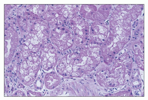
FIGURE 26.4 Tubular cells showing severe cell swelling, in some areas apparently obstructing the tubular lumen. (H&E; ×640.)


ISCHEMIC ACUTE TUBULAR INJURY

The histologic picture varies with the severity of renal failure and the evolution of the lesion. Early in the course, cellular changes can range from minimal alterations to severe cell swelling (Fig. 26.4) to individual cell necrosis with denudation of the basement membrane (Fig. 26.5). With injury, there may be shedding of both viable (Fig. 26.6) and necrotic epithelial cells (Fig. 26.7) into the tubular lumen. Exfoliated epithelial cells, some viable, can be demonstrated in the urine (Fig. 26.8) (180).

In sections stained with periodic acid-Schiff (PAS), the brush border of proximal tubules is often thinned or absent. Blebs of apical membrane and intact cells shed from their basement membrane anchor are present in the lumen of the tubules (Fig. 26.9). Focal lesions of individual cell necrosis with disruption of the basement membrane also occur in the ascending limb of the loop of Henle. Hyaline, granular, cellular, and/or pigmented casts are seen in the distal portions of the nephron and are often particularly prominent in the collecting ducts (Fig. 26.10). These casts consist of Tamm-Horsfall protein, which stains positively with PAS, mixed with cell debris (181). It is the relative prominence of these distal changes that gave rise to the term lower nephron nephrosis. In segments of the tubules that do not show significant necrosis, the tubules are often dilated and lined by flattened epithelial cells—so-called tubular simplification (Fig. 26.11). The denuded basement membrane sections are covered by proliferating adjacent viable epithelial cells. There may be evidence of transdifferentiation of tubular cells, which may express vimentin and other mesenchymal markers (Fig. 26.12). There is some evidence that transdifferentiated tubular cells may contribute to fibrogenesis in later stages (182).


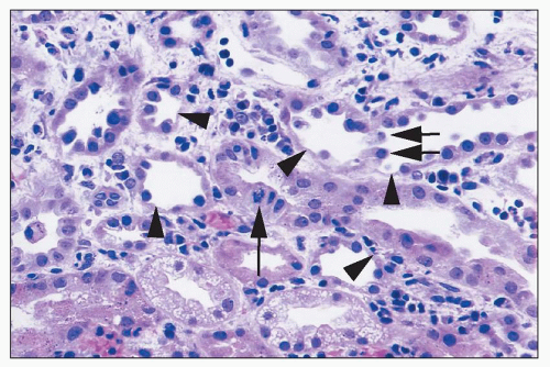
FIGURE 26.5 Areas of single tubular cell loss from a kidney with ischemic injury. Injured cells have detached, leaving areas of tubular basement membrane covered by a thin layer of cytoplasm from adjacent cells (arrowheads). A few detached cells can be seen in tubular lumina (short arrows). A mitotic figure can be seen in one tubular cell (long arrow). There are also interstitial edema and inflammatory cells largely marginating in capillaries. (H&E; ×400.)


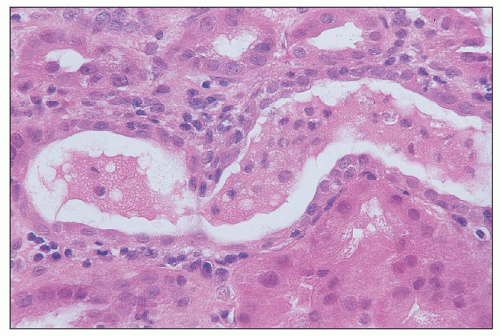
FIGURE 26.6 Intact exfoliated tubular cells in tubular lumen in a kidney with ischemic injury. (H&E; ×640.)

As the lesion progresses after the initial injury, evidence of tubular regeneration can be seen. Histologic indicators of cellular proliferation, such as mitoses, hyperchromatic nuclei, and a high nuclear-cytoplasmic ratio, may be seen (Fig. 26.13). Recent studies using genetic fate-mapping techniques in mice after ischemia-reperfusion injury (IRI) showed that most of the injured tubule cells were replaced within 2 days through extensive proliferation by surviving neighboring cells (183). These results indicate that regeneration of injured tubule cells through proliferation of surviving tubule cells is the predominant mechanism of repair after ischemic injury (183). Proliferation can be demonstrated by staining for transcription factors (Fig. 26.14) and other markers.

The injured tubules are separated by sometimes markedly edematous interstitium. There may be a mild interstitial inflammatory infiltrate with small numbers of lymphocytes, macrophages, and neutrophils or, occasionally, eosinophils. The cellular infiltrate tends to be clustered around necrotic and ruptured segments of tubules or where Tamm-Horsfall protein has been extruded, forming small granulomas. It is in these late stages that distinctions have to be made between ischemic ATI and acute tubulointerstitial nephritis, but in general, the infiltrate is much less prominent in cases of ATI.


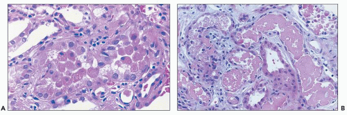
FIGURE 26.7 A: Detached necrotic tubular cells, several with pyknotic nuclei, in the lumen of proximal tubule. B: Granular casts with necrotic cell debris. Note flattened tubular epithelium in tubules containing necrotic debris. (H&E, ×640.)


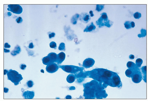
FIGURE 26.8 Intact tubular cells in the urine from a patient with ischemic injury. (Papanicolaou; ×1,000.)


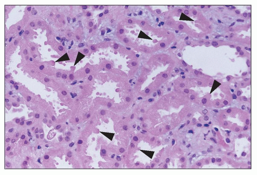
FIGURE 26.9 Apical blebbing from the surface of injured proximal tubular cells (arrowheads). Apical cytoplasmic blebs can be seen in tubular lumina. (H&E; ×640.)


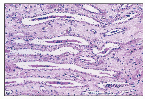
FIGURE 26.10 Tubular cell casts in collecting ducts in papilla. Injured cells have detached from sites in proximal nephron and aggregate into casts, often around a protein core. (H&E; ×400.)


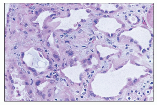
FIGURE 26.11 Dilated tubules with flattened epithelium in the regenerative phase after tubular injury. Marginating inflammatory cells can be seen in capillaries. (H&E; ×640.)


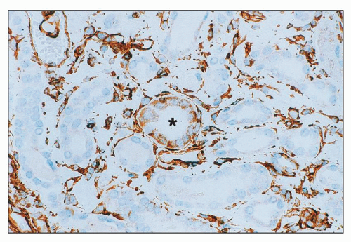
FIGURE 26.12 Injured tubule with cells staining for vimentin (center). Adjacent tubules do not stain. Note bright background staining for vimentin in interstitial areas. (Immunoperoxidase; ×400.)

Tubular cell death during ischemia/reperfusion occurs via apoptosis as well as coagulative necrosis and has been documented both in animal models and in clinical renal disease (184,185,186). It is possible to detect the nuclear and cytoplasmic condensation of cells undergoing apoptosis by light microscopy. Apoptotic cells may appear triangular and may be extruded from the epithelium into the tubular lumen (Fig. 26.15). Apoptotic bodies, representing membrane-bound nuclear fragments, may also be detected in adjacent tubular cells, which have engulfed these cell remnants. However, the most reliable methods of detection are by nick end labeling (TUNEL) of the chromatin that has been cleaved in a characteristic “ladder” pattern by the endonucleases or by staining for apoptosis-associated markers, such as cytochrome c or apoptosis-inducing factor (AIF) (187). Coagulative necrosis is characterized by eosinophilic cytoplasm and pyknosis and eventual disappearance of nuclei (Fig. 26.16).


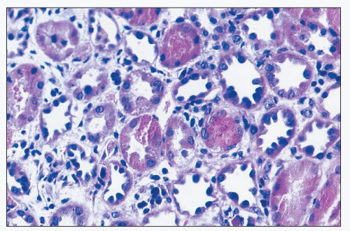
FIGURE 26.13 Striking regenerative changes in tubular cells, with pleomorphic hyperchromatic nuclei, with relatively little cytoplasm. (H&E; ×640.)


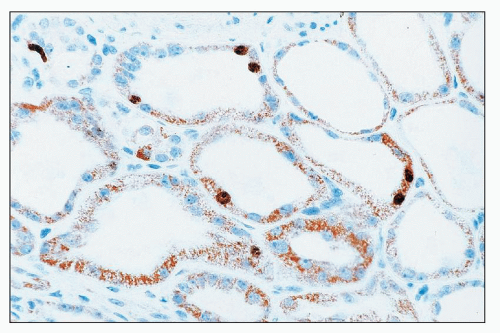
FIGURE 26.14 Immunostain for the transcription factor Ki-67. Note positive nuclear staining in several tubular cells. (Immunoperoxidase; ×640.)

ATI in renal allografts can show changes similar to those found in native kidneys, but more frank necrosis of tubular cross sections may be seen, and calcium oxalate deposits may be numerous in renal tubules (188) (Fig. 26.17). The cellular lesions are most prominent in the S3 segment of the proximal tubule and tend to be more uniform in character. Apical blebbing may be the only finding in milder forms, whereas the more severe cases also show focal necrosis with rupture of the tubular basement membrane. It is interesting to note that one study has shown a correlation of loss of Na+,K+-adenosine triphosphatase (ATPase) polarity with delayed graft function (189).

Electron microscopy has been helpful in evaluating the tubular epithelial changes in ATI (190,191,192). In ischemic ATI, scattered epithelial cell changes show a variety of different cytopathic alterations. These include loss of the apical brush border; blebbing of the apical membrane, with shedding of apical membrane blebs into the tubular lumina; high-amplitude swelling, with condensation of the cristae of the mitochondria; individual cell apoptosis, as demonstrated by cell shrinkage
with nuclear fragmentation; and a variety of other cytopathic changes leading to necrosis (Fig. 26.18). Detachment of tubular epithelial cells may be seen (Fig. 26.19).


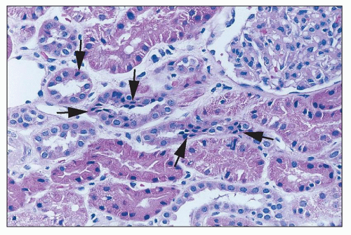
FIGURE 26.15 Focal tubular cell apoptosis, with condensed triangular cells in the epithelium (arrows), focally extruding from the epithelium. (H&E; ×640.)


FIGURE 26.16 Coagulative necrosis of focal tubular epithelial cells, with cell debris in the tubular lumina. (H&E; ×640.)

Interstitial inflammation is seen as a response to tubular injury. This inflammation is typically mononuclear and patchy. There is often associated interstitial edema, which may be severe (see Fig. 26.5). A particularly interesting and useful finding in cases of ARF is the accumulation of nucleated cells in the vasa recta of the outer medulla (193,194). This is a very common feature, and in many cases, it is the only histologic clue to the diagnosis of ATI (Fig. 26.20). The nature of the cells changes with progression of the ATI through its three different phases. Lymphocytes are predominant in the first 24 to 48 hours, followed later by immature cells of the myeloid series and eventually by nucleated red cells and red cell precursors. The accumulation of the larger nucleated cells in this location may be merely a reflection of the hemodynamic shifts that occur in ATI, with shifting of blood flow away from the superficial and midcortical glomeruli to the juxtamedullary glomeruli, resulting in a relative increase in blood flow to this nephron population, which gives rise to the vasa recta. The countercurrent nature of blood flow in the vasa recta would result in dilution of cellular elements as blood flows toward the hairpin turn, resulting in a concentration of cellular elements at the proximal end of the vasa recta vasculature in the outer medulla. There is also some evidence that up-regulation of adhesion molecules on ischemic endothelium leads to accumulation of leukocytes in the microvasculature, which may contribute to stasis and lack of reflow in ischemia-reperfusion injury (see section “The Inflammatory Response in Ischemic Injury”).


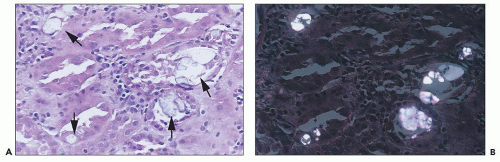
FIGURE 26.17 A: Oxalate crystal precipitates (arrows) in ATI in an allograft kidney. B: Oxalate crystals in polarized light. (H&E; ×640.)


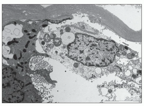
FIGURE 26.18 An electron micrograph shows a tubular epithelial cell in the process of desquamation into the lumen. The cell has separated from the basement membrane but is still adherent to the adjacent epithelial cell through the cell-to-cell junctions. (×3,000.)

At the molecular level in AKI, in addition to transcription factors and markers of cell differentiation/transdifferentiation, there is altered expression of a range of proximal and distal gene products in kidney tissue sections, among which KIM-1 and NGAL are most prominently expressed (195,196,197,198). KIM-1 is primarily expressed at the luminal side of dedifferentiated proximal tubules (198). ER stress markers such as CHOP and GRP94 can also be identified in renal tissue in injured cells (199). Tissue (and urine) detection of fibrinogen
has been proposed as a sensitive early marker of AKI, with markedly increased expression (199,200). The L-1 cell adhesion molecule has been identified as a potential biomarker of distal tubular injury in AKI, with loss of normal polarized distribution in the collecting duct, and induction of expression in medullary thick ascending limb and distal tubule with injury (201). Apoptotic cell death, difficult to detect by histology, can be detected in tissue using techniques such as terminal deoxynucleotidyl transferase dUPT nick end labeling (TUNEL) (201). An increase in progenitor cells (e.g., CD133+ CD24+ CD106- cells) can be identified in tissue sections by immunostaining as a correlate of injury (202). Other potential injury markers in tissues are described below in the section on “Pathophysiology.”


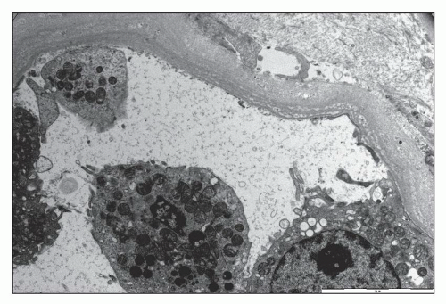
FIGURE 26.19 An electron micrograph demonstrates an area of loss of an array of tubular epithelial cells. (×3,000.)


NEPHROTOXIC ACUTE TUBULAR INJURY

Tubules Classically, toxic tubular injury may be associated with extensive epithelial necrosis, which tends to involve all nephrons more uniformly than in the ischemic form. However, a range of morphologic changes may be seen in the renal tubules as the result of toxic injury. The extent and severity of the changes will vary depending on the agent, the dose, and the timing of the morphologic assessment. Renal tubular cell changes detectable by light microscopy include the following:


  • Alterations in the surface of the cells, including loss of brush border (detectable on PAS), loss of basolateral infoldings, and blebbing of apical cytoplasm


  • Cytoplasmic swelling and vacuolation


  • Intracellular inclusions


  • Extensive tubular cell necrosis


  • Loss of individual tubular cells, with gaps along the tubular basement membrane or tubular profiles with fewer and attenuated cells lining the tubule


  • Intraluminal proteinaceous cellular debris, casts, or crystals


  • Tubular dilation with flattening of tubular epithelium


  • Tubular rupture with urinary extravasation


  • Regenerative changes, including flattening of epithelial cells, cytoplasmic basophilia, heterogeneity in cell size and shape, a higher nuclear-to-cytoplasmic ratio in individual cells, and cellular mitoses


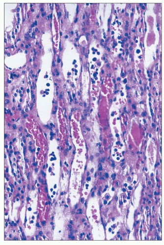
FIGURE 26.20 Erythrocyte congestion and nucleated cells in dilated vasa recta in the outer medulla of a kidney with ischemic injury. (H&E; ×400.)

Swelling and vacuolation of proximal tubular cells may be seen; cells appear large and pale and may contain discrete vacuoles of varying size. Hypertonic solutions, including IVIG preparations (203), have been reported to produce severe swelling and vacuolation of renal tubular cells. Intracellular inclusions are occasionally seen in renal tubular cells exposed to drugs. Giant mitochondria, appearing as bright eosinophilic inclusions, have been described after the administration of relatively high doses of CsA to humans. In gold-induced nephropathy, gold can be demonstrated in tubular cells (204). Calcification of tubular cells has been described in cases of severe toxicity caused by amphotericin or bacitracin (205).

Extensive coagulative necrosis of tubular cells has been seen in cases of poisoning by heavy metals such as mercuric chloride, rarely seen today, or in cases of poisoning due to chemicals such as diethylene glycol (206). More often, changes are more subtle, with individual tubular cell necrosis or loss, though there may be more obvious and extensive necrosis than is seen in ischemic
injury (207). It is clear that both necrosis and apoptosis occur in toxic nephropathies in experimental models as well as in clinical tubular injury (208,209). In vitro studies have documented apoptosis in cell culture on exposure to nephrotoxins. For example, LLC-PK1 cells exposed to sublethal doses of mercuric chloride in vitro undergo apoptosis (210), and apoptosis can be induced in Lewis lung cancer-porcine kidney-1 [LLC-PK1] cells by cisplatin as well via activation of caspases (211). Our understanding of the role of apoptosis in the pathogenesis of toxic renal injury has evolved over the past decade. ATI due to cisplatin therapy depends also partially on Fas-mediated apoptosis driven by Fas ligand (FasL) expressed on tubular epithelial cells (212). Moreover, cisplatin down-regulates the expression of the taurine transporter gene TauT in LLC-PK1 cells (213). Taurine is one of the organic osmolytes, which have important antiapoptotic properties in the kidney (214). The antiapoptotic function of organic osmolytes in kidney cells is mediated through suppression of efflux of proapoptotic molecules, such as cytochrome c, from the mitochondria.

Tubular casts, which may include cells and cell debris, are frequently seen with toxic tubular injury. In addition, tubular crystalline deposits are found in cases of renal toxicity produced by nephrotoxins. Anesthetic agents, including methoxyflurane and halothane, and antiretroviral agents such as indinavir may produce tubular crystalline deposits. Mechanical obstruction may also result from deposition of intratubular crystals in patients treated with sulfonamides or acyclovir. In addition, radiocontrast agents are uricosuric and oxaluric, and casts and birefringent crystals have been identified following administration of these agents. Uric acid lithiasis with tubular obstruction has been reported with phenylbutazone (215). Finally, pigmented casts may result from hemolysis in rare cases of fulminant drug reactions and with the rhabdomyolysis caused by cocaine.

Vessels Vessels usually show no remarkable features unless there is intercurrent disease. However, newer studies in experimental models have refocused attention on injury to the microvasculature, which may have been underappreciated in clinical specimens. Certainly, vascular congestion in the outer medulla with margination has been noted.

Electron Microscopy Electron microscopy of injured tubules reveals loss of brush border microvilli and basolateral infoldings in the proximal tubules. Cells may show rarefaction of the cytoplasm, with intracellular vacuoles and swollen organelles. Degenerative changes in mitochondria, including swelling and loss of cristae, loss of endoplasmic reticulum, or alterations in lysosomes, are often visible. Within the cells, membrane-bound structures consisting of concentrically arranged whorls of membrane may form, especially with exposure to aminoglycosides; however, these so-called myeloid bodies do not necessarily indicate toxicity.


Pathology of Specific Nephrotoxins

As a preface to this discussion, it should be recognized that it is often difficult to establish a pathogenetic link between a pathologic lesion and a particular drug or toxin. Several factors contribute to this uncertainty, including concurrent factors that may produce renal injury, such as administration of other potentially nephrotoxic drugs, lack of or inadequacy of morphologic data in reported cases, and the fact that some drugs may have multiple effects. Moreover, experimental models of toxicity may not be relevant to a particular clinical context owing to interspecies variation and markedly different dosing of drug or toxin in these models. In general, we limit our discussion to those drugs for which toxicity has been well documented in humans by disappearance of toxic effects when the drug is withdrawn, recurrence of symptoms after readministration of the drug, or both.

A range of chemotherapeutic agents and other toxins may produce direct injury to the renal tubular epithelium. These agents are outlined in Table 26.2. The focus in this discussion is on primary toxic tubular injury, recognizing that secondary injury to the renal tubule may also occur with other types of toxic renal injury, including tubulointerstitial nephritis, hemodynamic changes, and vascular disease.


ANTIBIOTICS

There have been many reports of renal damage associated with antibiotic therapy. Some drugs are more nephrotoxic and can promote acute renal injury even with brief exposure. One such example is the aminoglycosides (especially neomycin), which are classic nephrotoxins. However, in many cases, there is an association, but the causative role of the antibiotic in the etiology of tubular injury cannot be firmly established. There are several reasons for this. First, the infection for which the drug is being used may damage the kidney directly or indirectly. Second, infections are frequently treated with several agents, making it difficult to implicate a particular drug. Finally, the paucity of renal biopsy studies makes it difficult to define the pathologic changes produced by individual drugs and the pathogenetic mechanisms involved in producing renal injury.

Antiviral Agents In experimental animal models and in humans receiving acyclovir, indinavir, or ganciclovir, renal histologic examination often shows drug crystals in tubules, especially collecting ducts, with dilation of tubules reflecting obstruction (54,55,216,217) (Figs. 26.21 and 26.22). In other cases, there may be tubular dilation and tubular cell injury without detectable crystals in the urine or kidney (218); crystals, of course, might be missed if relatively few collecting ducts are sampled. Proximal tubular injury has been described,
especially with the nucleotide reverse transcriptase inhibitors adefovir and tenofovir, with degenerative changes, thinning and vacuolization of cytoplasm, loss of brush border, and even tubular cell necrosis, with nuclear changes reminiscent of viral inclusions (61,219,220,221). On electron microscopy, alterations in mitochondria have been observed, with swelling and dysmorphic changes. Changes include variable mitochondrial size, with some small and rounded and others swollen with irregular contours, clumping, loss and disorientation of cristae, and focal marked reduction of mitochondria (61). Giant mitochondria, in some cases, the size of nuclei, fuchsinophilic on trichrome stain but PAS- and silver-negative, may be seen in proximal tubular epithelial cells. Patchy interstitial inflammation has been described without crystals (222), and, occasionally, granulomas have been described. Renal tubular cell apoptosis has been detected in renal biopsy of a patient with irreversible cidofovir toxicity (223), and fibrosis and tubular atrophy have been described with tenofovir (61), with persistent renal dysfunction.


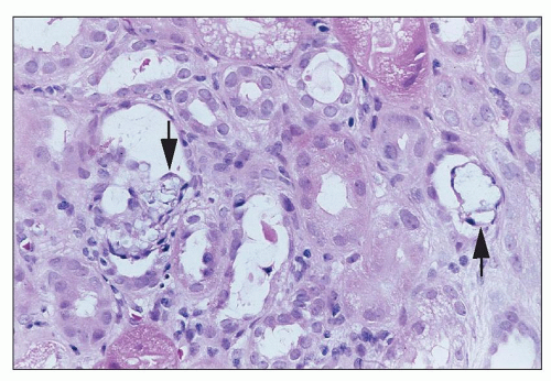
FIGURE 26.21 Crystalline precipitates (arrows) in tubules in a patient treated with intravenous acyclovir. (H&E; ×640.)


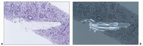
FIGURE 26.22 A: Indinavir crystal in papilla of a patient treated long term with highly active antiretroviral therapy. The patient also had crystals in the urine. B: Indinavir crystal under polarized light. (H&E; ×200.)

Aminoglycosides Aminoglycosides are classic nephrotoxic agents. Accumulation of high concentrations within lysosomes and release to the cell cytoplasm promotes phospholipid membrane rupture, oxidative stress, and mitochondrial injury. As a consequence, proximal tubule cells develop apoptosis and necrosis. The pathologic lesion most often reported with gentamicin is ATI (224), although in some cases, this lesion has been attributed to concomitant volume depletion and hypotension. Tubulointerstitial inflammation with tubular necrosis also has been reported (225). Myeloid bodies can be seen by ultrastructural examination in the tubular epithelium of patients receiving gentamicin, which predominantly reflect exposure to the drug (224) (Fig. 26.23).

Zager (226) has shown experimentally that gentamicin in a dose that does not by itself cause renal failure will trigger severe renal failure when combined with 1 hour of moderate renal hypoperfusion, which also does not produce renal failure on its own. In those studies, there was tubular necrosis in the S3 proximal tubule segment, a pattern of injury characteristic of renal ischemia rather than gentamicin toxicity; this suggests that in some instances, gentamicin may worsen ischemic injury rather than causing injury to the S1 and S2 segments, which is more typical of toxic doses of gentamicin (226).

There are few descriptions of renal pathologic lesions in patients receiving kanamycin or tobramycin. In one patient with oliguria who received 21 g of kanamycin over a 2-week period (227), a renal biopsy was done 25 days after the onset of oliguria (21 days after diuresis) and was reported to show some flattening of tubular epithelial cells.

In cases of ARF resulting from a combination of gentamicin and cephalothin, the pathologic features are those of ATI with normal glomeruli and vessels. However, experimental studies in the rat have found no potentiating effect of cephalosporins on gentamicin nephrotoxicity (228).

Amphotericin In human autopsy or biopsy specimens from patients treated with amphotericin, extensive calcification in tubules, presumably developing in the context of severe tubular cell injury, has been reported (229) (Fig. 26.24A). There may be vacuolation of smooth muscle cells in small arteries and arterioles (230) (see Fig. 26.24B). This is a change that potentially reflects direct toxic effects on the arterial wall, some element of intrarenal vasospasm, or both, and it may be very striking.

Cephalosporins Renal biopsies have been obtained in relatively few cases of cephalosporin-induced renal injury, usually in those in which the older cephalosporins were given. In cases of cephaloridine-induced ARF, biopsies have shown a picture of interstitial edema with variable numbers of chronic inflammatory cells accompanied by tubular dilation or necroses (231). The renal histologic features in these cases showed what is described as ATI, with interstitial fibrosis or edema and infiltration by lymphocytes and mononuclear cells. Pathologic changes in cases induced by cephalothin with or without other potential nephrotoxins consist of interstitial edema with variable numbers of lymphocytes and plasma cells; necrosis, swelling, and evidence of regeneration of tubular epithelium; and only trivial glomerular changes (232,233). Vacuolation of tubular cells has been evident on electron microscopy (232). A case of bilateral renal cortical necrosis (RCN) associated with cefuroxime has been reported (234). ATI has been described in patients treated with cephalexin (77,78).


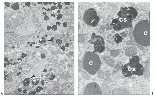
FIGURE 26.23 Electron micrographs of a rabbit killed 14 days after being given a high dose of gentamicin. A: Cytosegresomes (arrowheads) are scattered throughout the cytoplasm of the proximal convoluted tubule. The brush border can be seen at the top left (×5,700). B: Numerous cytosomes (C) and cytosegresomes (CS) are visible at higher power (×2,800) (lead citrate and uranyl acetate). (Courtesy of Dr. E. F. Cuppage.)

Polymyxin/Colistin On biopsy of patients treated with polymyxin, there is interstitial edema with eosinophils, plasma cells, lymphocytes, and, occasionally, neutrophils (235). The cellular reaction may have granulomatous characteristics. Tubules show swelling of the epithelium with intramuscular administration of colistin; the lesion described was ATI (236).


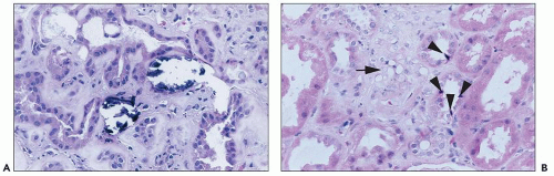
FIGURE 26.24 A: Calcification of tubular cells in a patient treated with amphotericin. B: Striking vacuolization of smooth muscle cells (arrow) in small vessels in the biopsy of a patient treated with amphotericin. Note apoptotic cells (arrowheads) in adjacent tubules. (H&E; ×640.)

Vancomycin While ARF with vancomycin is associated with interstitial nephritis, acute tubulopathy has also been described. Acute tubular necrosis (ATN) with anuria has been described associated with vancomycin and one dose of aminoglycoside (237). Another case with biopsy-proven ATN has been described in a child (238). Another biopsied case in a
child with elevated vancomycin levels and ARF revealed focal tubular dilation with attenuation of brush border, hyaline casts, and one neutrophil cast without interstitial nephritis (239).


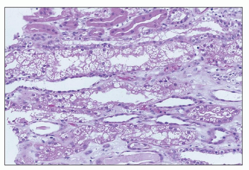
FIGURE 26.25 “Isometric” vacuolization, with many small equal-sized vacuoles in tubular cell cytoplasm, in a patient with high serum levels of calcineurin inhibitor. (H&E; ×400.)


IMMUNOSUPPRESSIVE/IMMUNOMODULATORY AGENTS

Cyclosporine Functional CsA nephrotoxicity can occur without any morphologic changes. The most common morphologic change observed in the kidneys of patients treated with CsA is isometric vacuolation of the proximal tubular cells (Fig. 26.25); this change is characteristic but not specific. Other changes include tubular epithelial cell necrosis with or without calcification, inclusion bodies corresponding to giant mitochondria, and giant lysosomes (240). The megamitochondria and microcalcification in tubular cells of CsA-treated patients do not correlate with dysfunction (241). Strong staining for osteopontin protein and mRNA has been demonstrated in tubular epithelium in clinical CsA toxicity (242).

Vessels in CsA-induced acute renal dysfunction may show only vasospasm and vacuolation of smooth muscle cells, changes that often reflect vasoconstriction. The onset of hyaline arteriolar thickening, especially with nodular accumulation of hyalin in the periphery of the arteriolar wall, has been associated with CsA-induced renal dysfunction, although dysfunction can also exist without this change. The juxtaglomerular apparatus may be hyperplastic; this finding is significantly more prominent in renal transplant patients with CsA nephrotoxicity than in other posttransplant groups, probably indicating activation of the renin-angiotensin system (243). Thrombotic microangiopathy may be seen in particularly severe cases of toxicity.

Descriptions of the pathologic characteristics of both clinical and experimental long-term CsA toxicity have focused on interstitial fibrosis and tubular atrophy, which appears in a “striped” pattern reminiscent of ischemic injury, and hyaline arteriolar change, as described earlier. The fibrosis involves medulla and medullary rays in the cortex (244). Bertani et al. (245) have reported the renal biopsy changes observed in cardiac allograft recipients with renal failure after they had received cyclosporine for 31 to 48 months. Obliterative arteriolopathy with ischemic glomerular changes was found. Serial reconstruction of the glomeruli showed the presence of populations of both abnormally small and abnormally large glomeruli. Sclerotic lesions were confined to the small glomeruli. Myers et al. (246) and Morozumi et al. (247) have also emphasized sclerosing glomerular changes with long-term CsA therapy. The pathologic findings and the differential diagnosis of CsA nephrotoxicity in renal transplant recipients are discussed in detail in Chapter 29.

Tacrolimus Pathologic changes owing to FK506 are very similar to those described for CsA. Changes include tubular cell vacuolization, calcification, myocyte vacuolization, necrotizing arteriolitis, thrombotic microangiopathy, arteriolar hyalinosis, and interstitial fibrosis. Morphologic changes with FK506 toxicity have been compared to those produced by CsA (248,249). Tubular cell vacuoles were small and focally confluent and involved proximal and distal tubules. Morozumi et al. (249) have suggested that FK506-related vacuoles are foamy and nonisometric and present in straight and convoluted portions of the proximal tubules. We have noted involvement of tubules in outer medulla as well (unpublished).

Glomerular capillary and arteriolar thrombi have been seen in renal allograft recipients (250,251) and a few kidney/liver allograft recipients (250). In several of these cases, other factors, including prior CsA therapy in three cases and fungal sepsis in a fourth case, may have contributed to the endothelial injury underlying thrombosis. An HUS-like syndrome with thrombotic microangiopathy may be seen (Fig. 26.26); the estimated incidence is approximately 1% (251), and cases are frequently associated with high serum levels of drug. Hyaline arteriolar change and interstitial fibrosis have been reported with long-term therapy (240).

An increase in BUN and sCr has been documented in rats treated with FK506, with evidence of proximal tubular cell vacuolation and megamitochondria on histologic examination (249). Pathologic changes, including proximal tubular cell vacuolation and tubular regeneration similar to those reported with CsA therapy, also have been reported in canine allografts (252). Stillman et al. (253) developed a rat model of prolonged FK506 toxicity by combining FK506 with a low-salt diet for 6 weeks. In this model, sCr and plasma renin levels were elevated, and there was tubular atrophy and fibrosis in
medullary rays and the inner stripe of the outer medulla. Tacrolimus alone produced increased juxtaglomerular apparatus granularity.


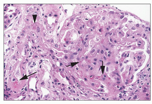
FIGURE 26.26 Thrombotic microangiopathy in a patient on tacrolimus. Note the arteriole with very focal intramural fibrin (arrowhead), focal erythrocyte extravasation into the intima (long arrow), and focal erythrocyte fragmentation in the glomerulus (short arrows). (H&E; ×640.)


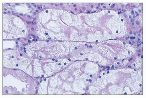
FIGURE 26.27 Severe cell swelling of tubular cells in the biopsy of a patient being treated with IVIG. Note persistence of brush border. (H&E; ×640.)

Intravenous Immunoglobulin Intravenous administration of immunoglobulins has been associated with severe swelling of tubular epithelial cells (254) (Fig. 26.27). Of note, the brush border of the cells is generally well preserved. Swelling may be severe enough to occlude the tubular lumen. In one series of transplant patients, isometric vacuolization appeared to precede the more severe cell swelling (255).

Sirolimus Rats given sirolimus (3 mg/kg orally) for 2 weeks on a low-salt diet developed magnesium wasting and structural renal lesions consisting of tubular collapse, vacuolization, and nephrocalcinosis (248). ATI has occasionally been described in patients (128). In one study of sirolimus-treated patients (also on FK506), there was a subset that developed striking intratubular cast formation, reminiscent of myeloma cast nephropathy (125). Vascular changes and glomerular disease have been reported with sirolimus. Some reports have appeared that sirolimus may delay recovery from tubular injury, exacerbate acute FK506 tubular cell toxicity (125,128), or exacerbate chronic calcineurin inhibitor toxicity.


CHEMOTHERAPEUTIC AGENTS

Cis-Platinum In the human kidney, focal necrosis of tubular cells is seen, primarily in the distal tubule and collecting ducts; cast formation and dilation of proximal tubules may be observed (256). In animals, tubular changes are found in the proximal nephron, with or without accompanying distal changes (257). Many patients show continuing damage and fail to regain pretherapy levels of renal function. Dobyan et al. (258) studied the effects of chronic administration in animals and found cyst formation, interstitial fibrosis, and tubular atrophy.

Other The pathologic picture produced by the alkylating agent ifosfamide is that of ATI or chronic tubulointerstitial changes (259). On pathologic examination of kidneys from patients with toxic injury caused by streptozotocin, there is ATI in the proximal tubules, with or without accompanying interstitial inflammation (142).


RADIOCONTRAST AGENTS

Because renal biopsies are not indicated in patients with transient renal failure after contrast media administration, human studies are scant. Patients who undergo biopsy are those who do not recover from renal failure, and in most of these cases, the histologic picture reveals an underlying (and most likely preexisting) renal disease. The majority of publications describing renal morphologic changes are based on experimental studies. The induction of renal failure in experimental animal models usually requires administration of additional agents (e.g., indomethacin, gentamicin, glycerol), ischemia, water or salt depletion, or a combination of these factors (225,260,261,262,263).

The overwhelming majority of both experimental and clinical studies report variable, transient proximal tubular vacuolation, which appears as soon as 30 minutes following administration, disappears within a few days (264,265), and seems to be dose dependent (266). One study emphasizes the selective injury of the thick ascending limb of Henle in the outer medulla after coadministration of indomethacin and iothalmate to unilaterally nephrectomized, salt-depleted rats (225). They describe mitochondrial swelling, pyknosis, cytoplasmic disruption, calcification, necrosis, and tubular collapse in the thick ascending limb of Henle in areas away from vascular bundles, suggesting hypoxic injury. They also report vacuolation of the proximal convoluted tubules.

Ultrastructural studies indicate that the vacuoles are membrane bound, probably representing lysosomes (225,264). The fine structure of the mitochondria and the endoplasmic reticulum remains intact. Tervahartiala et al. (264) believe that the vacuolation is caused by a nonspecific lysosomal injury and is not the consequence of osmotic diuresis. Autoradiographic and electron microscopic studies failed to demonstrate the presence of iodinated molecules within the vacuoles (225,267).

The most extensive studies in human beings come from the Necker Hospital in Paris, where radiocontrast examination of the kidney was routinely performed before renal biopsies in the 1970s (265,267). This group published the case studies of 211 patients who underwent biopsy within 10 days of urography or renal arteriography using ionic contrast media (267). Tubular vacuolation characteristic of “osmotic nephrosis” was found in 47 patients and was more severe in patients with preexisting renal failure; however, they did not find a correlation between the extent of tubular vacuolation and the degree of renal functional impairment. The same group later described osmotic nephrosis in 14 of 33 patients who received low-osmolality contrast media before renal biopsy (267). They reported the case of one patient who had evidence of ATI on initial biopsy and showed signs of advanced tubular atrophy and interstitial fibrosis on a second biopsy. Other patients in the Necker Hospital also had evidence of ATI in the renal biopsy, but it appears that these patients had ARF as a preexisting condition. They concluded that tubular vacuolation after radiocontrast administration probably does not represent true osmotic nephrosis and that it is not a reliable morphologic indicator of RN (267).

In 1970, two articles described hemorrhagic necrosis, primarily of the renal medulla, in six infants (268,269). Five of them underwent cardiac catheterization for heart problems, and one had excretory urography because of a flank mass. In these children, there were several confounding variables that might have contributed to the renal necrosis, such as seizures, cardiac developmental abnormalities, and sepsis. All six children died. Although
ATN has been reported only rarely in patients (265,267,270), recent studies have shown that radiocontrast agents induce apoptosis in proximal tubule cells (271). Increased ceramide synthesis, which stimulates apoptosis, is an important contributing factor to radiocontrast-mediated nephropathy (272).


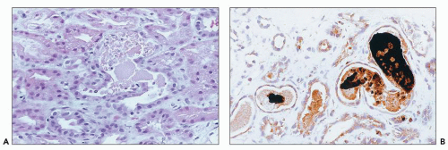
FIGURE 26.28 Pigmented casts in the tubular lumina of a biopsy from a patient with ARF who had overdosed on cocaine. A: Light microscopy. (H&E; ×640). B: Immunostain for myoglobin. (Immunoperoxidase; ×640.)


NARCOTICS AND MYOGLOBINURIC ACUTE RENAL FAILURE

The characteristic finding is the presence of pigmented casts, as in other forms of myoglobinuric ARF. Renal biopsy is rarely performed in affected patients, and for this reason, pathologic reports are uncommon (273,274). The characteristic casts show mild brown pigmentation and usually have a granular appearance with irregular globules. These casts are frequently bright red as seen by trichrome stain. Immunohistochemistry is helpful in identifying myoglobin casts (Fig. 26.28). Hyaline and granular casts not containing detectable myoglobin may be present. Other features of ATI, such as tubular epithelial damage with exfoliation of tubular epithelial cells, thinning of the tubular epithelium, and tubular calcification, are usually noted. Immunofluorescence is typically not helpful. On ultrastructural examination, the myoglobin casts frequently consist of very electron-dense, finely granular globules that may have a somewhat less electron-dense rim (Fig. 26.29). In addition, electron microscopic signs of ATI are readily visible.


ANESTHETICS

The renal lesion consists of interstitial edema with somewhat dilated tubules lined by flattened epithelium. In several cases, a striking degree of intratubular collection of oxalate crystals has been reported (275).


HERBAL MEDICATIONS

There are relatively few reports of biopsy findings in ARF caused by herbal medications. ATI has been described (reviewed by Isnard Bagnis et al. (169)). Pathology of the kidneys in patients with renal failure caused by Aristolochia species has shown hypocellular interstitial fibrosis and tubular loss, especially in the outer cortex, in lesions obtained later in the course of the injury (276).


Etiology and Pathogenesis

Although the number of disease entities that have been associated with ATI is large, the basic etiologic factors are very similar (Table 26.3; Fig. 26.30). Prolonged renal ischemia is the most common cause of ATI (277,278,279). In the hospital setting, it is frequently associated with major surgery, with extensive trauma such as crushing injuries and burns, or severe congestive heart failure and septic shock (280,281). The widespread use of nonsteroidal anti-inflammatory drugs (NSAIDs), which inhibit renal prostaglandins, is another potential mechanism through which renal ischemia can be initiated, and these drugs have been associated with the development of ARF, particularly in patients who are volume depleted or dehydrated.

The second major category of etiologic agents of AKI is exposure to nephrotoxic drugs. The kidney is uniquely susceptible to toxic injury, because it is the principal excretory organ of the body. Since metabolism and excretion of exogenously administered therapeutic and diagnostic agents are major functions of the kidney, the ingestion of drugs is significantly associated with kidney injury. A number of therapeutic agents have known nephrotoxic potential. Classic examples include antimicrobial agents, chemotherapeutic agents, analgesics, and immunosuppressive agents (44,65,282,283,284,285,286). A problem that has been observed in developing countries is the contamination of commonly used drugs by nephrotoxins during their preparation under less than stringent conditions. Examples include the sudden occurrence of unexplained ARF in children in Pakistan and Haiti, where the cause was found to be contamination of the liquid vehicle of paracetamol with diethylene glycol (287,288). Interaction of herbal products with conventional drugs is also a potential source of toxicity. Examples of nephrotoxic herbal products include aristolochic acid, Ephedra species, and Glycyrrhiza species (169,289). Adulteration of food products may be another cause of kidney injury. One example is the addition of melamine to baby formula to increase the protein content, which caused AKI and nephrolithiasis in neonates (290). In many cases involving the use of diagnostic and therapeutic agents, the known risk of nephrotoxicity is outweighed by the clinical benefits of using the drug. While the range of injurious compounds is diverse, there are a limited number of patterns of injury produced in the kidney; the focus of this section will be on agents and specifically drugs that produce ATI.

ARF in the newborn may have a prenatal onset associated with maternal hypotension or occur in the setting of congenital diseases, such as renal dysplasia or polycystic kidney disease (279).
In the postnatal period, hypoxic/ischemic injury and toxins are the most common etiologies. Toxic ARF is most commonly associated with administration of aminoglycoside antibiotics and NSAIDs given to close a patent ductus arteriosus. Decreased renal function can be documented in about 40% of premature infants receiving indomethacin; the decrease is usually reversible.

Only gold members can continue reading. Log In or Register to continue

Stay updated, free articles. Join our Telegram channel

Jun 21, 2016 | Posted by in UROLOGY | Comments Off on Ischemic and Toxic Acute Tubular Injury and Other Ischemic Renal Injuries

Full access? Get Clinical Tree

Get Clinical Tree app for offline access