Development of the Kidney


Development of the Kidney


Adrian S. Woolf

Dagan Jenkins


Developmental kidney disorders account for a wide spectrum of disease in fetuses, children, and adults (1,2,3,4,5,6). As assessed by registries from developed countries, such disorders typically account for about half of all children who require dialysis and kidney transplantation for end-stage renal disease. Renal agenesis (absent kidneys) and dysplasia (poorly differentiated and metaplastic tissues) represent profound defects of morphogenesis; renal hypoplasia (too few nephrons) is a term used to describe kidneys that have differentiated but contain significantly fewer nephron units than normal. Congenital anomalies of the kidney and urinary tract (CAKUT) is a useful phrase coined by Ichikawa et al. (7) that emphasizes that kidney malformations often coexist with lower renal tract anomalies. CAKUT encompasses the following: agenesis, dysplasia, hypoplasia, ectopia, and duplication of the kidney; ureter anomalies such as megaureter, ureteropelvic junction obstruction, ureterovesical junction obstruction, vesicoureteric reflux, duplication, ureterocoele, and ectopic termini; bladder anomalies such as exstrophy and persistent cloaca; and urethral malformations such as atresia and posterior urethral valves. On the other hand, the use of the CAKUT diagnostic label, while
convenient, should not obviate the need to document the exact renal tract dysmorphology found in a particular patient, nor should it be assumed that there will be one underlying pathogenesis for CAKUT. Thus, the use of the term is analogous to using a disease group designation such as “glomerulonephritis” or “tubulopathy.” As medical and surgical therapies are refined, a new cohort of young babies with severe kidney malformations and renal failure, who might otherwise have perished, are surviving childhood (8,9); as yet, it is too early to know how they will fare through adulthood.

It has been suggested that essential hypertension, which manifests later in life, is initiated before birth by a little understood phenomenon called fetal programming, hence suggesting a novel but unexplained link between development and adult disease (10). Environmental influences somehow transmitted from the mother to the fetus during gestation may alter the growth trajectory of the developing kidney. Indeed, rats born to mothers fed low-protein diets have fewer nephrons, and such diets can alter cell turnover and gene expression in the forming kidneys (11,12). Furthermore, adults with essential hypertension have fewer glomeruli per kidney versus individuals with normal blood pressure, and it is now appreciated that the numbers of glomeruli per kidney show a wide variation even within populations of “normal, normotensive individuals” (13,14).

It is therefore important to understand how such a range of kidney malformations might arise, and, to do this, one must not only address the nature of the malformations themselves but also understand normal kidney development. The morphology and histology of the key types of human renal malformations were first comprehensively defined by Edith Potter in her landmark book Normal and Abnormal Development of the Kidney (1), and this area is reviewed in detail in Chapter 4 in this book (15).

In this chapter, we summarize the anatomy of normal human kidney development; in addition, we present mechanistic and functional insights derived from both animal experiments and human genetic diseases that are accompanied by renal malformations. The main focus is on the metanephric kidney, which forms the adult mammalian organ; the section on anatomy features human data, although rodent genetic models and organ culture will also be used when they inform the mechanisms of renal tract development. Although the term nephrogenesis is often used as a substitute for all aspects of kidney development, we here use the term in its more exact meaning to refer to the generation of the nephron lineage. We also briefly allude to the development of the lower renal tract (i.e., the renal pelvis, ureter, and bladder). Because of space limitations, we are unable to review the expanding literature on renal tract differentiation in animals other than mammals, and here, the reader should seek recent reviews in relation to the fly, fish, and amphibians (16,17,18).

In the future, an understanding of the normal anatomy and mechanisms of kidney development will offer insight into human renal disease. For example, the Wilms tumor-1 (WT1) gene is not only involved in normal kidney development but has also been found to be mutated in certain types of renal malignancy and glomerular disease (19). In addition, in the polycystic kidney diseases (PKDs), renal epithelial biology appears to revert to a less differentiated state (15,20,21).

Detailed discussion of disorders is beyond the limits of this chapter, but they are addressed in detail elsewhere in this book (15,22).


CELLULAR EVENTS IN KIDNEY DEVELOPMENT

A number of key biologic events occur during kidney development. They can be broadly classified as the birth, death, migration, and specialization of individual cells, as well as the acquisition of complex shapes by groups of cells. In real life, several of these processes may be occurring at the same time, but it is useful to consider them separately here.


Cell Proliferation

Cell proliferation is required for growth of the developing kidney. Proliferation is prominent in the nephrogenic zone, which is located in the outer cortex of the metanephros (23). Although there are no published measurements of the numbers of cells in human metanephroi, it is of note that total cell numbers in the rat metanephros increase from approximately 2 × 10⁴ on the day of formation of the embryonic organ to 2 × 10⁶ only 2 days later (11); the morphologic changes during this period correspond to the period of growth of the human metanephros between about 5 and 7 weeks’ gestation. By contrast, proliferation is rare in the postnatal normal human kidney, although it is readily detected in cysts of dysplastic kidneys resected surgically in childhood (23). It is considered that stem cells are located in the outer cortex of the metanephros and are capable of division to generate a copy of themselves and also a different type of cell that will subsequently mature (24,25); both the stem cells and their early progeny are called precursor cells. Although stem cells were previously considered to be absent from the mature mammalian kidney, evidence suggests that they may exist as rare subpopulations in adult organs and that they may be mobilized to replenish the organ following renal injury (26,27).


Cell Death

Not all cells formed in the developing kidney are destined to survive the fetal period. In rodents, a considerable amount of programmed cell death occurs during normal kidney development, and these cells generally die by apoptosis (11,28). This term describes a form of death that is accompanied by nuclear condensation and fragmentation and that is associated with activation of caspase enzymes. Apoptosis probably plays a role in determining the final number of nephrons and also the cell content of individual nephron tubules; it may also have a role in the remodeling that occurs during formation of the collecting ducts, renal pelvis, and lower tract. Early kidney growth thus involves a fine balance between cell proliferation and cell death.

An excess of proliferation is associated with renal neoplasms (e.g., Wilms tumor) and epithelial cyst growth during cyst formation (23). Conversely, excessive apoptosis would inhibit overall kidney growth and has been reported in the human dysplastic kidneys (29), which generally undergo spontaneous regression either before birth or in the 1st year of postnatal life (30). Excess renal apoptosis has also been reported to occur in experimental kidney malformations induced by teratogens (e.g., retinoic acid [RA]) (31), maternal low-protein diets (11), obstruction of fetal urinary flow (32), and mutations of genes expressed in the first stages of metanephric development (33,34,35).


Morphogenesis

Morphogenesis describes the process by which groups of cells acquire specialized three-dimensional shapes during development. Two examples during kidney development include the formation of nephron tubules from renal mesenchymal cells, a mesenchymal to epithelial phenotypic conversion, and the serial branching of the ureteric bud to form the collecting duct system. These normal processes are described in detail below; both are profoundly disrupted in human dysplastic kidney malformations (1,3). Another example of morphogenesis is the restructuring of just-formed nephrons into longer, thinner tubules. In the embryonic fly kidneys, it is well established that “convergent extension” occurs, in which already existing cells undergo rearrangements to form a thinner, longer tubule (16).

Perhaps a similar phenomenon occurs, for example, as loops of Henle grow from the deep cortex into the forming medulla/papilla of the metanephic kidney. In mammals, however, rapid tubule extension appears at least in part driven by proliferation, with mitotic spindles orientated along the length of the growing tubule; notably, immature tubules become cystic when the orientation of spindles is randomized (36). In the adult mammalian kidney, de novo formation of nephrons does not take place; however, the damaged adult kidney is capable of regenerating epithelia within already existing tubules, and, in other types of animals, such as certain fish, new nephrons and tubules continue to be generated throughout normal life (37,38).


Migration

Cell migration, driven by rearrangements within the cytoskeleton, is a widespread event in mammalian development (39).

Within the developing kidney itself, migration of cells occurs as endothelial (40) and mesangial (41) cell precursors invade nascent glomeruli, attracted by growth factors secreted by immature podocytes.


Differentiation

As renal precursor cells become phenotypically specialized, they are said to undergo differentiation. The term lineage describes the series of phenotypes that are displayed by the progeny of a precursor cell during the formation of specialized renal epithelial, interstitial fibroblast, mesangial, smooth muscle, and endothelial cells. It is generally considered that cells derived from the ureteric bud are confined to epithelia of the collecting ducts and urothelium of the renal pelvis, ureter, and bladder trigone. By contrast, metanephric mesenchymal cells can, as assessed by various experimental strategies in animals, give rise to a wide range of cell types including nephron epithelia (from glomerulus to distal tubule), endothelia, and mesangial and vascular wall cells (25,42,43).


ANATOMY OF DEVELOPMENT OF THE HUMAN KIDNEY

The summary of anatomic events presented here is based, to a large extent, on previous accounts (1,2,44,45,46,47,48,49,50,51); other specific references will be cited, as appropriate. Unless otherwise stated, the specific timings refer to human kidney development.

The intermediate layer of the mammalian embryo that forms during gastrulation is called the mesoderm. The kidneys develop from the nephrogenic cords, masses of intermediate mesoderm located between the dorsal somites and the lateral plate mesoderm behind the embryonic coelom. Three successive excretory systems are formed: the pronephros from the most cranial (cervicodorsal) segments of the nephrogenic cord, the mesonephros from the intermediate (dorsolumbar) segments, and the metanephros from the most caudal (initially sacral) segments. The pronephros and the mesonephros are transient organs, although both are essential to the formation of the definitive kidney from the metanephros. Other mesonephric derivatives form structures in the fully developed organism. Although the pronephros, mesonephros, and metanephros form sequentially in the developing embryo, there is some overlap so that the mesonephros begins to develop before the pronephros has disappeared, and likewise, the metanephros forms before the mesonephros has entirely regressed. Some authorities (52) have regarded a strict delineation of the three sets of the embryonic kidneys as artificial, viewing the whole system as a single unit, the holonephros. However, in mammals, including man, the mesonephros and metanephros at least can be clearly recognized as separate organs.


The Pronephros

The most cranial portion of the nephrogenic cord between the 7th and 14th somites develops as the pronephros from the end of the 3rd week after conception. The pronephros consists only of a few rudimentary tubules opening proximally to the coelomic cavity by nephrostomes and coalescing distally to form the pronephric duct, which grows caudally to the cloaca. The pronephros rapidly involutes and cannot be identified by day 25 of gestation; however, the pronephric duct survives as the mesonephric duct.


The Mesonephros

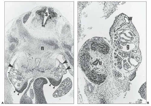
Just before the pronephros disappears, the mesonephros begins to develop from day 24 of gestation from the dorsolumbar segments of the nephrogenic cord below the pronephros, growing to form a prominent (urogenital) ridge that bulges into the coelomic cavity (Fig. 2.1A). A series of vesicles form, each of which develops into a nephron consisting of a glomerulus and a tubule with thicker-walled proximal and thinner distal segments (Fig. 2.1B). Capillaries forming each mesonephric segment establish connections with the primitive aorta by an afferent arteriole and with the posterior cardinal vein by an efferent arteriole. The tubules morphologically resemble proximal and distal tubules found in the metanephros, although there is no equivalent of the loop of Henle. Each mesonephric tubule joins an excretory duct (originally the pronephric duct, but now called the mesonephric or wolffian duct) that gradually extends caudally to communicate with the cloaca at the 26th to 28th somite stage (about 4 weeks postconception). Although it is possible that some excretory function is performed by the mesonephros in the human embryo, it is only transitory. By the end of the first trimester, all the mesonephric glomeruli involute, but some mesonephric structures do persist. In the male, remnants of a number of caudal mesonephric tubules develop as the efferent ducts of the epididymis. The mesonephric duct forms the duct of the epididymis, the seminal vesicle, and the ejaculatory
duct. In the female, apart from a few vestigial and inconstant structures, such as the epoophoron, the paroophoron, and Gartner duct, the whole mesonephros degenerates during the 3rd month of gestation.


FIGURE 2.1 Early human metanephros and the mesonephros. A: Transverse section of a 5- to 6-week gestation human embryo showing mesonephros (large arrowheads) and relatively undifferentiated gonadal ridge (small arrowheads). Also shown is the spinal cord (s) with the notochord (n) degenerating in a mass of cartilage that will form the vertebral body. (H&E, ×12.5.) B: Six-week human embryo showing the metanephros (left) and the mesonephros (right). The ureteric bud (u) in the center of the metanephros has branched twice, and the mesenchyme is condensing around the branch tips. In contrast, the mesonephros is much more differentiated and contains glomeruli (g) connected to tubules that drain into the wolffian duct (w). (H&E, ×50.) (Courtesy of Dr. P. J. D. Winyard, Institute of Child Health, London, UK.)


The Metanephros

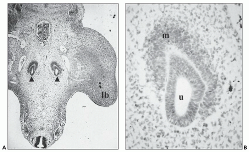
The metanephros forms the definitive kidney and is developed in two parts: the renal parenchyma from the caudal end of the nephrogenic cord, called the metanephric mesenchyme, and the collecting ducts, calyces, renal pelvis, and ureter from the ureteric bud. This is a hollow posteromedial offshoot arising during the 5th week (5-mm stage) from the caudal end of the mesonephric duct opposite the 28th somite, where it curves medially to join the cloaca. The proximal end of the ureteric bud grows dorsally and cranially toward the metanephric mesenchyme, while its distal end extends caudally as the embryo elongates. The ureteric bud develops a slightly swollen cranial tip, the ampulla, to be distinguished from the remaining tubular interstitial portion. When the ureteric bud impinges on the renal mesenchyme (Fig. 2.2), the latter is pushed upward to lie dorsal to the caudal end of the degenerating mesonephros.

Here, it is situated behind the peritoneum but ventral to the corresponding umbilical artery. As the ampulla of the ureteric bud contacts the metanephric blastema, which now forms a cap of densely packed cells, it undergoes a process of rapid dichotomous branching, each branch forming a new ampulla; each ampulla has the capacity for further dichotomous branching and ultimately to induce nephron formation from the metanephric mesenchyme (compare Fig. 2.2, the metanephros at 5 weeks’ gestation, with Fig. 2.1B, the metanephros at 6 weeks’ gestation).


Formation of the Human Renal Pelvis and Calyces

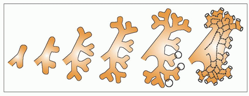
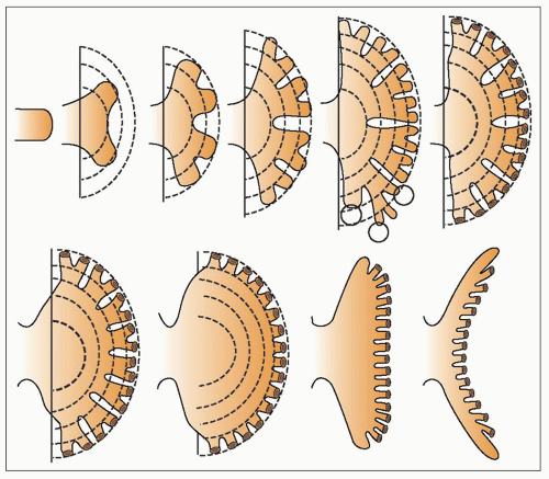
This process, together with the development of nephrons and collecting ducts, has been studied by Osathanondh and Potter (44,45) by microdissection of the fetal kidneys (Figs. 2.3 and 2.4). These authors showed that the renal pelvis and major calyces formed from the first three to six generations of ureteric bud branches (branching occurs more rapidly at the poles than in the midzone) and the minor calyces formed from the subsequent generation of branches. Because
branching is so rapid at this stage, the interstitial portions of the branches are very short, and indeed, sometimes a number of branches appear to arise from a single stem. Distension of the whole system, which is ascribed to the onset of urine production as nephrons start to function, results in the coalescence of the first generations of branches to give the more familiar appearance of the pelvicaliceal system seen postnatally. This is completed by about the 10th to 12th week of gestation. When the minor calyces are formed, some 20 or so ampullae are related to each minor calyx, and it is from these that the papillary collecting ducts originate. Further branching of these ampullae is associated with nephron formation, and these branchings proceed more slowly with longer intervening periods of interstitial growth. From about the 11th week, the spherical expansion of the calyces is limited by the development of nephrons associated with their own and surrounding collecting duct systems that form the developing renal papillae. The minor calyces are indented by the papillae and change from spherical to a wineglass shape, with the cup-shaped portion around the developing papilla and a narrower stem-like infundibular portion connecting to the major calyx. This process is achieved by about the 14th week of gestation
and can be appreciated in histologic sections of the fetal kidneys at this gestational age.


FIGURE 2.2 Human metanephros at the ureteric bud stage. A: Transverse section of a human embryo, approximately 5 weeks’ gestation, showing paired metanephric kidneys (arrowheads), one on each side of the midline. A lower limb bud (lb) is seen on the right (hematoxylin, ×12.5). B: High power of (A) shows one of the ureteric buds (u) capped by the metanephric mesenchyme (m), which is demarcating from the surrounding loosely packed, intermediate mesoderm.


FIGURE 2.3 Development of the renal pelvis. Diagram represents coalescence of the third through fifth generations of branches (circled) of the ureteric bud to form the renal pelvis.) (Modified from Osathanondh V, Potter El. Development of the kidneys as shown by microdissection. II. Renal pelvis, calyces and papillae. Arch Pathol 1963;76:277-289.


FIGURE 2.4 Development of renal calyces and papillae. Diagrams depict coalescence of the third to fifth generations of branches (circled) of the primordial calyx and inward prolapse of the renal papilla. (Modified from Osathanondh V, Potter El. Development of the kidneys as shown by microdissection. II. Renal pelvis, calyces and papillae. Arch Pathol 1963;76:277-289.)


Formation of the Human Collecting Duct System and Nephrons

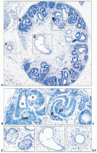
From the end of the 7th week (18- to 20-mm stage), the development of the collecting duct system and nephrons proceeds in parallel. The nephrons develop from oval condensations of metanephric mesenchymal cells that become related to the ampullary tip of each branch of the ureteric bud (Fig. 2.5). Some cells in each condensation form a hollow (nephrogenic) vesicle, while others form a separate solid cap for the next subdivision of the ampulla. Each hollow vesicle elongates and folds back on itself to become S-shaped (Fig. 2.6). The proximal end of the upper limb joins and becomes continuous with the lumen of the related ureteric bud ampulla. The upper and middle limbs of the nephrogenic vesicle elongate farther and differentiate to form the proximal and distal convoluted tubules and the loop of Henle. Adjacent to the lower limb of the vesicle, a tuft of capillaries develops, and this forms the glomerular tuft. This invaginates the lower limb, which becomes concave. The outer layer cells become flattened to form the parietal layer of the Bowman capsule, while the inner layer cells remain columnar and stretch over the developing tuft capillaries to form the visceral epithelial cells of the glomerulus. Since nephrons are attached to the growing tips of the ureteric bud branches at this stage, they advance progressively with the ampullae away from the future hilus of the kidney, the interstitial portions of the collecting ducts forming the future medullary collecting ducts.

After the 14th week of gestation, the first period of nephron induction ends and the second period commences. The second period of nephrogenesis, which lasts until about the 22nd week of gestation, is characterized by the formation of nephron
arcades. Each ampulla ceases to divide and becomes capable of inducing the formation of a further three to six nephrons. With the induction of further nephrons, the connecting tubule of the older nephron shifts the position of its point of attachment away from the ampulla to the connecting tubule of the next-formed nephron, so they are joined together in a string or arcade of between four and seven nephrons. The innermost and first-formed member of each nephron arcade is a nephron formed during the first period of nephrogenesis and becomes a juxtamedullary nephron in the fully developed organ. The loops of Henle are longest in these nephrons, extending almost to the papillary tip.
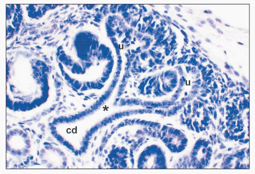


FIGURE 2.5 Metanephros from a 7-week human fetus. A branch point (asterisk) of the ureteric bud/collecting duct (cd) lineage leading to two ureteric bud ampullae (u). (hematoxylin, ×100.)


FIGURE 2.6 Metanephros from a 7-week human fetus. A: Low-power view of whole metanephros. Boxed areas are shown in higher powers in subsequent frames. (Hematoxylin, ×12.5.) B: Nephrogenic zone showing the ampulla of a ureteric bud (u) branch, condensed metanephric mesenchyme (cm), and a primitive nephron vesicle (v) that has just undergone the mesenchymal-to-epithelial transition. C: An S-shaped body: the primitive nephron is segmenting into the glomerulus, with cuboidal podocytes (p) with the adjacent proximal tubule (pt). D: The first layer of glomeruli is noted in the deep cortex. E: The renal pelvis lined by a monolayer of the urothelium. F: The ureter consists of a 1-2 cell thick layer of the urothelium surrounded by mesenchyme differentiating into smooth muscle.

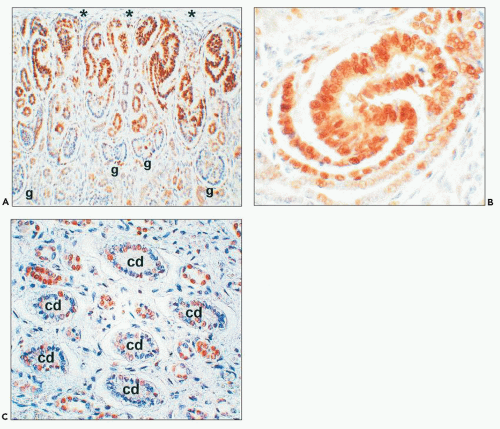
In the third period of nephrogenesis, from the 22nd to the 36th week of gestation, the ampullae advance to the peripheral cortex beyond the region of nephron arcades. A further four to seven nephrons form and are attached separately just behind the ampullary tips (Fig. 2.7). No further divisions of the ampullae occur, and as each new nephron is added, the ampullae advance farther toward the surface. Cell proliferation, as assessed by immunolocalization of proliferating cell nuclear antigen (PCNA), is depicted in the cortex and medulla of a late second trimester human fetal kidney in Figure 2.8. At about 36 weeks, the ampullae cease to function and disappear. No new nephrons are then formed, the last to develop
producing the subcapsular so-called nephrogenic zone that can be seen in the fetal kidneys up to 36 weeks and is useful in dating gestational age.


FIGURE 2.7 Arrangement of nephrons at birth as revealed by microdissection. A: Usual pattern with, sequentially, an arcade of four nephrons that drain into a tributary of the collecting duct and six nephrons that drain individually into the collecting duct. B: Possible variations. (Modified from Osathanondh V, Potter El. Development of the kidneys as shown by microdissection. II. Renal pelvis, calyces and papillae. Arch Pathol 1963;76:277-289.)

In the fourth period of kidney development, from 36 weeks to term, no new nephrons are formed, and this period is one of purely interstitial growth. Loops of Henle continue to lengthen, and the proximal and distal tubules become longer and more tortuous. In mature infants at birth, some 20% of the loops of Henle are still within the renal cortex (53). In recent years, dissector methodology has been used to quantify the number of glomeruli in the human kidneys (13,14,54). Depending on the laboratory making these measurements, mean numbers per kidney in “normal” individuals can vary, for example, between about 0.7 and 1.5 × 10⁶, and all studies have emphasized the wide range of nephron numbers measured within populations. There are indications that numbers may be reduced in some populations with essential hypertension and that glomerular volume tends to be inversely proportional to numbers per kidney. A potential criticism, however, of studies that have assessed adult kidneys would be that the results underestimate numbers present at birth if some glomeruli might be “lost” as the kidney ages. There are likely to be several causes for variations in numbers of glomeruli generated per kidney within normal populations. An environmental modulator may be poor nutrition during gestation, as well demonstrated in experimental animals (11,12), while genetic modifiers may include common variants, or “polymorphisms,” of genes that drive nephrogenesis (55).


The Lower Renal Tract: Urinary Bladder and Ureter

As the initial steps of metanephric development occur, the lower renal tract is beginning to form. By 28 days of gestation, the mesonephric duct drains into the urogenital sinus, which is forming as the cloaca is divided into the sinus and rectum by the caudal extension of the urorectal septum. The epithelia of the sinus and mesonephric duct fuse, and the ureteric bud arises as described above. By 33 days of gestation, the mesonephric duct below the ureteric bud fuses with the urogenital sinus and will contribute to the trigone. As part of these morphogenetic steps, the ureteric bud origin enters the bladder directly by day 37 to become the ureteric orifice. Between 28 and 35 days of gestation, the ureter appears to be patent, but from 37 to 47 days’ gestation, a membrane temporarily blocks the ureterovesical junction (56) and the ureter becomes occluded. This is followed by recanalization of the elongating ureter, which is complete by 8 weeks (57). By the end of the first trimester, the epithelium of the ureter differentiates into the pseudostratified urothelium, and the ureter has a submucosal course on entering the bladder. Myogenesis begins in the upper part of the ureter at 12 weeks (58). The first layer of vascularized glomeruli is present by 8 to 9 weeks and would be expected to filter blood to produce urine, which would enter the lower renal tract. The urogenital membrane ruptures on day 48 of gestation, thus providing a connection between the nascent bladder and outside of the body. At 7 weeks of gestation, the urinary bladder appears as a cylinder of epithelium surrounded by mesenchymal tissue (59). Within this urogenital sinus, mesenchyme differentiates into the smooth muscle layers of the detrusor, a process that commences in the ventral part of the bladder dome (59). The allantois, a second outflow tract on the anterior of the developing bladder, appears at 21 days of gestation; it regresses by the end of the first trimester by 12 weeks of gestation, and its remnant is marked by the median umbilical ligament.


STUDYING KIDNEY DEVELOPMENT


Descriptive Studies: Anatomy and Gene Expression Patterns

In descriptive studies, the anatomy of the kidney is documented through the embryonic and fetal periods. Patterns of cell division, programmed cell death, differentiation, and morphogenesis can be related to changing regional patterns of gene expression in terms of mRNA and protein using the respective techniques of in situ hybridization and immunohistochemistry. Such observations provide the essential foundations for the generation of hypotheses regarding the molecular mechanisms of kidney development. Most of the data on gene expression are derived from studies with experimental animals, and it is generally assumed that overall patterns will be the same in humans. The GenitoUrinary Development Molecular Anatomy Project (GUDMAP) provides an accessible and searchable database
that documents the expression patterns of many thousands of gene transcripts through mouse renal tract development (60).

Such databases can then be used to construct interconnecting networks of genes and their products, to help understand complex biologic pathways involved in renal tract development (61). Research collections of normal human fetal tissues have been made following ethical approval (62), and these are proving informative for gene expression studies in the developing renal tract (63,64,65,66).


FIGURE 2.8 Cell proliferation in a 25-week human fetal kidney. A: Low-power overview to show that proliferation is prominent in the nephrogenic zone below the renal capsule (asterisks) but is down-regulated deeper in the cortex: Glomeruli are indicated by g. (Section was immunoprobed for proliferating cell nuclear antigen [positive signal appears brown] and counterstained with hematoxylin, ×12.5.) B: Higher power: Most cells within this forming nephron (S-shaped body stage) are proliferating. C: Deep in the same kidney, subsets of nuclei in maturing medullary collecting ducts (cd) are proliferative, as are cells in smaller-caliber tubules, probably representing the loops of Henle, which are growing into the deeper medulla.

Three main classes of molecules are expressed during kidney development (Table 2.1). These are the transcription factors, growth factors, and cell adhesion/extracellular matrix proteins. It should be noted that italics are used when referring to the gene that codes for these molecules, whereas regular typescript is used when referring to the gene product (e.g., mRNA or protein). Strictly, capital letters are often used to abbreviate the human gene or gene product (e.g., WT1 gene and WT1 protein), whereas lower case is used for animal genes (e.g., wt1 gene and wt1 protein). In this chapter, for simplicity, we have generally used the human system when discussing the genes and their products.


Transcription Factors

These proteins have domains that bind DNA and that regulate the expression of other genes; in other words, they can enhance or switch off the transcription into mRNA. Because of these regulatory roles, the transcription factors have been likened to conductors of an orchestra, and the normal program of development can be perceived as being defined and directed by the sequential expression of these factors. These molecules can be classified into families that share similar DNA-binding protein motifs and domains. One of these motifs is called the zinc finger, which describes an elongated projection of the molecule that intercalates with DNA. An example of a transcription factor with multiple zinc fingers is the Wilms tumor 1 (WT1) protein, which is expressed in mesenchyme at the inception of the metanephros and also in mature podocytes (33,67,68). If the mouse gene is ablated by a null mutation, no kidneys are formed, while other types of mutation cause glomerular diseases (Denys-Drash and Fraser syndromes) and Wilms tumor (19). Some other examples of transcription factors expressed during renal tract development include the HOX family, which contain DNA-binding homeodomains

(69), and the PAX family, which contain DNA-binding paired domains (70,71,72) (Fig. 2.9). The specific gene targets of certain of these transcription factors are being investigated. For example, WT1 down-regulates PAX2 (73); up-regulates amphiregulin, a growth factor that stimulates tubule formation in the metanephros (74); and modulates the expression of WNT4, a growth factor implicated in early nephron differentiation (67). Another transcription factor called hepatocyte nuclear factor 1β (HNF1β) promotes the expression of a battery of genes that maintain the differentiated state of renal epithelia; when HNF1β is mutated, renal tubules become cystic (36,65,75,76).


TABLE 2.1 Genetic bases of mouse kidney defects based on mutants


Mouse gene


Full name


Disease descriptiona


Reference


Genes coding for transcription factors and related molecules


Bf2


Brain factor 2/homologue of Drosophila forkhead


Renal hypoplasia


151


Brn1(Foxd1)


Retarded loop of Henle development


152


Emx2


Empty spiracles 2


Renal agenesis


135


Foxc1


Forkhead box c1


Duplex kidney


153


Foxc2


Forkhead box c2


Renal hypoplasia


153


Foxd1


Forkhead box c2


Fused kidneys


154


Hnf1b


Hepatocyte nuclear factor 1β


Renal cysts


36,76


Hoxa11/Hoxd11b


Homeobox a11 and d11


Renal agenesis or hypoplasia


69


Lim1 (Lhx1)


Lim homeobox 1


Absent kidneys


155


Lmx1b


Lim homeobox transcription factor 1β


Glomerular podocyte immaturity


156


N-Myc


Avian myelocytomatosis viral oncogene homologue


Hypoplastic mesonephros


157


Pax2


Paired box 2


Renal agenesis in null mutant; renal hypoplasia in heterozygous mice


71


Pod1


Podocyte 1


Failure of renal epithelial differentiation


158


Rara/Rarb2b


Retinoic acid receptor α and β2


Kidney and ureter malformations


159,160,161


Sall1


sal-like 1/homologue of Drosophila spalt


Renal agenesis


162


Six1 and Six2


Homologues of Drosophila sine oculis


Renal agenesis


24,163


Wt1


Wilms tumor 1


Renal agenesis in null mutants; proteinuria in heterozygous mice


33,67


Genes coding for growth factors and molecules implicated in their signaling pathways


Agt


Angiotensinogen


Hypoplastic papilla and hypotension


91


Agtr1a/Agtr1bb


Angiotensin receptor type 1a and 1b


Hypoplastic papilla and hypotension


91,92


Agtr2


Angiotensin receptor type 2


Diverse renal tract malformations in males (gene on the X chromosome)


93


Angpt2


Angiopoietin 2


Disorganized peritubular capillaries


80


Bmp4


Bone morphogenetic protein 4


Wide range of kidney and ureter malformations in heterozygous effect/null mutants die before nephrogenesisb


94


Bmp7


Bone morphogenetic protein 7


Renal dysplasia


95


Fgf7


Fibroblast growth factor 7


Renal hypoplasia


164


Fgf10


Fibroblast growth factor 10


Renal agenesis (can substitute for Gdnf in ureteric bud growth)


81


Gdnf


Glial cell line-derived neurotrophic factor


Renal agenesis


77,78


Gremlin


Bmp antagonist


Renal agenesis


96,165


Notch2


Receptor for delta


Glomerular capillary defects in hypomorphs (not a complete null mutant)


166


Pdgfrb


Platelet-derived growth factor receptor B


Absent mesangial cells


41


Ret


Rearranged during transfection/Gdnf receptor


Renal agenesis or dysplasia


77


Robo2


Roundabout 2; receptor for slit 2


Duplex kidneys


167


Shh


Sonic hedgehog


Renal hypoplasia and ureter malformations


98


Slit2


Homologue of Drosophila slit


Duplex kidneys


167


Spry1


Homologue of Drosophila sprouty; modulates growth factor signaling


Duplex kidneys


168


Wnt4


Wingless-type MMTV integration site family member 4


Renal hypoplasia


103


Wnt11


Wingless-type MMTV integration site family member 11


Renal hypoplasia


104


Vegf


Vascular endothelial growth factor


Congenital glomerular disease


88,169


Genes coding for cell adhesion/extracellular matrix proteins


Fras1


Fraser syndrome 1


Renal agenesis


34,119


Frem2


Fras1-related extracellular matrix 1


Diverse renal malformations


170


Itga8


Integrin α8


Dysplastic kidneys


116


Lamab2


s-Laminin/laminin β2


Nephrotic syndrome


113


Nephrin


Congenital nephrotic syndrome 1; NPHS1


Congenital nephrotic syndrome


108


Genes coding for diverse other classes of proteins


Bcl2


B-cell CLL/lymphoma 2; cell survival molecule


Renal hypoplasia and apoptosis


121


Cox2


Cyclooxygenase 2; enzyme


Renal hypoplasia


122


Eya1


Eyes absent 1


Renal agenesis


123,124


Hs2st


Heparan sulfate 2-sulfotransferase


Renal agenesis


125


Mpv17


Peroxisomal membrane protein disrupted by viral insertion


Glomerulosclerosis


171


Pkd1


Polycystin 1


Metanephric cysts in null mutants; postnatal PKD in heterozygous mice


126


Pkd2


Polycystin 2


Metanephric cysts in null mutants; postnatal PKD in heterozygous mice


127


UpII


Uroplakin II


Hydronephrosis and vesicoureteric reflux


129


UpIII


Uroplakin III


Hydronephrosis and ureteric obstruction


130


a The disease is described in clinical terms rather than giving the mechanisms, many of which are outlined in the main text.


b Phenotype is present when two similar genes are knocked out. Unless otherwise stated, the malformations occur in null mutants in which there is no gene activity; in other cases, the disease occurs in mice with one copy of the gene ablated (i.e., heterozygous mice).

Only gold members can continue reading. Log In or Register to continue

Stay updated, free articles. Join our Telegram channel

Jun 21, 2016 | Posted by in UROLOGY | Comments Off on Development of the Kidney

Full access? Get Clinical Tree
Get Clinical Tree app for offline access