Anti-Glomerular Basement Membrane Glomerulonephritis and Goodpasture Syndrome


Anti-Glomerular Basement Membrane Glomerulonephritis and Goodpasture Syndrome


J. Charles Jennette

Volker Nickeleit


BACKGROUND INFORMATION


Anti-GBM Disease

In 1919, Ernest Goodpasture (1) reported an 18-year-old male patient who developed hemoptysis and acute renal failure after a flu-like illness. The patient died and at autopsy was found to have massive pulmonary hemorrhage and glomerulonephritis with “fibrinous exudates in the Bowman space” and necrotizing vasculitis in the spleen and gut. Four decades later, Stanton and Tange (2) coined the eponym Goodpasture syndrome for the association of pulmonary hemorrhage with glomerulonephritis. This eponym was initially used to indicate any form of glomerulonephritis combined with pulmonary hemorrhage. The use of the term Goodpasture syndrome now should be confined to those examples of pulmonary-renal syndrome that are caused by anti-glomerular basement membrane (anti-GBM) antibodies (3). Glomerulonephritis that is caused by anti-GBM occurs not only as a component of Goodpasture syndrome but also in the absence of pulmonary disease. In this setting, it should not be diagnosed as Goodpasture syndrome but simply as anti-GBM glomerulonephritis. A few patients have anti-GBM-mediated pulmonary hemorrhage without glomerulonephritis (4). Anti-GBM disease is the generic term for any clinical expression of disease caused by anti-GBM antibodies and includes Goodpasture syndrome, isolated anti-GBM glomerulonephritis, and isolated anti-GBM pulmonary hemorrhage (5). Grouping these clinically distinct syndromes in a unifying disease category is justified based on the finding that patients with these varied expressions of disease have autoantibodies with specificity for the identical portion of the α3 chain of type IV collagen in vascular basement membranes (6).

The term Goodpasture syndrome is used in this chapter only for anti-GBM antibody-mediated pulmonary-renal syndrome. However, the patient whom Goodpasture described in his publication probably did not have anti-GBM antibody-mediated pulmonary-renal syndrome. He more likely had pulmonary-renal syndrome caused by antineutrophil cytoplasmic antibodies (ANCA) because the pulmonary alveolar capillaries had more intense neutrophilic infiltration than is usually seen with anti-GBM disease, and, even more compelling, there was a necrotizing small-vessel vasculitis in the spleen and gut (7).

Anti-GBM antibodies were first detected by immunofluorescence microscopy using antibodies to immunoglobulins that revealed linear localization of IgG along GBMs in patients with pulmonary hemorrhage and crescentic glomerulonephritis (CGN) (Fig. 15.1). Scheer and Grossman, in 1964 (8), reported the linear immunostaining for γ globulin along the GBMs of two additional patients with Goodpasture syndrome. The next year, Sturgill and Westervelt (9) reported linear staining of pulmonary as well as glomerular capillary basement membranes for γ globulin in a patient with Goodpasture syndrome. This was followed by the landmark article by Lerner et al. (10) that established anti-GBM antibodies as a cause for pulmonary-renal syndrome as well as for CGN in the absence of pulmonary hemorrhage. In 1971, Martinez and Kohler (3) proposed that the term Goodpasture syndrome should be used only for patients with anti-GBM antibody-mediated pulmonary-renal vasculitic syndrome. At that time, it was clear that other pathogenic mechanisms could cause concurrent glomerulonephritis and pulmonary
hemorrhage. Unless Goodpasture syndrome is restricted to one pathogenic category, the term does not specify a clinically and pathogenetically distinct entity.


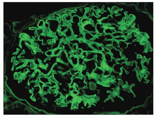
FIGURE 15.1 Global linear staining for IgG indicative of diffuse binding of anti-GBM antibodies to type 4 collagen in glomerular basement membranes. (FITC anti-IgG.)

Once immunofluorescence microscopy became a routine method for examining renal biopsy specimens, glomerulonephritis with intense linear staining of capillary walls that was indicative of anti-GBM disease was noted to be associated with glomerular crescent formation and thus CGN. CGN is not a specific disease but rather is a morphologic manifestation of severe glomerular injury that can be caused by many different etiologic and pathogenic mechanisms, one of which is anti-GBM disease. The definition of a glomerular crescent that is endorsed by the World Health Organization (WHO) is two or more layers of cells that are partially or completely filling the Bowman space (Fig. 15.2). The term crescentic glomerulonephritis often is used generically for glomerulonephritis with any degree of crescent formation; however, it is advisable to use this term on the diagnostic line only when 50% or more of glomeruli have crescents (11). Lesser degrees of crescent formation should be stated in the diagnosis as the percentage of glomeruli involved. By this approach, a specimen with anti-GBM glomerulonephritis that has crescents in 70% of glomeruli would be diagnosed as crescentic anti-GBM glomerulonephritis, whereas a specimen with anti-GBM glomerulonephritis that has crescents in 15% of glomeruli would be diagnosed as anti-GBM glomerulonephritis with 15% crescents.


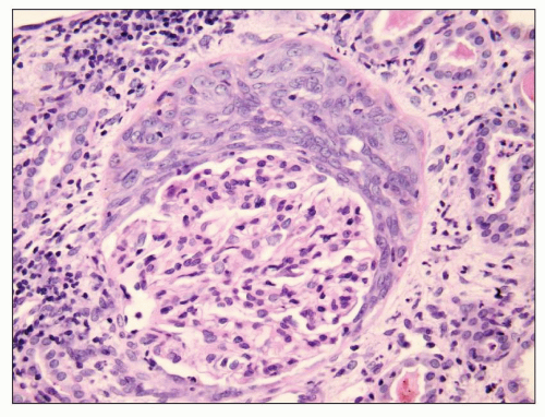
FIGURE 15.2 Anti-GBM glomerulonephritis with a large cellular crescent forming a cap over the glomerular tuft. Note also the edema and inflammation in the periglomerular interstitium. (H&E.)


Crescentic Glomerulonephritis

Glomerular crescents have been described in the medical literature since the 19th century. For example, Langhans (12) and Purdy (13) provided excellent illustrations of glomerular crescents in publications that appeared in 1879 and 1886, respectively. By the turn of the century, a number of investigators, including Volhard and Fahr (14), had recognized that glomerular crescents often correlated with poor outcome. This association was confirmed by many investigators, and the close relationship between rapid clinical progression of glomerulonephritis and the pathologic finding of glomerular crescents became well established. The term “rapidly progressive glomerulonephritis” has become the standard designation for clinical manifestations that suggest severe glomerulonephritis. Most patients with this clinical syndrome have severe glomerulonephritis with crescent formation, but some have other diseases that can mimic CGN, such as thrombotic microangiopathy. A conclusive diagnosis of CGN can be made only by evaluation of a renal biopsy specimen.

With the advent of immunofluorescence microscopy and electron microscopy, the heterogeneity of the glomerular diseases that cause crescent formation was recognized (15,16). This allowed Steen Olsen (15) to conclude that CGN “might constitute a common final and fatal pathway of several etiologically and pathogenetically different glomerular diseases.” Thus, the light microscopic recognition of CGN in a renal biopsy specimen is only the beginning of an adequate pathologic analysis. Further elucidation of pathogenic mechanisms that are causing the glomerulonephritis is equally or more important in predicting the prognosis and selecting the appropriate treatment strategy for the patient. A pathologic diagnosis of CGN is incomplete unless the disease is categorized further, which usually requires immunofluorescence microscopy (or immunohistochemistry) and electron microscopy. Accurate and prompt categorization of CGN is critical for optimal patient outcome, because one of the major positive prognostic factors in the most aggressive forms of CGN, including anti-GBM glomerulonephritis, is rapid institution of immunosuppressive treatment (17).

CGN is categorized by immunohistology into anti-GBM CGN with linear GBM staining for immunoglobulin, immune complex CGN with granular staining of glomeruli for immunoglobulin or complement, or crescentic glomerulonephritis with little or no glomerular staining for immunoglobulin or complement (i.e., pauci-immune CGN) (Fig. 15.3) (18,19). These three immunopathologic categories are sometimes designated types I, II, and III, respectively; however, this terminology is not widely used and thus may not be understood when used in a diagnosis. Anti-GBM disease is a special form
of immune complex disease caused by in situ binding of anti-GBM antibodies to GBM antigens, but it is traditionally separated from other forms of immune complex glomerulonephritis when categorizing CGN. Throughout this chapter, the term immune complex glomerulonephritis is used for glomerulonephritis caused by immune complexes other than anti-GBM immune complexes.


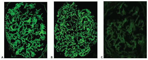
FIGURE 15.3 Immunofluorescence microscopy patterns of glomerular staining for IgG that are indicative of anti-GBM (A), immune complex (B), and pauci-immune (C) glomerulonephritis. Note the linear staining of anti-GBM disease compared with the granular staining of immune complex disease and the scanty background staining of pauci-immune disease. (FITC anti-IgG.)

Crescents also occur in categories of glomerular injury that are not included in the three major categories of anti-GBM, immune complex, or ANCA disease. For example, C3 glomerulopathy (including dense deposit disease and C3 glomerulonephritis), which is caused by dysregulated alternative pathway complement activation rather than immune complexes, may have crescent formation (20). Rarely, crescents occur with thrombotic microangiopathy and diabetic glomerulosclerosis apparently caused by capillary injury that results directly from these diseases. However, whenever crescents are observed in the context of a glomerular disease that rarely has crescents, the possibility of a concurrent disease (e.g., ANCA disease) that may be causing the crescents should be considered.

Table 15.1 lists the frequency of the categories of CGN among patients whose renal biopsies were evaluated in the University of North Carolina Nephropathology Laboratory (18). Anti-GBM glomerulonephritis is uncommon at any age, accounting for about 15% of CGN. Pauci-immune CGN is the most common category of glomerulonephritis in patients who have more than 50% of glomeruli involved by crescents. Approximately 90% of patients with pauci-immune CGN have circulating antineutrophil cytoplasmic autoantibodies (ANCA) (21). Pauci-immune CGN (i.e., ANCA-CGN) is by far the most common category of CGN in older patients (18). In contrast, immune complex glomerulonephritis with crescents is more common in younger patients, which is not surprising considering that many of the most common immune complex glomerulonephritides occur most frequently in children or young adults, such as IgA nephropathy, IgA vasculitis (Henoch-Schönlein purpura), lupus nephritis, poststreptococcal glomerulonephritis, and membranoproliferative glomerulonephritis. For example, in an analysis in London of 30 children with CGN, Jardim et al. (22) identified IgA vasculitis (Henoch-Schönlein purpura) glomerulonephritis in 9, membranoproliferative glomerulonephritis in 7, poststreptococcal glomerulonephritis in 2, and lupus glomerulonephritis in 1. The remaining patients had microscopic polyangiitis (MPA) in four, granulomatosis with polyangiitis (GPA) (Wegener granulomatosis) in one, anti-GBM disease in two, and idiopathic (pauci-immune) CGN in four.

Although reports from North America and Europe are generally in agreement with the data in Table 15.1 (23,24,25), the relative frequency of different types of CGN may vary substantially in different geographic locations. For example, a study of 172 patients with CGN from China noted 8.7% anti-GBM CGN, 22.7% pauci-immune CGN, and 68.6% immune complex CGN (half of which were lupus nephritis) (26). Another study of a cohort of 106 patients with CGN from China, although more recent and from a more industrialized region, observed 16% anti-GBM disease, 40.6% immune complex disease, and 43.4% pauci-immune disease (27). An analysis of 28 patients with CGN from the Republic of Macedonia identified 22 cases of poststreptococcal glomerulonephritis, 1 case of poststaphylococcal glomerulonephritis, 1 case of anti-GBM glomerulonephritis, and 4 cases of ANCA-positive pauci-immune CGN (28). This higher proportion of immune complex CGN from some regions may be caused by a higher frequency of nephritogenic infections in this region.

Even in the same country, demographic differences in cohorts affect the frequency of different causes for CGN. For example, in a study from India of all patients (predominantly adults) with ≥50% crescents, 72% (33/46) had pauci-immune CGN (29), whereas another study from India of only children (≤18 years old) with ≥50% crescents found that 86% (19/22) had immune complex glomerulonephritis (30).

In general, anti-GBM glomerulonephritis and ANCA glomerulonephritis have a higher frequency and severity of
crescent formation than immune complex glomerulonephritis (Table 15.2 and Fig. 15.4) (18,19). This finding suggests that the pathogenic mechanisms that underlie anti-GBM and ANCA glomerulonephritis are more destructive than the mechanisms that underlie immune complex glomerulonephritis. However, some categories of immune complex glomerulonephritis tend to have a greater frequency of crescent formation than do others. In general, the extent of glomerular subendothelial, as opposed to subepithelial or mesangial, immune complex localization correlates with severe inflammation and crescent formation among patients with immune complex glomerulonephritis. This suggests that the proximity of subendothelial immune complexes to the inflammatory mediator systems in the circulation is more effective at causing the glomerular injury that results in crescent formation than are immune complexes in the mesangium or subepithelial zones of glomeruli. This could in part explain the severity of anti-GBM glomerulonephritis because the autoantibodies form immune complexes in situ with the GBM antigens at the subendothelial interface between the GBM and the plasma.


TABLE 15.1 Frequency of different types of Crescentic Glomerulonephritisa in renal biopsy specimens evaluated by the University of North Carolina Nephropathology Laboratory


Age (years)


n


Anti-GBM crescentic glomerulonephritis (%)


Pauci-immune crescentic glomerulonephritis (%)


Immune complex crescentic glomerulonephritis (%)


Other crescentic glomerulonephritis (%)


All


632


15


60


24


1


1-20


73


12


42


45


0


21-60


303


15


48


35


3


61-100


256


15


79


6


0


aAnti-GBM crescentic glomerulonephritis was defined as glomerulonephritis with ≥50% crescents and ≥2+ linear GBM staining for IgG by direct immunofluorescence microscopy. Pauci-immune crescentic glomerulonephritis was defined as glomerulonephritis with ≥50% crescents and ≤2+ staining of glomeruli for any immunoglobulin. Immune complex crescentic glomerulonephritis was defined as glomerulonephritis with ≥50% crescents and ≥2+ nonlinear GBM staining for any immunoglobulin by direct immunofluorescence microscopy. Crescentic membranoproliferative and postinfectious glomerulonephritis, which typically have substantial staining for C3 but little or no staining for immunoglobulin, also were included in the immune complex category. The other category includes all other glomerular diseases, such as thrombotic microangiopathy, diabetic glomerulosclerosis, monoclonal immunoglobulin deposition disease, and so on.


Modified from Jennette JC. Rapidly progressive crescentic glomerulonephritis. Kidney Int 2003;63:1164.


TABLE 15.2 Frequency of glomerular crescents, necrosis, and endocapillary hypercellularity in different types of glomerular disease evaluated by the University of North Carolina Nephropathology Laboratory


Type of glomerular disease


% With any crescents


% With >50% crescents


Average % glomerular crescentsa


Glomerular necrosis (0-4+)


Glomerular hypercellularity (0-4+)


Anti-GBM glomerulonephritis


97.1


84.8


77


1.7+


0.8+


ANCA glomerulonephritis


89.5


50.3


49


1.2+


0.8+


Lupus glomerulonephritis (III and IV)


56.5


12.9


31


1.7+


2.2+


Henoch-Schönlein purpura glomerulonephritis


61.3


9.7


27


0.4+


1.5+


IgA nephropathy


32.5


4.0


21


0.1+


1.4+


Postinfectious glomerulonephritis


33.3


3.3


19


0.3+


2.7+


Type I membranoproliferative glomerulonephritis


23.8


4.6


25


0.2+


2.8+


DDD/C3 glomerulopathy


43.8


18.8


48


0.2+


1.8+


Fibrillary glomerulonephritis


22.8


5.0


26


0+


0.6+


Monoclonal immunoglobulin deposition disease


5.6


0


13


0+


0.3+


Thrombotic microangiopathy


5.6


0.9


26


0.4+


0.3+


Diabetic glomerulosclerosis


3.2


0.3


20


0+


0.3+


Nonlupus membranous glomerulopathy


3.2


0.1


15


0+


0.1+


a ANCA glomerulonephritis was defined as glomerulonephritis with ≤2+ staining of glomeruli for any immunoglobulin in a patient who is positive for MPO-ANCA or PR3-ANCA by ELISA.


Modified from Jennette JC. Rapidly progressive crescentic glomerulonephritis. Kidney Int 2003;63:1164.


This chapter reviews the clinical and pathologic features of anti-GBM disease, including Goodpasture syndrome and anti-GBM glomerulonephritis in the absence of pulmonary disease. In the context of this discussion of an archetype of CGN, the pathogenesis of crescent formation that is initiated by many aggressive forms of glomerulonephritis in addition to anti-GBM disease is summarized. The specific pathogenic events that are unique to anti-GBM disease are reviewed.


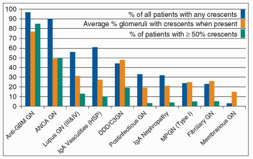
FIGURE 15.4 Frequency of glomerular crescents in different types of glomerular disease evaluated in the University of North Carolina Nephropathology Laboratory. For each category of disease, the data are expressed as (i) the % of patients who had any crescents in biopsy specimens, (ii) the average % of crescents in specimens that had crescent formation, and (iii) the % of patients who had specimens with ≥50% crescents. (Data derived from Jennette JC. Rapidly progressive crescentic glomerulonephritis. Kidney Int 2003;63:1164.)


CLINICAL PRESENTATION

Anti-GBM disease is rare with an incidence of approximately 1/1,000,000 (compared to greater than 1/100,000 for ANCA disease), an equal male-to-female ratio, and a broad bimodal age of onset with one peak in older children and young adults and another in older adults (31,32,33,34,35,36,37,38). Anti-GBM GN is rare in children but does occur (39). Glomerulonephritis is the most common initial clinical manifestation of anti-GBM disease. Anti-GBM glomerulonephritis usually manifests with rapidly progressive glomerulonephritis, and renal biopsy reveals more than 50% glomerular crescents in most patients. For example, a cohort of 28 patients with anti-GBM but no ANCA (see Table 15.2) had more than 80% of patients with crescents in 50% or more of glomeruli in the initial renal biopsy (18). Multiple other studies confirm that more than 80% of patients with anti-GBM disease have more than 50% of glomeruli with crescents in the initial renal biopsy (34,35,36,37,38). The data in Tables 15.2 and 15.3 demonstrate that anti-GBM glomerulonephritis is pathologically and clinically the most severe form of glomerulonephritis (18). Hematuria is virtually always present. Proteinuria is common. Although most patients with anti-GBM disease present with severe renal disease, severe pulmonary disease, or both, patients will rarely present with hematuria and proteinuria in the absence of renal insufficiency or pulmonary hemorrhage (40).


TABLE 15.3 Features at the time of presentation of different types of Crescentic Glomerulonephritisa Evaluated by the University of North Carolina Nephropathology Laboratory


Mean age


Male:female


Creatinine


Proteinuria (g/24 h)


Anti-GBM crescentic glomerulonephritis


52 ± 21 (14,15,16,17,18,19,20,21,22,23,24,25,26,27,28,29,30,31,32,33,34,35,36,37,38,39,40,41,42,43,44,45,46,47,48,49,50,51,52,53,54,55,56,57,58,59,60,61,62,63,64,65,66,67,68,69,70,71,72,73,74,75,76,77,78,79,80,81,82,83,84)


1:1.0


45:47


9.7 ± 7.2 (0.8-50)


1.67 ± 3.35 (0.20-16.20)


Pauci-immune crescentic glomerulonephritis


56 ± 20 (2,3,4,5,6,7,8,9,10,11,12,13,14,15,16,17,18,19,20,21,22,23,24,25,26,27,28,29,30,31,32,33,34,35,36,37,38,39,40,41,42,43,44,45,46,47,48,49,50,51,52,53,54,55,56,57,58,59,60,61,62,63,64,65,66,67,68,69,70,71,72,73,74,75,76,77,78,79,80,81,82,83,84,85,86,87,88,89,90,91,92)


1:0.9


202:177


6.5 ± 4.0 (0.8-22.1)


1.94 ± 2.95 (0.11-18.00)


Immune complex crescentic glomerulonephritis


33 ± 17 (4,5,6,7,8,9,10,11,12,13,14,15,16,17,18,19,20,21,22,23,24,25,26,27,28,29,30,31,32,33,34,35,36,37,38,39,40,41,42,43,44,45,46,47,48,49,50,51,52,53,54,55,56,57,58,59,60,61,62,63,64,65,66,67,68,69,70,71,72,73,74,75,76,77)


1:1.6


61:95


4.9 ± 3.8 (0.8-21.7)


4.39 ± 4.77 (0.30-22.00)


aAnti-GBM crescentic glomerulonephritis was defined as glomerulonephritis with ≥50% crescents and ≥2+ linear GBM staining for IgG by direct immunofluorescence microscopy. Pauci-immune crescentic glomerulonephritis was defined as glomerulonephritis with ≥50% crescents and ≤2+ nonlinear staining of glomeruli for any immunoglobulin. Immune complex crescentic glomerulonephritis was defined as glomerulonephritis with ≥50% crescents and ≥2+ nonlinear GBM staining for any immunoglobulin by direct immunofluorescence microscopy (plus membranoproliferative and postinfectious glomerulonephritis).


Jennette JC. Rapidly progressive crescentic glomerulonephritis. Kidney Int 2003;63:1164.


The frequency of pulmonary hemorrhage among patients with anti-GBM disease varies among cohorts, but most series show a frequency of 50% ± 15% (33,34,35,36,37,38). For example, in a cohort of 71 patients from the United Kingdom evaluated by Savage et al. (33), 35% had Goodpasture syndrome at presentation, although approximately 50% eventually had some pulmonary hemorrhage. In a cohort of 53 North American patients with native kidney anti-GBM disease reported by Wilson and Dixon (31), 60% had Goodpasture syndrome. In a series of 40 patients from Ireland, 62% had pulmonary involvement (34). In a large cohort from China, 45% (80/176) of patients with anti-GBM disease had hemoptysis (37). Pulmonary involvement typically causes hemoptysis, dyspnea, rales, and rhonchi. Most patients with extensive pulmonary hemorrhage have pallor because of anemia. Many patients have a history of a recent flu-like illness or other infection before the onset of anti-GBM disease (8,31,37,41), and a minority of patients have a history of hydrocarbon exposure although this frequency is higher than in the general population (42,43,44). Patients have a higher frequency of cigarette smoking than does the general population. For example, in a study of 23 anti-GBM disease patients from New Zealand, 50% were regular
smokers compared to 17% of the general population, and only 14% had never smoked compared to 56% of the general population (P < 0.001) (38). Savage et al. (33) observed that there is a tendency for young men with anti-GBM disease to present with Goodpasture syndrome (i.e., combined pulmonary hemorrhage and nephritis) and for older women to present with glomerulonephritis alone. We have observed a similar pattern in the patients at the University of North Carolina. Overall, however, the male-to-female ratio is equal (see Table 15.3). Anti-GBM glomerulonephritis is rare in African Americans.

Approximately one quarter to one third of anti-GBM patients have ANCA, usually MPO-ANCA (Table 15.4) (18,37,45,46,47,48,49). Most patients with concurrent anti-GBM and ANCA have renal limited disease or microscopic polyangiitis (MPA); however, some have granulomatosis with polyangiitis (GPA) (Wegener’s) (50) or eosinophilic granulomatosis with polyangiitis (EGPA) (Churg-Strauss) (51). In a study of anti-GBM patients from China, patients who were 65 years or older at the time of diagnosis were more often ANCA positive (46.0% vs. 14.6%; P < 0.001) and had less hemoptysis (26.0% vs. 46.2%; P = 0.01) (52).

Only gold members can continue reading. Log In or Register to continue

Stay updated, free articles. Join our Telegram channel

Jun 21, 2016 | Posted by in UROLOGY | Comments Off on Anti-Glomerular Basement Membrane Glomerulonephritis and Goodpasture Syndrome

Full access? Get Clinical Tree

Get Clinical Tree app for offline access