Acute Postinfectious Glomerulonephritis and Glomerulonephritis Caused by Persistent Bacterial Infection


Acute Postinfectious Glomerulonephritis and Glomerulonephritis Caused by Persistent Bacterial Infection


Anjali A. Satoskar

Tibor Nadasdy

Fred G. Silva


Bacterial infection outside the kidney can lead to glomerulonephritis either after the infection has been cleared, in which case the glomerulonephritis is appropriately called postinfectious glomerulonephritis, or while the active, persistent infection is still ongoing, in which case the terminology of postinfectious glomerulonephritis is obviously incorrect and the glomerulonephritis is best designated as glomerulonephritis associated with active/persistent bacterial infection. From the clinical point of view, it is relevant to differentiate these two forms of bacterial infection-related glomerulonephritis because, in postinfectious glomerulonephritis, antibiotic treatment is not necessary and immunosuppressive medications can be given. In contrast, if a bacterial infection is still active, the glomerulonephritis should be treated primarily with antibiotics, and immunosuppression should be avoided. If the previous or ongoing infection is known to the clinician and pathologist, the diagnosis is usually easy. However, in real-life situations, it is common that patients with bacterial infection-related immune complex glomerulonephritis do not display obvious clinical symptoms of infection at the time the glomerulonephritis is diagnosed. Such subclinical infections may be easily overlooked, and if the slightest suspicion for an underlying infection emerges, the pathologist has to raise this possibility to avoid immunosuppressive treatment before the infection can be safely excluded (1). There are several publications in the literature where postinfectious glomerulonephritis is defined
quite liberally and include cases of glomerulonephritis with a preceding infection, but the infection is still present. Such cases include glomerulonephritides associated with staphylococcal infection (see section on Glomerulonephritis associated with staphylococcal infections). However, if we consider a glomerulonephritis following an infection postinfectious, then even HIV-associated nephropathy or hepatitis C-related cryoglobulinemia could be considered postinfectious glomerulonephritis, which would clearly be inaccurate. In our opinion, poststreptococcal glomerulonephritis is the best example for postinfectious glomerulonephritis. Most other infection-related glomerulonephritides are not postinfectious; they are associated with ongoing infection.

This chapter deals first with acute postinfectious (poststreptococcal) glomerulonephritis, which is the type of glomerulonephritis that follows infections. The incidence of poststreptococcal acute glomerulonephritis (PSAGN) is declining in well-developed countries, but it used to be the most common and the most studied form of acute postinfectious glomerulonephritis. Therefore, it is described first in this chapter. In spite of the declining incidence of PSAGN, it is important to discuss it in detail, because the extensive studies on this glomerular disease provide invaluable insight into the pathogenesis of acute glomerulonephritis, in general, and the disease is still prevalent in many parts of the world. Major additions have been made to the section on Staphylococcus aureus infection-associated glomerulonephritis, which is now emerging as the most common form of “infection-related glomerulonephritis” in the United States, especially in the elderly, debilitated and diabetic population (2,3,4). The remainder of the chapter describes glomerulonephritis that accompanies persistent bacterial infections such as infective endocarditis, deep-seated abscesses, and infected ventriculoatrial shunts inserted for the relief of hydrocephalus.


ACUTE POSTSTREPTOCOCCAL GLOMERULONEPHRITIS


Historical Perspective

Acute glomerulonephritis has long been known to follow certain acute infections. Although Hippocrates, more than two millennia ago, described the occurrence of back pain and gross hematuria leading to oliguria or anuria (5), it was Wells (6) who noted bloody urine in patients with scarlet fever and postscarlatinal anasarca. Bright (7), too, noted the association with scarlatina and described the finding of blood in the urine and swelling of the face in what were probably attacks of PSAGN.


According to Bright:

The history of this disease, and its symptoms, is nearly as follows: a child or adult is affected with scarlatina or some other acute disease; or has indulged in the intemperate use of ardent spirits for a series of months or years: he is exposed to some casual cause or habitual source of suppressed perspiration: he finds the secretion of his urine greatly increased, or he discovers that it is tinged with blood; or, without having made any such observation, he awakes in the morning with his face swollen, or his ankles puffy, or his hands oedematous.

Bright further observed that, with or without treatment, the patient’s urine lost its “red particles” and that the swelling subsided, although in some instances, he recorded that the patient returned at a later time with what we would now regard as chronic renal failure.

With the introduction of microscopic examination of the kidney over the ensuing decades, it became apparent that changes took place in the glomeruli, and Langhans (8) described a category of Bright disease with glomerular inflammation. Schick (9) commented on the similarity of the latent period in serum sickness to that of acute glomerulonephritis.

The first major clinicopathologic treatise on renal disease appeared in 1914 with the publication of the work of Volhard (the clinician) and Fahr (the pathologist), Die Brightsche Nierenkrankheit (10). They classified Bright disease into various categories, including inflammatory diseases of the glomerulus. Longcope (11,12) recognized two general forms of glomerulonephritis, the first of which was associated with preceding bacterial infections and had a quick recovery and a good prognosis (acute glomerulonephritis). Ellis (13) also distinguished two types of glomerulonephritis. Type 1 nephritis, usually seen in young patients, was characterized by an abrupt onset, constitutional symptoms, gross hematuria following infection, and a high recovery rate; this condition corresponded to the first type described by Longcope (11).

Scarlet fever was noted to be associated with acute glomerulonephritis in the late 1800s and early 1900s, and it soon became apparent that there was a common association with a previous infection of group A streptococcus (GAS). Varying attack rates of acute glomerulonephritis were noted in patients with scarlet fever, and a greater attack rate of nephritis was found in children compared with adults. These varying rates were probably largely dependent on the strain of Streptococcus causing the infection. Ophüls (14), as early as 1917, had suggested that only certain strains of streptococci appeared to have a nephritogenic capacity.


Nephritogenic Strains and Epidemiology

Most cases of acute glomerulonephritis seen worldwide are caused by the streptococcus, without the clinical signs and symptoms of scarlet fever. In colder northern climates, most cases of acute glomerulonephritis follow upper respiratory tract infections, such as pharyngitis or tonsillitis, but cases also can follow skin infections, especially in warmer climates (15,16,17). Most cases of PSAGN are caused by group A streptococci, which are also responsible for rheumatic fever. Although rare cases of concomitant rheumatic fever and PSAGN have been noted (18,19), there are many differences between the epidemiology of acute glomerulonephritis and rheumatic fever. A likely explanation for the different patterns is that only certain (nephritogenic) strains of group A streptococci lead to acute glomerulonephritis (20,21,22,23,24,25,26). Types 12, 4, and 1 are more likely to cause acute glomerulonephritis after throat infections than other types (25,26). Type 12 is especially common as a strain leading to acute glomerulonephritis. However, even with the same type of streptococcal organism, there are differences in the attack rate. Siegel et al. (27) showed that only 1 of 58 patients with well-documented type 12 streptococcal infection progressed to acute glomerulonephritis; this was a convincing demonstration that not all strains of type 12 are nephritogenic. The attack rate (variably defined and detected) with certain nephritogenic strains ranges from 1% to 33% of patients (28,29). Differences in the host, such as different immune responses, abnormalities in the alternate complement pathway activation, could lead to
this variability. Of all children infected with the various strains of streptococci, it appears that less than 2% show clinically obvious signs of acute glomerulonephritis.

Acute glomerulonephritis resulting from skin infections is not uncommon, especially in warm climates (16,17,22,30,31,32,33,34). Streptococcal infections of the throat are more common in the winter and early spring, whereas streptococcal pyoderma or impetigo tends to occur in temperate climates in the late summer and early fall (34). The same seasonal pattern is found for acute glomerulonephritis. When skin infections lead to acute glomerulonephritis, certain serotypes of streptococci are isolated more commonly than others; streptococcal M types 49, 42, 2, 57, and 60 seem to be predominant, and types 49, 42, and 2 are particularly potent (34). The nephritogenic strains from the throat and skin show, in general, comparable attack rates of glomerulonephritis (34). However, type 49 infections of the skin have quite different attack rates of acute glomerulonephritis (24%) than type 49 that appears in the throat (5%) (34). Frequently, the same bacteria are found in the throat and the skin; in these instances, the skin infection typically precedes that in the throat (35).

Many of the important early observations on the epidemiology of acute postinfectious glomerulonephritis were made on the Red Lake Indian Reservation in Minnesota, where outbreaks of acute glomerulonephritis occurred in 1953 and again in 1966 (35,36). A study of the second epidemic (both caused by type 49) showed that all cases occurred in children too young to have been infected in the first epidemic (35) because of the lack of type-specific immunity. Epidemic outbreaks tend to occur in closed communities (20,36,37) or in highly populated areas in which poor hygiene, malnutrition, anemia, and parasites are common (17,20,22,38,39,40). PSAGN epidemics related to skin infections may be associated with outbreaks of scabies (38). Some epidemics in certain communities tend to recur and are separated by 5 to 7 years (17,20).

From the 1940s to the mid-1980s, the incidence of the suppurative and nonsuppurative complications of group A β-hemolytic streptococcal infections, such as glomerulonephritis and rheumatic fever, all but disappeared in the United States and throughout much of Europe (41,42). However, in 1987, the first of several outbreaks of rheumatic fever was reported (43). The concern about these infections increased when the outbreaks were followed by severe systemic streptococcal illnesses, invasive skin diseases, and a toxic shock-like syndrome (42). Despite these outbreaks and the continued prevalence of certain major nephritogenic strains (M type 12) (44), PSAGN has apparently continued the same sharp decline in incidence it had pursued before the mid-1980s. This decline is not thought to be caused by primary prophylaxis with penicillin but by a changing epidemiology of the disease that may stem from either changes in the nephritogenic potential of certain strains (e.g., M type 12) or changes in susceptibility of the host.

Roy and Stapleton (18) noted a changing perspective in the occurrence of PSAGN in one Tennessee hospital during the course of two decades. Although they noted a marked decline in the prevalence of PSAGN, they also noted a decline in urban patients and an increase in rural patients with PSAGN. In the last decade, they noticed a predominance of antecedent pharyngeal infection in children older than 6 years of age and a predominance of antecedent pyoderma in African American children (18). Postinfectious glomerulonephritis, however, continues to have a high incidence in other parts of the world, especially in areas with tropical climates, where skin infections are common, such as Africa (19), South America (20,38,39), the Caribbean (17), New Zealand (45), India, and in indigenous communities (Aborigines in Australia) (46). Incidence and prevalence of PSAGN in selected countries worldwide, based on biopsy studies published after 1985, are shown in Table 10.1 (68). A Kuwait study described 234 patients over a 9-year period, and these patients showed a high prevalence of certain nephritogenic strains (M types 12 and 49) (67). The possibility of reemergence of poststreptococcal glomerulonephritis in the United States and Europe cannot be excluded (41). In 1995, an epidemic outbreak was reported in Armenia following serious deterioration of the living conditions there. In this epidemic, most children had upper respiratory tract infections or scarlet fever preceding PSAGN, and only 13% of them had skin infections (40). There are two recent reviews describing the global burden of PSAGN/postinfectious glomerulonephritis worldwide, one from Bangkok, Thailand, and the other a collaborative work from the United Kingdom, Australia, and Canada (46,68). The calculations of incidence and prevalence of PSAGN are based on large population-based studies by Carapetis et al. (69) and Rodŕiguez-Iturbe et al. (70,71). Carapetis et al. calculated an incidence of approximately 24.3 cases per 100,000 person-years in children and 2 cases per 100,000 person-years in adults in the developing world versus 6 and 0.3, respectively, in the developed world. They calculated a prevalence of GAS disease of at least 18.1 million cases, with 1.78 million new cases each year and 517,000 deaths per year due to severe group A streptococcal (GAS) diseases (acute rheumatic fever, rheumatic heart disease, PSAGN, and invasive infections). There is significant global variation with the highest incidence of 239 per 100,000 in Australian Aborigines and the lowest incidence of 0.04 per 100,000 in an Italian study of people under the age of 60. Still all these statistical calculations are likely to be underestimations since they cannot account for the vast majority of subclinical disease that is thought to be 4 to 19 times more common than symptomatic disease. The estimates are even higher in the reports by Rodŕiguez-Iturbe et al. (70,71).

The epidemiology of postinfectious glomerulonephritis in the developed world is undergoing rapid changes. In the United States, most infection-related glomerulonephritis cases occur in adults, and staphylococcal infection-associated glomerulonephritis (usually not postinfectious) is more common than PSAGN (2,3). The type 2 diabetes “epidemic” and rapid increase in obesity substantially contribute to these changes (2,3,68).


General Properties of Streptococci, Antibody Formation, and Complement Changes


Streptococcus Pyogenes

Streptococcus pyogenes (GAS) produces many virulence-enhancing extracellular products and toxins, including erythrogenic toxin, DNase, hyaluronidase, streptokinase, NADase, proteinases, and the hemolysins streptolysin-O (oxygen labile) and streptolysin-S (oxygen stable). GAS is the etiologic agent of a number of suppurative infections, including pharyngitis, cellulitis, necrotizing cellulitis, scarlet fever, erysipelas, pyoderma, puerperal sepsis, toxic shock-like syndrome, and impetigo. Ferretti (72) reviewed the molecular basis of virulence and antibiotic
resistance in GAS. Certain GAS organisms have surface receptors that bind selectively to the key fibrinolytic enzyme, plasmin (73). The bacterium-bound plasmin retains its enzymatic ability to cleave substrates and hydrolyze a fibrin clot, which may in part contribute to its tissue-invasive properties (73). GAS infection can be diagnosed and monitored with many laboratory procedures that detect the organism, its antigens, or its antibodies.


TABLE 10.1 Incidence and prevalence of PSAGN worldwide


Country


Ref.


Incidence


Prevalence


Period of study


Australia


(47)


7.33


0.16


1995 and 1997


Brazil


(48)


22.86


0.23b


1999-2005


China


(49)


13.25


0.04


1979-2002


Czech Republic


(50)


4.43


0.04


1994-2000


Denmark


(51)


7.54


0.14


1985-1997


France


(52)


0.6


0.15


1981-1985


Hong Kong


(53)


2.6


0.04


1993-1997


India


(54)


39.24


10.14b


1986-2002


Italy


(55)


37


0.06


1996-2000


Japan


(56)


25


0.02


1970-1980


Jordan


(9)


2.6


0.26


1993-1997


Korea


(57)


6


0.06


1973-1995


Macedonia


(58)


3.26


0.16b


1975-2001


Nigeria


(59)


0.9


0.04


1994-2003


Peru


(60)


4.82


0.06b


1985-1995


Portugal


(61)


2.67


1.01


1977-2003


Romania


(62)


7.7


0.12b


1995-2004


Saudi Arabia


(63)


25


0.11


1979-1983


Serbia


(64)


0.95


0.06b


1987-2006


Thailand


(65)


2


0.04b


2001-2004


Tunisia


(66)


44


0.41


1975-2005


USA


c


0.78


0.13a


1999-2006


a Data from children.

b Data from adults.

c Data provided by Dr Tray Hunley, Division of Pediatric Nephrology, Vanderbilt Children’s Hospital, USA.


Estimates of the incidence and prevalence of postinfectious glomerulonephritis in selected countries, based on data from biopsy studies published after 1985 (68). Incidence is given as cases per year; prevalence is given as cases per 100,000 population.


Conventional methods for the identification of viable organisms include finding β-hemolytic colonies on 5% sheep blood agar with a subsequent presumptive identification based on sensitivity to bacitracin on streptococcal-selective agar or hydrolysis of L-pyrrolidonyl-β-naphthylamide. Serologic procedures to identify streptococcus include latex agglutination, coagglutination, immunofluorescent antibody staining, and the Lancefield precipitin test, in which cell wall antigens must be extracted by heating or chemical treatment prior to testing. Throat cultures are a reliable method for finding GAS, but they have a long turnaround time (74).

Rapid methods for the detection of organism antigen directly from throat swabs include latex agglutination, coagglutination, and enzyme immunoassay (74,75). These rapid methods are truly helpful only if results are positive; negative results do not necessarily mean that the specimen collection site was free of S. pyogenes. In addition, cultures with fewer than 10 colonies (with false-negative rapid test results) have yielded positive serologic test results (i.e., a fourfold or greater rise in antibody titers). Therefore, small numbers of group A streptococci can be meaningful.

There is much information on the biologic characteristics of the streptococcal organism as well as the strains that lead to acute glomerulonephritis. Poststreptococcal glomerulonephritis is almost always caused by strains of the serogroup A; however, several well-documented outbreaks have been caused by group C organisms in patients with septic arthritis, pneumonia, and septicemia (76,77) and to group G streptococci (skin infections) (78). Milk-borne Streptococcus zooepidemicus infection from unpasteurized milk and cheese has been reported with septicemia and clinical evidence of PSAGN (39,79).

Streptococcal M proteins are dimeric, alpha-helical, coiled molecules on the surface of the bacteria and function as the major antiphagocytic factor. Despite differences in the amino acid sequences of different serotypes of the M molecules, there are certain regions that show important conserved architectures (80). Manjula et al. (80) demonstrated distinct differences within this conserved molecular architecture between nephritis-associated and rheumatic fever-associated strains; the functional significance of these differences is unclear. Historically, GAS vaccine development has focused on the N-terminus of the M protein. This has led to clinical trials of the 26-valent recombinant M protein vaccine; however, no vaccines using antigens
contained in the most conserved region of the M protein have yet been possible (81,82). Molecular typing of the M protein has been used to investigate the molecular epidemiology of GAS as well as group C and G streptococcal disease. A systematic review of the global distribution of GAS M types revealed that the epidemiology of GAS disease in Africa and the Pacific region seems to be different from that in other regions, particularly the United States and Europe. In Africa and the Pacific regions, there is more diversity of M types. The M types (including 1, 4, 6, and 12) that are more common in the highincome countries were found to be less common in Africa and the Pacific region. This has implications for the development of multivalent GAS vaccines. One vaccine may not provide good coverage worldwide (81). Nontypeable GAS organisms also have been cultured from patients with acute glomerulonephritis, which presumably represent unclassified nephritogenic strains (20). It is likely that multiple factors of the bacteria and the host contribute to the differences in the attack rates for different strains of streptococcal organisms. Numerous proteins/antigens were described that are characteristic of nephritogenic strains of streptococci. These nephritogenic proteins are discussed later in this chapter.


Antibody Formation

Both intracellular and extracellular antigens of the streptococcus stimulate the production of antibodies in the infected patient. Antibodies against the various streptococcal products are of great help in clinical medicine because their presence provides evidence of a preceding streptococcal infection, although probably not all are involved with conferring immunity. However, the specificity of these antibodies can be questionable. These antibodies include antistreptolysin O (ASO), antistreptokinase (ASK), antihyaluronidase (AH), antideoxyribonuclease-B (anti-DNase-B), antidiphosphopyridine nucleotidase (anti-DPNase), and anti-nicotinamide adenine dinucleotidase (anti-NADase). The “streptozyme” antibody test was introduced as a latex agglutination method in a kit intended to simultaneously measure antibodies to five streptococcal extracellular antigens (exoenzymes), including streptolysin, streptokinase, hyaluronidase, DNase, and NADase. It should be noted, however, that approximately 20% of healthy children have elevated streptozyme titers. Also, some investigators believe that the reliability of the streptozyme test is not as good as that of conventional methods for single-antibody determinations.

Two of the most useful antibody determinations in the evaluation of patients with recent streptococcal infections are the ASO and anti-DNase-B assays. The ASO assay is the best known and the one most frequently ordered. In one study (83), the ASO titer was greater than 160 units in 92% of patients with acute glomerulonephritis; ASK and AH titers were raised in less than 50%. The ASO titer is usually greater than 250 Todd units, and most patients have a threefold or greater rise. A rising titer provides the best proof of a streptococcal infection. The ASO titer begins to climb within a few days of infection and reaches a peak level after several weeks; as a rule, it then declines. However, the ASO titer does not increase in all patients with streptococcal infections; thus, the absence of a high titer does not exclude a streptococcal infection. This is especially true of patients with skin infections (pyoderma).

Conversely, the ASO titer can be modestly elevated in patients with nonstreptococcal disease, and up to one third of patients with other forms of nonstreptococcal glomerulonephritis may have mild elevations of ASO. Because the ASK or AH titer may be elevated when the ASO titer does not rise (as in skin infections), some researchers have suggested that a combination of all three tests is of advantage in diagnosing PSAGN. False-positive results are caused by β-lipoprotein in liver disease, some other bacteria, and oxidation of streptolysin O. False-negative results may be obtained after antibiotic treatment of the patient. AH and anti-DNase-B titers are commonly raised in patients with skin infections, more so than ASO, as indicated earlier. Anti-DNase-B testing is more sensitive than AH and is the test of choice in the investigation of skin infections. The anti-DNase-B titer remains elevated longer than the ASO titer. Zymogen is the precursor of cationic streptococcal proteinase (erythrogenic toxin B). Researchers in a multicenter study from South America concluded that detecting antibodies to streptococcal zymogen is superior compared with ASO and anti-DNase-B titers in the detection of PSAGN (84).

It is unusual for patients to experience a second attack of poststreptococcal glomerulonephritis. This finding is probably owing to the relatively limited number of nephritogenic strains of streptococci and to the acquisition of type-specific protective immunity to the serotype of Streptococcus that elicited the initial attack. Other factors probably contribute to the relative resistance to another nephritic attack, such as the presence of antibodies to the specific nephritogenic factor(s). Cross-reacting neutralizing antibodies may be directed at one potential factor but may cross-react with another (85). When second attacks occur (86), they are similar clinically and morphologically to the initial attack. Penicillin interferes with the production of both type-specific antibody and ASO when significant dosages are given (27). Experimental evidence in a rabbit model of poststreptococcal glomerulonephritis using viable group A streptococci suggested that penicillin therapy within the first 3 days of infection prevents the acute nephritic process (87).


Clinical and Laboratory Findings

PSAGN most commonly affects children and young adults, although no age group is exempt. Although the peak incidence is in the first decade of life, occurrence in older patients has been noted, particularly in the diabetic population (2,3,38,68,88,89). According to an epidemiologic study of the Italian Registry of Renal Biopsy, the incidence of PSAGN in the elderly is higher than in the adult general population (90). Haas et al. (91) reviewed 259 renal biopsies from elderly patients with acute renal insufficiency at the University of Chicago and found that 5.5% of them had some ultrastructural evidence of possible postinfectious glomerulonephritis. Males are affected more commonly than females, the ratio often being 2:1 (92). This ratio is in marked contrast to that for patients with rheumatic fever, which affects both sexes equally. The distribution of PSAGN glomerulonephritis is worldwide. In North America, it affects groups equally in the northern and southern parts, also in contrast to rheumatic fever. PSAGN may appear in either sporadic or epidemic form; children are the group most usually affected in the epidemic forms.

For diagnosis of “acute postinfectious glomerulonephritis,” clear evidence that infection preceded the glomerulonephritis
is required. A preceding infectious episode (such as pharyngitis, tonsillitis, mastoiditis, peritonsillar abscess, otitis media, or pyoderma) is the sine qua non for clinical diagnosis of PSAGN (93,94). Skin infection also may lead to nephritis (13,16,17,22,31). This is most often associated with epidemics, particularly in humid warm climates. The offending organism is virtually always a GAS; types 12, 4, 1, and 49 appear to be the most typical nephritogenic types.

There is a delay, or latent (or gap), period between the streptococcal infection and the onset of signs and symptoms of acute glomerulonephritis. This period is usually 1 to 4 weeks (average 10 to 11 days) before the onset of the acute nephritic syndrome (hematuria, edema, hypertension, acute renal dysfunction). In general, the latent period is 1 to 2 weeks after a throat infection but may be longer (3 to 6 weeks) after a skin infection. Onset of signs and symptoms of nephritis at the same time as a respiratory tract infection (so-called synpharyngetic nephritis) is more likely to be immunoglobulin A (IgA) nephropathy than postinfectious glomerulonephritis.

The onset of clinical symptoms is typically abrupt. At the onset of clinical symptoms, the urine becomes dark, smoky, or Coke or coffee colored. Puffiness of the face or eyelids as a manifestation of edema is sudden and common; in some cases, there also may be edema of the legs and sacral region. Periorbital edema is characterized by prominence on awakening in the morning and a tendency to subside or decrease if the patient is up and about in the morning. Edema, as well as other features of circulatory congestion, such as dyspnea, cardiomegaly, and increased venous pressure, is the result of a defect in the renal excretion of sodium and water, although heart failure is also a factor both in children and older patients (95). The severity of edema in poststreptococcal glomerulonephritis is often disproportionate to the degree of renal impairment.

Compromise of the intraglomerular blood flow caused by glomerular endocapillary hypercellularity resulting in progressive encroachment on the patency of the glomerular capillaries, in part, leads to reduced blood flow that is manifested by low fractional excretion of sodium, low urinary sodium concentrations, and concentrated urine (96,97). This salt and water retention often results in dilutional anemia, hypertension, and edema. In patients with impaired cardiac function, this sodium retention may lead to pulmonary edema.

In patients with severe proliferative glomerulonephritis, progression of the lesion may result in oliguria or even anuria. This is particularly common in elderly patients with PSAGN. Oliguria may either be short lived or persistent, and it is possibly indicative of a severe form of glomerular disease (i.e., the crescentic glomerular form). Oliguria tends to be transient, with diuresis usually occurring within 1 to 2 weeks. Anuria is less common. During the onset of oliguria/anuria, proteinuria may actually diminish because of a decrease in the glomerular filtration rate (GFR) (98). With resolution of the glomerular inflammation, increasing proteinuria may parallel an increasing GFR or even precede it.

Hypertension occurs in half of the children (98) but is more common in adults, especially in elderly patients (99). It is usually transient with a rapid return to normal levels of blood pressure on normalization of the GFR, loss of edema, and normalization of the plasma volume. Plasma renin activity is usually low. Although hypertension is generally thought to be the result of sodium and water retention, studies by Parra et al. (100) have suggested that inhibition of the angiotensin-converting enzyme by captopril could be an effective short-term treatment of low-renin hypertension in this disease. However, hypertension may persist, and when it does, it indicates either progression to a more chronic stage (the likelihood of this happening is discussed later) or that the disorder is not acute postinfectious glomerulonephritis.

Hypertensive encephalopathy is noted in no more than 5% to 10% of patients. There is usually clinical improvement without any neurologic deficit. Despite sodium retention during the acute phase of PSAGN, investigators (101) have found increased plasma levels of atrial natriuretic peptide. This finding suggests unresponsiveness of the kidneys to atrial natriuretic peptide in this condition. The accompanying increased plasma levels of endothelin may also contribute to this condition (53,60). Endothelin-1 not only is a potent vasoconstrictor but also facilitates sodium reabsorption in the proximal tubule that results in increased blood volume. Patients with PSAGN have experienced successful pregnancies (48).

Subclinical forms of PSAGN are probably more usual than symptomatic disease (21,102,103,104,105,106). In a prospective study of 248 children with GAS infections, Sagel et al. (103) measured serum complement levels and ASO titers and performed urinalysis. They found that subclinical disease was almost 20 times more common than overt acute glomerulonephritis. Over a 6-week period, 54 children showed either transient depression of serum complement (19 children) or mild urinary abnormalities (proteinuria, hematuria, or leukocyturia—15 children) or both (20 children). All patients were asymptomatic, except for one who had edema; hypertension was present in half the patients. Renal biopsy was performed in 20 patients in whom depressed complement and urinary abnormalities were present, and a proliferative glomerular change, ranging from mild to severe, was noted. A granular glomerular immunofluorescence pattern for complement C3 was noted in 15 of the 20 biopsies.

Few patients may develop left ventricular dysfunction during the acute congestive and convalescent phases of PSAGN. This dysfunction may not be associated with hypertension or pericardial/pleural effusions (107). PSAGN rarely shows initial signs of pulmonary hemorrhage (108,109). PSAGN may be seen in alcoholics with or without cirrhosis (110). PSAGN has often been reported to be superimposed on diabetic nephropathy (Poststreptococcal and other infection-related glomerulonephritides superimposed on diabetic nephropathy, p. 412). A number of other diseases have been associated with PSAGN, but these case reports probably describe by chance associations.

Elevations in blood urea nitrogen (BUN) and serum creatinine levels reflect the decrease in GFR, and they are often noted during the acute stages. Lack of normalization of these values within several weeks or a few months suggests that one may not be dealing with a true case of acute postinfectious glomerulonephritis. Elderly patients have a higher rate of major elevations of serum creatinine (90,99). BUN and serum creatinine levels may remain elevated in those patients who have the crescentic form of postinfectious glomerulonephritis (111,112).

Proteinuria may be mild or so severe as to cause hypoalbuminemia and severe edema. Proteinuria is less than 3 g/24 hours in most cases. Nephrotic syndrome occurs in approximately 5% to 10% of patients (113); in one series (114), it was noted in 20%. Proteinuria usually disappears within 6 months and cleared with clinical healing in all but 4 of the 23 patients in the studies
of Jennings and Earle (115). Proteinuria may persist for longer periods, but complete clinical recovery has been noted after proteinuria has been maintained for as long as 26 months (115). McCluskey and Baldwin (114) described a well-documented case of one patient with disappearance of proteinuria after 6 years. Symptoms, including proteinuria, hypertension, and renal insufficiency, are more severe in adults and, in particular, in the elderly with postinfectious glomerulonephritis (2).

The urine of patients with PSAGN has a high specific gravity. The brown/smoky color is caused by hemolysis of erythrocytes that have penetrated the GBM and have passed into the tubular system. Both are particularly common in patients in whom urine volume is reduced. The urinary sediment has red blood cells, red blood cell casts, granular casts, and sometimes leukocyte casts. Microscopic hematuria often persists longer than proteinuria and may be present in patients in whom the disease has otherwise completely disappeared clinically (115). Hematuria may persist for as long as 18 months, and in a few patients, it persists for much longer periods, even up to 5 years. The presence of red blood cell casts is of great importance because they indicate that the bleeding is of glomerular origin. Such casts are best discovered in first early morning urine specimens studied by the physician shortly after voiding. Dysmorphic red blood cells are also indicative of glomerular disease. A report on 152 patients with PSAGN from Turkey indicates that microscopic hematuria is more common than gross hematuria in patients with severe systemic manifestations (defined by the authors as pulmonary edema, cardiac failure, and severe hypertension with signs of encephalopathy), and, in contrast, gross hematuria is more than three times more common than microscopic hematuria in patients with renal symptoms only (116).

Albuminuria and microhematuria can be detected in the interval between infection and nephritis in up to half the patients with streptococcal upper respiratory tract infections and are thought by some to be more likely to occur in patients who progress to acute glomerulonephritis. In renal biopsies taken shortly after the onset of infection, only mild focal hypercellularity of the glomeruli was noted (92). The serum albumin level is sometimes low because of the loss of albumin in the urine in those patients with severe proteinuria. The serum cholesterol level may be elevated in some children as well as adults.

As noted earlier, antibodies to various streptococcal products can be found in patients with acute glomerulonephritis and are used diagnostically to establish the presence of a preceding streptococcal infection. Comparisons of acute and subsiding titers are of importance clinically. A rise in serum titer of two or more dilution increments between the acute and the convalescent serum is usually considered significant regardless of the magnitude of the titer. The upper limit of the normal range varies with the season, geographic area, and age of the patient; thus, each laboratory should establish its own reference range. Patients who are treated with antibiotics early in the course of the infectious episode or elderly patients may not exhibit a significant rise in antibody titers; thus, the diagnosis may be difficult to make in the absence of positive cultures.

Anemia is commonly noted in the early stages. This feature is thought to be primarily a dilutional phenomenon as a consequence of the expanded extracellular fluid, although cases of hemolytic anemia (117) and hemolytic uremic syndrome have been reported (118,119,120,121).

Serum complement is decreased during the acute episode in almost all patients with PSAGN (52,65,118) and is considered evidence in favor of the diagnosis and of an antigen-antibody reaction. Serum complement levels usually return to normal within 6 weeks of the acute onset of the nephritis. In patients in whom the serum complement level is apparently normal, serial determinations will often show an increase during the recovery stage, suggesting that there was in fact a decrease associated with the glomerulonephritis. There is activation of both the classic and the alternative pathways of the complement cascade. Levy et al. (57) suggested that although both pathways are implicated in the early stages of the disease, continued C3 depression is probably through the alternative pathway. Serum complement abnormalities in PSAGN will be discussed more in detail later in this chapter (see Pathogenesis).


Pathologic Findings

As with most of the glomerulonephritides, our early knowledge of the pathology of acute glomerulonephritis was derived mostly from autopsy material. With the advent of renal biopsy, it became possible to compare the morphologic pattern of the glomerular involvement in the living patient with that seen in patients dying with glomerulonephritis. Renal biopsy studies have shown that the morphologic picture described from autopsy material is virtually identical to that found on renal biopsy. This is probably because death in patients with acute postinfectious glomerulonephritis was attributable to congestive heart disease, not to renal injury. Recently, fewer biopsies showing acute glomerulonephritis have been available, since the clinician (especially the pediatric nephrologist) is less likely to conduct a biopsy on a patient with classic or typical signs and symptoms of acute postinfectious glomerulonephritis.

Indications for considering renal biopsy in children with acute nephritic syndrome include (a) persistent severe (gross) hematuria longer than 1 month or persistent hypocomplementemia longer than 6 weeks, (b) progressive deterioration of the GFR, (c) hypertension persisting longer than 2 months, (d) extrarenal manifestations of systemic disease, (e) family history of renal disease, (f) age younger than 2 years, (g) onset of nephritis within 48 hours of pharyngitis (54,55), or (h) nephrotic syndrome (Table 10.2). The indications for renal biopsy in adults are not as clear but are probably more liberal because of the less common occurrence of PSAGN in the adult. A number of glomerular diseases may appear in a manner clinically resembling PSAGN, including C3 glomerulopathy (including dense deposit disease and C3 glomerulonephritis), membranoproliferative glomerulonephritis (MPGN), IgA nephropathy, IgA vasculitis (Henoch-Schönlein purpura), and renal-limited forms of anti-GBM (antiglomerular basement membrane) disease and ANCA (antineutrophil cytoplasmic autoantibody) disease. The most typical examples of disease processes masquerading as acute postinfectious glomerulonephritis in a child are C3 glomerulonephritis and MPGN, which may present with acute nephritis and hypocomplementemia.


Gross Appearance

The kidneys are symmetrically enlarged, generally 25% to 50% larger than normal. They are pallid in appearance, and the cut surfaces bulge because of interstitial edema. The main thickening is in the cortex. The glomeruli may stand out as reddish or gray translucent dots. The capsular and cut surfaces
may have tiny red speckles caused by red blood cells in the lumens of the Bowman space and tubules.


TABLE 10.2 Atypical features that suggest a need for renal biopsy


Absence of evidence of streptococcal infection or immune complex disease


No rise in antistreptococcal antibodies


Normal serum complement level


Atypical early features


No latent period


Anuria


No improvement or continued decrease in GFR at 2 wk


Persistence of hypertension longer than 2 wk


Nephrotic syndrome


Atypical features during presumed recovery


Failure of normalization of GFR by 4 wk


Depression of serum complement level longer than 6 wk


Persistence of proteinuria longer than 6-8 mo


Persistence of hematuria longer than 18 mo


GFR, glomerular filtration rate.


Modified from Nash M. Renal Biopsy in Medical Diseases of the Kidney. New York: Postgraduate course, Department of Pathology, Columbia-Presbyterian Medical Center, 1989.


Microscopic Findings


GLOMERULI

Hypercellularity and Other Common Findings The glomeruli are all affected (diffuse involvement) and usually to an approximately equal degree (Figs. 10.1, 10.2, 10.3). The glomerular tufts are larger than normal, and the cells are more numerous. Many cell types contribute to the hypercellularity, including proliferating endothelial and mesangial cells from the tuft itself and influx of inflammatory cells, among them polymorphonuclear leukocytes and monocytes (Figs. 10.4 and 10.5). The extent to which native endocapillary cells contribute to the hypercellularity is a subject of debate and is discussed later. In most specimens with acute disease, polymorphonuclear leukocytes are the most easily identified cells and may be present in large numbers-hence the term exudative glomerulonephritis (although many of the neutrophils are marginated in the lumens of capillaries rather than exuding from the capillaries). In other cases, they are inconspicuous. It has been suggested by Jennings and Earle (115) that polymorphonuclear leukocytes may be more frequently found in biopsies performed shortly after the clinical onset of the disease. Obviously, as the acute inflammation resolves, the number of neutrophils will decline progressively. Occasionally, other inflammatory cells, such as eosinophils (59,98) and lymphocytes, are noted, but this is unusual. Necrosis in the glomerular tuft is rare (Fig. 10.6).


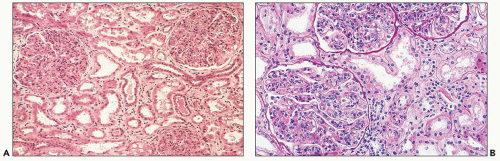
FIGURE 10.1 Three enlarged glomeruli show diffuse endocapillary hypercellularity with numerous neutrophils and closure of all the glomerular capillaries. The glomeruli are increased in size and cellularity. Although both (A) and (B) show very similar changes, the images were taken from biopsies of two different patients with PSAGN. (A, H&E, ×200.) (Courtesy of Dr. J. Charles Jennette.) (B, PAS, ×200.)


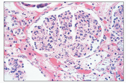
FIGURE 10.2 A glomerulus with endocapillary hypercellularity and closure of the glomerular capillaries. Because of the increased cellularity within each lobule, there is an accentuation of the lobularity. (H&E, ×400.)

The individual lobules of the glomeruli are wider than normal and sometimes assume a clubbed appearance. The glomerular capillary lumina are often reduced by the hypercellularity so that erythrocytes may be difficult to see. The stalk region of the mesangium may be quite hypercellular (115). The glomerular capillary walls are generally not thickened, although there may
sometimes be mild thickening visible on light microscopy. The combination of expansion of the lobules, hypercellularity of the tuft, and localized thickening of the glomerular capillary walls may produce a picture mimicking MPGN. Ultrastructural and immunofluorescence studies and clinical findings at the time of biopsy and follow-up allow easy separation of these morphologically and clinically distinct entities.


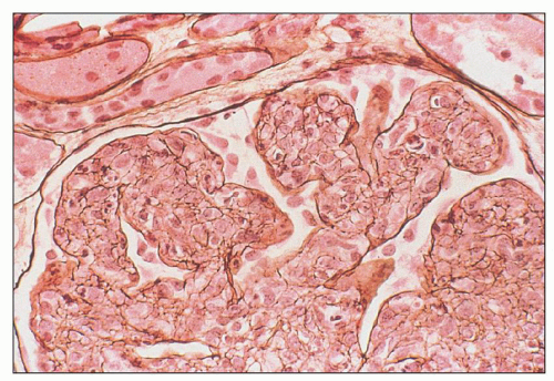
FIGURE 10.3 Silver stain of a glomerulus from a patient with PSAGN. Note that all the hypercellularity is confined within the glomerular tuft (endocapillary hypercellularity). (Jones silver methenamine, ×600.)

In some patients, it is possible, especially with the oil immersion lens, to detect tiny nodules on the epithelial side of the glomerular capillary wall. These nodules can be identified as fuchsinophilic dots with the trichrome stain (Masson or Mallory) either on thin (3-µm) sections or on 0.5-µm plastic-embedded sections stained with toluidine blue (Fig. 10.7). These minute structures correspond to the subepithelial deposits (humps) noted by electron microscopy. For optimum detection, it is important to study these renal biopsies with the oil-immersion lens and the trichrome stain because these small deposits may be overlooked on casual examination with the high-dry (40× or 60×) objective alone. Detection of those deposits by light microscopy is especially useful if there are no materials (i.e., glomeruli) for study by electron microscopy or immunofluorescence. Although the glomerulonephritis is diffuse (involving all or almost all glomeruli equally), there may be focal and segmental variability of the lesions among glomeruli, but this is unusual (Fig. 10.8).


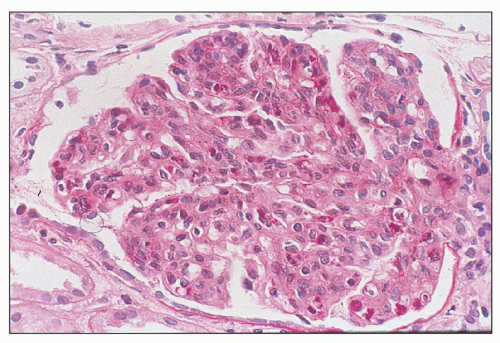
FIGURE 10.4 This glomerulus shows a broadening of the lobules, increase in cellularity with moderate numbers of neutrophils with segmented nuclei, and reduction of the capillary lumens. (PAS, ×400.)


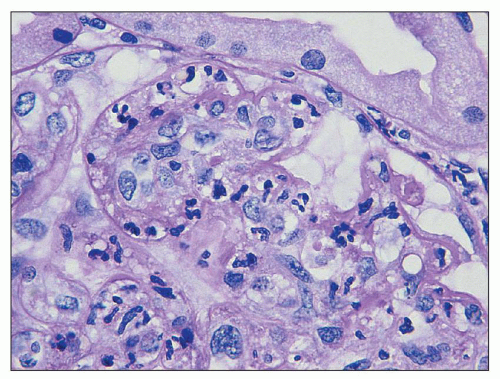
FIGURE 10.5 Acute diffuse proliferative glomerulonephritis with considerable infiltration of the glomerulus by neutrophils, which is common in acute postinfectious glomerulonephritis. (PAS, ×1000.)

In some patients, there may be crescent formation (2,66) or small adhesions (synechiae) (Fig. 10.9). In a few patients, crescent formation is so prominent that the term crescentic glomerulonephritis may be used, but usually only a small percentage of glomeruli are affected by crescents. The Bowman space may contain erythrocytes, which is evidence in a percutaneous renal biopsy that hematuria is caused by glomerular bleeding. Polymorphonuclear leukocytes also may be seen in the Bowman space.

Cell Types Both infiltrating leukocytes (neutrophils and monocytes) and proliferating glomerular cells (mesangial, endothelial, and epithelial cells) contribute to the glomerular
hypercellularity. The old term proliferative glomerulonephritis implies that the increased cellularity is restricted to native glomerular cells, either endocapillary cells (endothelial or mesangial) or extracapillary cells (visceral and parietal epithelial cells). However, current evidence suggests that much of the glomerular hypercellularity (as well as some of the crescent formation in the Bowman space) stems from infiltrating leukocytes from the peripheral circulation. Langhans (8) initially proposed that the major cause of the cellular increase in the glomerular tuft was a proliferation of endothelial cells. This concept was responsible for the designation endocapillary proliferative glomerulonephritis.


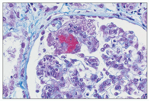
FIGURE 10.6 A glomerulus from a patient with clinically classic PSAGN. Note the focal segmental fibrinoid necrosis at approximately 10 o’clock as well as the endocapillary hypercellularity. (Masson trichrome, ×600.)


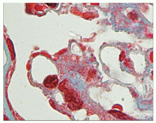
FIGURE 10.7 High magnification of a Masson trichrome-stained section from a renal biopsy with PSAGN. Note the fuchsinophilic (red) subepithelial deposits along the glomerular capillary walls at the periphery of the glomerular capillaries. (Masson trichrome, ×1000.)


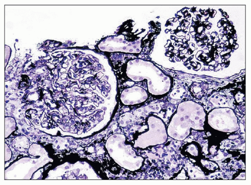
FIGURE 10.8 Renal biopsy from a patient with PSAGN. Most of the glomeruli showed global severe endocapillary hypercellularity such as the glomerulus on the left. An occasional glomerulus (the glomerulus on the right) showed only mild and/or segmental hypercellularity. Note that the glomerular capillaries in the glomerulus on the right are patent. (Jones silver methenamine, ×200.)


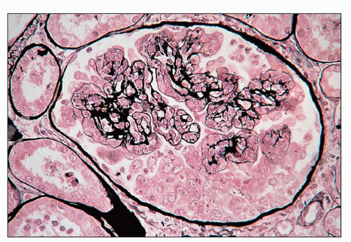
FIGURE 10.9 A glomerulus, with early crescent formation. Note that in the underlying glomerular capillary tuft, there is global increase in cellularity including polymorphonuclear leukocytes. (H&E, ×400.) (Courtesy of Dr. Vivette D’Agati.)

Jennings and Earle (115) favored the notion that hypercellularity was owing to native intraglomerular cells because of the presence of mitotic figures in “cells inside and attached to the glomerular capillary basement membrane (by definition, endothelial cells).” These authors also used electron microscopy to strengthen their view and described cells in the resolving phases of the disease process that had inclusions of electron-dense material resembling GBM. They stated that such inclusions were not evident in mononuclear inflammatory cells (115). This argument, however, did not resolve the question regarding which endocapillary cells (mesangial or endothelial) participate in the proliferation. Ia (MHC class II antigen)-bearing mesangial cells (53), which might be resident macrophages, also could play a role in hypercellularity, but studies in humans have not been forthcoming.

Grishman and Churg (60) described proliferation of cells in the mesangium but did not commit themselves as to the nature of the cells. Many other investigators have mentioned the great difficulty in distinguishing between endothelial and mesangial cells in light microscopy of a hypercellular glomerulus.

Experimental studies also confirmed the idea that mononuclear phagocytes appear in the glomerular tuft in various forms of experimentally induced glomerulonephritis. Although most of these studies used models of Masugi nephritis and Habu snake venom, some used acute serum sickness models in the rabbit (48,63). Monocytes were found to be present in the latter model of acute glomerulonephritis by Hunsicker et al. (63) and by Holdsworth et al. (48), using electron microscopy and staining for nonspecific esterase. Thus, it appears that bone marrow-derived mononuclear cells play a significant part in the hypercellularity noted in the glomeruli of certain experimental diseases.

In human studies using staining for nonspecific esterase and electron microscopy, monocytes are identified in less than half the biopsies of acute glomerulonephritis examined, especially in the early stages of the disease (50,58). Specimens with
mesangial hypercellularity, but less exudative change, had fewer esterase-positive cells. It was suggested that early glomerular hypercellularity is owing to an influx of blood-borne cells, but at a later stage, it is caused mainly by proliferation of intrinsic glomerular cells (62,64). Magil et al. (62,64) verified the presence of glomerular intraluminal monocytes that correlated positively with the presence of deposits. They also described dissection of the glomerular endothelium from the capillary wall adjacent to deposits by the monocytes. Ferrario et al. (58) showed that the degree of proteinuria correlated well with the degree of glomerular mononuclear cell infiltration.

Chung and Kim (47) performed immunohistochemical studies with a monoclonal antibody to Ki-67 (a cell proliferation marker) on renal biopsies from 21 children with PSAGN. They found that the active phase of the disease was associated with more prominent glomerular Ki-67 expression compared with the convalescent phase of PSAGN. However, one has to note that even if in the active disease group, only 11 of 13 biopsies showed Ki-67-positive glomerular cells. Therefore, their results clearly indicate that although some degree of proliferation of endogenous glomerular cells takes place, the bulk of the hypercellularity is secondary to infiltrating inflammatory cells (47). Unfortunately, the study does not clarify the exact cell populations that undergo proliferation in these biopsies with PSAGN.

Inflammatory cells in acute diffuse proliferative glomerulonephritis have been studied by immunophenotyping, but only a few studies concentrated on PSAGN. Hooke et al. (56) found significantly increased numbers of glomerular monocytes and granulocytes, but no significant increase in the number of glomerular T cells or B cells in cases of acute postinfectious glomerulonephritis when compared with normal glomeruli. There was a rise in the number of interstitial T lymphocytes compared with the normal interstitial cell population, but the OKT4/OKT8 ratio was the same as in normal cell populations (56).

Parra et al. (51) studied the cell populations in glomeruli of patients with PSAGN using monoclonal antibodies and indirect immunofluorescence. They found infiltration of the glomeruli by monocytes, granulocytes, and lymphoid cells. T cells were noted adjacent to the Bowman capsule. CD4+ (helper/inducer) lymphocytes were found in the glomeruli early in the course of the disease, whereas CD8+ (cytotoxic/suppressor) cells were found later. Yoshizawa et al. (61) evaluated the role of the cell-mediated immune response in the kidneys of 22 patients with PSAGN. They found a substantial increase in the total number of granulocytes and monocytes/macrophages with only a slight rise in T cells. The number of cells correlated well with time after clinical onset. A positive linear correlation was confirmed between helper/inducer T cells and monocytes/macrophages. As with the study of Parra et al. (51), helper T cells tended to increase to a higher proportion early on, although suppressor T cells remained constant throughout the course of the disease. Although there were fewer total leukocytes than in patients with clinically overt disease, in asymptomatic patients with PSAGN, the proportions of infiltrating cells were similar (61).

Polymorphonuclear leukocytes often are numerous, although large numbers of these cells do not indicate a poor outcome. The leukotactic properties of complement have been verified. Lewy et al. (98) documented a case with a predominantly polymorphonuclear leukocytic reaction in which there were large numbers of glomerular subepithelial humps. These authors commented that in other cases, the greatest numbers of polymorphonuclear leukocytes were found when there were large numbers of humps.

Other Mesangial Changes Several probes have found increases in some naturally occurring substances in the glomeruli in postinfectious glomerulonephritis. Actomyosin has been shown to be mildly elevated in the mesangial regions (49), but the significance of this finding is unclear. Likewise, fibronectin has been demonstrated in the mesangial areas (122,123). This protein is present in the normal mesangium and is significantly elevated in a variety of conditions associated with mesangial increase. Mosquera and Rodriguez-Iturbe (124) found glomerular-binding sites for fluoresceinated peanut agglutination lectin in renal biopsies showing evidence of PSAGN. Peanut agglutination lectin has specificity for galactosyl radicals that are exposed after sialic acid removal, as one would see with neuraminidase. These findings suggested to the authors that sialic acid-depleting material (probably neuraminidase) was present in glomeruli early in the course of the glomerulonephritic process.

Several immunopathologic studies have been performed on renal biopsy material taken from patients with PSAGN. Parra et al. (125) confirmed enhanced expression of intraglomerular ICAM-1 (intercellular adhesion molecule 1) in early-stage biopsies; this expression decreased with time. The numbers of cells expressing lymphocyte function-associated antigen 1 (LFA-1) in glomeruli were also elevated in early biopsies. Levels of VCAM-1 (vascular cell adhesion molecule 1) were not higher. This study suggested that ICAM-1 (and possibly other adhesion molecules) is important in the recruitment/influx/localization of leukocytes in acute postinfectious glomerulonephritis. The study by Lhotta et al. (126) of ICAM-1 expression in various proliferative glomerulonephritides demonstrated that the changes in staining intensity were observed mainly on the glomerular endothelial cells with less mesangial staining. However, it should be noted that induction of E-selectin (ELAM-1) is not invariable in acute glomerulonephritis (127).

Subclinical and Resolving Glomerulonephritis Renal biopsies in patients with minimal urinary changes have been performed (usually in prospective studies) and show differing results. Some find no substantial abnormalities. Increased cellularity of the glomeruli also has been noted, as have changes indistinguishable from characteristic acute diffuse proliferative poststreptococcal glomerulonephritis (105,128,129). In renal biopsies taken several weeks after the clinical onset of disease, there is diffuse hypercellularity in mesangial regions of the glomeruli; the glomerular capillaries are patent, and the capillary walls appear thin and delicate (130) (Fig. 10.10). Some degree of resolution of the hypercellular process takes place, and the number of polymorphonuclear leukocytes is diminished. Mesangial hypercellularity appears to persist for many months in patients who eventually experience complete resolution of the glomerular lesion (131).

An old term for this morphologic picture is chronic latent glomerulonephritis. However, caution needs to be exercised for two reasons. First, an unusually thick paraffin section may give the false appearance of diffuse mesangial hypercellularity.
Second, many cases termed chronic latent glomerulonephritis may not be resolving/resolved PSAGN, in our opinion, but rather represent a nonspecific histologic pattern associated with various renal injuries unrelated to previous infections (132). In one long-term follow-up study (more than 5 years) of 26 patients suffering from PSAGN, Buzio et al. (133) found diffuse mesangial hypercellularity in those patients with persisting urinary albumin or proteinuria.


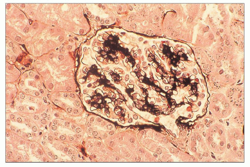
FIGURE 10.10 Resolving PSAGN shows mesangial expansion. Increased mesangial cellularity and mesangial matrix increase may persist for years. (Jones methenamine silver, ×200.)

It has been proposed that patients with well-documented PSAGN who show a mesangial proliferative picture on subsequent biopsy have a worse renal prognosis than those with typical resolving streptococcal glomerulonephritis. Patients with isolated mesangial C3 deposits in association with mesangial proliferative glomerulonephritis may actually have poststreptococcal glomerulonephritis, but there is little proof in most patients. Multiple entities may show this pattern, since the kidney responds in a limited fashion to many different injurious stimuli. The recently characterized entity, C3 glomerulopathy, should be considered in proliferative glomerulonephritis with isolated C3 deposits, particularly in the absence of an obvious preceding infection (134). Also, patients with underlying abnormalities of the alternative complement pathway activation may have an atypical course of postinfectious glomerulonephritis (135). Complete morphologic resolution occurs following PSAGN, but follow-up biopsies in such patients, obviously, are not performed. In fact, “incidental healed” postinfectious glomerulonephritis may be more common than anticipated. Haas (136), in a study from the Johns Hopkins Hospital, reviewed 1112 consecutive renal biopsy specimens and found 57 biopsies in which ultrastructural findings indicated resolving/healed PSAGN. According to Haas, resolving or largely healed postinfectious glomerulonephritis was present in 10.5% of renal biopsy specimens, excluding biopsies with a primary diagnosis of immune complex glomerulonephritis (136). The study was based on ultrastructural findings (subepithelial deposits in glomerular mesangial notch regions); therefore, this incidence may be somewhat overestimated because subepithelial deposits in the mesangial notch region are not specific for resolving postinfectious glomerulonephritis because they can be seen in other glomerular diseases, for example, in C3 glomerulopathy (including dense deposit disease and C3 glomerulonephritis). Interestingly, in Haas’ study, 50% of the biopsies showing some evidence of incidental healing postinfectious glomerulonephritis had evidence of mesangial hypercellularity (136).


TUBULES

The tubular changes are not as pronounced as those involving the glomeruli. When proteinuria is present, there may be hyalin droplets (protein reabsorption droplets, phagolysosomes) or vacuoles (dissolved lipid droplets) in the proximal convoluted tubular epithelium. Erythrocytes may be seen in the lumen of the tubules, and they are sometimes mixed with eosinophilic cast-like material. Polymorphonuclear leukocytes also can be present in the lumens, especially in the first portions of the proximal tubules. This feature is most commonly seen in patients with severe exudation or infiltration of polymorphonuclear leukocytes in the glomeruli. In a few patients, polymorphonuclear leukocytes can be seen between the tubular basement membrane (TBM) and the overlying tubular epithelial cells (Fig. 10.11). In patients who develop severe renal insufficiency, classic changes of acute tubular necrosis (ATN) are usually evident. With serial renal biopsies, these changes resolved. There was no apparent relationship between the morphologic tubular changes and TmPAH. In the most florid cases of acute glomerulonephritis with extensive crescent formation, there may be progressive tubular injury with tubular atrophy and loss, as well as tubulitis characterized by inflammatory cells between the TBMs and the tubular epithelium or within the tubular epithelium.


INTERSTITIUM

The degree of interstitial involvement is variable. The interstitial areas may show edema with separation of the tubules from one another. Scattered foci of inflammatory cells, made up of mixtures of polymorphonuclear leukocytes, monocytes, and lymphocytes, are sometimes present (Fig. 10.11). There may occasionally be severe interstitial mononuclear cell infiltration
and scattered regions of interstitial fibrosis. Usually, however, the interstitial changes are not remarkable or severe. As noted earlier, interstitial changes may be found in relation to tubular changes (137). Bohle et al. (138), using morphometric methods on tissue sections, showed that the level of serum creatinine correlated with the increase in interstitial volume. They explained this finding on the basis of a reduction of renal blood flow and, hence, the GFR, brought about by compression of the postglomerular vasculature.


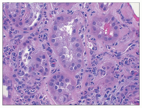
FIGURE 10.11 Unusually pronounced neutrophilic infiltration in the interstitium in this biopsy from a patient with PSAGN. Note that some neutrophils infiltrate the tubular epithelium. (H&E, ×600.)


BLOOD VESSELS

The arteries and arterioles generally do not show changes. In older patients, preexisting vascular abnormalities, such as arterial and arteriolar sclerosis, may be seen, and, according to Gallo et al. (139), these may be accentuated and lead to greater renal parenchymal injury. Arteritis has been described (140), but in such cases, systemic necrotizing vasculitis must be excluded. There are other accounts of arteritis (141,142) as well, but they are rare. Fibrinoid necrosis of the arterioles may be associated with severe hypertension. As noted earlier, in rare instances, vascular changes of thrombotic microangiopathy (TMA) may be seen (143,144).


Immunofluorescence Findings

Immunofluorescence studies have been reported by many investigators (59,96,131,145,146,147,148,149,150,151,152). Classically, in biopsies taken early in the clinical course (first 2 or 3 weeks) of the illness, small, granular deposits are noted along the glomerular capillary walls following immunofluorescence studies with anti-IgG and anti-C3 fluoresceinated antisera (Figs. 10.12 and 10.13). The pattern is granular (“lumpy-bumpy”) and usually more coarse than in patients with membranous glomerulonephritis. Large, coarsely granular immune complex deposits are usually easy to visualize using immunoperoxidase methodology as well on paraffin sections (Fig. 10.14). These deposits may assume a somewhat linear or band-like (garland) pattern in some areas, owing to the confluence of subepithelial deposits. The granular deposits correspond to the glomerular subepithelial deposits evident on electron microscopy, although there has been controversy over this feature in the past (153).


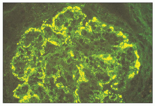
FIGURE 10.12 Immunofluorescence microscopy for IgG in a patient with PSAGN shows a coarsely granular pattern along the capillary walls and a less prominent granular mesangial pattern. (×400.)


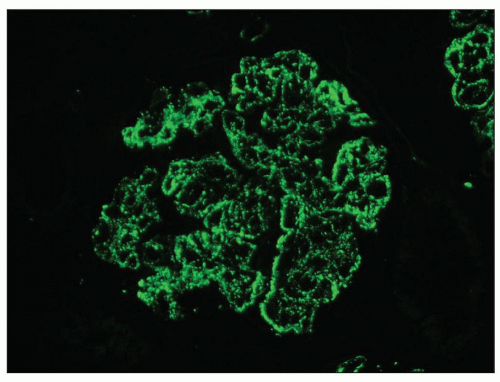
FIGURE 10.13 “Lumpy bumpy” coarsely granular C3 deposits along the glomerular capillary loops in PSAGN. (×400).

Sorger et al. (147,148,149) have described different immunofluorescence microscopy patterns called the garland pattern (Fig. 10.15), the starry sky pattern (Fig. 10.16), and the mesangial pattern (Figs. 10.17 and 10.18). The garland pattern has a discrete, more densely packed and sometimes confluent heavy disposition of IgG and C3, corresponding to numerous humps noted on the subepithelial side of the glomerular capillary wall (147,149). This arrangement is most often seen in patients with acute glomerulonephritis who have severe proteinuria (often with the nephrotic syndrome) (see Fig. 10.15). Other glomerular deposits are rare. The starry sky pattern has a more irregular, finely granular pattern, with the deposits (IgG, IgM, C3, IgA) being smaller and often situated on the GBM overlying the mesangial regions. This arrangement was most commonly seen in early cases (see Fig. 10.16). Only a few large, typical humps were noted in these cases. This picture may turn into the mesangial pattern, characterized by a granular deposition of IgG and C3 (usually with predominance of C3). It seems to be most closely related to a resolving pattern (see
Fig. 10.17). The deposits are generally noted in the mesangial matrix of the glomerulus and are accompanied by mesangial hypercellularity.


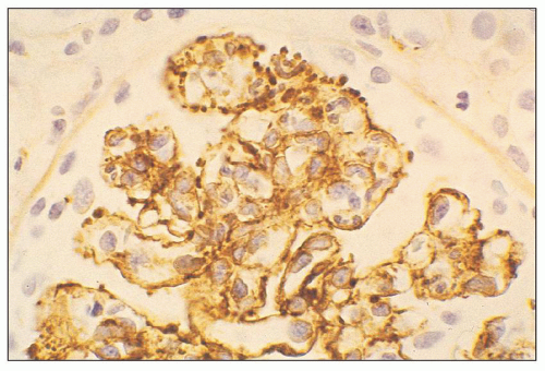
FIGURE 10.14 Immunoperoxidase method on paraffin section with an antibody to C3 highlights the coarsely granular large subepithelial deposits in this case of PSAGN. (×600.) (Courtesy of Dr. J. Charles Jennette.)


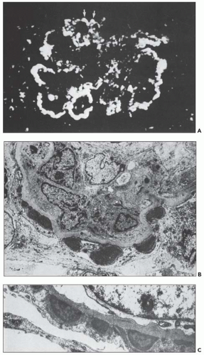
FIGURE 10.15 A: Immunofluorescence shows a garland pattern. The garland-like outline of the glomerular loops is due to large subepithelial deposits on the outer side of the GBM (arrows). The mesangial regions appear to be largely empty. (IgG, ×450.) A,B: Accompanying electron micrographs show part of a glomerulus from a patient with a garland pattern. In B, there are hypercellularity and numerous subepithelial deposits of varying density. (×5700.) In C, taken at a higher magnification, there are variegated deposits of different sizes. (×9000.) From a 37-year-old man with a 2-week history of poststreptococcal glomerulonephritis. (From Sorger K. Postinfectious Glomerulonephritis. Stuttgart, Germany: Gustav Fischer Verlag, 1986.)

Edelstein and Bates (154) studied 42 adult patients with characteristic acute postinfectious glomerulonephritis and divided the biopsies into these three subtypes. There was no significant difference among the three subgroups of patients with regard to age, blood pressure, serum creatinine, ASO titers, or decreased serum C3 levels at presentation. Patients with the garland pattern had significantly more proteinuria, whereas the renal biopsies with the mesangial pattern had a lesser degree of glomerular hypercellularity and leukocytes.


The starry sky pattern was noted in four of five patients with a crescentic pattern and six of seven patients with a chronic course (154). There is no evidence so far that different etiologic factors are responsible for these three subtypes (148). The individual immune response of the host and the stage of the disease are likely to play a role in their genesis. A diffuse granular pattern for IgG and, usually, C3 is also found in patients with subclinical glomerulonephritis (146,150,151) and in those with minimal urinary changes.


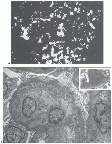
FIGURE 10.16 A: Immunofluorescence shows a starry sky pattern. There are diffuse and irregularly distributed fine and coarse granular deposits in the glomerular capillary walls and in the glomerular mesangial regions. (C3, ×704.) B: The accompanying electron micrograph shows a segment of a glomerular capillary loop from a patient with a starry sky pattern. The lumen of the glomerular capillary is totally occupied by a monocyte and by endothelial cells with prominent nuclei and swollen cell bodies. Numerous subendothelial deposits are present. (×5280.) Inset: A pointed, arch-like glomerular subepithelial deposit with sparse areas of higher electron density. (×17,600.) From a 56-year-old man with a 3-week history of poststreptococcal glomerulonephritis. (From Sorger K. Postinfectious Glomerulonephritis. Stuttgart, Germany: Gustav Fischer Verlag, 1986.)


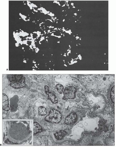
FIGURE 10.17 A: Immunofluorescence shows a mesangial pattern. Granular deposits are found in the mesangial regions, although the glomerular capillary wall remains largely negative. (C3, ×704.) B: The accompanying electron micrograph shows the corresponding ultrastructural appearance. A portion of the glomerulus shows marked proliferation of mesangial cells but free and open glomerular capillary lumens. Mesangial deposits are located in the mesangial matrix, and individual subepithelial deposits are present in the region of the mesangial waist (arrow). Less frequently, glomerular subendothelial deposits are situated along the loop. (×5280.) Inset: A glomerular subepithelial deposit with an almost homogeneous, comparatively pale density. (×17,600.) From an 18-year-old boy with a 5-week history of illness. (From Sorger K. Postinfectious Glomerulonephritis. Stuttgart, Germany: Gustav Fischer Verlag, 1986.)


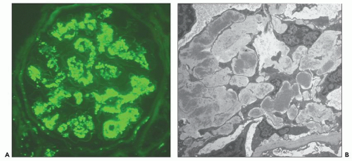
FIGURE 10.18 A: Coarsely granular predominantly mesangial C3 deposits in PSAGN (×400). B: Accompanying electron micrograph from the same patient showing mesangial electron-dense immune-type deposits. (Uranyl acetate and lead citrate, ×3000). Few subepithelial humps were also seen. The patient was a 51-year-old diabetic male with low C3, normal C4, very high antistreptolysin-O titer after a sore throat. The biopsy showed diffuse endocapillary proliferative glomerulonephritis with polymorphonuclear leukocytes in the glomeruli as well as in the interstitium. The biopsy findings suggested a very active disease. The biopsy was performed 2 months after the episode of streptococcal infection. The clinical history is supportive of PSAGN.

There is usually more intense and more constant staining with anti-C3 than with anti-IgG (145,146,150,151). In fact, it is common to see granular glomerular C3 without any demonstrable IgG. Some authors have noted the combination of granular and patchy, interrupted linear staining along the glomerular capillary wall and in the mesangial regions (59,88). At times, there seems to be an exclusively patchy, interrupted linear pattern for C3 along GBMs close to the mesangium as well as in the mesangium, with no staining for IgG (154). This interrupted linear pattern has been found most commonly either in patients in whom the initial renal biopsy was performed at a later stage than usual or in subsequent biopsies (59). C3 without the presence of IgG has been recorded in the mesangial areas with no capillary wall deposits (149). This pattern also tends to be seen in patients who undergo biopsy later than usual (i.e., several weeks after the clinical onset of disease).

Fish et al. (59) have advanced various explanations as to why C3 is present in the absence of IgG. They suggested that IgG may have been covered up by C3, thus preventing its detection. Another explanation could be that C3 reaches detectable levels for the immunofluorescence technique, while IgG remains below the threshold level of detection. It was thought that the interrupted linear deposits of C3 in the absence of IgG could be the result of a direct toxic effect of the organism (i.e., Streptococcus) on the glomerulus and that only a certain percentage of patients (especially those destined to experience severe acute nephritis) proceed to nodular deposits. As we will discuss briefly later, another explanation for the absence of IgG is that PSAGN may represent a transient form of C3 glomerulonephritis induced by streptococcal infection/antigens. In C3 glomerulonephritis, the glomerular deposits do not truly represent immunoglobulin-containing immune complexes.

IgM is frequently present and was recorded in more than 50% of cases in one series (131). Other authors (146) have not found IgM. IgA is usually absent (59,145,146), but it has been noted from time to time (131,150,151). If IgA immunofluorescence is strong in postinfectious glomerulonephritis, the possibility of an underlying staphylococcal infection has to be considered irrespective of presence or absence of diabetes mellitus (section on Glomerulonephritis associated with staphylococcal infections). IgE has seldom been sought; in one series of 10 patients, it was present in 5 biopsies (151). Fibrin/fibrinogen-related antigen also can be detected in the mesangial regions (as well as in the Bowman space in the crescentic form) (59,131,146).

Westberg et al. (155) noted properdin in the glomeruli in eight cases of postinfectious glomerulonephritis. In three of these patients, the pattern was granular along the glomerular capillary walls, whereas in five, it was noted in the mesangium together with C3. Various other authors (146,150) have also found properdin in several biopsies. Classic pathway components of complement, such as C1q and C4, are generally lacking (146,150,151). These various observations support the role of the alternative pathway of complement activation in postinfectious glomerulonephritis (151).


Parra et al. (51) studied the membrane attack complex in glomeruli of patients with PSAGN using monoclonal antibodies and indirect immunofluorescence. Membrane attack complex was noted along the GBM early and within the mesangial regions later in the course of the disease (a distribution similar to the deposition of C3 and C5). Rarely, immunoglobulins and complement components may be detected in renal arterioles, especially in those rare patients with necrotizing arteritis. Immunohistochemical studies by Kamitsuji et al. (156) localized intraglomerular deposits of fibrin and cross-linked fibrin in the proliferative glomerulonephritides.

As noted earlier, attempts to identify and localize the streptococcal antigen have usually failed. However, studies by Seegal et al. (157) demonstrated streptococcal antigen in over half the cases studied. Andres et al. (158), using ferritin-conjugated antibodies to type 12 streptococcal products, confirmed the presence of labeled antibody in the GBM, mesangium, and arterioles. No staining was noted in the glomerular subepithelial deposits. Some studies identify the streptococcal pyrogenic exotoxin B (SPEB) and nephritis-associated plasmin receptor (NAPlr) in the glomeruli of biopsies with PSAGN (159,160,161). The discrepancy between the results of various studies suggests that either the wrong antibody or the wrong antigen is being studied or that the antigen is being lost or masked in the glomeruli in these studies or that streptococcal antigens are not present in the glomerular deposits at all.

Deposits of immunoglobulins and, especially, complement may be detected in the glomeruli for months to years after apparent clinical resolution (136). The intensity of the immunofluorescence staining usually correlates with the severity of the glomerular lesion, although severe diffuse glomerulonephritis may be accompanied by unimpressive or negligible deposits.


Electron Microscopic Findings

There are many reports of the ultrastructural findings in acute glomerulonephritis (59,136,149,162,163,164). Many of the findings merely confirm what has long been noted at the light microscopic level, that is, increased numbers of endocapillary and infiltrative inflammatory cells in the glomerular tuft. There is swelling of both endothelial and mesangial glomerular cells, with closure of the capillary lumens by the increased numbers of cells, or swelling of the native glomerular cells. The GBMs generally appear normal in contour, thickness, and texture, although patchy thickening may occasionally be noted. There may be widening of the lamina rara interna by subendothelial electron-lucent “fluff” or fibrillar/finely granular amorphous material. The outer layer of the GBM may show “scalloping” or irregularity of the lamina rara externa and lamina densa. The GBM may contain lucent areas that may represent resolving deposits that are described and discussed later. Often, the glomerular endothelium is focally disrupted and denuded, with polymorphonuclear leukocytes directly adjacent to the denuded GBMs.

The most consistent classic diagnostic change is the presence of glomerular subepithelial electron-dense deposits, often referred to as “humps” (Figs. 10.19 and 10.20). A “hump” is a term used in renal pathology to describe subepithelial electron-dense immune-type deposits that bulge outward toward the Bowman capsule beyond the boundary of the glomerular basement membrane. They can be large or small in size. Typically, they are more unevenly distributed and more heterogeneous in size than the subepithelial deposits of membranous glomerulopathy. They are especially abundant in the first few weeks of acute postinfectious glomerulonephritis, and they decline in number afterward. They are usually less than 1 µm wide and long, but they sometimes are up to 3 µm wide and 6 µm long. In the study of Lewy et al. (98), two patients had quite elongated cigar-shaped glomerular subepithelial humps; these patients also had the nephrotic syndrome. The subepithelial humps are sometimes separated from the lamina densa by a zone of translucence that is continuous with the lamina rara externa; on other occasions, they merge with the underlying lamina densa. The deposits often bulge or project toward the cytoplasm of the overlying podocyte that often shows effacement of foot processes just above the deposit. There is frequently condensation of the microfilaments (especially actin) at the base of the effaced podocyte adjacent to the hump. The electron density of the deposits is variable, and the granularity may range from fine to coarse (165) (Figs. 10.21 and 10.22). Occasionally, the subepithelial deposits are markedly variegated with an irregular admixture of dense and less dense zones. Although there is no direct correlation between the fine ultrastructural appearance of the deposit and the clinical or nonultrastructural morphologic findings, Tornroth (164) has suggested that electron-lucent regions in the deposit may represent regions of resolution.

The deposits are usually plentiful and discrete and are most commonly found on that part of the GBM overlying the mesangial regions (i.e., the paramesangial GBM). West and McAdams (166) described a population of pediatric patients with PSAGN who had prominent hypoalbuminemia and edema with no or only very few subepithelial deposits along the glomerular basement membrane covering the mesangium. At times, the subepithelial deposits may be confluent along short stretches of the basement membranes. Similar discrete electron-dense immune-type deposits may be seen in the lamina densa and the subendothelial regions (59,66,149,163,164) (Fig. 10.23). Although the glomerular subepithelial hump is the most characteristic lesion by electron microscopy, similar subepithelial deposits are seen in various other glomerular disorders, such as membranous glomerulonephritis, MPGN, systemic lupus erythematosus, IgA vasculitis (Henoch-Schönlein purpura), and especially C3 glomerulopathy (including dense deposit disease and C3 glomerulonephritis). The subepithelial humps of C3 glomerulopathy have the greatest resemblance to the humps of PSAGN.

Discrete, electron-dense, immune-type deposits collect in the mesangial regions in classic postinfectious glomerulonephritis (149,167) (see Fig. 10.18B). The mesangial regions contain large numbers of cells whose identity is often elusive. In addition to native mesangial cells, there appear to be varying quantities of infiltrating inflammatory cells. Polymorphonuclear leukocytes are the easiest cells to identify in the glomerular mesangial regions, and they may be extensive in the exudative form of glomerulonephritis. There may be a mild increase in the amount of mesangial matrix. The Bowman space may contain debris, fibrin, large epithelial cells, erythrocytes, and polymorphonuclear leukocytes, particularly if crescents are present.

During the recovery phase (based on observations of patients undergoing serial biopsy), the glomerular subepithelial humps usually disappear within 6 weeks of the clinical


onset of disease (162). Sometimes, they persist for longer periods of time (163), but the clinical course in such cases is not clear. The fate of the glomerular subepithelial deposits has been studied by Tornroth (164), who has shown that the electron density (osmiophilia) of the deposits diminishes with time, so that electron-lucent regions are formed in the subepithelial zone that eventually disappear. The deposits may disappear by dissolution and passage into the blood or urinary ultrafiltrate or by pinocytotic removal by podocytes. Glomerular intramembranous electron-lucent regions have been seen in later biopsies (after 1 month) and, in some cases, these regions protruded toward the epithelium and were covered on that side by a thick layer of basement membrane-like material.


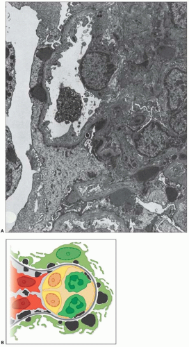
FIGURE 10.19 Electron micrograph and drawing showing the ultrastructural features of PSAGN. A: This electron micrograph shows a number of discrete electron-dense osmiophilic deposits in the subepithelial portions of the glomerular capillary walls. Some of the glomerular capillaries are narrowed or compressed, but one is patent. (Uranyl acetate and lead citrate, ×6930.) (Courtesy of Drs. William Murphy and Lillian Gaber.) B: Drawing depicting a single glomerular capillary with features of PSAGN including variably sized subepithelial, small subendothelial, and multiple mesangial electron-dense deposits (black), mesangial hypercellularity (red), endothelial hypercellularity (yellow), capillary margination of neutrophils (dark green), and effacement of podocyte foot processes (light green). Compare this drawing with features in the electron micrographs in Figures 10.15, 10.16, 10.17, 10.18, 10.19, 10.20, 10.21, 10.22, 10.23.


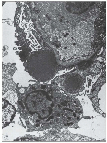
FIGURE 10.20 Glomerular capillary wall from a patient with PSAGN. There is a large, discrete, electron-dense (osmiophilic) immune-type deposit in the glomerular subepithelial region of the capillary wall. This hump, or deposit, is large and abuts on the glomerular capillary wall. There is effacement or fusion of the glomerular visceral epithelial cell over the deposit. (Uranyl acetate and lead citrate, ×10,125.) (Courtesy of Drs. Tito Cavallo and Srinivasan Rajaraman.)


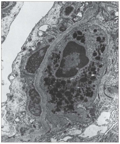
FIGURE 10.21 Electron micrograph of a segment of the glomerular capillary wall shows large, discrete, subepithelial electron-dense deposits. Note the heterogeneous or variegated appearance of these humps (i.e., varying degrees of osmiophilia or electron density). There is loss of the glomerular endothelium, and a polymorphonuclear leukocyte abuts the naked, or denuded, GBM. Polymorphonuclear leukocytes are often found near these glomerular subepithelial deposits. (Uranyl acetate and lead citrate, ×14,400.)


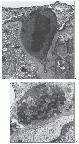
FIGURE 10.22 A: Electron micrograph of a segment of the glomerular capillary wall from a patient with typical PSAGN. Note that the electron density (i.e., osmiophilia) of the glomerular subepithelial deposit is not homogeneous. B: A similar variegated appearance of the deposit. These regions of greater electron lucency suggest to some investigators that the deposit is being “washed out” or is in the process of dissolution. (Uranyl acetate and lead citrate, A, ×20,100; B, ×23,450.) (Courtesy of Drs. William Murphy and Lillian Gaber [A] and Drs. Conrad L. Pirani and Vivette D’Agati [B].)


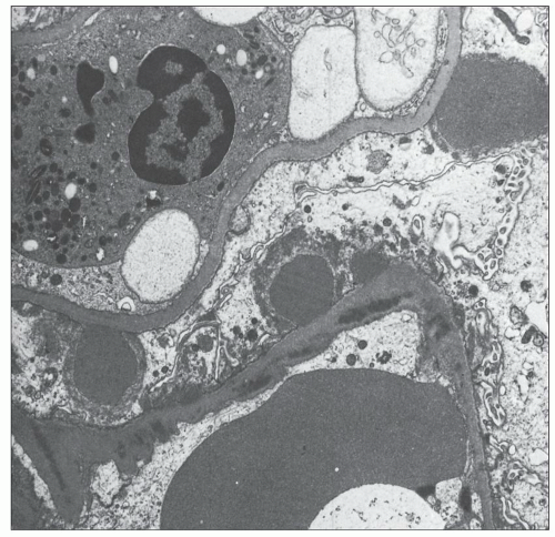
FIGURE 10.23 Electron micrograph of a segment of glomerular capillary wall from a patient with PSAGN. Glomerular subendothelial deposits can sometimes be found in these patients, although the predominant deposit is usually the typical subepithelial deposit. (Uranyl acetate and lead citrate, ×6090.) (Courtesy of Drs. Conrad L. Pirani and Vivette D’Agati.)

Other deposits were found deeper in the lamina densa, giving it a somewhat mottled appearance (164). Kobayashi et al. (163) showed that the deposits became buried in the GBM and also acquired a fine granularity with an electron density less than that of the original humps. Glomerular subendothelial deposits that were present early disappeared with time (163,164). The GBM became irregular in thickness and contour. Increased cellularity may persist in the mesangial regions for many months, even in those patients in whom the clinical picture and urinary sediment have returned to normal. In patients who have chronic proteinuria, an increase in mesangial matrix is often found. It appears that there are more subepithelial humps in those patients with a severe, protracted clinical picture than in those with immediate clinical recovery (163). Size of the deposits does not seem to correlate with clinical course or outcome (168).

Haas (136) emphasized the significance of scattered intramembranous and subepithelial remnant deposits following possible history of PSAGN. Using careful ultrastructural studies, Haas identified 57 renal biopsies with such deposits out of 543 biopsies that did not have a primary diagnosis of immune complex glomerulonephritis. Haas emphasizes the diagnostic significance of subepithelial deposits in the mesangial notch region. The mesangial notch region represents a fold of the glomerular basement membrane overlying the mesangium. Interestingly, 40% of the biopsies that, according to Haas, revealed incidental healed postinfectious glomerulonephritis also showed evidence of diabetic nephropathy (136). The author recognizes that the shortcoming of the study is that the diagnosis of incidental healed postinfectious glomerulonephritis was entirely based on morphologic (ultrastructural) findings. Authors of this chapter agree that scattered intramembranous deposits of variable electron density and deposits in the mesangial notch region occur in biopsy specimens, but these deposits are not specific for postinfectious glomerulonephritis.


Ultrastructural studies in prospective series of patients with minimal urinary findings have shown either a lack of electron-dense deposits (169) or glomerular subepithelial or subendothelial deposits. In one case, the deposits were confined to the glomerular subendothelial zone. In another series, the ultrastructural findings in patients with subclinical glomerulonephritis were studied (103). The only biopsies in which glomerular subepithelial deposits were documented were from cases overt acute glomerulonephritis by light microscopy.


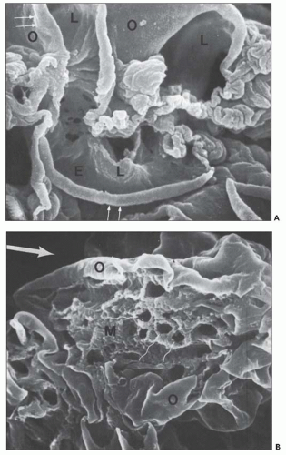
FIGURE 10.24 Scanning electron micrographs using the technique of Bonsib (170), in which the cellular elements have been removed by enzymatic digestion, leaving behind only the basement membrane, mesangial matrix, and other basement membrane-like substances. A: A segment of a normal glomerulus for purposes of orientation shows the open capillary lumen (L) and endothelial (E) aspect of the GBM (thin arrows). The subepithelial outer (O) surface of the GBM is also identified. B: A segment of a glomerulus from a patient with acute proliferation, as manifested by alterations in the mesangial matrix (M); holes (wavy white arrows) represent the former site of either proliferating cells or deposits. Orientation is provided by the outer (O) aspect of the GBM and urinary space (thick white arrow). C: A segment of a glomerulus from a patient with later changes seen in PSAGN, showing the subepithelial aspect of the GBM (curved white arrows) with reaction (dark arrows) to the site of deposit. (A, ×6375; B, ×2550; C, ×13,125.) (Courtesy of Dr. Steven M. Bonsib.)

Focal disruptions or gaps of the GBM have been identified (170,171,172) and, rarely, mononuclear cells and erythrocytes can be seen migrating through these gaps (171). Usually, however, the gaps are covered by glomerular endocapillary cells and podocytes. Bonsib (170) conducted interesting scanning electron microscopic studies following the selective removal of glomerular visceral epithelial cells by a sequence of lytic and solubilization procedures. The changes seen in acute postinfectious glomerulonephritis are illustrated in Figure 10.24. There are several distinct crater-like GBM formations (Fig. 10.24B)
that are empty, reflecting the solubilization of the subepithelial immune complexes. The craters are uniform in size and shape. Every glomerulus studied contained at least several craters that were located at various sites within the glomerular tuft. The case from which Figure 10.24C was prepared was unique because of the presence of subepithelial humps several years after the acute nephritic episode and because of the large size of GBM craters.


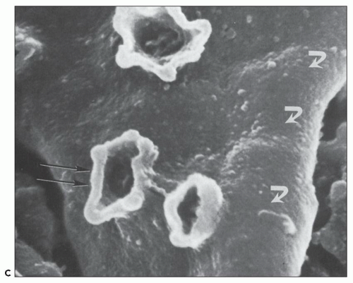
FIGURE 10.24 (Continued)


Etiology and Pathogenesis

The relationship between streptococcal infection and acute glomerulonephritis is well established and much has been learned about the mechanism of action by which the infection leads to the characteristic glomerular changes (173). It has been known for a long time that the blood and urine are sterile in patients with acute glomerulonephritis (11), and the kidney parenchyma is also sterile. The renal changes in acute glomerulonephritis were noted to be unlike those in patients with streptococcal septicemia, in which the major changes are interstitial nephritis and abscess formation. However, some studies have shown that streptococcal septicemia can lead to proliferative glomerulonephritis, a finding that is especially common in patients with acute bacterial endocarditis. Although streptococcal toxins could play a role in acute glomerulonephritis, it is unlikely, because the renal injury would be expected to occur at the height of the infection (whereas it takes place during the subsidence of the infection). Moreover, acute proliferative glomerulonephritis is not the type of morphologic change usually noted in patients with various circulating toxins; additionally, it would be anticipated that the renal changes would be proportional to the severity of the infection, which is not the case.


Immune-Mediated Disease and Experimental Studies—Historical Perspective

It is now widely accepted that acute poststreptococcal and other forms of postinfectious or infection-associated glomerulonephritis stem from an immunologic phenomenon. There is much to support an immune complex mechanism of action. Schick (9) noted the latent interval between clinical signs of infection and the onset of acute glomerulonephritis and likened it to the course of events in acute serum sickness and other allergic states. The latent interval after infection has been well documented a long time ago and usually ranges between 7 and 21 days (average, approximately 10 to 11 days).

Much of the support for immune complex pathogenesis comes from the analogy to acute “one-shot” serum sickness in the rabbit. When human acute postinfectious glomerulonephritis is compared with acute serum sickness, there are obvious similarities. Von Pirquet (174), in 1911, thought that the similarities between postinfectious glomerulonephritis and acute serum sickness syndrome supported his concept of allergy. Longcope (175) showed that parenteral administration of foreign protein in experimental animal models could induce glomerulonephritis. There is a latent interval between the injection of foreign protein and development of acute glomerulonephritis that is quite similar to the latent interval between streptococcal infection and the clinical onset of human renal disease. By immunofluorescence microscopy techniques, it is possible to demonstrate antigen, antibody, and complement in the glomeruli. This finding correlates with the formation of a variety of circulating immune complexes.

Ultrastructural studies in both human beings and experimental animal models also show similar glomerular subepithelial electron-dense “immune-type” deposits (176,177,178,179). Low levels of serum complement are found in both instances. As in the human counterpart, experimental acute serum sickness is a self-limited disease that generally resolves over a period of weeks.

Unfortunately, there is no perfect animal model for PSAGN. Many attempts have been made to induce an animal model of PSAGN by the injection of intact streptococci (180,181,182,183), crude culture supernatants (94), or specific components of the streptococci (47,48,49,50,51,52,53,54,55,56,57,58,59,60,61,62,63,64,65,66,85,86,87,88,89,90,91,92,93,94,95,96,97,98,99,100,101,102,103,104,105,106,107,108,109,110,111,112,113,114,115,116,118,122,123,124,125,126,127,130,131,132,133,134,135,136,137,138,139,140,141,142,143,144,145,146,147,148,149,150,151,152,153,154,155,156,157,158,159,160,161,162,163,164,165,166,167,168,169,170,171,172,173,174,175,176,177,178,179,180,181,182,183,184,185,186).
Although some of these experimental manipulations produced histologic lesions somewhat similar to the disease pattern in man, most called for the periodic administration of the putative factors thought to be involved; this, of course, does not precisely mimic the gradual release of streptococcal products that probably occurs at the site of infection in the clinical condition in humans (85). Also, many of the experimental studies were performed at a time when electron and immunofluorescence microscopy and other biochemical determinations were not available, making it difficult to carry out an adequate comparison (85).

Holm (85) and Nordstrand et al. (187) developed animal models in rabbits and mice that permitted the establishment of a focal infection using subcutaneous tissue cages and continuous monitoring of the infection and the release of streptococcal factors. According to the authors, these models simulate PSAGN clinically, histologically, and immunologically; however, not all investigators agree that this is a solid model of the human disease. Yoshizawa et al. (188) developed a rabbit model with the infusion of a peculiar streptococcal antigen called preabsorbing antigen (PA-Ag). After infusing 18 mg of this antigen into rabbits for 8 days, the animals developed proliferative immune complex glomerulonephritis with glomerular deposition of C3 without deposition of immunoglobulins. Electron microscopy revealed hump-like subepithelial deposits. Burova et al. (189) induced proliferative glomerulonephritis in the rabbit following repeat infusions of GAS strains bearing IgG-binding M family proteins. The animals had glomerular IgG and C3 deposition. Infusion of mutant bacterial strains, lacking the IgG-binding proteins, did not result in glomerulonephritis. The authors postulate that streptococcal IgG-binding proteins have an important role in triggering PSAGN in their model (189).


Search for the Antigen

The identity of the nephritogenic fractions of the bacteria is the subject of controversy. Rodriguez-Iturbe (20) has suggested two major possibilities: that the nephritogenic antigen is a specific component of some streptococci or that the streptococcal infection itself triggers an autoantigenic reactivity in the host. However, as noted later, it has been very difficult to establish the presence of streptococcal antigen either within the presumed immune complexes in the glomeruli or in the circulating immune complexes. The potential nephritogenic streptococcal antigens that have been proposed to play a causative role are summarized in Table 10.3.

Treser et al. (194) described how IgG fractions from the serum of patients with acute postinfectious glomerulonephritis caused staining of the glomerular capillaries and mesangium of patients with early-stage acute glomerulonephritis. This staining was abolished if the serum fraction had been previously absorbed with frozen and thawed nephritogenic β-hemolytic streptococcal organisms. The plasma membrane appeared to be the fraction responsible. This staining activity was not abolished by absorption with other bacteria, and the antisera against streptococcal plasma membranes had staining properties similar to those of the sera of the patients with acute postinfectious glomerulonephritis. The conclusion drawn was that the streptococcal plasma membrane constituents were present in the glomeruli of patients with PSAGN (195). These same workers (195) were able to show antigenic sites in the mesangial matrix and on the glomerular subendothelial region using immunoferritin ultrastructural techniques (195). There is some evidence that antibodies to streptococcal cell membrane antigens may cross-react with antigens in the glomerular basement membrane (221). Studies by Lange et al. (196) and others (158,222,223) have suggested that free antigen may be found in situ in the glomeruli and is available for the deposition of circulating antibodies. Cationic antigens, which are able to penetrate the fixed glomerular polyanionic charge barrier of the capillary wall, could be candidates for triggering an in situ immune complex reaction (119,224,225).


TABLE 10.3 Potentially nephritogenic streptococcal antigens


Antigen


References


Streptococcal M protein and its fractions


(190,191,192,193)


Endostreptosin


(194,195,196,197,198,199,200,201)


Streptokinase


(202,203,204)


Nephritis strain-associated protein or streptococcal pyrogenic exotoxin exotoxin B or Nephritis plasmin binding protein


(85,186,205,206,207,208,209,210,211,212)


Nephritis-associated plasmin receptor or streptococcal glyceraldehyde 3-phosphate dehydrogenase


(161,213,214,215,216,217)


Preabsorbing antigen


(188,218,219,220)


Streptococcal M protein is a strong candidate for the relevant antigenic bacterial fraction (190). M protein fractions can complex with fibrinogen and localize in glomeruli (226), and glomerulonephritis can be induced with injection of M protein-M protein/fibrinogen complexes. M protein may be antigenically cross-reactive with the GBM (191). However, Treser et al. (227) have suggested that the nephritogenic fraction is different from the M protein. Serum from patients convalescing from poststreptococcal glomerulonephritis, when labeled, could identify free antigenic sites in renal biopsy specimens showing poststreptococcal glomerulonephritis; the fact that this serum had these antibodies independent of the M type of the original infection suggested that a non-M antigen was present in the glomerulus. In contrast, Mori et al. (192) found that IgG titers against the C region of the M protein of group A streptococci are elevated in patients with PSAGN compared with patients with uncomplicated streptococcal pharyngitis, chronic glomerulonephritis, and healthy controls. IgG titers against the A and B regions of streptococcal M protein were not different between these groups.

Several streptococcal fractions have been studied in search of the trigger for glomerulonephritis. One streptococcal fraction, endostreptosin, has been extensively studied (194,195,196,197,198,199,200,201). This antigen is demonstrable in the glomerulus only during the initial phase of acute glomerulonephritis and reacts with antibodies present in the convalescent sera of patients with acute glomerulonephritis. In the late phases of the disease, the antigen can no longer be detected, presumably because all the previously noted antigenic sites have been covered by the specific antibody.

Endostreptosin’s molecular weight is between 40 and 50 kDa (186) and is most likely derived from the streptococcal cytoplasm. Seligson et al. (200) have suggested that acute
elevations of endostreptosin titers are generally diagnostic of PSAGN. Although low titers of antibody have been found in as many as 70% of normal individuals, significantly higher titers of antibodies are found in patients with poststreptococcal glomerulonephritis (200). Most patients with acute rheumatic fever do not have these high levels of antibody titer. Thus, Lange et al. (198) believe that elevated levels of antibody to endostreptosin are diagnostic of postinfectious glomerulonephritis and correlate well with the course of the pathologic disease process. Experimental studies by Cronin and Lange (197), using Wistar Furth rats, showed deposition of endostreptosin along the GBMs 1 day after injection of immunoaffinityisolated endostreptosin. Rats killed on days 8 to 12 showed increasing deposition of IgG and C3 with diminished staining for endostreptosin. No antiendostreptosin antibodies were detected in the sera in the first 3 days, whereas rats from day 4 onward had low levels of these antibodies. According to these authors, endostreptosin does not appear to be immunologically related to streptococcal exoenzymes or the streptococcal cell wall (197). Endostreptosin is similar to the preabsorbing antigen described by Yoshizawa et al. (188,201,218) and Holm et al. (228).

Yoshizawa et al. (218) isolated a 43-kDa protein from nephritogenic streptococci (“preabsorbing antigen”) and noted identical precipitation lines by immunodiffusion between rabbit antisera against preabsorbing antigen and the sera of patients with PSAGN. Antibodies to preabsorbing antigen were found in 30 of 31 patients with acute glomerulonephritis but only very rarely in control groups. The preabsorbing antigen is present in the glomeruli in the early phases of human PSAGN and appears to activate C3 by the alternate pathway (factor B). These authors developed a rabbit glomerulonephritis model by administering preabsorbing antigen for 8 days (188). Light microscopy revealed proliferative glomerulonephritis; immunofluorescence showed glomerular capillary and mesangial C3 deposits; and electron microscopy revealed occasional subepithelial hump-like deposits. Interestingly, IgG and preabsorbing antigen were not detected in the glomerular deposits (188).

Villarreal et al. (206) identified an extracellular protein unique to nephritogenic strains from cultures of type 12 organisms. This fraction (called nephritis strain-associated protein, NSAP) was noted in 56% of renal biopsies with signs of poststreptococcal glomerulonephritis; it was not found in biopsies from patients with other forms of nonstreptococcal glomerulonephritis or rheumatic fever. The vast majority of patients with glomerulonephritis had serum antibodies to the fraction (205). Holm et al. (85,207) suggested that the ability of NSAP to convert plasminogen to plasmin (possibly in situ) might be related to many of the pathologic events taking place in PSAGN. The plasmin formed by the interaction between NSAP and plasminogen splits the C3 molecule and activates the alternative pathway of complement activation, thereby initiating the inflammatory glomerular response. Interaction with plasmin or plasminogen can cause glomerular damage by degrading the GBM through the activation of latent metalloproteinases or collagenases. The circulating or in situ immune complexes can then move across the altered GBM and accumulate as subepithelial “humps.” NSAP (also called streptococcal pyrogenic protease exotoxin B [SPEB] or nephritis plasmin-binding protein [NPBP]) can directly cause tissue destruction by cleaving extracellular matrix proteins including fibronectin and vitronectin and might aggravate inflammation via superantigenic effects on the immune system, similar to staphylococcal enterotoxins A and C. SPEB can directly bind to class II MHC molecules on antigen-presenting cells and specific Vβ chain of T-cell receptors causing proliferation and massive activation of T cells, liberating copious amounts of Th1 cytokines. Antibodies to streptococcal glyceraldehyde 3-phosphate dehydrogenase (GADPH) and SPEB (NSAP) have been found specifically in patients with poststreptococcal glomerulonephritis and persist for 10 years and 1 year, respectively, after acute attack, thought to give long-lasting immunity (208). The genes for both these proteins are highly conserved among isolates of group A streptococci. Studies using double immunofluorescence staining methods for NSAP and collagen type IV demonstrate that NSAP localizes to the inner side of the glomerular capillary walls (209,210,229).

NSAP has a subunit of 46 kDa. The molecule has been isolated and purified (186); it has antigenic, biochemical, and structural similarities to streptokinase from group C streptococcal organisms, and it binds to plasmin and is a plasminogen activator. This protein is not related to group A streptokinase (230) or to a recently described streptococcal dehydrogenase protein according to these authors (213,230). Amino acid sequence analysis and immunologic reactivity studies suggest that this protein is the SPEB precursor (previously termed zymogen streptococcal proteinase precursor) (213).

Vogt et al. (214,231) isolated and identified a number of different cationic proteins from nephritogenic streptococci. Cationic moieties are known to have affinity for the GBM. Publications from the group at the Rockefeller University indicate that the cationic protein described by Vogt et al. (214,231) is structurally identical to SPEB (159). This group and other investigators suggest an important role of SPEB in PSAGN (159,232). Cu et al. (159) found that SPEB antibodies were present in the sera of patients with PSAGN significantly higher than in patients with acute renal failure, scarlet fever, and normal sera. Following injection of SPEB into male Sprague-Dawley rats, Romero et al. (232) found that inflammatory cells are accumulated in the glomeruli and in the interstitium. The same authors also found elevated levels of apoptosis in human SPEB-treated leukocytes (233). Savill (234) also proposes a role of apoptosis in the pathogenesis of PSAGN.

Glurich et al. (235) found that a number of surface proteins from nephritogenic streptococcal strains (M type 12) bind to the rabbit kidney in vitro and in vivo. Streptococcal components bound in vitro to several constituents of the glomerular capillary walls (heparan sulfate, laminin, and collagen IV), suggesting that bacterial proteins, when released by these bacteria during infection, become planted antigens that contribute to the pathogenesis of acute glomerulonephritis.

Some promising studies have suggested that streptokinase is the most important bacterial product leading to PSAGN (202,203,204). Holm et al. (202), in a steel net tissue cage model of “slow release” in a rabbit model of acute glomerulonephritis, showed loss of nephritogenic potential by deletion of a streptokinase gene (in a nephritogenic type 49 strain) in a molecular construct prepared by electrotransformation.

Nordstrand et al. (203,204) demonstrated an important role for streptokinase in the pathogenesis of PSAGN in their mouse model. Nephritogenic group A streptococci could cause glomerulonephritis only if they contained the nephritis-associated
streptokinase gene (SKA1). Strains with deleted SKA1 gene did not cause glomerulonephritis in their mouse model (203,204). Studies conducted by Mezzano et al. (236) and others (237,238) have failed to make a strong connection between streptokinase and PSAGN. Mezzano et al. (236) did not find any unique reactivity to group A streptokinase in the sera of patients with PSAGN, and they also failed to establish the presence of streptokinase in renal biopsies early in the course of disease in 10 patients with PSAGN. Okada et al. (238) studied the major variable region of streptokinase genes of S. pyogenes strains isolated from patients with and without PSAGN. The major variable region of the streptokinase gene did not show any apparent relationship to poststreptococcal glomerulonephritis, suggesting that unique classes of streptococcal streptokinase do not play a role in the pathogenesis of PSAGN (238).

Considerable attention has been given to NAPlr in PSAGN (213,214,217). NAPlr is proved to be homologous to streptococcal glyceraldehyde-3-phosphate dehydrogenase (GAPDH). Yoshizawa et al. (161) found that 92% of patients with early PSAGN had anti-NAPlr in their serum and 80% of the renal biopsies of early cases showed deposits of NAPlr. In a subsequent study, authors showed that distribution of glomerular plasmin-like activity and glomerular NAPlr is identical and postulated that NAPlr traps and maintains plasmin in the active form in the glomeruli, which, in turn, induces glomerular damage (215). Those authors propose that following infection with a nephritogenic strain of group A streptococci, NAPlr will be released into the circulation, which will bind to the glomerular mesangium and the glomerular basement membrane. This bound NAPlr traps plasmin and maintains its activity, which in turn may degrade the glomerular basement membranes by itself or through activation of matrix metalloproteinases (216). Plasmin activity may also attract neutrophils and macrophages to the site of inflammation. The circulating immune complexes, therefore, can easily pass the damaged glomerular basement membrane and accumulate along the subepithelial surface as large subepithelial deposits (215). Although these studies and the hypothesis are very attractive, Rodriguez-Iturbe points out that Yoshizawa et al. studied patients with upper respiratory infection; therefore, the results may not be applicable to PSAGN following skin infections (239). Transient immunostaining for NAP1r antigen has been demonstrated in the glomeruli during the early stages of PSAGN, and the staining diminishes within several months. This antigen is reported to be localized in mesangial cells, endothelial cells, and neutrophils, similar to the localization of SpeB antigen (215,239). However, glomerular NAP1r deposition has also been found in other glomerular diseases including IgA vasculitis (Henoch-Schönlein purpura), lupus glomerulonephritis, and dense deposit disease (240,241). Therefore, the specificity of this nephritogenic antigen for PSAGN is somewhat questionable.

In summary, the search for antigens responsible for the development of PSAGN continues. In fact, there is still no proof that immune complexes containing streptococcal antigens are causing PSAGN. A large number of streptococcal proteins have been hypothesized to be important in the causation of PSAGN, through their binding to plasmin, release of matrix metalloproteinases, destruction of glomerular capillary basement membranes, and recruitment of inflammatory cells. Lack of specificity of these proteins to PSAGN alone is what plagues the findings. Another important obstacle is the fact that not only Streptococcus but a large number of other infectious agents can cause immune-mediated glomerulonephritis, suggesting that not one but a large spectrum of bacterial proteins may be capable of binding to glomerular matrix and basement membranes and causing tissue injury, complement activation and recruitment of inflammatory cells to the site.


Circulating Immune Complexes and Cryoglobulins

Ninety percent of patients with PSAGN have elevated serum levels of IgG and IgM. Various techniques have been used to detect circulating immune complexes (201,242,243,244,245,246). Circulating immune complexes (as measured by C1q-binding activity) are found in the serum of two thirds of patients in the 1st week of the disease. After 4 weeks, they are evident in approximately 20% of patients (246,247). It has been suggested that circulating immune complexes correlate with the severity of renal disease and with the detection of renal immune deposits (224). Rodriguez-Iturbe et al. (83,239) and others (245), however, did not find a correlation between this assay and the intensity of the clinical manifestations. Lin (244) found that patients with poststreptococcal glomerulonephritis had significantly elevated levels of circulating immune complexes during the acute phases; 6 months later, the levels were only slightly elevated, and by 9 months after the initial attack, no circulating immune complexes were detectable. In patients who had persistent hematuria and proteinuria, however, immune complexes continued to be detected during this time.

Cryoglobulins (usually type III) are frequently found in patients with PSAGN (131,145,248). In fact, Rodriguez-Iturbe (20) noted cryoglobulins in two thirds of patients in the first 2 weeks of the disease. Most of these studies have found that the cryoglobulins contain combinations of IgG, C3, and/or IgM. IgA is less commonly found in precipitates. Streptococcal antigens are not generally evident in the cryoprecipitates (248). McIntosh et al. (248) suggested that the detection of serum cryoglobulins is a better indicator of clinical and morphologic renal disease than measurement of serum complement.

Serum from patients with PSAGN contains components of mesangial matrix and GBMs (194,195,196,221,223), and nephritogenic antigens have been observed in circulating immune complexes in patients with PSAGN (but not in patients with acute rheumatic fever) (219,248). Yoshimoto et al. (249) noted high levels of antibodies to streptococcal cell membrane antigens. Kefalides et al. (223) recorded that the sera from patients with postinfectious glomerulonephritis contained antibodies against major macromolecular components of the GBM (i.e., type IV collagen, laminin). Because circulating immune complexes of various types have been observed in patients with streptococcal infections alone (without glomerulonephritis), it is possible that these complexes represent a systemic inflammatory response rather than being the cause of glomerular damage (201).


Complement and Complement Receptors

The fact that PSAGN is associated with low levels of serum complement has been proven by many investigators (98,106,199,250,251,252). Serum complement levels are almost always low in the acute stages of PSAGN. Rodriguez-Iturbe (20) noted depressed levels in 93% of patients. Lange et al. (196) suggest that in the acute stage of a clinical disease thought
to be PSAGN, the absence of a low complement level indicates that the patient does not have PSAGN. It is worth noting that patients with postinfectious glomerulonephritis, not related to streptococcal infection, may have normal serum complement levels more commonly than patients with PSAGN (2). Serum complement levels rise to normal levels after several weeks and almost always return to normal within 6 weeks. In more than half the patients of Fischel and Gajdusek (251), normal serum complement levels had been attained within 3 weeks of the acute clinical onset of disease. All 32 patients studied by Cameron et al. (250) had regained normal serum complement levels within 4 months and all but 2 within 2 months.

It has been suggested that the persistence of low serum complement levels is associated with a poor prognosis (252); however, in such patients, renal biopsy must be performed to exclude other glomerular diseases, such as MPGN or C3 glomerulopathy. Most authors have not found a correlation between the level of serum complement and the degree of proteinuria (251), indicating that complement was not diminished because of loss of complement in the urine. As noted earlier, the low serum complement level is evidence in favor of an antigen-antibody reaction. Because the serum complement level rises soon after the acute phase of the disease, it is generally not thought that there is a generalized disorder in the synthesis of complement (251). One study (253), however, did show that children with postinfectious glomerulonephritis had depressed synthesis of C3 relative to normal subjects. Serum complement levels are low even in patients with subclinical glomerulonephritis (103,106).

The serum complement studies have measured either total hemolytic complement or components of the complement cascade, such as C3. Many studies have shown that although serum levels of C3 are depressed, the classical pathway components of the complement cascade, such as C1q, C2, or C4, have been normal or only slightly depressed (52,65,250). These studies suggest that the alternative pathway of complement activation is operating, a suggestion strengthened by immunofluorescence studies that show the deposition of properdin in the glomeruli (131,155). Other studies that reveal C3 deposition, but no apparent IgG (59,145,150,151,254), also suggest that the alternative pathway often operates in acute postinfectious glomerulonephritis. Matsell et al. (255) have found terminal complement pathway activation and the local generation of terminal complement complexes in patients with PSAGN. All patients had elevated plasma C5b-9 concentrations at the onset of clinical nephritis; there was an inverse linear relationship with time after onset of clinical disease. Renal biopsies of five patients established colocalization of C5b-9, S-protein, and C3 deposition along the glomerular capillary walls and mesangial areas (255).

Other studies, by Tanuma et al. (256), suggest the possibility of accelerated decay of the cell-bound C4b2a complex by serum of patients with both PSAGN and MPGN. This accelerated decay of C42 convertase might interfere with the clearing and processing mechanism(s) of circulating immune complexes. C3 nephritic factor (C3NeF) autoantibody activity (which stabilizes the alternative pathway convertase complex) has been detected in the serum of children with PSAGN. This finding was associated in the acute phase with decreased plasma levels of C3. C3NeF activity declined within weeks as the plasma levels of C3 progressively returned to normal. C3NeF activity was undetectable within 1 to 4 months following normalization of the plasma C3 levels (257).

Only gold members can continue reading. Log In or Register to continue

Stay updated, free articles. Join our Telegram channel

Jun 21, 2016 | Posted by in UROLOGY | Comments Off on Acute Postinfectious Glomerulonephritis and Glomerulonephritis Caused by Persistent Bacterial Infection

Full access? Get Clinical Tree

Get Clinical Tree app for offline access