Renal Involvement in Systemic Lupus Erythematosus


Renal Involvement in Systemic Lupus Erythematosus


Brad H. Rovin

Daniel J. Birmingham

Tibor Nadasdy


The kidney is affected in a clinically important way in about 38% of patients with systemic lupus erythematosus (SLE), although renal involvement varies considerably by race and ethnicity. Caucasians (European, European Americans) have an incidence of renal lupus of 12% to 33%, whereas black (African American, Afro-Caribbean), Hispanic, or Asian patients have a 50% or greater incidence.1,2,3,4 Of the patients who eventually have clinical renal involvement, 40% to 60% have overt findings of kidney disease at the time of initial diagnosis of SLE.1,2,4

Kidney damage in SLE is most often due to lupus nephritis (LN) in which glomerular immune complex accumulation leads to an inflammatory response that damages glomeruli and eventually the renal interstitium. LN is associated with a worse outcome in SLE, in part due to the development of chronic kidney disease (CKD) or end-stage renal disease (ESRD).5,6 The incidence of ESRD attributed to LN in adults from 1996 to 2004 was 4.4 to 4.9 cases per million in the general population according to the United States Renal Data Service.7 However, in blacks and Hispanics, the incidence of ESRD was 6 to 20 per million compared to Caucasians (2.5 per million). Similarly, in the United Kingdom 19% of Caucasians versus 62% of blacks with LN progressed to ESRD.3 The prevalence of CKD in patients with SLE is difficult to estimate, but because current therapies induce complete remission in 50% or fewer LN patients, CKD is likely to be high in the lupus population.

LN is generally treatable. Presently this requires intense, nonspecific immunosuppression, which confers considerable risk of severe infection and other morbidities. Efforts are under way to develop new LN therapies that have greater efficacy and less toxicity. These new therapies are based on our current understanding of the pathogenesis of LN.


THE PATHOGENESIS OF LUPUS NEPHRITIS


Overview

SLE occurs when there is a loss of tolerance to self-antigens, and autoantibodies to these antigens are produced. Although the exact etiology of SLE remains unknown, a number of pathogenic mechanisms are thought to be involved. These include defects in the clearance of cellular debris and immune complexes (IC) that lead to enhanced self-antigen presentation, HLA-based polymorphisms that reduce the tolerogenic presentation of self-antigen, defects in B and T lymphocytes that facilitate their activation, and overproduction of cytokines that affect lymphocyte activation. In addition to their role in breaking tolerance, many of these pathways also directly contribute to the clinical manifestations of SLE.

The pathogenesis of LN mirrors, in many respects, the pathogenesis of systemic lupus, particularly immune complex (IC)-driven inflammation. Inflammatory kidney injury occurs following intrarenal IC accumulation. However, there appears to be qualitative differences between the 30% and 40% of the SLE patients who develop LN and those who do not. Most patients without kidney involvement in the first few years of the disease will never develop LN, and younger age has been shown to be a risk factor for LN. Thus, certain aspects of the pathogenic pathways of SLE are manifested only in the subset of SLE patients that develop LN. The following discussion will highlight some of these aspects, focusing in particular on what is known about human LN, with animal models of LN cited for support where appropriate.


Autoantibodies and Immune Complexes

One of the earliest demonstrations of loss of tolerance in SLE was the discovery of autoantibodies in lupus, in particular antinuclear and anti-double-stranded (ds)DNA antibodies.8 Antinuclear antibodies (ANAs) are the most prevalent, appearing in over 95% of SLE patients. However, over 100 self-antigens have been identified in SLE patients that are targets of autoantibodies, including dsDNA, single-stranded (ss)DNA, nucleoproteins, RNA-protein complexes, ribosomes, phospholipids, carbohydrates, cell cytoplasm and cell surface molecules, blood components, and endothelial cells.9 The fact that autoantibodies to all of these antigens are not present in every patient suggests that autoantibody specificities may define which organs are affected. Two antibody specificities seem to be particularly relevant to LN
pathogenesis, those against dsDNA and those against the complement component C1q.

Two lines of evidence have historically suggested a specific role for anti-dsDNA in the development of LN. First, numerous studies found an association of high titer anti-dsDNA with active LN.10 Second, anti-dsDNA antibodies can be isolated from the glomeruli of LN patients.11 Why these antibodies, and IC containing these antibodies, target renal tissue is not completely clear, although two main mechanisms are proposed that focus on the nature of the dsDNA antigen. One mechanism involves nucleosomes, which are composed of DNA in association with a core of positively charged histone proteins. Nucleosomes are released by cells undergoing apoptosis, and can be trapped in the glomeruli, perhaps facilitated by interactions between the positively charged histones and the negatively charged glomerular basement membrane.12 Anti-dsDNA can recognize the DNA in nucleosomes, and the binding of anti-dsDNA in lupus renal tissue occurs at the site of glomerular nucleosome deposition.13 Another mechanism is based on cross-reactivity between anti-DNA and one or more renal tissue antigens. Many potential tissue antigens have been implicated, and two of the more relevant candidates are alpha-actinin expressed in both glomerular podocytes and mesangial cells,14 and annexin II on mesangial cells.15 Regardless of which mechanism predominates, the result is localized anti-dsDNA-containing IC with the potential to drive local tissue inflammation. Anti-dsDNA autoantibodies appear to be predominantly immunoglobulin G1 (IgG1)16 which is an inflammatory IgG subtype due to its ability to activate complement and engage Fc receptors for IgG.

Antibodies to C1q, the first component of the classical complement pathway, have been strongly associated with LN in so many studies17 that some investigators feel they are required for active nephritis.18 However, this does not seem to be true in all cases.19 Nevertheless, the high prevalence of anti-C1q antibodies in active LN patients suggests an important pathogenic role. Anti-C1q does not appear to cause an acquired deficiency of circulating C1q because anti-C1q binding requires a neoepitope formed when it becomes fixed to its target substrate. Rather, injury is likely related to interaction of anti-C1q with C1q already present in the kidney, such as in IC bound to nucleosomes.20,21 The resulting C1q/anti-C1q IC could focus on inflammatory response to renal tissue, similar to anti-dsDNA/nucleosome IC, leading to nephritis. It should be noted, however, that unlike anti-dsDNA antibodies, most anti-C1q antibodies appear to be IgG2,22 which is a poor activator of complement and binds Fc receptors with low affinity. Other IgG subtypes (mainly IgG1) can be present in these IC, so the role of anti-C1q in LN pathogenesis may depend on the relative amounts of each anti-C1q IgG subtype.


The Complement and Fcγ Receptor Systems

The formation of IC leads to the activation of both the complement cascade and cells bearing Fc receptors for IgG (known as Fcγ receptors, or FcγR). The activation of complement and FcγR by IC can provide protective effects against SLE, mainly by promoting proper clearance of circulating IC. However, once IC are deposited in tissue, both of these pathways can drive tissue inflammation and damage, either through direct effects on tissue (complement membrane attack complex) or by activating cells to produce proinflammatory cytokines and toxic mediators.

Complement is thought to provide protection from SLE in a few different ways. First, classical complement activation by IC results in a more soluble, less phlogistic form of IC that is less likely to be trapped in tissue.23 Second, complement contributes to clearance of apoptotic debris through opsonization by C1q, thus removing a highly immunogenic source of self-antigen.24 Third, IC opsonization by other complement components (C4b and C3b/bi) that result from complement activation promotes IC clearance through C4b/C3b/C3bi receptors.25 The type one complement receptor (CR1, CD35), which binds C4b, C3b and C3bi and acts as a regulator of complement activation, is expressed in the circulation predominantly on erythrocytes (E-CR1), and mediates the binding of complement-opsonized IC to erythrocytes (a process known as immune adherence).26 This binding allows erythrocytes to shuttle IC through the circulation, minimizing glomerular trapping of IC, and promoting IC delivery to the liver and spleen for safe removal.26 The evidence that all of these complement functions protect against SLE include studies showing that individuals with homozygous deficiencies of classical pathway components have an increased risk for developing SLE and SLE-like diseases,27 and that E-CR1 levels are decreased in SLE and fluctuate in chronically active disease.28,29

In contrast, several observations suggest complementmediated inflammation and direct tissue damage contribute to the pathogenesis of LN:


  • Circulating levels of C3 and C4 are lower in active LN compared to inactive LN or nonrenal SLE, indicating ongoing complement activation.30,31


  • Complement components, including the membrane attack complex, are deposited in LN kidneys.30,32,33


  • Longitudinal assessment of circulating C3 and C4 levels during SLE flare showed that levels decrease significantly at the time of a renal flare, but not at nonrenal flare, even if the nonrenal flare occurred in patients with a history of LN.29


  • Renal tubular production of C3 and complement factor B occurs in LN patients but not healthy controls.34,35


  • The inflammatory receptor for C3a (C3aR), absent from healthy kidneys, becomes expressed in glomerular endothelium in association with IC deposits in LN, and the expression level correlates with LN severity.36


  • The inflammatory receptor for C5a (C5aR), although present in normal kidneys, is greatly upregulated in the mesangium and podocytes of LN kidneys.37


  • The expression of CR1 is decreased in LN glomeruli, compared to its normal expression on podocytes.38


  • The expression of another complement regulator, decay accelerating factor (DAF, CD55), is also reduced in LN patients from its normal expression in the juxtaglomerular apparatus, and appears de novo in the renal vasculature, interstitium, and mesangium.39

Although there have been no human studies of complement inhibition in LN to verify its pathogenic role, such experiments have been done in experimental animals. For instance, in the NZB/NZW murine lupus model, anti-C5 antibody blocks the development of glomerulonephritis, suggesting C5a and/or the membrane attack complex are critical nephritic factors.40 In the MRL/lpr mouse model of SLE, the administration of a rodent inhibitor of complement activation (Crry) was effective at protecting against glomerulonephritis.41 Interestingly, nephritis in the MRL/lpr model appears to be dependent on the alternative pathway of complement activation, as deleting either the factor B or factor D genes significantly reduced the degree of renal injury.42,43 The alternative complement pathway is an amplification pathway that is tightly regulated, suggesting that renal damage in LN is due to amplified complement activation occurring in the face of inadequate or overwhelmed complement regulation.

The role of FcγR in the pathogenesis of LN, although perhaps not as complex as complement, is similarly confounding. Like complement, IC activation of FcγR can provide protection by mediating IC phagocytosis and clearance, but can also induce inflammatory responses by activating the cells expressing FcγR.44 Studies of polymorphic forms of FcγR have clarified which role has the most influence in SLE pathogenesis. There are three classes of FcγR (FcγRI, FcγRII, and FcγRIII), with different genes that produce full length products for FcγRII (FcγRIIA, FcγRIIB, FcγRIIC) and FcγRIII (FcγRIIIA and FcγRIIIB). Single nucleotide polymorphisms (SNPs) that affect the peptide sequence have been identified in some of these genes that influence binding affinity for IgG, including the FcγRIIA 491G>A SNP (amino acid 131R>H) and the FcγRIIIA 559T>G SNP (amino acid 158F>V).45,46 Although not unequivocal, most studies have reported that the lower affinity forms of FcγRIIa (R131) and FcγRIIIa (F158) are associated with SLE, and particularly with LN.47,48 The fact that the forms of these receptors that bind IC more efficiently are associated with protection against SLE suggest that their overall function is to promote IC clearance rather than drive tissue inflammation, and that relative deficiencies in this function contribute to LN.

It should be noted that there is an extensive body of work in mouse models of SLE that suggests IC inflammation is mainly FcγR-mediated, with little contribution from the complement system.49 This includes nephritis in the NZB/NZW model, where deleting the signaling unit of FcγRI and FcγRIII, which also prevents expression of these FcγR, significantly reduces proteinuria, and increases survival time.50 Although these studies support the potential of FcγR to drive inflammation, they do not negate the contributions of complement to this process. Caution must also be taken in their interpretation, as the relative contribution of complement and FcγR to mouse models of IC inflammation, including LN, depends on the mouse strain that is being tested.51,52 Finally, if the role of FcγR, particularly FcγRIIIa, in lupus and LN is mainly to drive inflammation, higher affinity forms of the receptor should be associated with worse IC inflammation and LN. However, the studies in human lupus discussed previously demonstrate the opposite; higher affinity forms of FcγRIIIa and FcγRIIa are associated with protection against SLE and LN. Thus the extent to which these models recapitulate the complex nature of human SLE and LN must be considered.


Renal Chemokines, Cytokines, and Cellular Infiltrates

The presence of IC and the activation of the complement system are key initiators of inflammation that define LN. One consequence of complement activation is the deposition of the membrane attack complex, which directly induces cell membrane damage through the formation of transmembrane pores.33 Another consequence of IC and complement activation is more indirect, and is mediated by the induction of chemokines and cytokines that induce infiltration and activation of proinflammatory cells. These chemokines and cytokines can be initially produced by renal parenchymal tissue, including glomerular endothelial cells, mesangial cells, podocytes, and tubular epithelium.53 Once leukocytes containing chemokine receptors are drawn into the kidney, inflammation is accelerated through leukocyte secretion of additional chemokines and inflammatory cytokines. Some notable examples of upregulated chemokines and cytokines in kidneys of LN patients include monocyte chemoattractant protein-1 (MCP-1), and macrophage inflammatory protein-1-alpha (MIP-1α); interleukin (I)L-6, IL-10, IL-12, IL-17, IL-18; interferon (IFN)-gamma (IFN-γ); tumor necrosis factor (TNF)-alpha (TNF-α); and Eta-1/osteopontin.53,54,55,56,57

In support of a role for chemokines and cytokines in the pathogenesis of LN, deletion or inhibition of their expression substantially reduces kidney injury in mouse models of lupus. For example, deletion of the genes for MCP-1 or its receptor (CCR2) in the MRL/lpr mouse,58,59 or predisease treatment of the mouse with a MCP-1 antagonist,60 reduced infiltration of macrophages and T cells and attenuated clinical and histologic measures of injury, despite accumulation of renal IC comparable to wild-type animals. In both MRL/lpr and NZB/NZW mice, anti-IL-6 antibody treatment reduced anti-dsDNA antibodies and glomerulonephritis, as reflected by near normal renal function and glomerular histology.61,62 Anti-IL-18 antibodies, induced in MRL/lpr mice through IL-18 cDNA vaccination, attenuated LN.63 Anti-TNF-α treatment of NZB/NZW mice reduced proteinuria, renal inflammatory infiltrates, and glomerulosclerosis, despite increasing circulating anti-dsDNA levels.64 These data suggest that the renal expression of proinflammatory chemokines and cytokines is an integral step in the pathogenesis of LN.
Some of these may specifically mediate kidney damage (e.g., MCP-1 and TNF-α), whereas others may predispose to kidney injury through general effects on autoimmunity.

Infiltrating neutrophils and monocytes/macrophages can cause direct renal tissue damage by secreting mediators like reactive oxygen species and proteolytic enzymes. The effect of infiltrating T cells is less direct, and is reflected by the cytokine profile of these T cells. During proliferative LN the intrarenal production of Th1 cytokines appears to predominate over Th2 cytokines and correlates with histologic activity. Th1 responses are associated with activated macrophages, and with the production of IgG capable of activating complement and FcγR pathways. Specifically, relatively high levels of IL-12, IFN-γ, and IL-18 are present, although IL-10, a Th2 cytokine, has also been shown to increase. This leads to an overall higher Th1/Th2 cytokine ratio.55,56,65,66 Th1-dominant expression can also be observed in serum, urine, and circulating T cells of LN patients.66 The Th1 dominance displayed in LN patients, both locally in the kidney and systemically in the circulation, suggests that this may be an important prerequisite for developing LN.

IL-17 may also play a particularly important role in the pathogenesis of LN. As mentioned previously, IL-17 is found in the kidney in LN, and two major cell sources of IL-17, Th17 cells and CD4-CD8 T cells, have been observed in renal biopsies of LN patients.57 Local production of IL-17 may drive inflammatory cytokine and chemokine expression by resident glomerular and tubular cells having the IL-17 receptor,67 leading to activation of neutrophils and monocytes.68 The presence of IL-17-producing cells in the LN kidney may also represent a shift away from natural regulatory T cells capable of suppressing immune responses.69 The role of regulatory T cells is discussed later.

Although not usually prevalent, infiltrating B cells have also been described in LN kidneys. Their presence may directly target autoantibodies to the kidney, as has been shown in NZB/NZW mice.70 B cells in renal tissue may also present kidney antigens to intrarenal T cells. Recent work has shown that intrarenal B and T cells associate with various degrees of organization, including structures resembling germinal centers with central follicular dendritic cells.71 Interestingly, these structures appear to occur mainly outside of the glomeruli, and are associated with tubular basement membrane IC.71 These may contribute specifically to tubulointerstitial inflammation in LN.


Intrinsic Regulatory T Cells

Human regulatory T cells (Treg), characterized as CD4+ CD25hiFoxP3+, inhibit immune responses through effects on T and B cells, and particularly autoantibody production.72,73 Studies in the NZB/NZW mouse suggest a role for Tregs in lupus pathogenesis, with an inverse correlation between circulating Treg numbers and circulating anti-dsDNA levels,74 and suppression of lupus-like disease activity, including glomerulonephritis by adoptive transfer of Tregs.75 More than 25 studies have been done on human SLE and the majority of these indicate lower circulating levels of Tregs in SLE, although there is no clear consensus.76 With regard to the role of Tregs in human LN, one study demonstrated an increase in Treg markers following rituximab-induced B cell depletion in LN patients (n = 7) that correlated with clinical remission,77 whereas a second study showed no relationship between circulating Treg numbers or function and active LN.78 Although Tregs are likely involved in SLE pathogenesis, the specific nature of that involvement, especially with respect to LN, remains to be determined.


Interferon-α and Plasmacytoid Dendritic Cells

IFN-α has recently taken a central role in the proposed paradigms of SLE pathogenesis.79 This pathway is initiated when IFN-α is produced in response to a variety of stimuli, most involving nucleic acids. Plasmacytoid dendritic cells (pDCs) are the major sources of IFN-α following engagement of their endosomal toll-like receptors 7 and 9 (TLR7, TLR9) by ssRNA and unmethylated CpG in DNA, respectively.80,81 Both TLRs are intracellular. Other cell types can produce IFN-α following engagement of different receptors, such as TLR3 in myeloid dendritic cells, or non-TLR pattern recognition receptors such as the helicases RIG-I and MDA5 in a variety of cells.82 All these receptors sense various viral and bacterial nucleic acids and activate signaling cascades that end in the production of IFN-α. The effects of IFN-α on the immune response includes driving maturation of conventional dendritic cells into potent antigen presenting cells,83 inducing B cell differentiation to plasma cells,84 and contributing to the development of CD4 helper T cells85 and CD8 central memory T cells.86

The IFN-α response receptors theoretically are important in discriminating between self and nonself. For example, TLR7 shows specificity for guanosine/uridine rich ssRNA such as viral ssRNA, whereas TLR9 shows specificity for unmethylated CpG that occurs mainly in nonmammalian DNA. Both receptors also can recognize mammalian nucleic acid in the form of IC containing RNA/protein (e.g., anti-RNP IC) or anti-dsDNA containing IC.87 The presence of autoantibody may be crucial for this recognition, as RNA and DNA in the form of IC allow phagocytosis of the nucleic acids via FcγRIIa expressed on pDCs.88 By generating increased IFN-α through this mechanism, an enhanced immune response can occur that may break tolerance to RNA and DNA-containing antigens, resulting in the types of autoantibody that are prevalent in SLE. Initiation of SLE strictly by this mechanism would require a baseline level of IgG against nucleic acids, which is reasonable as ANA positivity occurs in >1% of the general population.89 Whether the IFN-α /pDC pathway initiates SLE or not, evidence suggests that the pathway is important to the pathogenesis of SLE. This evidence includes the observation that patients treated with IFN-α can develop a lupuslike disease,90,91 the identification of a number of IFN-α-related
genes as susceptibility genes for SLE onset,79 an increase in IFN-α induced gene expression (the IFN-α signature) associated with active SLE,92 and the number of known SLE autoantigens that can drive IFN-α secretion.

There is also evidence that IFN-α may be particularly involved in the pathogenesis of LN. Serum levels of IFN-α correlate directly with anti-dsDNA and inversely with C3 levels,93,94 markers that are associated with LN. Peripheral blood cell levels of the IFN-α signature are associated with LN patients.92,95 IFN-α-inducible chemokines, including MCP-1, correlate negatively with C3 levels, and are associated with active LN,94 and with risk for renal flare.96 During severe LN pDC disappear from the circulation and accumulate in glomeruli, due in part to glomerular expression of IL-18 and pDC expression of the IL-18 receptor.97 It is plausible that the presence of renal IC containing dsDNA (e.g., nucleosomes) could drive glomerular pDCs to produce IFN-α, thus amplifying the autoimmune response to local glomerular antigens and contributing to the formation of local germinal centers. Studies in mouse models also generally support a role for IFN-α in LN pathogenesis. Experimental LN is reduced by deletion of the IFN-α receptor or by administration of TLR7 or TLR9 antagonists, whereas LN is worsened by administration of an IFN-α-producing vector or an agonist of TLR7 or 9.98 One exception is seen in the MRL/lpr model, in which LN is significantly worsened following deletion of the IFN-α receptor,99 suggesting that IFN-α protects against LN in this mouse strain.

The realization of the importance of the IFN-α pathway in SLE pathogenesis has reinvigorated the concept of microbial pathogen involvement in SLE pathogenesis. The activation of TLRs and other sensors that stimulate IFN-α by viral and bacterial nucleic acids may be important in initiating the break in tolerance, or in accelerating the autoimmune response.


The Genetics of Lupus Nephritis

Much effort has gone into identifying the basis for genetic susceptibility to SLE, using genomewide and candidate gene studies.100 Over 30 genes have been identified that appear to be related to specific pathogenic pathways in SLE. These include IC clearance/inflammatory pathway genes, immune response genes, and IFN-α signaling and response genes. A number of these impart particular susceptibility to LN.101 Examples include genetic variation in the FcγRIIA and FcγRIIIA genes described previously, in which the higher affinity variants are associated with protection against LN.47,48 Two cytokines previously discussed as important for cell infiltration into the kidney, the chemokine MCP-1 for monocytes/T cells and IL-18 for pDCs, have promoter polymorphisms that influence expression levels. The MCP-1 variant that results in higher expression levels is associated with LN.102 Similarly, the IL-18 variant that causes higher expression is associated with diffuse proliferative LN.103 Also of interest, the HLA DR3 allele (DRB1*0301) correlates with renal disease,104 and with anti-dsDNA antibodies,104 supporting a genetic contribution to a type of autoantibody that may target renal tissue. For the IFN-α pathway, STAT4, which is important for transmitting the IFN-α signal, has a genetic variant that is associated with increased STAT4 RNA levels, and with SLE, particularly LN.105

Genome studies have identified six quantitative trait loci (QTLs) that are linked to LN, supporting the fact that LN has a specific genetic component.106,107 Three of these regions are linked to LN in European Americans, and three are linked to LN in African Americans. One of the loci for European Caucasians occurs on chromosome 4, at q13.1, a region that contains the gene for IL-18. This may account for the relationship between this QTL and LN.


A Composite Picture of Lupus Nephritis Pathogenesis

Considering all of the LN-specific “traits” of the various pathways that contribute to SLE pathogenesis, a picture emerges as to what may be the important steps that culminate in clinical LN (Fig. 53.1). Clinically active LN is always associated with IC accumulation and complement deposition in the kidneys, and often with corresponding evidence of systemic complement activation. The IC that are perhaps most relevant to LN are those containing nuclear antigens. These can arise due to deficiencies in clearance of IC containing nuclear antigens from microbes or apoptotic debris. Deficiencies in the clearance of apoptotic debris may also lead to glomerular accumulation of self-antigen, such as nucleosomes, that can target autoantibody directly to renal tissue. Initial accumulation of glomerular IC sets the stage for an escalating cascade of events that includes local complement activation and chemokine/cytokine production, leading to infiltration and activation of inflammatory (monocytes, neutrophils) and immune cells (pDCs, T cells), and a heightened intrarenal Th1-dominant immune response with significant Th17 contributions. This then leads to an escalation of autoantibody production targeted to the kidney, and inflammation driven primarily by complement and FcγR activation. Many of the mediators derived from this activation contribute to kidney injury, including direct tissue damage by complement proteins and toxic factors produced by inflammatory sells, such as reactive oxygen species and proteolytic enzymes. Continued inflammation can lead to matrix expansion, fibrosis, scarring, and eventually ESRD.

Why LN occurs only in some SLE patients remains an unknown, although the data discussed previously point to the existence of specific LN genes, including those that favor inefficient IC clearance, exuberant chemokine/cytokine production, and loss of tolerance and activation of T and B cells. Environmental contributions such as exposure to certain microbial infections may also be involved in the development of LN. As the specifics of how genetic
and environmental factors interact and contribute to LN become clearer, so too will our understanding of the pathogenesis of LN.


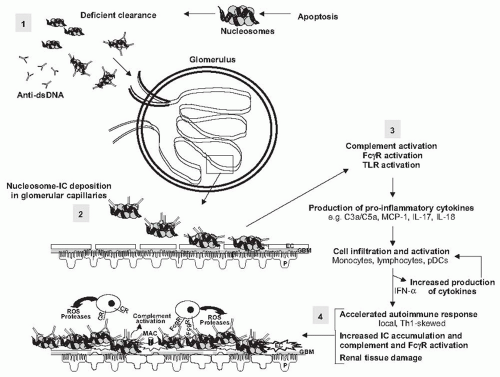
FIGURE 53.1 A paradigm for lupus nephritis pathogenesis. The onset of lupus nephritis likely begins with initial accumulation of self-antigen and immune complexes. In the model presented in the figure, the self-antigens are nucleosomes that can persist due to deficient clearance or overwhelming production, and which in turn can drive anti-dsDNA production (step 1, in shaded box). The resulting immune complexes (IC) are prone to deposit in the renal vascular beds (step 2), in part due to the positively charged core histones of the nucleosome. Once deposited, the IC can activate the complement system, activate circulatory leukocytes via expressed FcγR, and activate resident cells expressing TLRs (step 3). This establishes a cascade of inflammatory cytokine and chemokine production that recruits and activates inflammatory cells, lymphocytes, and pDCs. These infiltrating cells further amplify the production of cytokines and chemokines in the kidney microenvironment. The result is a locally driven and accelerated autoimmune response with Th1 characteristics, and increased IC accumulation and accompanying complement and FcγR activation. This response culminates in the production of inflammatory mediators of tissue damage (step 4). One immediate consequence is destruction of the glomerular filtration barrier through the damaging effects on glomerular endothelial cells (EC), glomerular basement membrane (GBM), and podocytes (P), which leads to proteinuria and hematuria, the hallmark clinical manifestations of active lupus nephritis. CR, complement receptor; MAC, complement membrane attack complex.


DIAGNOSIS OF LUPUS NEPHRITIS

Preservation of kidney function in patients with LN is best achieved with early diagnosis and treatment.108,109,110 This requires a high index of suspicion for renal involvement in all patients with SLE. Although some patients may present with overt clinical signs of renal disease, such as edema secondary to nephrotic syndrome, or severe hypertension, it is more likely that the initial evidence of kidney involvement will be an abnormality of serum creatinine and/or the urinalysis. An approach for evaluating the SLE patient for kidney involvement is presented in Figure 53.2. Considering serum creatinine, it is important to recognize that a normal range value may be abnormally high for a woman with small-moderate muscle mass and low rates of creatinine production. Also, hypoalbuminemic patients with severe nephrotic syndrome may have increased tubular creatinine secretion, lowering serum creatinine, and leading to an impression of better renal function than in actuality.111 Finally, in addition to LN, SLE patients may develop acute renal insufficiency because
of infection, medications, nephrotoxins, hemolysis, thrombosis, and cardiac failure.


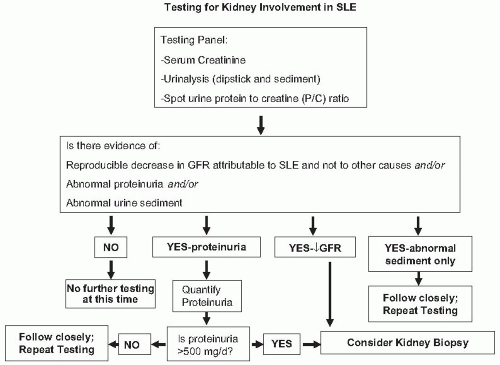
FIGURE 53.2 An algorithm for the evaluation of the kidney in patients with systemic lupus nephritis. Note that patients with a history of lupus nephritis and previous kidney biopsy may not need a repeat biopsy (see text). Kidney biopsy should be done for all new diagnoses of kidney involvement.

Urinalysis is a useful screening test for patients with SLE. A urine dipstick positive for blood and/or protein suggests possible LN; however, a systematic study of the accuracy of the urine dipstick as a screening tool found a false-negative rate in up to 30% of SLE patients and a falsepositive rate in about 40% of patients.112 Therefore the urine sediment should be evaluated for evidence of glomerulonephritis. Glomerular bleeding is suggested by acanthocytes and/or red blood cell (RBC) casts. White blood cells (WBCs) and white blood cell casts in the absence of infection are indicative of renal inflammation, and support a diagnosis of glomerulonephritis.

Proteinuria is a key indicator of kidney injury in SLE. It has prognostic importance because proteinuria may injure the kidney, and it is used as a clinical biomarker of relapse, remission, and successful treatment. Therefore accurate measurement of protein excretion is crucial to the ongoing management of LN.

Random spot urine protein-to-creatinine (P/C) ratios can be used in addition to urine dipsticks to screen patients, but are not accurate enough to be used to make therapeutic decisions or to follow changes in proteinuria magnitude in response to therapy. The most reliable method to quantify proteinuria is to measure the P/C ratio of a 24-hour urine collection, or an intended 24-hour collection that is at least 50% complete.113 Measuring the P/C ratio reduces confounding the assessment of proteinuria by errors in collecting the 24-hour urine. A 12-hour overnight urine collection that includes the first morning void urine also provides an accurate measure of proteinuria magnitude, and may be easier for patients to collect.114

Ultimately a kidney biopsy is essential for the optimal diagnosis and management of most cases of LN. A biopsy is not necessarily required if the only abnormalities are isolated hematuria, or low level proteinuria in the absence of hematuria and an active urine sediment. A biopsy should be considered when proteinuria is above 500 mg per day, as this degree of proteinuria has been associated with significant renal injury.115,116,117

There is some controversy surrounding the utility of kidney biopsies in LN. The main argument against biopsy is a prevalent notion that most patients can be treated with mycophenolate mofetil (see later), and biopsy information would not change the approach to therapy.118 There are, however, several important reasons to obtain a biopsy:


  • Not all kidney disease in SLE patients is classic, IC-mediated glomerulonephritis (LN), so one therapy does not fit all patients. For example non-LN glomerular diseases have been reported in SLE patients.119,120,121 This literature is mostly case reports, but in a series of 252 patients, 5% were found to have changes consistent
    with focal segmental glomerulosclerosis, minimal change disease, thin glomerular basement membrane disease, hypertensive nephrosclerosis, and amyloidosis.119 The incidence of podocytopathies in lupus patients appears to be greater than in the general population, suggesting a causal link to the immune dysregulation of SLE.122,123 Amyloid A (AA) amyloidosis has also been reported frequently in some series.120,121 Finally, there are other important kidney lesions found in SLE patients that are treated differently than LN, such as interstitial nephritis without glomerulonephritis121 and thrombotic microangiopathy with or without LN.124,125


  • The kidney biopsy, especially if performed serially, assesses the degree of chronic kidney injury, and therefore the risk of progressive renal failure that is not related to active LN. If extensive scarring is the dominant process found on biopsy even with some areas of active inflammation, the risk of immunosuppression may outweigh its benefits in terms of renal survival. Such patients may be more appropriately treated with kidney-protective therapies alone.


  • In the context of LN therapeutics, kidney biopsies can and should be exploited in novel ways to better inform future drug development. For example, leukocyte subsets can be analyzed by specific staining in lupus kidneys and may yield new insights on renal inflammation.126 Proteomic techniques can be used to look for patterns of protein expression in LN.127,128 Gene expression in biopsies can be analyzed with microarray techniques.128,129 These technologies are just being applied to kidney biopsies, but have the potential to greatly enhance the amount of information available from renal tissue.


KIDNEY PATHOLOGY IN SYSTEMIC LUPUS ERYTHEMATOSUS

Although the gold standard for the exact diagnosis and classification of LN is the renal biopsy, it should be emphasized that LN is not a renal biopsy diagnosis. Renal biopsy changes, although characteristic, are not specific and the diagnosis of LN cannot be made unless the patient fulfills the American College of Rheumatology criteria for SLE. In the absence of a concurrent clinical diagnosis of SLE, only a diagnosis of immune complex glomerulonephritis can be made, with the suggestion that the glomerulonephritis, in the appropriate clinical setting, could be associated with SLE.

The clinical utility of the kidney biopsy depends on obtaining an adequate sample of renal cortex (at least 10 glomeruli) and examination by a renal pathologist.130 In as much as every biopsy is a clinicopathologic correlation, the nephropathologist must be given all relevant clinical information in order to properly interpret the tissue and integrate the microscopic findings with the clinical data. Furthermore, it is essential that the clinician and pathologist review the findings together before initiation of therapy to ensure that specific clinical concerns have been addressed and that the lesions have been contextualized appropriately.

The first renal biopsy of a patient with LN, although important diagnostically and therapeutically, has somewhat limited prognostic value because most of the active lesions are reversible with treatment. However, a follow-up biopsy performed after several months or years may provide important prognostic information.131,132,133 If the degree of chronic injury in the follow-up biopsy does not change substantially, and the patient had a good response to treatment, outcome is likely to be favorable. In contrast, if the degree of chronic injury is substantially more prominent in a follow-up biopsy, a progressive decline in the disease course can be anticipated.


Classification Schemes for Lupus Nephritis

Renal biopsy findings in LN involve the entire spectrum of renal pathology. Therefore, it became necessary to develop a pathologic classification of LN. A first attempt was made in 1974 by a group of pathologists under the auspices of the World Health Organization (WHO), and was later designated as the WHO classification. This was further modified in 1982 and 1995.134 The original WHO classification was relatively simple, with five classes of LN (Table 53.1). Subsequent modifications made the WHO classification more complicated and cumbersome to use, leading a group of nephrologists and pathologists to develop a new classification of LN (Table 53.1) in 2003 under the auspices of the International Society of Nephrology (ISN) and the Renal Pathology Society (RPS).135

Similar to the previous WHO classification, the ISN/RPS classification is based primarily on characteristic light microscopic patterns of glomerular injury:


  • Mesangial hypercellularity. Mesangial hypercellularity is almost always present in LN, except in Class I (Fig. 53.3), and is the basic, and probably the earliest LN lesion which is later combined with other pathologic patterns of injury.


  • Endocapillary hypercellularity. Endocapillary hypercellularity is the hallmark lesion in forms of proliferative LN (Figs. 53.4 and 53.5). Intracapillary cells usually are infiltrating inflammatory cells (including monocytes/macrophages, polymorphonuclear leukocytes, lymphocytes, and rarely eosinophils or basophils). There may also be a component of endothelial cell proliferation.


  • Extracapillary hypercellularity. Extracapillary proliferation results in crescent formation (Fig. 53.6), and is common in proliferative forms of LN. It is frequently associated with glomerular capillary rupture, Bowman’s capsular basement membrane rupture, fibrin in Bowman’s space, and fibrinoid necrosis of the glomerular capillary tuft.


  • Karyorrhexis with or without associated fibrinoid necrosis of the glomerular capillary tuft (Figs. 53.7 and 53.8). Karyorrhexis in glomeruli usually reflects apoptosis, a common finding in LN. The apoptotic cells may be infiltrating inflammatory cells or native glomerular cells. Hematoxylin bodies (Fig. 53.9), seen occasionally in

    biopsies, most likely represent a tissue equivalent of the LE cell phenomenon.


  • Wire loop lesions. These lesions are due to large subendothelial immune complex deposits, visible even with light microscopy (Fig. 53.10). If these subendothelial deposits are large enough, they may occlude the entire glomerular capillary lumen and appear as “hyalin thrombi” (Figs. 53.7 and 53.10). Wire loop lesions are positive for periodic acid-Schiff (PAS), negative with methenamine silver stain, and red with Masson’s trichrome stain. Wire loop lesions are much more common in LN with global glomerular hypercellularity than with biopsies showing mainly segmental hypercellularity and/or necrosis.


  • Spikes. Diffuse uniform glomerular capillary loop thickening with “spike” formation on methenamine silver stain (Figs. 53.11 and 53.12) is the main light microscopic pattern of injury if the immune complex deposits are subepithelial in membranous lupus nephritis.


TABLE 53.1 Classification of Lupus Nephritis


Original World Health Organization Classification


Simplified ISN/RPS Classification


Class I


Normal: No pathologic findings, no glomerular IC


Minimal mesangial LN: Mesangial IC


Class II


Mesangial LN: Mesangial IC, normal or hypercellular mesangium


Mesangial proliferative LN: Mesangial IC, hypercellular mesangium


Class III


Focal LN (<50% of glomeruli) Glomerular lesions mainly segmental


Focal LN (<50% of glomeruli)


– III (A): active lesions


– III (A/C): active and chronic lesions


– III (C): chronic lesions


Class IV


Diffuse LN (>50% of glomeruli) Glomerular lesions mainly global


Diffuse LN (≥50% of glomeruli involved, lesions may be segmental [S] or global [G])


– IV (A): active lesions


IV-S(A); IV-G(A)


– IV (A/C): active and chronic lesions


– IV-S(A/C); IV-G(A/C)


– IV (C): chronic lesions


IV-S(C); (IV-G(C)


Class V


Membranous LN


Membranous LN


Class VI


Advanced sclerosing LN


IC, immune complex; LN, lupus nephritis.


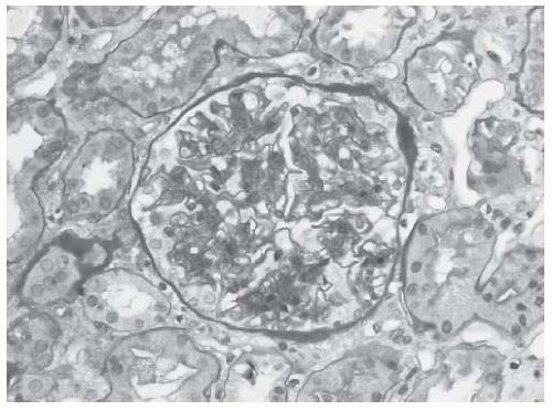
FIGURE 53.3 Mesangial hypercellularity in a case of class II lupus nephritis. Note that the glomerular capillaries are patent. (Periodic acid-Schiff [PAS] X400.)


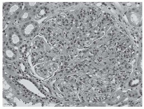
FIGURE 53.4 Global endocapillary hypercellularity with obliteration of the glomerular capillaries in a case of class IV lupus nephritis. The hypercellularity is the result of infiltrating inflammatory cells, including occasional polymorphonuclear leukocytes, as well as proliferating glomerular cells, including endothelial cells and mesangial cells. (Hematoxylin and eosin [H&E] X400.)


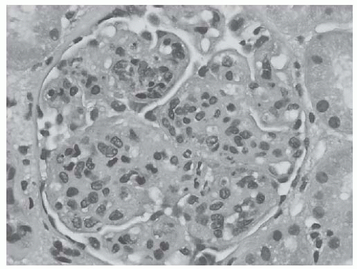
FIGURE 53.5 Global endocapillary hypercellularity with accented lobularization of the glomerular capillary tuft, resembling a membranoproliferative glomerulonephritis in a case of class IV lupus nephritis. (H&E, X400.)


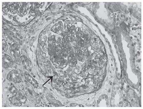
FIGURE 53.6 A cellular crescent in a case of class IV lupus nephritis. Note the compressed glomerular capillary tuft and the rupture in the Bowman’s capsule (arrow). (PAS X400.)


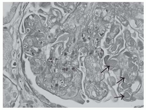
FIGURE 53.7 Apoptotic debris (karyorrhectic nuclei) in the glomerular capillaries in a case of class IV lupus nephritis. In this glomerulus, large subendothelial deposits (“wire loop” lesions) and intracapillary hyalin thrombi (arrows) are also present. (PAS X600.)

The ISN/RPS classification (Table 53.1) retained the main subclasses of the modified WHO classification, but introduced several modifications: The ISN/RPS classification
differentiates active (A) and chronic (C), and segmental (S) and global (G) glomerular lesions. Active glomerular lesions include glomerular endocapillary hypercellularity with or without leukocyte infiltration and with substantial luminal reduction, karyorrhexis, fibrinoid necrosis, rupture of the glomerular basement membrane, cellular or fibrocellular crescents, wire loop lesions, and large intraluminal immune complexes (hyalin thrombi) (Figs. 53.4,53.5,53.6,53.7,53.8 and 53.10). Chronic lesions include glomerular sclerosis (segmental or global), fibrous adhesions, and fibrous crescents (Figs. 53.13,53.14,53.15). Segmental lesions involve less than half of the glomerular capillary tuft area; global lesions involve more than 50% of the glomerular capillary tuft area.


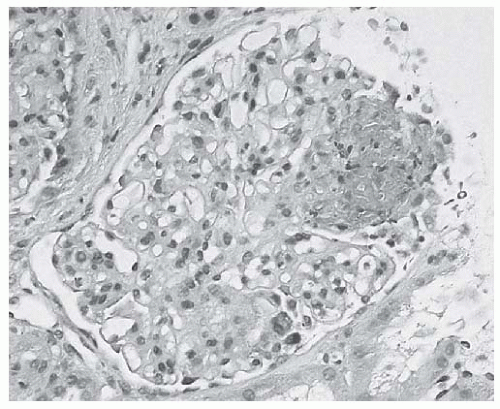
FIGURE 53.8 Segmental glomerular capillary tuft necrosis associated with karyorrhectic/apoptotic debris in a case of focal lupus nephritis. (H&E X400.)


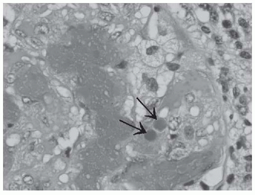
FIGURE 53.9 Hematoxylin bodies in a glomerular capillary (arrows) in a case of active class IV lupus nephritis. (H&E X1000.)


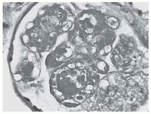
FIGURE 53.10 Large PAS positive deposits along the glomerular capillary loops (“wire loop” lesions) as well as extensive mesangial deposits and glomerular capillary hyalin thrombi in a case of class IV lupus nephritis. (PAS X600.)


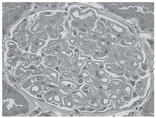
FIGURE 53.11 Diffuse uniform glomerular capillary thickening without hypercellularity in a case of membranous class V lupus nephritis. (H&E X400.)


Class I: Minimal Mesangial Lupus Nephritis

In class I LN, the glomeruli appear entirely normal by light microscopy. However, immunofluorescence and electron microscopy reveal obvious mesangial immune complex deposits (Fig. 53.16).


Class II: Mesangial Proliferative Lupus Nephritis

In class II LN, there is pure mesangial hypercellularity (Fig. 53.3) without glomerular endocapillary hypercellularity or crescents. Immunofluorescence and electron microscopy reveal mesangial deposits (Figs. 53.17 and 53.18) as in class I LN. By electron microscopy a few isolated glomerular capillary deposits may be seen. If many peripheral
glomerular capillary immune complex deposits are present, the diagnosis of class II LN should not be made.


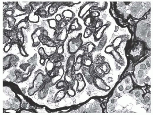
FIGURE 53.12 Methenamine silver stain reveals extensive spike formation along the glomerular capillary loops in the same biopsy shown in Figure 53.11. (Jones’ methenamine silver X600.)


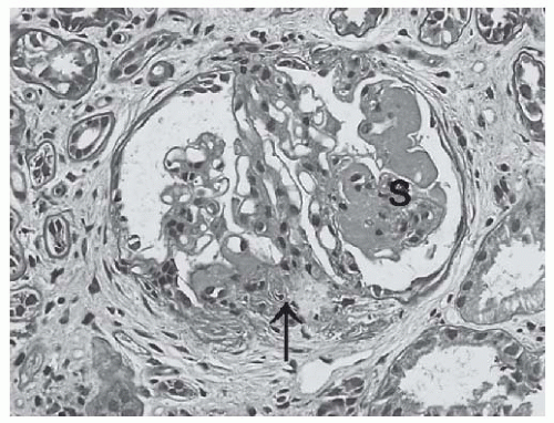
FIGURE 53.13 Segmental sclerosis (S) and glomerular capillary adhesion (arrow) in a glomerulus from a biopsy with class III lupus nephritis. (PAS X400.)


Class III: Focal Lupus Nephritis

In class III LN, obvious endocapillary or extracapillary (crescents) proliferative lesions are seen (Figs. 53.7,53.8, and 53.19), but in less than 50% of all glomeruli, including sclerotic glomeruli, which are also taken into account. Glomerular lesions in focal LN are almost always segmental (Fig. 53.8). By immunofluorescence and electron microscopy, abundant mesangial immune complex deposits are seen, usually associated with segmental glomerular capillary deposits (Fig. 53.20). There are three possible subclasses of focal LN.


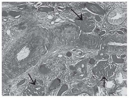
FIGURE 53.14 Globally sclerotic glomeruli (arrows) in a biopsy with advanced sclerosing (class VI) lupus nephritis. (PAS X200.)


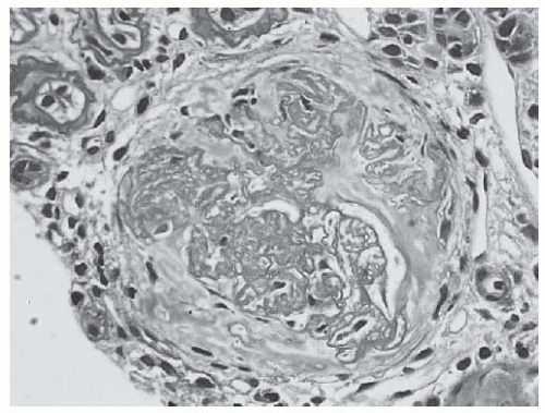
FIGURE 53.15 A fibrous crescent from biopsy with class IV lupus nephritis with moderate to advanced chronicity and mild activity. Note the disrupted Bowman’s capsule and the separation of sclerosing glomerular lobules by faintly PAS positive interstitial type collagen. (PAS X400.)


  • In class III (A) there are only active lesions (focal proliferative LN).


  • In class III (A/C) both active and chronic lesions are present (focal proliferative and sclerosing LN). In such cases, focal or segmental sclerosing glomeruli coexist with glomeruli with active proliferative/necrotizing lesions.


  • In class III (C) only focal sclerosing glomerular lesions are noted with glomerular scars and segmental or global sclerosis (focal sclerosing LN). Active lesions are not seen.


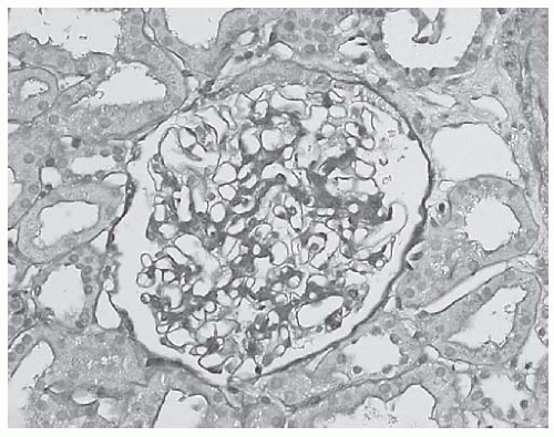
FIGURE 53.16 A light microscopically unremarkable glomerulus in a biopsy with class I lupus nephritis. Immunofluorescence and electron microscopy revealed mesangial immune complex deposits. (PAS X400.)


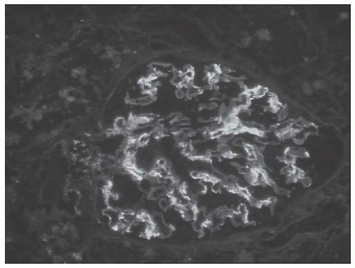
FIGURE 53.17 Mesangial immune complex deposits in a case of class II lupus nephritis. (Direct immunofluorescence with an antibody to IgA, X400.)


Class IV: Diffuse Lupus Nephritis

In this class of LN, segmental or global endo- or extracapillary glomerular proliferative lesions are seen in more than 50% of all glomeruli (Figs. 53.4,53.5,53.6,53.7,53.8). Large subendothelial deposits, visible under the light microscope (wire loop lesions) (Figs. 53.7 and 53.10), are common. In class IV LN, the glomerular lesions can be global or segmental. Also, active and chronic glomerular lesions are evaluated separately. Immunofluorescence and electron microscopy reveal abundant glomerular mesangial and capillary loop deposits. The glomerular capillary loop deposits are mainly subendothelial, and frequently quite large (Figs. 53.21 and 53.22). Scattered intramembranous and subepithelial deposits are common. Therefore, there are six possible subclasses of diffuse LN.


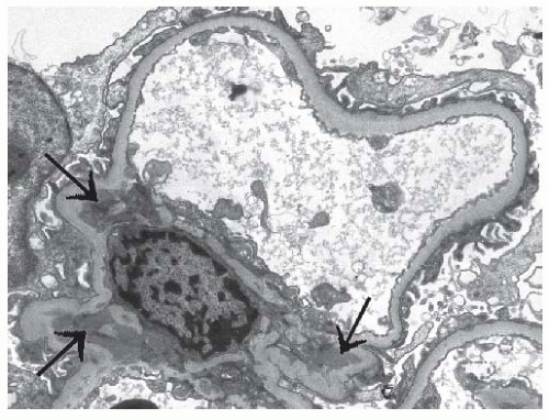
FIGURE 53.18 Mesangial electron dense immune type deposits (arrows) in a case of class II lupus nephritis. (Uranyl acetate, lead citrate X8000.)


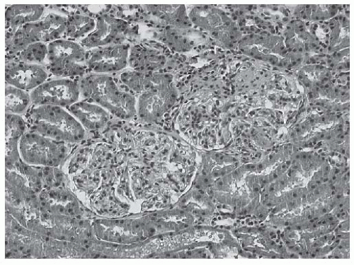
FIGURE 53.19 Two glomeruli from a biopsy with class III lupus nephritis. Note that the left lower glomerulus is light microscopically unremarkable whereas the right upper glomerulus reveals segmental proliferative lesions. (H&E, X200.)


  • Class IV-S(A) indicates active diffuse segmental endocapillary or extracapillary proliferative glomerular lesion or necrosis involving more than 50% of the glomeruli.


  • Class IV-G(A) shows diffuse global LN with active endocapillary or extracapillary proliferative glomerular
    lesions and/or necrosis involving more than 50% of glomeruli.


  • Class IV-S(A/C) indicates diffuse segmental proliferative and sclerosing LN. In such biopsies, active segmental proliferative lesions coexist with chronic sclerosing glomerular lesions.


  • Class IV-G(A/C) indicates diffuse global proliferative and sclerosing LN. These biopsies show active global proliferative lesions with chronic sclerosing glomerular lesions.


  • Class IV-S(C) indicates diffuse segmental sclerosing LN. In this subclass, no active lesions are present; only inactive, mainly segmental glomerular lesions are seen, such as segmental sclerosis/scarring.


  • Class IV-G(C) shows diffuse global sclerosing LN. In such biopsies, glomeruli reveal global sclerosis or scarring with or without fibrous crescents, involving more than 50% of all glomeruli, in the absence of active proliferative lesions.


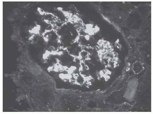
FIGURE 53.20 Granular mesangial and segmental glomerular capillary loop deposits in a case of class III lupus nephritis. Also note the subtle granular tubulointerstitial staining. (Direct immunofluorescence with an antibody to IgG, X400.)


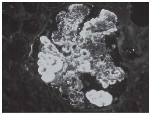
FIGURE 53.21 Diffuse granular glomerular deposits with large subendothelial deposits (“wire loop” lesions) in a case of class IV lupus nephritis. (Direct immunofluorescence with an antibody to IgG, X400.)


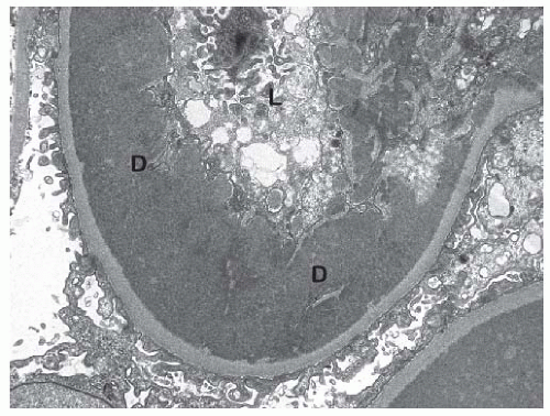
FIGURE 53.22 This electron micrograph shows a large subendothelial electron dense deposit (d) in the same biopsy shown in Figure 53.21. L, glomerular capillary lumen. (Uranyl acetate, lead citrate, X8,000.)


Class V: Membranous Lupus Nephritis

In class V LN the glomeruli do not reveal endocapillary hypercellularity; the mesangium may be normocellular or hypercellular. The glomerular capillaries are uniformly and diffusely thickened (Fig. 53.11), except in very early stages of the disease. Spike formation on methenamine silver stain is common, just like in idiopathic membranous glomerulonephritis (Fig. 53.12). Glomerular subepithelial immune complex deposits involve over 50% of the glomerular capillary tufts (Figs. 53.23 and 53.24). In contrast to idiopathic membranous glomerulonephritis, in class V LN the immunofluorescence frequently, shows a “full house” pattern (see later text), and the IgG deposits contain mainly IgG1 and IGg3 as opposed to IgG2 and IgG4 (see later). However, we encountered several cases of class V LN with IgG4 predominant glomerular capillary deposits. Mesangial immune complex deposits are almost invariably present. A few small subendothelial deposits are possible. Electron microscopy usually reveals endothelial tubuloreticular inclusions (TRIs) (Fig. 53.25).


Class VI: Advanced Sclerosing Lupus Nephritis

In class VI LN over 90% of the glomeruli are globally sclerosed without residual activity (Fig. 53.14). There has to be clinical or morphologic evidence that the advanced glomerular
sclerosis is secondary to LN. Immunofluorescence and electron microscopy still frequently reveal mild glomerular immune complex deposits in the few nonsclerotic glomeruli.


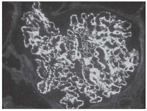
FIGURE 53.23 Granular mesangial and glomerular capillary fluorescence with an antibody to IgG in a case of membranous (class V) lupus nephritis. Note that over 50% of the glomerular capillaries contain granular deposits. (Direct immunofluorescence, X400.)


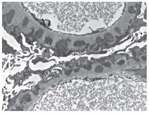
FIGURE 53.24 Subepithelial electron dense immune type deposits along the glomerular basement membrane in a case of class V (membranous) lupus nephritis. Note that occasional deposits are already completely incorporated into the glomerular basement membrane. (Uranyl acetate, lead citrate X15,000.)
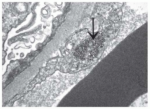

Controversies with the ISN/RPS Classification

Although several follow-up studies emphasize the benefits of the ISN/RPS classification of LN,136,137 not all investigators share this enthusiasm.138,139 The classification is based purely on morphologic findings and arbitrary definitions. For example, the classification of proliferative LN into focal and diffuse forms is based on an arbitrary cut off value of 50% glomerular involvement. It is hard to imagine that a patient with LN and 40% glomerular involvement would be treated and respond differently than a patient with 60% glomerular involvement.


Get Clinical Tree app for offline access