Alport Syndrome, Fabry Disease, and Nail-Patella Syndrome


Alport Syndrome, Fabry Disease, and Nail-Patella Syndrome


Godela M. Brosnahan

Martin Gregory

Imed Halal

Berenice Y. Reed


Hereditary glomerular diseases have diverse and often covert patterns of inheritance and varied clinical presentations. They are more common than is generally appreciated. Vigilance and awareness of these subtleties are important for prompt and accurate diagnoses, which allow for optimal management and timely genetic counseling.


ALPORT SYNDROME

Alport syndrome (AS), formerly called hereditary nephritis, is a disorder characterized by hematuria, proteinuria, progressive renal failure, variable sensorineural hearing loss, and ocular abnormalities. On a histologic examination, irregularities of the glomerular basement membrane (GBM) constitute the primary disease feature. Cecil Alport’s archetypal kindred1 had dominantly inherited kidney disease that was characterized in both sexes by hematuria and urinary erythrocyte casts, variable proteinuria, and by hearing loss and renal failure in males. Affected males died in adolescence of uremia, whereas females lived to old age.


The Present Definition of Alport Syndrome and the Molecular Defect Definition

For this chapter, AS will be defined as progressive, hereditary, hematuric, nonimmune glomerulonephritis that is characterized ultrastructurally by progressive irregular thickening, thinning, and lamellation of the GBM and genetically by a mutation in COL4A3, COL4A4, or COL4A5.


The Genetic Classification of Alport Syndrome

Between 2% and 5% of males with end-stage renal disease (ESRD) and probably less than 1% of females have AS. Various estimates place the gene frequency between 1:5,000 and 1:53,000, respectively.2 The true prevalence may be higher because affected patients with subtle hearing loss are easily overlooked. There are three main genetic types of AS: X-linked Alport syndrome (XL-AS), autosomal recessive Alport syndrome (AR-AS), and autosomal dominant Alport syndrome (AD-AS), which are classified based on the mode of inheritance as described in Table 17.1. The severity of symptoms varies from person to person and with age and gender. Large kindreds show modes of inheritance and kindred-specific phenotypes that clearly reflect the genetic heterogeneity of AS. Thus, although the demonstration of a family history of glomerulonephritis among affected persons within a kindred may be helpful to ascertain, demonstration of a causative mutation remains the gold standard for genetic diagnosis.


X-Linked Alport Syndrome Caused by a Mutation in COL4A5 (OMIM 301050)3

XL-AS is the most common form of AS, accounting for 80% of cases. XL-AS results from a mutation in the COL4A5 gene located at Xq22.3, which encodes the α5 chain of type IV collagen (α5[IV]). Hematuria from birth occurs in 100% of hemizygous males4,5,6 and 90% to 100% of heterozygous female carriers of XL-AS.5,7,8 Disease expression is phenotypically very heterogeneous. ESRD is inevitable in males but occurs at widely different ages in different families. The age of ESRD tends to run true within a family, but even within a family there can be quite wide variability. Knowing the mean age of ESRD in males in a family is useful from a prognostic standpoint. Moreover, extrarenal manifestations, such as hearing loss and ocular defects, tend to occur more commonly, more severely, and at an earlier age in kindreds whose males develop ESRD early.4

Because they are late symptoms, it is unwise to equate chronic renal failure and ESRD with Alport gene penetrance. We define penetrance of ESRD as the fraction of a population at risk in whom ESRD eventually develops. For males in each Alport kindred, ESRD penetrance generally coincides with hematuria penetrance. In Figures 17.1 and 17.2, representations of the probability of ESRD and hearing loss in boys and girls with XL-AS are shown. Inactivation of the X chromosome likely explains both incomplete hematuria penetrance among females heterozygous for XL-AS and their low probability of ESRD and hearing loss.9 Jais et al.7 found a cumulative prevalence of ESRD of 12% in female carriers of XL-AS by the age of 40 years.


TABLE 17.1 Genetic Classification of Alport Syndrome


Description


OMIM


Gene


Comment


X-Linked Alport Syndrome


301050


COL4A5


Most common form of Alport syndrome accounting for 80% of cases. A heterogeneous condition with progressive renal insufficiency and timing of ESRD occurring between childhood and adult. Hearing loss and eye problems are common but severity is variable. More severe phenotype in affected males.


COL4A5 and Contiguous Gene Defects


Alport Syndrome with Diffuse Leiomyomatosis


308940


COL4A5-COL4A6


Alport syndrome with diffuse leiomyomatosis.


Alport Syndrome, Mental Retardation, Midface Hypoplasia, Elliptocytosis (AMME)


300194/95


COL4A5 with FACL4/AMMECRI


Contiguous gene deletion affecting genes located 3′ of the COL4A5 gene.


Autosomal Recessive Alport Syndrome (ARAS)


203780


COL4A3/COL4A4


This form of Alport syndrome accounts for 15% of cases. Most cases result in the early onset of ESRD. Hearing and eye problems are common, with both males and females being equally affected.


Autosomal Dominant Alport Syndrome (ADAS) without Hematologic Defects


104200


COL4A3 or COL4A4


A milder disease with the later onset of renal impairment, and a lower incidence of hearing loss and eye problems.


Familial Thin Basement Membrane Disease and Benign Familial Hematuria


141200


COL4A3 or COL4A4


A milder disease, with no hearing loss or occurrence of eye problems.


One consequence of X linkage is that twice as many females as males with a nephritis gene will be born if there are no prenatal effects and if reproductive fitness is independent of gender of the gene-carrying parent. In kindreds with XL-AS and early onset of ESRD, most affected children obtain the gene from their mothers, and the sex ratio of genecarrying newborns approaches 1:1.


X-Linked Alport Syndrome Caused by a Deletion in COL4A5 with Damage to Contiguous Genes (OMIM 308940 and 300194/5)

In this form, AS is associated with other features caused by an extension of the deletion outside COL4A5. Alport syndrome with diffuse leiomyomatosis (smooth muscle tumors) (Online Mendelian Inheritance in Man [OMIM] 308940)3 stems from a deletion embracing the 5′ ends of COL4A5 and COL4A6.2,10 The AMME syndrome (OMIM 300194/5)3 consists of AS, midface hypoplasia, mental retardation, and elliptocytosis and has been described in several families with deletions of COL4A5 that extend beyond the 3′ end of the gene.11,12,13,14


Autosomal-Recessive Alport Syndrome Caused by Homozygous Mutations Mutations in COL4A3 or COL4A4 (AR-AS, OMIM 203780)

AR-AS is allelic with familial thin basement membrane nephropathy (TBMN). This form of AS accounts for 15% of

AS cases. AR-AS results from mutations affecting both alleles of the COL4A3 gene or the COL4A4 gene. To date, most examples of AR-AS syndrome have resulted in early ESRD, but this may reflect ascertainment bias.15,16,17 Males and females are equally severely affected, and hearing and ocular defects are usual. It is not yet clear whether all AR-AS or all TBMN is caused by mutations of these genes.


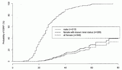
FIGURE 17.1 Probability of end-stage renal disease (ESRD) in 315 boys and men and 288 girls and women with the COL4A5 mutation. In the third curve, girls and women with incomplete clinical data were added because they were not in ESRD at last follow-up. (Reprinted from Jais JP, Knebelmann B, Giatras I, et al. J Am Soc Nephrol. 14:2603-2610;2003 with permission from the publisher, American Society of Nephrology.)


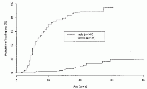
FIGURE 17.2 Probability of hearing loss in 144 boys and men and 151 girls and women with the COL4A5 mutation. (Reprinted from Jais JP, Knebelmann B, Giatras I, et al. J Am Soc Nephrol. 14:2603-2610;2003 with permission from the publisher, American Society of Nephrology.)


Autosomal-Dominant Alport Syndrome without Hematologic Defects (AD-AS, OMIM 104200)

AD-AS is a relatively rare genetic form of AS. It is linked with a heterozygous mutation of either the COL4A3 gene or the COL4A4 gene. Only a few families have been described to date. One well described family had relatively mild renal impairment and no hearing loss, eye signs, platelet abnormalities, or leiomyomatosis.18 The mutation in this family is a splice site mutation in COL4A3.19 In a study of eight families with heterozygous mutations affecting the COL4A4 gene none had ocular problems, and a low incidence of hearing anomalies was observed.20 Loss of renal function occurred at a later age than the X-linked form.


Familial Thin Basement Membrane Nephropathy or Benign Familial Hematuria (OMIM 141200)

In 1997, Lemmink et al.21 linked the occurrence of heterozygous mutations in the COL4A3 and COL4A4 genes with familial TBMN. These conditions are associated with the maintenance of long-term normal renal function and the absence of hearing loss or ocular problems. More recently, Pierides et al.22 described 11 large pedigrees in which heterozygous COL4A3/COL4A4 mutations were associated with microscopic hematuria before the age of 30 and late development of proteinuria and ESRD due to focal segmental glomerulosclerosis.


Genotype-Phenotype Correlation in Alport Syndrome COL4A5 Mutations

XL-AS is both clinically and genetically heterogeneous. To date, more than 590 mutations and potential mutations have been described in the COL4A5 gene associated with XL-AS.23,24 Among affected male patients, the age at ESRD typically ranges between the second and third decades of life. However, in milder cases, ESRD may be delayed until the fifth or sixth decade. Similarly, deafness occurs at variable ages and a wide variety of ocular abnormalities have been reported among patients.2 Several large studies from Europe, the United States, and China have assessed a genotypephenotype correlation in XL-AS.2,4,25,26,27 The COL4A5 mutation type appears to be one factor associated with renal disease severity and extrarenal manifestations. In male-affected patients large deletions, non-sense, and frame shift mutations have been associated with more severe disease manifestations, such as earlier age at ESRD onset, hearing loss, and the occurrence of eye abnormalities compared to patients with missense mutations.4,25,26 Early renal failure and retinopathy have also been reported to associate with the occurrence of certain specific mutations, including some missense mutations.28 The position of the mutation and the affected domain of collagen α5(IV) also appears to affect disease severity. An earlier age at onset of ESRD was shown to associate with a more 5′ gene location in one study.26 The distance of the mutation from the NC1-domain was shown to affect severity.25 Glycine substitutions occurring in exons 1 through 20 resulted in a less severe phenotype compared to those affecting exons 21 through 47.25 This effect was attributed to the fact that the triple helix formation starts at the C-terminal of the NC1-domain and proceeds in a zipperlike manner to the N-terminal end.29 Similar studies in affected women and girls have failed to demonstrate any genotype-phenotype correlation.7 However, it should be stated that phenotypic variability is also common among affected family members of the same gender who carry the same germline mutation. This intrafamilial variability may be attributed to both the environment and the effect of other genes.


COL4A3 and COL\4A4 Mutations

To date, 71 mutations or potential disease-associated sequence variants have been described in the COL4A3 gene, and 56 have been described in the COL4A4 gene.23 Although ARAS associated with homozygous mutation in either COL4A3 or COL4A4 results in the early onset of ESRD with typical hearing loss and eye problems, heterozygous mutations in the COL4A3/COL4A4 genes are associated with the less severe phenotype associated with benign familial hematuria and familial TBMN. Due to the innate genetic complexity of these disorders, not surprisingly, no genotype-phenotype correlation has been described.

Clinical variability in disease expression has been described for both male and female patients with AD-AS who carry a heterozygous mutation in either COL4A3 or COL4A4. However, no significant genotype-phenotype correlations have been described in AD-AS.20 This may relate to the small number of families that have been identified with this genetic form of AS.


Pathogenesis

Type IV collagen, a major constituent of basement membranes, is comprised of six chains: α1(IV) through α6(IV). The type IV collagen chains assemble into three different heterotrimers in the mammalian basement membrane: α112345; or α556, respectively. The α345 form is synthesized by the podocytes in the glomerulus, and this heterotrimer of type IV collagen is also the predominant form of collagen found in the basement membrane of the ears, eyes, and lungs.30 Mutation affecting any of the corresponding genes—namely, COL4A3, COL4A4, or COL4A5—will have a consequential effect on the integrity of the type IV collagen. In the GBM in Alport syndrome, the normal α345 collagen network is replaced by the fetal α112 network, which
is less resistant to degradation, thus resulting in the gradual deterioration of the GBM and in the clinical features of AS.31


Pathology


Kidney

There are no pathognomonic lesions seen with light microscopy in AS.32,33 Lipid-laden interstitial foam cells are seen in the cortex of some but not all biopsy specimens. Foam cells are typically absent from biopsies taken early in the disease process.34,35 Direct immunofluorescence is initially negative, whereas a faint deposition of immunoglobulin (Ig)G, IgM, and/or C3 may be observed with the progression of the glomerular segmental lesions.36 The most definitive diagnostic information is provided by electron microscopy (EM) and by differential immunostaining for collagen α(IV) chains with specific monoclonal antibodies, which is discussed further under immunopathology.

Electron microscopy. The classic ultrastructural lesion of AS is characterized by an irregular thinning and thickening of the GBM, splitting and lamellation of the GBM with the loss of the normal lamina densa, small granules within the GBM, and an irregular outer and inner contour of the GBM (Fig. 17.3).36 Distortion of the lamina densa may be extreme at times, amounting to a basket weave appearance in which the lamellae branch and rejoin in a complex triangle.37 Focal or diffuse foot process fusion is common. The ultrastructural changes are common to all variants of AS. However, the extent of GBM thickening and lamellation is gender and age dependent. Moreover, there is considerable variability between affected individuals regarding the presence of these characteristic ultrastructural findings and even between affected individuals from the same family.36 It should be emphasized that GBM splitting with variable thickness and an irregular outer contour is not specific for AS; such changes may be seen with other renal injuries, as reviewed by Haas.36 Therefore, the combined use of EM and immunohistology for the detection of the collagen α3(IV), α4(IV), and α5(IV) chains increases the specificity for AS diagnosis. Use of these combined methods permits AS diagnosis in most cases.36


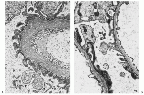
FIGURE 17.3 A: Glomerular ultrastructure in Alport syndrome. Electron micrograph of a renal biopsy specimen from a man in Utah kindred M, illustrating a widened lamina densa of the GBM (GBM). The lamina densa is split into several layers, between which may be seen numerous small electron-dense granules. B: Electron micrograph, at same magnification as (A), from an affected woman with familial thin GBM disease. The uniform thinning of the GBM can be appreciated by comparison with the width of the epithelial foot processes. (Electron micrographs courtesy of Dr. M.E. Hammond, University of Utah, Salt Lake City.)

Immunopathology. Staining for α3(IV), α4(IV), and α5(IV) collagen chains with monoclonal antibodies distinguishes between the various forms of AS including XL-AS, AR-AS, and TBMN as depicted in Table 17.2. In particular, the absence
of GBM staining for α3(IV) and α5(IV) is indicative of AS: either XL-AS in male subjects or AR-AS in both genders.36 It should be noted that in 15% to 20% of XL-AS kindreds, α3(IV) and α5(IV) staining remains normal. This may result from the presence of certain missense mutations that cause only minimal disruption to the collagen chain structure. Both α3(IV) and α5(IV) are normally expressed in TBMN. Readers are referred to a comprehensive overview of differential α(IV) staining patterns for differentiation of the various forms of AS and TBMN by Haas.36


TABLE 17.2 Staining for α3(IV) and α5(IV) in Thin Basement Membrane Nephropathy (TBMN) and Alport Syndrome Variants


α3(IV)


α5(IV)


GBM


BC


TBM


GBM


BC


TBM


EBM


Normal/TBMN


+


+


+


+


+


+


+


Alport variantsa


X-linked carrier (heterozygote)


Discont


Discont


Discont


Discont


Discont


Discont


Discont


X-linked male


b


Autosomal recessive


+


+


+


a Infrequent exceptions to these patterns have been noted on renal biopsies.

b Up to approximately 50% of individuals in each of these categories will show normal staining for α5(IV) on skin biopsy. GBM, glomerular basement membrane; BC, Bowman capsule; TBM, distal tubular basement membrane; EBM, epidermal basement membrane; Discont, discontinuous staining (mosaic pattern). Reproduced from Haas M. Arch Pathol Lab Med. 2009;133:224-232 with permission from the publisher American College of Pathologists.


Skin

Skin biopsies are far less invasive than renal biopsies and may provide useful diagnostic information based on immunofluorescence analysis in certain patients. A normal epidermal basement membrane (EBM) contains α5(IV) but not α3(IV) or α4(IV) chains. Thus, in AR-AS, α5(IV) is present in EBM.38 Absence of staining for α5(IV) is highly specific of XL-AS in male patients. A pronounced pattern of discontinuous α5(IV) staining is observed in some heterozygous carriers of XL-AS. However, it should be noted that a significant proportion of XL-AS patients, both male and female, retain a normal EBM staining of α5(IV).36 Thus, the diagnostic value of skin biopsies that demonstrate a positive staining for α5(IV) is limited. Moreover, skin biopsy staining cannot be used for the diagnosis of AR-AS as expression of α3(IV), and α4(IV) is normally absent in EBM.


Cochlea

The inner ear is much less amenable to a histopathologic study than the kidney. Specific lesions in AS include a zone of separation between the basilar membrane and the overlying basement membrane of the organ of Corti and the presence of cells filling the tunnel of Corti and the extracellular spaces of Nuel.39 Cellular infilling of the tunnel of Corti and spaces of Nuel likely represents a persistence of the fetal cochlear structure.39


Eye

In the anterior lenticonus, the basement membrane of the anterior lens capsule is thinned and more fibrillar than normal,40,41 allowing for an anterior bulging of the cortex of the lens, most prominently in the pupillary region. The collagen α3(IV), α4(IV), and α5(IV) chains may be present or lacking in the anterior lens capsule of AS patients.42

Another common ocular manifestation is perimacular “dot and fleck” retinopathy, which consists of whitish or yellowish flecks or granulations in a perimacular distribution.43


Smooth Muscle

In Alport-leiomyomatosis syndrome, the orderly hyperplasia of smooth muscle may involve the trachea and bronchi, all muscle layers of the esophagus, the clitoris, and the uterus. True leiomyomas, characterized by disorderly smooth muscle proliferation, have been found in the trachea and lungs, the esophagus, the upper part of the stomach, the clitoris, the vagina, the vulva, and the perineum.10 Malignant transformation has not been observed. Ultrastructurally, basement membranes are normal in the esophageal tissue.10


Clinical Features of X-Linked and Autosomal Recessive Alport Syndrome


Renal Symptoms and Signs

Hematuria is the cardinal feature, persistent and present from birth, in 100% of affected males4 and in 90% to 100%
of heterozygous females.7 Hematuria is a sine qua non for the diagnosis of AS in males. Single or recurrent episodes of gross hematuria have been reported in 60% to 70% of males.4 They may follow sore throats or other infections in children and may be the presenting symptom.44

Microscopy reveals dysmorphic red cells, renal tubular cells, and red cell casts in the urine. Urinary tract infections are no more frequent than in the general population. Proteinuria develops in 95% of affected males and in 75% of female heterozygous patients and is of variable degrees, ranging from barely detectable in early stages to nephrotic range4 in some patients in advanced stages. As in other renal diseases, heavy or increasing proteinuria implies a worse prognosis.4,44 Nephrotic syndrome may occur in severely affected patients. The serum complement concentration is normal.

Renal function initially remains normal for years and then wanes inexorably to renal failure. The age at which ESRD develops is very variable and spans from childhood or adolescence in families with severe (usually truncating) mutations to the fourth or fifth decade in some families with very mild (usually missense) mutations.4,26 In the past, families in which ESRD developed in males at a mean age of 30 years or younger were classified as “juvenile” Alport families (about 70% of XL-AS), whereas others in which ESRD occurred at a mean age over 30 years were called “adult” Alport families (about 30% of XL-AS).45 However, even within a family, the age of ESRD may be widely variable. As renal function declines, hypertension develops and worsens. Rarely, crescentic glomerulonephritis has been described and accompanied by rapidly progressive renal failure.46

In XL-AS, renal failure is inevitable for affected males, whereas only 30% to 40% of females develop ESRD, generally in later adulthood. In a European study of 195 XL-AS families, 12% of gene-carrying women developed ESRD by the age of 40 years, 30% developed ESRD by age 60, and 40% developed it by age 80 years (Fig. 17.1).7 However, this may be an overestimate because in that study, about a third of gene-carrying women, possibly the less severely affected, were lost to follow-up. Many females remain asymptomatic carriers, only manifesting microscopic hematuria with or without low-grade proteinuria.7,9 There is no correlation between the severity of the disease in women and men from the same family, and no genotype-phenotype correlations in women, which is likely due to the random inactivation of X-chromosomes in women.7,9 The clinical manifestations of AR-AS are indistinguishable from XL-AS, with hematuria, proteinuria, and renal failure developing equally in males and females. ESRD occurs in all affected men and women, usually before the age of 30 years.2,8


Sensorineural Hearing Loss

Sensorineural hearing loss occurs in 50% to 80% of males with XL-AS and in 20% to 30% of heterozygous females, as shown in Figure 17.2.4,7,8 It is never present at birth but starts to develop in childhood; initially, there is high-frequency hearing loss that is detectable only by audiometry, but it progresses and becomes clinically detectable in boys at an average age of 11 years.2 Deafness develops at varying ages, often concurrently with the progression to ESRD, but in some cases 10 or more years later.4,47 There is some correlation between the severity of the renal disease and the severity of hearing loss; families without hearing loss appear to have less severe renal disease than those with hearing loss.44 However, there is also variability within AS families; while some affected family members develop hearing loss, other affected members may have apparently normal hearing even after ESRD.47 Hearing loss generally occurs less frequently, less severely, and at an older age in carrier females (Fig. 17.2),7,8,48 although some women and girls may have a profound loss. In the European study of XL-AS families, the risk of deafness by the age of 40 years was only 10% for women, but after the age of 60 years, 20% of the women had developed hearing loss.7

There is no anatomic abnormality of the tympanic membrane or ossicular chain; middle ear pressures are normal and air conduction is normal. Hearing loss is usually worse above 1,000 Hz, with an abnormal short increment sensitivity index, negligible tone decay, and normal brainstem auditory evoked responses,48 thus proving cochlear rather than neural dysfunction. Caloric test results are normal, but more subtle testing reveals impaired vestibular function48; flat intensity function curves locate the lesion in the end organ itself. Hearing loss is thought to be due to structural lesions of the capillary basement membrane of the stria vascularis in the cochlea where the collagen α3(IV), α4(IV), and α5(IV) chains are normally expressed.

Renal disease with hearing loss, even if familial, should not be equated with AS. Several genetic diseases affect the ear and the kidney, and chronic renal failure itself has been associated with impaired hearing. Hearing loss from the time of birth is unlikely to be due to AS.4,47,49


Ocular Features

A wide range of eye abnormalities have been reported, including anterior lenticonus, dot and fleck retinopathy, corneal endothelial vesicles (posterior polymorphous dystrophy) and erosions, macular holes, retinal detachment, and more recently, bull’s eye and vitelliform maculopathy.50 Eye abnormalities are usually not observed in children but develop in late adolescence and young adults. The most frequent are anterior lenticonus and dot and fleck retinopathy, which appear to be specific for AS.2,28 The retinopathy consists of yellowish or whitish spots around the macula, sparing the fovea. Loss of the foveal reflex with alterations in macular pigmentation may be observed, as well as more peripheral pigmentary disturbances, either white or dark.50 Visual acuity is unaffected by the presence of these retinal lesions. Their reported frequency varies and depends in part on whether a thorough eye exam is performed. In a smaller study, the retinopathy was found in 90% of affected males,2 whereas other studies report it in 50% to 60% of men and in about 15% of women in XL-AS.4,7,28


Anterior lenticonus is a conical protrusion on the anterior aspect of the lens due to thinning of the lens capsule; it is less common than the dot and fleck maculopathy, occurring in 20% to 40% of men with XL-AS and is usually, but not invariably, bilateral. It is easy to recognize when it is fully developed. The red reflex is present, but it is impossible to see the fundus clearly because of the severe refractive error. Examination through a dilated pupil with a strong convex lens in the ophthalmoscope reveals an “oil drop” bulging the anterior surface of the lens. Lesser degrees of lenticonus may be difficult to diagnose even by slit-lamp examination.51 Severe degrees of lenticonus cause grave visual impairment, are not correctable by glasses or contact lenses, and require lens replacement. Lenticonus is usually associated with early onset renal failure and more severe mutations in COL4A5.4,28 It is rare in women with XL-AS.7,8 Lenticonus is almost always accompanied by dot and fleck retinopathy,52 but the retinopathy can be found in the absence of lenticonus. Ocular findings in AR-AS are similar to those seen in XL-AS men.2 In one study, 91% of subjects with AR-AS had retinopathy and 82% had lenticonus.8


Autosomal Dominant Alport Syndrome

AD-AS is characterized by wide intra- and interfamilial variability in severity, but usually it is a milder disease.2,19,20,53 It has been described in more detail in recent years, and causative heterozygous mutations have been shown in both COL4A3 and COL4A4 genes.2,19,20,53 Although microhematuria is present in 95% to 100% of gene carriers, proteinuria is observed in about 50% of carriers at an age between 20 and 40 years, and ESRD is observed in 24% at a mean age of 51 years, but increases to 80% among patients older than 60 years.20 ESRD has not been documented before the age of 31 years, and the overwhelming majority (93%) occurs after the age of 40 years.20 Manifestations are similar in men and women. Hearing loss occurs in about 20%, with the onset usually after the age of 40 years. Ocular lesions have so far not been observed in AD-AS.20


Esophageal and Genital Leiomyomatosis

In several kindreds and in isolated patients, hematuric nephropathy was associated with striking muscular hypertrophy or leiomyomas of the esophagus.54,55,56,57 In females, there was also hypertrophy of the clitoris, vulva, and adjacent structures. Hearing loss and cataracts were common, and anterior lenticonus was occasionally present.57 Cataracts, which are not a feature of AS alone, were frequently severe and of early onset, and were sometimes congenital. Alport-leiomyomatosis syndrome has been comprehensively reviewed.55,57

Inheritance is X-linked dominant with deletions having been shown in the adjacent 5′ ends of COL4A5 and COL4A6.56,58,59,60 In cases examined by electron microscopy, lamellation and granulation were seen in renal but not in esophageal basement membranes.55 Clinically, affected patients suffer from dysphagia, odynophagia, regurgitation with respiratory symptoms, and bleeding. Occasionally, esophageal leiomyomatosis may be asymptomatic. The renal disease is similar to that in XL-AS men or women.55


Aortic Abnormalities

Aside from isolated case reports of aortic disease in males with AS, recently five males with a severe form of XL-AS were described who manifested thoracic aortic dissection at ages 25 and 32 years (two cases), ascending aortic aneurysm with rupture at age 32 (one case), aortic insufficiency requiring a replacement of the aortic root and valve at age 23 (one case), or asymptomatic dilatation of the ascending and descending aorta at age 21 years.61 All five patients had ESRD by age 20 years, three had sensorineural deafness, and two had anterior lenticonus. Supporting the contention that aortic disease is a manifestation of AS was the finding of absence of collagen α5(IV) from the aortic media in transgenic mice with XL-AS. A ruptured abdominal aortic aneurysm at age 36 and a ruptured intracranial aneurysm at age 14 years had been previously reported in two males with XL-AS.


Rare or Chance Associations

Many strange associations with AS have been described. Often, the diagnosis of AS was insecure, and in others, a coincidence noted on a few cases was not confirmed in larger studies. Further examples are needed to confirm these associations.


May 29, 2016 | Posted by in NEPHROLOGY | Comments Off on Alport Syndrome, Fabry Disease, and Nail-Patella Syndrome

Full access? Get Clinical Tree

Get Clinical Tree app for offline access