Pathophysiology of Nephrotoxic Cell Injury


Pathophysiology of Nephrotoxic Cell Injury


Brian S. Cummings

Rick G. Schnellmann


Nephrotoxic epithelial renal cell injury is induced by a variety of stimuli, including chemical exposure, which can lead to acute kidney injury (AKI). Chemicals can induce cell injury either directly or indirectly. Examples of chemicals that directly induce renal cell injury include chemotherapeutics, antibiotics, oxidants, metals, and cysteine conjugates. In contrast, indirect chemical insults are initiated at sites removed from renal epithelial cells. Processes that induce indirect renal epithelial cell injury include decreased renal blood flow, renal ischemia, and reperfusion-induced cell injury and death. Inflammatory cells also indirectly and secondarily induce renal epithelial cell injury in a number of models.1,2,3,4

A secondary effect of nephrotoxicant-induced cell death is the generation of “backleak.” After injury, epithelial cells can be released from the basement membrane and adhere to each other via integrins.5,6,7,8 These cellular aggregates form tubular casts that block the flow of filtrate and increase intraluminal pressure, decreasing the single nephron glomerular filtration rate.5 In addition, the loss of epithelial cells leaves gaps in the basement membrane, allowing tubular filtrate to backleak into the circulation, further decreasing the single nephron glomerular filtration rate. Thus, backleak and the loss of epithelial cells contribute to decreased renal function (Fig. 30.1). Tubular cast formation can be induced by both direct and indirect chemical injury.

After either direct or indirect injury, renal epithelial cells can die or repair and regenerate. The processes involved in renal cell regeneration have been reviewed,9 and they are categorized into four different mechanisms that include dedifferentiation, proliferation, migration, and redifferentiation—each having defined morphologic characteristics and activation of differential cell signaling pathways. Processes of renal cell regeneration are somewhat similar to epithelial-mesenchymal transition (EMT) in embryonic development and cancer9 and EMT is hypothesized to mediate fibrosis during chronic kidney injury induced by multiple stimuli.10,11

Significant controversy exists concerning the source of postinjury regenerating renal epithelial cells. Although some researchers have suggested that the majority of regenerating epithelial cells are derived from stem cells, present in either the kidney or the bone marrow, most recent studies provide convincing evidence that extratubular cells do not appreciably contribute to epithelial repair and regeneration after AKI.12 Furthermore, there was no evidence of intratubular “progenitor cells.”

The process of renal cell repair begins when cells adjacent to the injured area dedifferentiate, proliferate, and migrate into the denuded areas. Ultimately, the cells differentiate, and tubular structure and function are restored. Of course, such renal cell regeneration is not applicable for all nephrotoxicants. For example, repair of renal proximal tubule cell necrosis induced by the aminoglycoside tobramycin is initiated 4 days after treatment, with cells resuming normal morphology after 14 days.13 In contrast, a 4-day regimen of the anticancer agent cisplatin also resulted in renal dysfunction with proximal tubular necrosis; however, renal dysfunction persisted.14

Several chemicals mediate AKI by inhibiting repair via alteration of differentiation, migration, and proliferation or dedifferentiation. Inhibition of repair with these compounds occurs at concentrations that do not induce overt cell injury. For example, Counts and colleagues studied renal repair and regeneration in vitro in renal proximal tubular cells (RPTCs) using a model that involved mechanical injury15 and showed that HgCl2; the mycotoxin fumonisin B1; and the haloalkene cysteine conjugate, S-(1,2)-dichlorovinyl-L-cysteine (DCVC), inhibited the normal proliferative and migratory renal cell responses in the absence of overt cytotoxicity. Thus, mechanisms involved in the pathophysiology of nephrotoxic-induced AKI are not always directly related to cell death.

The goal of this chapter is to review mechanisms by which chemicals produce renal epithelial cell injury and death. Other chapters in this volume, and several excellent reviews, discuss renal cell repair and regeneration, as well as mechanisms of renal cell death and AKI produced by specific chemicals.


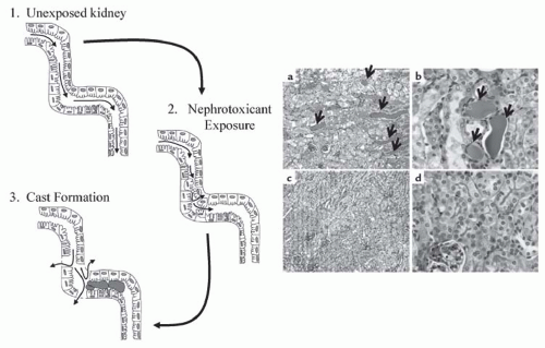
FIGURE 30.1 Cast formation in the nephron. Left: 1.Filtrate flow (as represented by the small arrows) through the tubules is constant and unobstructed in unexposed kidneys. 2. Exposure of the kidney to nephrotoxicants results in cell injury and death, and can induce detachment of the cells from the basement membrane. 3.Detached cells can adhere to each other and form casts (pink), which obstruct filtrate flow and increase intraluminal pressure. This increases permeability in the basement membrane and back leak of filtrate into the interstitium. Right: Cisplatin-induced cast formation in wild-type mice (a and b) and mice mutant for tumor necrosis factor (TNF)-α (c and d) as determined by PAS staining. The magnification in panels a and c is ×100, and that in panels b and d is ×400. Cast formation is visible in panels a and b as indicated by the pink/purple aggregates between the tubules (arrows). In contrast, little cast formation can be seen in TNF-α knockout mice (panels c and d). (Adapted from Ramesh G, Reeves WB. TNF-alpha mediates chemokine and cytokine expression and renal injury in cisplatin nephrotoxicity. J Clin Invest. 2002;110(6):835-842, with permission.) (See Color Plate.)


SUSCEPTIBILITY OF THE KIDNEY TO INJURY

The kidney is highly susceptible to numerous agents because of several functional properties of this organ. These include: (1) receiving 20% to 25% of the cardiac output, ensuring high levels of toxicant delivery over a period of time; (2) extensive reabsorptive capacity with specialized transporters promoting cellular uptake of the toxicant; (3) concentrating abilities resulting in high concentrations of toxicants in the medullary lumen and interstitium; (4) biotransformation enzymes for the formation of toxic metabolites and reactive intermediates; (5) high metabolic rate and workload of renal cells causing increased sensitivity to toxicants; and (6) sensitivity of the kidney to vasoactive agents.

Nephrotoxicants can target specific nephron segments. The proximal tubule epithelial cell is typically the primary target; however, other parts of the nephron can also be affected by chemicals with a specificity that is concentration-dependent. For example, nonsteroidal anti-inflammatory drugs (NSAIDs) and acetaminophen target the collecting ducts at low concentrations, but also induce damage to the proximal tubules at higher concentrations.16,17 Furthermore, different segments of the proximal tubule (S1, S2, and S3) are targets for different nephrotoxicants. For example, aminoglycoside antibiotics, chromate, cadmium chloride, and the mycotoxin citrinin primarily target the S1 and S2 segments, whereas cyclosporine, HgCl2, uranyl nitrate, cisplatin, bromobenzene, and cysteine conjugates of halogenated hydrocarbons target the S3 segment.17,18 Interferon-α, gold, and penicillamine can target cells in the glomeruli, whereas angiotensin-converting enzyme (ACE) inhibitors can target cells in the renal vasculature.17 Clostridium perfringens B and D and trichloroethylene can target distal tubules, and radiocontrast media, triethanolamine, amphotericin, and nystatin tend to target the loop of Henle. Studies suggesting that trichloroethylene targets the distal tubules are derived from in vitro models only at high doses,19 whereas agents that target the loop of Henle also can also affect the proximal tubules.16

These segmental differences in chemical sensitivity may be attributed to: (1) differences in toxicant delivery to a given segment, (2) differences in transport and uptake among segments, and (3) differences in biotransformation enzymes among segments. Once again, concentration may be a deciding factor.


NEPHROTOXICANT TRANSPORT

Many nephrotoxicants require transport into epithelial cells to induce injury, either by passive diffusion or by active or facilitated transport. Increased accumulation typically correlates to increased injury and decreased cellular function, which leads to AKI.


Several transporters are expressed in the kidney for the purpose of ensuring renal cell homeostatic functions, such as reabsorption and secretion; however, these proteins can also transport nephrotoxicants.20 Major transporters found in renal cells include, but are not limited to, the organic cation transporters (OCTs),21,22 organic anion transporters (OATs),23,24 the organic anion transporting polypeptide (OATP) family,25,26 and transporters involved in multidrug resistance (MDR) such as P-glycoprotein.27


Organic Anion Transporters and Organic Cation Transporters

OATs and OCTs are members of the solute carrier superfamily group 22A (SLC22A: human nomenclature) as assigned by the human genome organization (HUGO) nomenclature committee.28 Several members in the SLC22A family have homologs with human, mouse, rat, and rabbit kidneys, in addition to having overlapping substrate specificity with each other and with other transporter families. Other than physiologic substrates,28,29 these proteins also transport drugs, natural products, industrial chemicals, and pollutants.20,23,30,31,32

The OCT family of proteins typically transports small, hydrophobic, positively charged chemicals. Major isoforms include OCT1 (SLC22A1), OCT2 (SLC22A2), OCT3 (SLC22A3), OCT6 (SLC33A16), OCTN1 (SLC22A4), OCTN2 (SLC22A5), and OCTN3 (SLC22A21).28 Nephrotoxicants transported by OCTs include the chemotherapeutic cisplatin, which is a substrate for OCT2 in humans and rats.28,33,34,35 OCT2 may also mediate proximal tubule cell death induced by paraquat, a commonly used herbicide known to induce AKI. OCT1 mediates the toxicity of platinum compounds including cisplatin, oxaliplatin, and carboplatin in Madin-Darby canine kidney (MDCK) cells,22 and both OCT1 and OCT2 mediate the transport of 1-methyl4-phenyl-pyridinium, disopyramide, and chlorpheniramine into renal cells.36 OCT2 was also demonstrated to mediate the nephrotoxicity of ifosfamide both in vitro and in vivo.37

OCT1 and OTC2 were recently reported to mediate the transport and toxicity of several antiretroviral drugs used to treat human immunodeficiency virus (HIV) in human embryonic kidney 293 (HEK293) cells.38 Furthermore, OCT1, OCT2, and OCT3 were reported to mediate the transport of tyrosine kinase inhibitors (TKIs), such as imatinib, in HEK293 cells.39 More in vivo studies are needed to fully determine the role of OCTs in nephrotoxicity induced by both antiretroviral drugs and TKI.

The OAT families of proteins typically transport small organic anions into cells. Major isoforms include OAT1 (SLC22A6), OAT2 (SLC22A7), OAT3 (SLC22A8), OAT4 (SLC22A11), OAT5 (SLC22A19), OAT6 (SLC22A20), OAT7 (SLC22A9), OAT8 (SLC22A25), OAT10 (SLC22A13), and URAT1 (SLC22A12).28 Nephrotoxicants reported to be transported by OATs include the mycotoxin ochratoxin A, which is transported into renal tubular cells by OAT1, OAT 3, and OAT 5.30,31,40 Ochratoxin A transport into renal cells is inhibited by probenecid,41 an inhibitor of most OAT proteins. Recent studies also suggest that aristolochic acid, an inducer of both acute renal failure (ARF) and cancer, is transported into HEK293 cells via OAT1, OAT3, and OAT4.42 Other nephrotoxicants whose toxicity is mediated by OCTs include methotrexate (OAT1, OAT2, and OAT3), uremic toxins such as hippuric acid and indoleacetic acid (OAT1 and OAT3), and NSAIDs (OAT1, OAT2, OAT3, and OAT4).28

The ability of Hg+2 and its cysteine conjugates to induce cell death in vivo and in MDCK cells is altered by inhibitors or substrates of OAT proteins, suggesting that the nephrotoxicity of this environmental contaminant is regulated by these transporters.23,24 This hypothesis was confirmed by studies demonstrating that overexpression of human OAT1 in MDCK cells altered the nephrotoxicity of these compounds.43 Other compounds for which toxicity is suggested to be mediated by OATs include the trichloroethylene metabolite DCVC, some chlorinated phenoxyacetate-based herbicides, antiviral drugs, and β-lactam-based antibiotics.28,44


Organic Anion Transporting Polypeptides

OATPs are members of the solute carrier O family (SLCO, formerly referred to as the SLC21 family45). Endogenous substrates for these proteins include bile acids, hormones, and eicosanoids.26 Currently, genes for 11 human OATPs, 15 rat OATPs, and 15 mice have been identified.25,45 Not all of these are expressed in kidney. Furthermore, several OATPs expressed in humans are not expressed in rodents, such as OATP1A2, OATP1B1, and OATP1B3.45 Additionally, there are several rodent OATPs that do not have a human homolog. Such differences should be taken into account when assessing the role of OATPs in nephrotoxicity.

OATPs demonstrated to be expressed in human kidneys include OATP1A2, OATP2A1, OATP2B1, OATP3A1, OATP4A1, and OATP4C1.45 Rat and mouse kidneys are reported to express Oatplal, Oatpla6, Oatp2al, Oatp2bl, Oatp3a1, Oatp4a1, and 4c129,46,47 (the lowercase denotes rodent genes). Oatpla3 is reported to be a rat specific isoform.29 The expression of several mouse kidney oatp, such as Oatplal, Oatp3al, and Oatp4cl, are reported to differ depending on gender,47 but it is not known if this trend is replicated in human kidneys.

Several studies demonstrate that OATPs mediate the transport of nephrotoxicants. For example, ochratoxin A (OATP1A2, Oatplal), methotrexate (OATP1B1, Oatpla3), and digoxin (OATP4C1) are known substrates.29 Studies also suggest that the expressions of OATPs are altered by nephrotoxicants. For example, treatment of mice with nephrotoxic doses of cisplatin for 4 days increases the expression of Oatp2al and Oatp2bl mRNA.48 Future studies are needed to fully understand the role of OATPs in the pathophysiology of nephrotoxic renal cell injury.


Maillard Reaction Products

Maillard reaction products (MRPs) are members of the ATP-binding cassette super family (ABCC).29 Substrates for these proteins include hydrophobic molecules, such as
the chemotherapeutics vincristine and doxorubicin. At least six different MRP genes have been identified (designated MRP1-6, and MDR1) and all are expressed in the kidney.49

P-glycoprotein is perhaps the most well known MRP. Localized to the apical membrane of proximal tubule cells, MRP is believed to mediate the efflux of organic anions from the kidney. Known substrates for P-glycoproteins include methotrexate and cisplatin.49,50 The nephrotoxicity of cisplatin is altered by overexpression of P-glcyoprotein.50 P-glycoprotein may also mediate the nephrotoxicity of diallyl disulfide and S-allyl-cysteine, HgCl2, calcineurin, and cyclosporine.51,52,53,54


CELL DEATH

The mechanisms by which chemicals induce epithelial cell death are as varied as the chemicals themselves; nevertheless, some commonalities do exist. For example, many chemicals require transport into cells to induce death. Furthermore, regardless, of how nephrotoxicants gain entry into cells, cell death is thought to occur through one of three mechanisms: apoptosis (type I cell death), autophagy (type II cell death), or necrosis (type III cell death).55,56 Other commonalities that exist in injured and dying cells include activation of proteases, increases in cytosolic Ca2+, changes in mitochondrial function and morphology, and changes in nuclear morphology and chromatin/DNA structure.


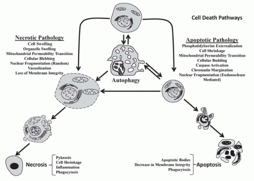
FIGURE 30.2 Schematic comparing the pathologic and morphologic features of necrosis, apoptosis, and autophagy. At the top middle, a normal cell is shown; below the normal cell is an autophagic cell demonstrating mass vacuolization; and autophagosomes (not shown). Left: Cell and organelle swelling, followed by vacuolization, blebbing, and increased membrane permeability (lysis) and finally necrotic changes (i.e., coagulation, shrinkage, and karyolysis). Right: Cell shrinkage followed by budding and karyorrhexis and finally necrotic changes (i.e., breakup into cluster of apoptotic bodies).The pathologies necrosis and apoptosis are listed. Double arrows represent the hypothesis that select pathways can switch. For example, autophagy can lead to cell survival and also progress to apoptosis.

Necrosis, apoptosis, and autophagy can be identified by assessing differences in cellular and nuclear morphology. In fact, some suggest that morphology is standard for delineating mechanisms of cell death; however, it is becoming evident that morphology alone is not the best way to identify the mechanisms of cell death.55 This reflects the fact that the mechanism of death induced by a given chemical is dependent on multiple factors such as the cell type being injured, the time of compound exposure, and the compound dose. Such multiple dependencies are typified by arsenic, which can induce all three types of cell death in a given cell.55 Additionally, the cell death mechanism may change midway through any series of postinsult events (Fig. 30.2). Thus, a chemical may initiate autophagy, but this pathway may switch to apoptosis as the dose and time of exposure increases, or if p53 is released to the cytosol. Furthermore, apoptosis may switch to necrosis as ATP decreases or if cytosolic Ca2+ increases high enough to activate select proteases or induce membrane rupture. Thus, a particular mechanism of cell death cannot always be directly linked to a specific morphology and, possibly, multiple cell death pathways may be activated in a single cell.55


Necrosis

Necrosis affects masses of contiguous cells and is characterized by swelling of organelles and increases in cell volume, after which the cell membrane becomes more permeable and ruptures with the release of cellular contents, followed by inflammation. Historically, necrosis has been used to describe drastic tissue changes occurring after cell death. These include karyorrhexis, karyolysis, pyknosis, condensation of the cytoplasm, and intense eosinophilia.

Morphologic markers for cellular necrosis include a loss of membrane and organelle integrity, cell swelling, and swelling of the endoplasmic reticulum (ER) and mitochondria (Figs. 30.2 and 30.3). Nuclear morphology in necrotic cells is usually typified by pyknosis (nuclear condensation without fragmentation); however, DNA fragmentation can occur in some cases, especially when agents that target the DNA are used. This can give rise to chromatin margination. Cellular blebs also form, but unlike apoptosis, necrotic cell blebs do not typically contain organelles. Necrosis usually induces inflammation, often with the infiltration of neutrophils and inflammatory cells in vivo (Fig. 30.3).


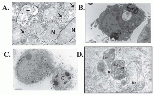
FIGURE 30.3 Comparison of the morphologic features of necrosis, apoptosis, and autophagy in tissues and cells. A: Hematoxylin and eosin staining of human kidney tissue after arterial embolization for treatment of renal cancer demonstrating intact tubules (T) and necrotic tubules (N). Arrows represent neutrophils and mononuclear inflammatory cells. (Modified from Hotchkiss RS, Strasser A, McDunn JE, et al. Cell death. N Engl J Med. 2009;361:1570, with permission.) B: Transmission electron microscopy (TEM) of necrotic human embryonic stem cells showing loss of membrane integrity without chromatin margination, cytosolic vacuolization, and spilling out of intracellular constituents. (Modified from Heng BC, Vinoth KJ, Lu K, et al. Prolonged exposure of human embryonic stem cells to heat shock induces necrotic cell death. Biocell. 2007;31(3):405, with permission.) C: TEM human HEK293 cells undergoing lysosomal mediated apoptosis. The cell on the right is relatively healthy whereas the cell on the left has shrunken and exhibits chromatin and nuclear condensation and is beginning to lose the membrane integrity (late apoptosis). (Modified from Heng BC, Vinoth KJ, Lu K, et al. Prolonged exposure of human embryonic stem cells to heat shock induces necrotic cell death. Biocell. 2007;31(3):405, with permission.) D: TEM of autophagic primary cultures of normal human renal cells exposed to cyclosporine demonstrating formation of the autophagosomes (Aut) with a double membrane (long arrow), next to a lysosomes (Lys) and a mitochondria (Mit).The arrowhead represents a cytoplasmic organelle. (Pallet N, Bouvier N, Legendre C, et al. Autophagy protects renal tubular cells against cyclosporine toxicity. Autophagy. 2008;4(6):72,with permission.)

Biochemical markers for necrosis include a drastic and rapid loss of ATP (greater than 70%-80%), rapid and sustained increases in cytosolic Ca2+, leakage of intracellular constituents such as lactate dehydrogenase, DNA fragmentation, and protease activation. A hallmark of necrosis is that it does not require ATP, separating it from both apoptosis and autophagy. DNA fragmentation also occurs in apoptosis, but DNA fragmentation that occurs during necrosis is random and not usually inhibited by caspases. Proteases activated during necrosis include select types of calpains, which are usually activated due to high concentrations of cytosolic Ca2+. Evidence exists that calpains can also be activated during apoptosis (see below); thus, calpain activation alone is not a valid marker for necrosis.


Apoptosis

Apoptosis usually affects scattered individual cells and, morphologically, the cell shrinks whereas organelle integrity is initially retained (Figs. 30.2 and 30.3). Next, chromatin become pyknotic and marginate against the nuclear membrane and, ultimately, the cell shrinks to a dense, round mass
(apoptotic body) or forms pseudopodia (i.e., buds) containing nuclear fragments and/or organelles that break off into small fragments (apoptotic bodies). In either case, adjacent cells or macrophages phagocytize the apoptotic bodies, and inflammation typically does not occur.

A key difference between necrosis and apoptosis is the activation of caspases in the latter. Caspases are cysteinyl aspartate-specific proteases that belong to an 18-member family.57,58,59 Caspases can be divided into three groups based on structural differences and substrate preferences: initiator caspases (caspase -2, -8, -9, -10, and possibly -12), executioner caspases (caspases -3, -6, and -7), and cytokine processors (caspases -1, -4, -5, -13, and -14). Caspases-15 to 18 have been identified in numerous mammals, but not in humans, with the exception of caspase 16.59

Initiator caspases are activated by numerous processes including receptor-directed mechanisms and chemical exposure. They mediate chemical-induced apoptosis in numerous cell types, including proximal tubular cells,60,61,62 glomerular cells,63 medullary cells,64,65 and cells present in the collecting ducts.66,67,68 Activation of initiator caspases results in the activation of executioner caspases, which leads to several of the biochemical characteristics of apoptosis. Initiator caspases can also be substrates for executioner caspases.69

Caspase-8 is an initiator caspase that plays an integral role in receptor-mediated apoptosis.69,70,71 It is activated by membrane receptors such as Fas-ligand and tumor necrosis factor (TNF)-α receptors71,72 and, in turn, cleaves the Bcl-2 family protein Bid to form tBid.70 tBid acts on mitochondria to cause the release of pro-apoptotic proteins and results in the activation of caspase-9 and caspase-3. In contrast, caspase-8 can directly activate caspase-9 or caspase-3, independently of the mitochondria (Fig. 30.4).

Caspase-8 can be activated by nephrotoxicants independent of receptor-mediated mechanisms. For example, cisplatin and etoposide activate caspases-8, -9, and -3 in LLC-PK1 cells in the absence of receptor-stimulation.73 In contrast, cisplatin and cyclosporine activate caspase-3 in the absence of caspase-8 in mouse and rabbit RPTC.74,75,76 Thus, the role of caspase-8 in chemical-induced renal cell apoptosis is variable.

Executioner caspases cleave numerous substrates that ultimately result in the morphologic features of apoptosis. Perhaps the most important substrates are proteins that control DNA degradation (DNAase). Caspases are known to mediate the activation of the nuclease DNA fragmentation factor (DFF). DFF is composed of two subunits: a 40-kDa DNAase subunit (CAD/DFF40) and a 45-kDa inhibitor of caspase-activated deoxyribonuclease (ICAD/DFF45).69 Caspase-3 cleaves ICAD/DFF45 during apoptosis, which results in the release and activation of CAD/DFF40. Active CAD/DFF40 results in double-stranded DNA breaks in chromosomes, giving rise to the characteristic nonrandom DNAladderlike pattern seen with apoptosis on agarose gels.69

Caspases can also mediate DNA degradation by cleaving poly(ADP-ribose) polymerase (PARP). PARP is involved in DNA repair and maintenance of stability, and regulates DFF40 activity.69 Caspases-3 and -7 can cleave and inactivate PARP.77 PARP cleavage is used as a marker for apoptosis in renal cells, including apoptosis induced by antimycin A and DCVC.78,79 Cleavage of DNA repair enzymes (such as PARP) by caspases is thought to prevent cells from making a futile repair attempt.

Caspases have numerous other substrates other than DNAases. Initiator caspase substrates include other caspases, the pro-apoptotic protein Bid, α-tubulin, and vinculin.58 Cytokine caspase substrates include inflammatory mediators such as such 11-18, Pro-IL-1B, and 11-17, whereas executioner caspase substrates include protein kinase C (PKC), focal adhesion kinases (FAK), and the cell cycle regulator p21.58 Cleavage of these proteins is believed to inhibit futile repair attempts, facilitate apoptosis signaling cascades, and allow for cytoskeleton reorganization and packaging of cell constituents into apoptotic bodies.80

Caspase-3, perhaps the best studied executioner caspase, can also cleave receptors, such as type 1 inositol(1,4,5) P4 receptor, Ca2+-ATPase, the Na+/Ca+ exchanger, and the Na+/K+-ATPase pump.55 The Na+/K+ ATPase may also be cleaved by initiator caspases, such as caspase-8 and -9.81 Cleavage of these receptors is believed to alter ion homeostasis and facilitate decreases in intracellular K+, which further promotes caspase activation. Cleavage of these receptors also leads to cell size alterations, such as cell shrinkage, early during apoptosis after cleavage of Na+/K+ ATPase, or cell rupture due to swelling after Ca2+-ATPase inactivation during late-stage apoptosis/secondary necrosis.


Role of Mitochondria in Apoptosis

The role of mitochondria in cell death cannot be understated, especially for apoptosis. Mitochondria regulate apoptosis by at least two major processes: maintenance of ATP production and release of pro-apoptotic proteins, such as cytochrome c, Bcl-2 family proteins, and DNAases. In addition, mitochondria regulate apoptosis by participating in Ca2+ signaling cascades and mediating protease activation.55

ATP is considered to be a requirement for both the initiation and execution of apoptosis.55 It is required for formation of an apoptosome protein complex (see later), which facilitates the activation of caspase-9. It may also be required for transport of pro-apoptotic proteins into the nucleus.55 ATP may also represent an important switch point between apoptosis or necrosis; depleting ATP below 30% transforms apoptotic liver cell death to necrotic death patterns.82 In addition, ATP is needed to maintain Na+/K+ ATPase pumps on the plasma membrane, and pump inactivation will eventually lead to cellular swelling, pathologic increases in intracellular Ca2+, and cellular lysis, which is typical of necrosis.

Cytochrome c is a heme protein bound to the inner mitochondrial membrane, transferring electrons between complexes III and IV of the electron transport chain. Release of cytochrome c from mitochondria activates the intrinsic pathway of apoptosis. Cytosolic cytochrome c will bind
to apoptotic protease activating factor 1 (APAF-1), which promotes the binding and proteolytic cleavage of procaspase-9 to caspase-9 (the apoptosome),83 and then activated caspase-9 cleaves and activates executioner caspases (i.e., caspases-3, -6, and -7) (Fig. 30.4). Nephrotoxicants known to induce cytochrome c release in correlation with apoptosis include cisplatin and DCVC.71,84 Cytochrome c release from the mitochondria is associated with a decrease in the mitochondrial inner membrane potential and the accumulation of several pro-apoptotic proteins such as Bad, Bax, and Bax at the mitochondria (Fig. 30.4). Other proapoptotic proteins released from the mitochondria include apoptosis-inducing factor (AIF), Smac/Diablo, Omi, and Endo G (Fig. 30.4).70,71,85,86,87,88,89,90,91,92


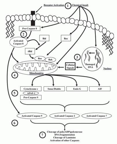
FIGURE 30.4 Cell signaling cascades involved in the activation of caspases and apoptosis. 1: Receptor-mediated death signals or chemicals can initiate apoptosis through multiple mechanisms. 2: Pro-caspase 8 is activated by receptor-mediated signals at the cellular membrane or directly by chemicals. Once activated, caspase-8 cleaves Bid to t-Bid, which interacts with Bax/Bak to induce mitochondrial-mediated apoptosis or directly activates caspase-9 and other caspases. 3: Some chemicals cause DNA damage that signals the release of pro-apoptotic proteins from the mitochondria. 4: Receptor-mediated signals, direct chemical injury, or signals resulting from DNA damage can all cause cytochrome c, Smac/Diablo, Endo G, and AIF release from the mitochondria. 5: Released cytochrome c forms a complex with APAF-1 and pro-caspase 9, resulting in caspase-9 activation. 6: Activated caspase-9 cleaves and activates procaspase-3 and -7, which can also be activated by caspase-8 independently of cytochrome c. 7: Activated caspases (e.g., 3 and 7), AIF, and Endo G cause the classical markers of apoptosis such as cleavage and activation of poly(ADP)polymerase, inactivation of inhibitors of DNases leading to DNA fragmentation, cleaved laminins, and the activation of other caspases.


Bad, Bak, Bax, and Bid belong to the Bcl-2 family of pro-apoptotic proteins, which are characterized by specific regions of homology, termed Bcl-2 homology domains.93 Under nonstressed conditions, these proteins exist bound to proteins in the mitochondria and cytosol.70 After toxicant exposure, Bax, Bid, or Bak can dissociate and translocate to the mitochondria which initiates the formation of a pore complex that causes membrane rupture55 and subsequent loss of mitochondrial membrane potential, facilitating the release of cytochrome c, Endo G, Smac/Diablo, Omi, and AIF (Fig. 30.4).68,70,74 Bid mediates apoptosis induced by hypoxia and ATP depletion in cultures of rat RPTC94; Bax mediates proximal tubular apoptosis in mice treated with cisplatin in vivo65; and Bak is elevated during apoptosis in primary bovine glomerular endothelial cells induced by TNF-α or lipopolysaccharide (LPS95) or during ischemiareperfusion-induced renal cell apoptosis in mice.96

In contrast to Bax, Bid, and Bak, Bcl-2 is an anti-apoptotic protein.60 Increased Bcl-2 prior to toxicant exposure protected numerous cells, including renal cells,96 from toxicant-induced apoptosis.96 The protective effect of Bcl-2 may be the result of its ability to bind Bax, Bid, and Bak, preventing them from inducing mitochondrial pore formation, altering mitochondrial membrane permeability, initiating the release of mitochondrial pro-apoptotic proteins, and activating caspases.97 Overexpression of Bcl-2 protected against ATP-depletion-induced apoptosis in cultures of rat RPTC,94 and upregulation of Bcl-2 protected kidney epithelial cells both in vitro and in vivo against apoptosis induced by hypoxia, azide, cisplatin, and staurosporine.98

AIF is released from mitochondria in response to decreases in the mitochondrial membrane potential induced by ATP depletion86,99; ischemia-reperfusion; anti-fas antibodies100; or exposure to high concentrations of Ca2+,101 t-butyl hydroperoxide,101 or atractyloside.101 Cellular pathologies associated with AIF release are similar to those seen with caspases (chromatin condensation and oligonucleosomal DNA fragmentation).100 Recent studies suggest that increases in cytosolic Ca2+ and calpain activation also facilitate the release of AIF from mitochondria102; and studies in LLC-PK1 cells support this hypothesis.103

AIF is a protease with properties similar to caspases, including being inhibited by N-benzyloxycarbonyl-Val-Ala-Asp-fluoromethylketone (Z-VAD-fmk), a commonly used broad spectrum caspase inhibitor.86 Thus, the decrease in renal cell death observed in the presence of Z-VAD-fmk may be a result of AIF or caspase inhibition. AIF can induce DNA fragmentation independently of caspases.69 AIF is released in opossum kidney (OK) cells after ATP depletion-induced by sodium cyanide and 2-deoxy-D-glucose.87,99 AIF is also activated in HEK293 cells after exposure to cadmium,104 in LLC-PK1 cells after exposure to cisplatin,105 and in OK cells after exposure to the peroxisome proliferator-activated receptor agonist ciglitazone.106

Smac/Diablo is a pro-apoptotic protein released from the mitochondria to the cytosol during apoptosis. It blocks antiapoptotic activity of inhibitors of apoptosis proteins (IAP), which increase apoptosis.89 The ability of Smac/Diablo to promote apoptosis is not exclusively a result of its ability to bind IAP.107 Smac/Diablo functions at the same level of executioner caspases, but downstream of the Bcl-2 family of proteins.90

Smac/Diablo is expressed in the mouse kidney and in several renal cell models.108 It mediates apoptosis in vivo in mice after treatment with high concentrations of folic acid or after exposure of cultures of renal epithelial cells to TNF-α.89 Increased expression of Smac/Diablo potentiates TNF-α- and etoposide-induced apoptosis in HEK293 cells107; however, similar to several other pro-apoptotic proteins, expression of Smac/Diablo is not essential for apoptosis in kidney cells. For example, acetaminophen-induced renal cell apoptosis proceeds in a caspase-dependent manner in the absence of Smac/Diablo activity.109

Omi is a mammalian serine protease homologous to bacterial HtrA endoprotease.110 Omi localizes to the mitochondria and is expressed ubiquitously in a number of cell types including RPTC.91 Omi is released from the mitochondria after exposure to apoptotic stimuli and binds to, and cleaves, IAP.91 Omi-directed degradation of IAP facilitates caspase activation and the subsequent biochemical and morphologic features of apoptosis. In addition, Omi can translocate to the nucleus and activate the transcription factor p73, which induces pro-apoptotic proteins such as bax.69 Omi participates in both caspase-independent and caspase-dependent cell death,69,111 an event that has been proven using either siRNA against Omi, or a synthetic inhibitor, called ucf-101, in in vitro and in vivo models, including primary cultures of mouse RPTC.91,111 More work is needed to determine if Omi can mediate cell death induced by other nephrotoxicants.


Autophagy

Autophagy is essentially “a cell eating itself.”112,113 This process was originally thought to be a cell survival pathway activated to produce energy during times of metabolic stress, such as starvation.113 Some cells undergoing autophagy can recover; however, ample evidence exists that autophagy itself leads to cell death, specifically referred to as type II cell death.55,112,113

Significant evidence shows that autophagy mediates renal cell death.56,114,115 The role of autophagy in renal cell death differs depending on the experimental conditions.56 Further, it is difficult to determine if autophagic cells are a result of a cell death mechanism, or a failure in repair or survival mechanism.56 Complicating this issue is that induction of autophagy is cell- and toxicant-dependent. Nevertheless, clear correlations exist between nephrotoxicity and autophagy. Autophagic cells are present in vivo in rat renal cells after ischemia-reperfusion and after treatment of mice with tunicamycin, a stimulant of ER Ca2+ release.56 In vitro, autophagy was identified in HK-2 cells after exposure to H2O2, in RPTC cultures after exposure to cisplatin, and in primary cultures of human renal cells treated with cyclosporine A.56,116


Morphologic markers for autophagy include the presence of autophagic vacuolization of the cytoplasm and the autophagosome, which forms near the lysosome and can contain cytosolic organelles (Figs. 30.2 and 30.3).55 This occurs in the absence of chromatin condensation. Double-membrane vesicles, autophagosomes, are formed and fuse with lysosomes to facilitate protein degradation112 and other morphologic changes that are best identified using transmission electron microscopy.

Biochemical markers of autophagy include expression of microtubule-associated protein-1 light chain 3 (LC3), and degradation of the cell signaling adaptor p62.112 LC3 is only considered an autophagic cell marker when it is cleaved to a lower molecular weight protein called LC3II. Cleavage allows LC3 to bind to phosphatidylethanolamine, which facilitates the formation of autophagosomes. Autophagosome formation is also facilitated by two kinases: autophagy-specific phosphatidylinositol 3-kinase (PI3K) Vps34 (also called human class III PI3K) and target of rapamycin (TOR) kinase.56,112

Beclin-1 is another protein whose expression is critical for autophagy. Beclin-1 facilitates formation of autophagosomes by regulating Vps34 (human class III PI3K56). It contains a BH-3 only domain, and is inhibited by other BH-3 only domain containing proteins called Bcl-2 and Bcl-XL.55 Proteins containing BH3-only domains are typically pro-apoptotic; however, beclin-1 does not induce apoptosis. In fact, beclin-1 is cleaved by caspases. Cleavage of beclin-1 by caspases is believed to be an important switch point used by cells to inhibit autophagy and stimulate apoptosis.55

p53, another regulator of autophagy, is a tumor suppressor protein found in the cytosol of living cells in an inactivated state and bound to the co-repressor Mdm2 (see later). The release of p53 from Mdm2 is stimulated by ionization radiation, DNA damage, oxidative stress, and several other death-inducing stimuli. Released p53 can translocate to the nucleus and induce apoptosis, cell cycle alterations, and the transcription of several proteins, including those that mediate autophagy.55 Interestingly, cytosolic p53 (unbound to Mdm2) appears to inhibit autophagy in nonrenal cells.117 It is not known if p53 can regulate autophagy in renal cells using similar mechanisms.


INITIATORS OF CELLULAR INJURY

Nephrotoxicants initiate renal cell injury by a variety of mechanisms. Some initiate toxicity directly because of their reactivity with selected cellular macromolecules, such as observed with the antifungal drug amphotericin B, which increases the permeability of the plasma membrane to cations,118 the mycotoxin fumonisin B1 that inhibits sphinganine (sphingosine) N-acyltransferase,119 and aminoglycosides that bind initially to cellular anionic phospholipids.120 Other nephrotoxicants initiate toxicity following biotransformation to a reactive intermediate or a stable metabolite, and nephrotoxicants can initiate toxicity indirectly through the production of reactive oxygen species.


Role of Biotransformation

Renal xenobiotic metabolism contributes significantly to whole-body metabolism and/or renal toxicity of numerous chemicals because of the role of the kidney as a primary route of xenobiotic excretion. Some chemicals require metabolism or biotransformation to a toxic reactive intermediate for cellular injury to occur (Fig. 30.5). Then the reactive intermediate binds covalently to critical cellular macromolecules, which are thought to interfere with the normal functioning of the macromolecules and thereby initiate cellular injury. Often, these reactive intermediates or “alkylating” agents are electrophiles that bind to cellular nucleophiles. The renal xenobiotic-metabolizing enzymes found in experimental animals and humans have been reviewed by Lock121 and are summarized in Table 30.1. These include cytochrome P-450, flavin containing monooxygenase (FMO), and glutathione S-transferase (GST).


Cytochrome P-450

The kidney contains many of the xenobiotic-metabolizing enzymes found in the liver; however, in general, their concentration within the kidney is lower. For example, renal cytochrome P-450 ranges between 0.1 and 0.2 nmol per mg microsomal protein across a variety of species, which represents approximately 10% of hepatic cytochrome P-450.121 The distribution of cytochrome P-450 also varies along the nephron, with the highest levels typically found in the S2 segment, followed by the S3 and S1 segments, with the other tubular segments having less than 10% of that of the S1 segment.121

The renal cytochrome P-450 system is active against a variety of endogenous and exogenous compounds, and numerous cytochrome P-450 isoforms have been identified in renal tissue. For example, cytochromes P-450 1A1, 1A2,1B1, 2A, 2B1, 2B2, 2B6, 2B9, 2B10, 2C2, 2C11, 2E1, 2J2, 2J3, 2J5, 2J9, 3A1, 3A4, 4A1, 4A2, 4A3, 4A5, 4A6, 4A8, 4A11, 4F1, 4F4, 4F5, 4F6, 4F11, and 4F12 have been identified in renal cells of the human, mouse, rat, and rabbit kidney.121,122,123,124,125

Cytochrome P-450 expression depends on the species and sex being studied, as well as the site along the nephron. For example, cytochrome P-450 2A, 2C, and 2E are present in male mouse kidneys but are barely detectable in female mouse kidneys.121 Several studies suggest differences in the expression of cytochrome P-450 isoforms between human and rodent kidneys. An important example is the expression of cytochrome P-450 2E1, which has been detected in renal proximal and distal tubular cells of mice and rats, but not human kidneys.121,122,126 In contrast, both human and rodent kidneys express high amounts of cytochrome P-450 4A isoforms. However, rat kidneys express 4A1, 4A2, and 4A3, whereas the human kidney appears to express 4A11.122,127 Such differences in xenobiotic expression must be considered when assessing the role of biotransformation in chemical-induced nephrotoxicity.

In contrast to the liver, fewer compounds are documented to produce nephrotoxicity through renal cytochrome P-450
bioactivation, although renal cytochrome P-450 contributes to the nephrotoxicity of chloroform128,129 by metabolizing it to the unstable trichloroethanol, which releases HCl to form phosgene. Phosgene reacts with: (1) two molecules of glutathione to produce diglutathionyl dithiocarbonate, (2) water to produce two molecules of HCl and CO2, (3) cysteine to produce oxothizolidine-4-carboxylic acid, or (4) cellular macromolecules to initiate toxicity.128,130,131


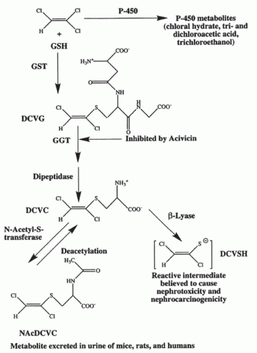
FIGURE 30.5 The bioactivation of trichloroethylene by the glutathione-(GSH-) conjugation pathway. Trichloroethylene (top left) can be metabolized by either cytochrome P-450 to the compound listed (top right) or be conjugated to GSH by the glutathione S-transferase (GST) to form S-(1,2)-dichlorovinyl-glutathione (DCVG).These reactions can occur either in the liver or in the kidney. DCVG formed in the liver is delivered to the kidney via the bile or the blood where the high concentrations of γ-glutamyltransferase (GGT) and dipeptidase in the kidney results in the cleavage of the GSH moiety and the formation of S-(1,2)-dichlorovinyl-L cysteine (DCVC). Metabolism of DCVC by N-acetyl-s-transferase produces N-acetyl-s-(1,2)-dichlorovinyl-L-cysteine (NAcDCVC), which is excreted in the urine of mice, rats, and humans exposed to trichloroethylene. NAcDCVC also can be deacetylated back to DCVC. Metabolism of DCVC by cysteine-conjugate β-lyase results in the formation of a reactive thiol that can rearrange to form a protein acylating species. (From Cummings BS, Parker JC, Lash LH. Role of cytochrome P450 and glutathione S-transferase alpha in the metabolism and cytotoxicity of trichloroethylene in rat kidney. Biochem Pharmacol. 2000;59:531, with permission.)

Chloroform bioactivation by renal cytochrome P-450 is sex- and species-dependent. The marked sex difference in the nephrotoxicity of chloroform is reversed by castration of males or treatment of females with testosterone, suggesting that the renal cytochrome P-450 responsible for chloroform bioactivation is under androgenic control.130,132 Because cytochrome P-450 isozymes 2B1 and 2E1 are present in male mice and are expressed in female mice treated with testosterone, these isozymes may be responsible for renal chloroform bioactivation.131

Acetaminophen is metabolized in the mouse kidney by cytochrome P-450 2E1 to the reactive intermediate N-acetyl-p-benzoquinoneimine (NAPQ), which binds to cellular proteins.132,133 In the liver, NAPQ binds to a selenium binding protein (58 kDa),134,135 microsomal glutamine synthetase (44 kDa),136 cytosolic N-10-formyl tetrahydrofolate dehydrogenase (100 kDa),135,137 and mitochondrial glutamate dehydrogenase (50 kDa).137 It is possible that similar protein binding may occur in renal cells.

Studies also suggest that acetaminophen mediates renal cell death in mouse RPTC by inducing ER stress.109 In this model, acetaminophen treatment increased the expression of GADD153, an ER stress protein, and induced caspase-12 cleavage and apoptosis—independently of caspase-3, -9,
or the release of the mitochondrial pro-apoptotic protein Smac/Diablo.


TABLE 30.1 Expression of Selected Xenobiotic Biotransformation Enzymes in the Kidney


Enzyme


Cell Type


Species


References


Cytochrome P450 monooxygenases


IA


Proximal tubules


Rat, mouse, human


390,391


IA2


Proximal tubules


123


IIB


Proximal tubules


Rat and mouse


122


Distal tubules


Rat and mouse


122


IIC2


Proximal tubules


IIC9


Unknowna


Human but not rat


390


IIC11


Distal tubules


Male rat


122


IID


Proximal tubules


125


IIE1


Proximal tubules


Rat, mouse, not human


122,127,392


Distal tubules


122


IIJ


Proximal tubules


Human, rat, mouse


123,393,394,395


IIIA1


Glomerulus


Rat, mouse, not human


122,390,391


IIIA4


Proximal tubules


Human, not rat or mouse


390


IVA2


Proximal tubules


Rat, mouse, not human


122,390


Distal tubules


122,390


IVA3


Proximal tubules


Rat, mouse, not human


122,390


Distal tubules


122,390


IVA11


Proximal tubules


Human, not rat or mouse


126,127


IVF


Proximal tubules


Human and mouse


124,127


Distal tubules


Mouse


124


Flavin-containing monooxygenases


FMO1


Unknowna


Rat, mouse, and human


141,390


FMO3


Unknowna


Rat, mouse, and human


141,390


FMO5


Unknowna


Human


141


Glutathione S-transferases


GST α


Proximal tubules


Rat, mouse, and human


19,126,148


Distal tubules


GST µ


Proximal tubules


Rat, mouse, not humanb


19,126,148,396


GST π


Proximal tubules


Rat, mouse, and human


19,126


GST θ


Proximal tubules


Human


126


a Activity and expression have been measured in kidney microsomes only.

b GSTµ is expressed in some human kidney malignancies.


Flavin-containing Monooxygenase

Flavin-containing monooxygenase (FMO) oxidizes the nucleophilic nitrogen, sulfur, and phosphorus moieties of a number of chemicals, including DCVC, tamoxifen, and cimetidine.121,138,139 The role of FMO in nephrotoxicity has received less attention than cytochrome P-450, but several FMO are expressed in the kidney. Like cytochrome-P450, renal FMO expression and activity is species- and sexdependent. For example, rabbit kidneys express FMO1, 2, 4, and 5, but not 3, and FMO1 is expressed in the female, but not the male kidney.121 FMO3 activity is detected in the kidneys of rats, dogs, mice, rabbits, and humans.140 Rat kidneys appears to have two- to sixfold greater activity levels (as determined by methionine S-oxidase activity) than other species, including humans.141 Studies in human kidney microsomes demonstrate that FMO1, FMO3, and FMO5 are all expressed, but at different levels.141 Furthermore, samples from African American patients had significantly more FMO1 activity compared to their Caucasian counterparts, suggesting that the expression of renal FMO isoforms may differ depending on race.141 Studies in mice
suggest that sex- and age-dependent differences exist for the expression of FMO mRNA in the kidney142; however, no differences in the expression of FMO1, FMO3, or FMO5, and overall FMO activity were detected between human male and female kidney microsomes.141 Thus, more work is needed to determine if FMO expression is sex-dependent in human kidneys.

In vitro, FMO1, FMO3, FMO4, and FM05 metabolize cysteine S-conjugated S-allyl cysteines, whereas FMO3 metabolizes DCVC.141 However, little DCVC was metabolized in human kidney microsomes, even though FMO3 was expressed in these tissues, suggesting that FMO may not contribute to the nephrotoxicity of this compound in human renal cells. In contrast, treatment of human proximal tubular cells with the FMO inhibitor methimazole decreased DCVC-induced apoptosis.143 Studies also suggest that FMO catalyzed sulfoxidation of the sevoflurane (a commonly used anesthetic) degradation product fluoromethyl-2,2-difluoro-1-(trifluoromethyl)vinyl ether mediates its renal toxicity.144 Other nephrotoxicants suggested to be metabolized by FMO include 4-amino2,6-dichlorophenol.145 4-amino-2,6-dichlorophenol is a metabolite of 3,4-dichloroanaline, a common industrial manufacturing intermediate. Finally, FMO may also mediate the nephrotoxicity of some pesticides such as organophosphate thioether compounds.146


Glutathione S-Transferase

The conjugation enzymes glucuronosyltransferases, sulfotransferases, and glutathione S-transferases (GST) are located in the kidney where they conjugate both endogenous and exogenous compounds. These enzymes increase the water solubility, excretion, and elimination of several nephrotoxicants.121 Although this typically decreases renal cell injury, some nephrotoxicants are bioactivated by these enzymes.

GST mediates the conjugation of the tripeptide glutathione (GSH, γ-glutamylcysteinlyglycine) to compounds with electrophilic centers.121 They are considered phase II biotransformation enzymes and are divided into cytosolic, membrane associated, and mitochondrial members. There are seven different cytosolic subfamilies (A, alpha; M, mu; P, pi; T, theta; O, omega; S, sigma; and Z, zeta). Microsomal GST is referred to as membrane-associated proteins in eicosanoid and glutathione metabolism (MAPEG), whereas mitochondrial GST is called K (kappa) GST.121,147 All of these GSTs, with exception of GST S, are expressed in rat and human kidneys,19,121,126,147 and GST expression in normal human RPTC appears to be similar to those observed in rat RPTC.126

GST expression can differ among nephron segments. For example, in the rat kidney, GST A is expressed primarily in proximal and distal tubules, whereas GST M and P are primarily expressed in the distal tubules.19,121,126 These expression patterns differ from a previous study, especially with regard to GST A in distal tubules.121,148 Thus, more research is needed to resolve this discrepancy.

As mentioned previously, conjugation of toxicants to GSH is normally a detoxification pathway in which electrophiles are neutralized and made more amenable for excretion. Unfortunately, numerous extrarenally formed glutathione conjugates are nephrotoxic. For example, the extrarenal conjugation of GSH is important for the nephrotoxicity of HgCl2,149 halogenated alkenes, and aromatics, and possibly acetaminophen.131,150,151 The nephrotoxicity of the halogenated alkene trichloroethylene in rats and humans is believed to be a direct result of its conjugation with GSH to form S-(1,2)-dichlorovinyl-glutathione, and the subsequent processing of the glutathione-conjugate to DCVC in RPTC (Fig. 30.5).19

In vivo, trichloroethylene is conjugated with GSH in the liver and delivered via the bile or blood to the kidney. The expression of enzymes, such as γ-glutamyl transferase and dipeptidase in the RPTC and biliary and intestinal tract, results in the cleavage of the γ-glutamyl and glycyl moieties, respectively, and the formation of DCVC. Metabolism of DCVC by N-acetyl-s-transferase produces N-acetyl-s-(1,2)-dichlorovinyl-L-cysteine, which is excreted in the urine of mice, rats, and humans exposed to trichloroethylene.152 N-acetyl-s-(1,2)-dichlorovinyl-L-cysteine also can be deacetylated back to DCVC. Metabolism of DCVC by cysteine-conjugate β-lyase results in the formation of a reactive thiol that can rearrange to form a protein acylating species. A strong correlation exists between increases in markers of renal injury (proteinuria, creatinine clearance, glucosuria) and GSH metabolites of trichloroethylene in the blood and urine of humans exposed to high amounts of trichloroethylene.153

Only gold members can continue reading. Log In or Register to continue

Stay updated, free articles. Join our Telegram channel

May 29, 2016 | Posted by in NEPHROLOGY | Comments Off on Pathophysiology of Nephrotoxic Cell Injury

Full access? Get Clinical Tree

Get Clinical Tree app for offline access