Renal Function and Disease in the Aging Kidney


Renal Function and Disease in the Aging Kidney


Ramesh Saxena

Andrew Fenves

Xueqing Yu

Nosratola D. Vaziri

Fred G. Silva

Xin J. Zhou


INTRODUCTION

The growth of an aging population in the world is on an explosive path and the cost of care for the elderly could overwhelm the budget of many countries in the next 40 years. The worldwide population of persons aged 65 years or more is estimated to be 420 million, or about 7% of the population, and is projected to increase to more than 1.5 billion by 2050.1,2,3,4,2a,2b In the United States, the percentage of people over 65 years of age has more than tripled since 1900 and continues to grow. The number of elderly has increased almost 15% (from 35 million to 40.5 million) from 2000 to 2009.1 The growing number of older adults has drastically increased healthcare costs. In fact, healthcare cost per capita for persons over 65 years is three to five times greater than those under 65 years of age.5

Aging is a complex process driven by diverse molecular pathways and biochemical events culminating in profound anatomic and functional changes in the kidneys. Additionally, older individuals have diverse chronic diseases that can accelerate the age-related renal changes. In particular, the prevalence of chronic kidney disease (CKD) is greatest in the elderly and they have the fastest growth of end-stage renal disease (ESRD) at 11% for age 65 to 74 and 14% for age 75 and older.6 This is a pressing problem that contributes substantially to disability, diminished quality of life, and enormous healthcare costs and begs for a massive effort to study the effect of aging on the kidney and the predisposing factors for nephropathy in the elderly population. This chapter provides an overview of the recent advances in the understanding of age-related changes in renal structure and function, the molecular pathways mediating these changes, and possible therapeutic interventions to mitigate age-related renal changes.


STRUCTURAL CHANGES OF THE AGING KIDNEY

Many studies have described the progressive structural and functional deterioration of kidneys with aging. Most of these studies are old and did not exclude patients with confounding comorbidities that might affect renal structures. Nevertheless, it appears that renal masses (i.e., weight of the kidney) progressively regress with advancing age.7 The average kidney weight increases from ˜50 g at birth to ˜200 g during the fourth decade, after which it progressively declines (about 20% to 30%) by the ninth decade.7,8 This loss of kidney mass is primarily cortical with relative sparing of the medulla, leading to thinning of the renal cortical parenchyma.7,8 Although many morphologic changes are observed in the aging kidney, none is specific or pathognomonic.


The Aging Glomerulus

With aging, a number of morphologic changes emerge in the human glomerulus (Table 65.1). These include a decrease in the number of identifiable glomeruli and an escalation in the proportion of globally sclerotic glomeruli, which is associated with a progressive increase in the size of intact glomeruli (Fig. 65.1).

The number of glomeruli is extremely variable in individuals, ranging from 333,000 to 1,100,000 in each kidney and vary with age (inversely), gender (15% lower in females), and race (lower in Australian Aboriginals).7,8,9 As renal cortical mass decreases with increasing age, the glomeruli decrease in number. There seems to be a direct correlation between the number/percentage of globally sclerotic glomeruli and increasing age and with intrarenal vascular disease, especially outer cortical vascular disease.10 In general, globally sclerotic glomeruli comprise less than 10% of the total glomeruli under the age of 40 years and increase thereafter so that by the eighth decade as much as 30% of glomeruli may be globally sclerotic. However, the estimation of “normal” sclerosed glomeruli is difficult in the elderly due to confounding effects of comorbid conditions such as diabetes and hypertension. In such situations, “pathologic” glomerulosclerosis should be considered when the number of globally sclerosed glomeruli exceeds the number calculated by the formula: (patient’s age/2) -10.7,11,11a

The pathogenesis of aging-associated global glomerulosclerosis is not completely understood and is likely multifactorial. Increasing oxidative stress that accompanies
aging can result in endothelial dysfunction and changes in vasoactive mediators resulting in atherosclerosis, hypertension, and glomerulosclerosis.12 Furthermore, age-related changes in cardiovascular hemodynamics, such as reduced cardiac output and systemic hypertension, may contribute to glomerular changes.13 Moreover, dysautoregulation of the afferent and efferent arterioles may increase glomerular plasma flow, glomerular capillary pressure, and “hyperfiltration,” leading to mesangial matrix accumulation.14 A morphometric study showed dilatation of the afferent arterioles, increased glomerular capillary lumens (especially hilar), and enlarged glomeruli, which suggested a discordance between the afferent and efferent arterioles.15 The vascular adaptations to functional or structural nephron loss may help preserve glomerular filtration rate (GFR) by producing hyperperfusion and hyperfiltration in the surviving nephrons. This local glomerular hypertension and hypertrophy may lead to cytokine-mediated mesangial matrix expansion and, eventually, glomerulosclerosis. Such hyperperfusion-associated glomerular injury is seen with oligomeganephronia, diabetic nephropathy, morbid obesity, sickle cell anemia, and reflux nephropathy. It has been suggested that the vascular/ischemic changes seen in aging kidneys first cause cortical glomerulosclerosis and consequent juxtamedullary glomerular hypertrophy, followed by juxtamedullary glomerulosclerosis.16


TABLE 65.1 Morphological Changes of the Aging Kidney


Glomerulus


Increased number of globally sclerotic glomeruli; initially the glomeruli in the outer cortical regions


Progressive decline in the number of intact/normal glomeruli


Abnormal glomeruli with shunts between the afferent and efferent arterioles, especially those in the juxtamedullary region


Progressive decrease, and then later increase, in the size of intact glomeruli with higher filtration surface area


Tubulointerstitium


Decreased tubular volume, length, and number


Increased number of tubular diverticula, especially the distal convoluted tubules


Tubular atrophy, often with simplification of the tubular epithelium and thickening of the tubular basement membranes


Increased interstitial volume with interstitial fibrosis and, sometimes, inflammatory cells


Decreased peritubular capillary density


Vasculature


“Fibroelastic hyperplasia” of the arcuate and subarcuate arteries


Tortuous/spiraling interlobar arteries with thickening of the medial muscle cell basement membrane


Intimal fibroplasia of the interlobular arteries


“Hyaline” change/plasmatic insudation of the afferent arterioles


Vascular “simplification” with direct channels forming between the afferent and efferent arterioles


Modified from: Zhou XJ, Rakheja D, Silva FG. The aging kidney. In: Zhou XJ, Laszik Z, et al., eds. Silva’s Diagnostic Renal Pathology. Cambridge, UK: Cambridge University Press; 2009.


The Aging Tubules and Interstitium

Several tubulointerstitial alterations parallel glomerular changes in the aging kidney. Three types of tubular atrophy can be seen in the aging kidney (Fig. 65.1). These include the classic form with wrinkling and thickening of the tubular basement membranes and simplification of the tubular epithelium; the endocrine form with simplified tubular epithelium, thin basement membranes, and numerous mitochondria in the tubular epithelial cells; and the thyroidization form with hyaline cast-filled dilated tubules.17 Although there is an overlap, the endocrine form is classically seen with vascular ischemia, and the thyroidization form is considered to be characteristic but not pathognomonic of chronic pyelonephritis. With tubular atrophy, the distal renal tubules develop diverticula that increase in number with increasing age. These diverticula in distal and collecting tubules may be precursors of simple cysts that are increasingly observed in the aging kidney.18 The diverticula may promote bacterial growth and contribute to the frequent renal infections in the elderly.


The Aging Renal Vasculature

Several changes in the renal vasculature have been documented in the aging human kidney, none of which are specific for aging (Fig. 65.1). Arterial sclerosis denotes thickening of the

wall and narrowing of the arterial lumen produced by thickening of the medial smooth muscle layer, fibrosis of the media, and/or intimal thickening. These changes may be seen with hypertension, diabetes, and aging, with the prevalence of arterial sclerosis increasing with advancing age.7,19,20 Intimal fibroplasia or collagenous fibrosis of the arterial intima may be associated with thinning of the media and is found uniformly in older kidneys with or without underlying cardiovascular disease. Intimal fibroplasia is seen primarily in arteries that are 80 to 300 µm in diameter, such as the interlobular arteries. The regional heterogeneity of intimal hyperplasia may account for the heterogeneity of ischemic nephrons. Although the etiology of aging-associated intimal fibroplasia is not entirely clear, it starts early in life and is accelerated by hypertension. Intimal hyperplasia in the interlobular arteries may allow the transmission of the pulse wave into the smaller distal branches leading to arteriolar hyaline changes, which may in turn accelerate the proximal intimal fibrosis. Global glomerulosclerosis appears to be associated with arterial intimal fibrosis rather than with arteriolar hyaline change.21
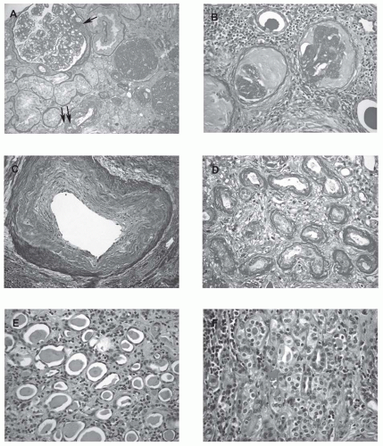


FIGURE 65.1 Morphological changes in the aging kidney. A: There are two glomeruli displaying solidified global glomerulosclerosis in which the sclerotic tufts fill the entirety of the Bowman space, often representing the sclerosis caused by focal segmental glomerulosclerosis. The nonsclerotic glomerulus shows ischemic changes with a segmental adhesion to the Bowman capsule (arrow). Significant tubular atrophy and interstitial fibrosis are also noted. A few arterioles demonstrate significant hyalinosis (double arrows, PAS, periodic acid-Schiff; × 200). B: The two glomeruli show ischemic obsolescence characterized by shrunken and globally wrinkled and thickened capillary tufts with the loss of most cells. The Bowman space is filled with collagenous material that stains less intensely than the capillary tuft (PAS; × 400). This type of global glomerulosclerosis is often secondary to ischemic vascular disease. C: An interlobular artery shows intimal fibrosis (arteriosclerosis) characterized by fibrous thickening and migration of medial muscle cells into the intima with an atrophic muscle layer (Trichrome; × 400). D: Classic type tubular atrophy: There is severe tubular atrophy with thickening and lamellation of the tubular basement membranes (PAS; × 400). E: A thyroidization type tubular atrophy: The atrophic tubules have a thin epithelium and contain homogeneous casts resembling thyroid tissue (H&E, hematoxylin and easin; × 400). F: An endocrine type tubular atrophy: The small tubules reveal cuboidal cells with pale-staining cytoplasma (containing abundant mitochondria) and virtually no lumens, reminiscent of endocrine glands (H&E; × 400). (See Color Plate.)

In aging kidneys, the thickening and folding/wrinkling of glomerular basement membrane (GBM) is accompanied by glomerular simplification and the formation of anastomoses between glomerular capillary loops. Frequently, afferent arteriole dilatation near the hilum is observed at this stage. The afferent arterioles commonly develop hyalinosis (accumulation of plasma proteins in the intima and/or media of small arteries and arterioles), a change that is less well correlated with systemic hypertension than with arterial intimal fibrosis in most, but not all studies. It may also be seen with diabetes mellitus; in fact, it is most severe and pronounced in patients with uncontrolled diabetes mellitus with or without hypertension.17

In the aging kidneys, the sclerosis and eventual loss of the glomerular tuft is often associated with a direct communication between afferent and efferent arterioles (“aglomerular arterioles”), particularly the juxtamedullary glomeruli.19 These aglomerular arterioles are rarely seen in the kidneys of healthy adults but are observed with increased frequency both in aging and CKD kidneys.19 The age-related vascular changes of intimal fibrosis and hyaline arteriolosclerosis are accentuated by hypertension, and probably diabetes mellitus as well. On the other hand, it has been suggested that aging-related interlobular arterial sclerosis may precede rather than follow systemic hypertension. Mean blood pressure rises by 1.6 mm Hg for each 1 µm increase in intimal thickness in a 100 µm diameter artery because of microischemia in scattered nephrons. This source of hypertension may account for the rise of blood pressure with age. The rate of decrease of renal plasma flow is accelerated by hypertension. Mean arterial blood pressure is directly proportional to the rate of decline of creatinine clearance. An increase in hypertension is a strong independent risk factor for ESRD, especially in African Americans. Thus, hypertension and morphologic vascular changes are not easily separable at this time.20,21,22


FUNCTIONAL CHANGES OF THE AGING KIDNEY

Aging is commonly associated with a decline in renal function. Unfortunately, most studies showing a diminished renal function with aging are confounded by comorbidities such as diabetes, hypertension, obesity, and smoking. It is therefore nearly impossible to separate the effect of physiologic (aging, per se) from pathologic (due to comorbidities) aging on renal function.23 This section summarizes our current understandings of the effects of aging on renal function (Table 65.2).


Renal Hemodynamics

The morphologic changes of aging are accompanied by parallel changes in renal function and hemodynamics. Using various techniques, several studies conducted in elderly individuals without significant renal disease have demonstrated that renal blood flow (RBF) decreases with advancing age. In a review of 38 renal hemodynamic studies including 634 healthy subjects with wide age range, Wessen24 described that total RBF was well maintained through approximately the fourth decade and progressively declined by approximately 10% per decade thereafter. In a study of 207 healthy kidney donors, Hollenberg et al.25 demonstrated an explicit and progressive reduction in mean blood flow per
unit kidney mass with advancing age, suggesting that the decrease in RBF does not simply reflect the decline in the renal mass with aging. In addition, they demonstrated that the fall in renal perfusion with aging is most profound in the cortex, with relative sparing of flow to the medulla. This redistribution of blood flow from the cortex to the medulla may explain the slight increase in filtration fraction observed in the elderly population. Studies on the morphology and histology of the renal vasculature by postmortem angiograms and histologic sections demonstrate increased irregularity and tortuosity of the preglomerular vessels and tapering of afferent arterioles. However, no characteristic histologic lesion of aging has yet been identified in the renal vasculature.


TABLE 65.2 Altered Glomerular, Tubular, and Vascular Functions in the Aging Kidney


Glomerulus


Decreased renal blood flow


Decreased glomerular filtration rate


Higher single-nephron ultrafiltration coefficient


Tubulointerstitium


Impaired urine concentrating and diluting ability


Impaired ability to maintain fluid and electrolyte balance


Reduced activity of the renin-angiotensin-aldosterone system (RAAS)


Reduced erythropoietin production


Reduced level/activation of 1,25 vitamin D


Increased renal calcium loss


Vasculature


Loss of compliance and increased stiffness of major arteries


Impaired angiogenesis


Impaired endothelial function


The precise mechanisms of reduced RBF with aging are not yet known. Aging is associated with changes in vascular tone, which is determined by the balance between vasoconstrictors and vasodilators. In aging, there is an attenuated response to vasodilators such as nitric oxide (NO), endothelial-derived hyperpolarizing factor (EDHF), and prostacyclin, and an enhanced responsiveness to vasoconstrictors such as angiotensin II (Ang-II).26 This may result in enhanced vasoconstrictive responses in aging, which can potentially cause renal damage and an ultimate fall in GFR. Although the renin-angiotensin system (RAS) is suppressed in aging, the intrarenal RAS may be relatively spared. In fact, the pharmacologic blockade of RAS has been shown to slow the progression of age-related CKD in experimental animals.27 In addition to the suppression of RAS, there is significant decrease in NO production and availability that leads to renal vasoconstriction and sodium retention. Several potential mechanisms contribute to the reduction of NO with aging. Chief among them is oxidative stress that can reduce NO availability by the inactivation of NO; the inhibition of NO synthase (NOS) via the depletion of the NOS cofactor, tetrahydrobiopterin; the uncoupling of endothelial NOS; the accumulation of the endogenous NOS inhibitor, asymmetric dimethyl arginine; and by limiting uptake of NOS substrate, L-arginine, by endothelial cells via downregulation of cationic amino acid transporter-1.28

There is also evidence that angiogenesis is attenuated in aging. In this context, vascular endothelial growth factor (VEGF) and angiopoietin-1 are altered both systemically and in the kidney with the aging process. Although levels of VEGF29 have been shown to be reduced in the aging rat, a profound upregulation in protein levels of angiopoietin-1 in the kidney cortex has been observed in aged versus young rats.30 Because angiopoietin-1 can stabilize blood vessels, its increase in aging may serve to counter the mechanisms leading to impaired angiogenesis and endothelial dysfunction. Taken together, these data indicate that strategies aimed at protecting the endothelium may help to mitigate the adverse renal effects of aging.

Recently, the role of arterial aging or arteriosclerosis in the pathogenesis of senescent changes in various organs, including the kidney, has become a major focus of interest. It has been observed that elastic arteries undergo two distinct physical changes, namely, dilation and stiffness with age.31,32 This is due to fatigue and fracture of the medial elastin, mainly of the elastic arteries, with little aging change occurring in the distal muscular arteries.33 Thus, dilation and stiffening are most marked in the proximal aorta and its major branches, namely, the brachiocephalic, carotid, and subclavian arteries. Increased arterial stiffening results in an increase in pulse wave velocity (PWV).31,34 Aortic PWV is the speed with which pulse waves travel along the artery. A typical value is 5 m per second in a 20 year old and 12 m per second in an 80-year-old person, representing a 2.5-fold increase in 60 years.31 The elastic properties of the aorta in the young serves to partially maintain blood volume and pressure during systole and then release them during diastole via the recoil process. This phenomenon helps to protect the vital organs by sustaining blood flow during diastole and blunting the damaging effects of high pressure waves during systole. In addition, the microcirculation, which comprises small arteries, arterioles, and the capillaries and constitutes the greatest resistance to blood flow, participates in transforming pulsatile flow to steady flow by reflecting the pulsations that enter from the larger arteries. With aortic stiffening and the consequent increase in aortic PWV, the transmission of flow pulsations downstream into various organs, principally the brain and kidney, can damage microvessels.35,36 This mechanism may account for asymptomatic cerebral microvascular disease associated with microaneurysms and infarcts. The lesions comprise damage to the medial smooth muscle and the endothelium (which is not attributable to atherosclerosis), and in their chronic form are described as lipohyalinosis.37 More recent studies have shown that amyloid plaques in older persons are probably a consequence of medial damage to small vessels and hemorrhage from damaged vessels.38,39 It is thought that the neurofibrillary tangles of dementia may have a similar microvascular etiology.40 Less data on pulsatile microvascular damage are available for the kidney, but one can expect this to emerge. The kidney afferent arterioles and glomeruli are exposed to the same high pulsatile microvascular stress and strain as in the brain. Recent studies have shown that independent of conventional brachial systolic and diastolic pressure values, measures of arterial stiffness are closely related to outcomes attributable to microvascular damage to vital organs, particularly the brain and the kidney.35,36 Furthermore, measures of large artery stiffness are closely related to effects of microvascular changes in the kidney, including albuminuria.35,36 Interventions aimed at reducing the ill effects of arterial stiffening by reducing the extent and frequency of the stretch cycles in order to minimize fatigue and fracture of the medial elastin of aorta can reverse or delay progression of cerebral and renal damage. These maneuvers entail a reduction in early wave reflection achieved through regular exercise, and the use of drugs such as angiotensin-converting enzyme inhibitors
(ACEI), angiotensin receptor blockers (ARB), calcium channel blockers, and nitrates, which relax smooth muscle in large and small conduit arteries throughout the body, thus resulting in arterial dilation.37,41


Glomerular Filtration Rate

GFR gradually increases after birth, approaching adult levels by the end of the second decade. It remains stable until the age of 30 to 40 years and then declines linearly at an average rate of about 8 mL per minute per decade, a phenomenon that can be partially explained by age-associated glomerulopenia.7,8,42 However, about one-third of elderly individuals show no change in GFR.43 This variability suggests that factors other than aging may be responsible for the apparent reduction in renal function. For instance, an increase in blood pressure, still within the normotensive range, is associated with an accelerated age-related loss of renal function.

The use of creatinine clearance in a timed urinary sample is commonly used as an estimate of GFR. Inulin and iothalamate clearance are very accurate measurements of GFR, but are clinically cumbersome to perform.44 To obviate the need for a timed urine collection, various equations have been developed and are increasingly used to estimate GFR (Table 65.3). In adults, creatinine clearance is often estimated by the Cockcroft-Gault (CG) equation, and GFR is estimated by the Modification of Diet in Renal Disease (MDRD) formula.45,46 It is important to point out that the CG equation estimates GFR in milliliters per minute, whereas the MDRD formula expresses GFR in milliliters per minute per 1.73 meters squared.


TABLE 65.3 Formulas to Estimate Glomerular Filtration Rate


Formula


Strengths


Weakness


1. Cockcroft-Gault


Simple


Estimates Ccr, not GFR


Estimates lower value in older subjects


Falsely elevated in chronic kidney disease


Not validated for elderly population


2. MDRD


Simple with use of calculators


Estimates GFR and not Ccr


Estimates higher GFR in the elderly


Not validated for elderly population


Not validated for higher levels of GFR


Not validated to measure normal kidney function


3. CKD-EPI


Simple with use of calculators


Estimates GFR and not Ccr


More accurate estimates of high GFR


Better estimation of GFR in elderly than 1&2


May still overestimate GFR in the elderly


4. Cystatin C


Less dependent on muscle mass


Better GFR estimate in elderly


Better predictor of adverse outcomes in CKD


Not available readily


Clinical role yet unclear


Ccr, creatinine clearance; GFR, glomerular filtration rate; MDRD, Modification of Diet in Renal Disease; CKD-EPI, Chronic Kidney Disease Epidemiology Collaboration.


However, neither MDRD nor the CG equation was developed for elderly individuals, and reduced reliability would be expected when used in this population. In a study involving 100 individuals aged 65 to 111 years, GFR values calculated with the MDRD formula were much higher than those obtained with the CG equation. Moreover, no correlation was observed between these two predictions. Additionally, the difference in GFR values between MDRD and CG increased dramatically with aging and decreased with higher body mass index and serum creatinine values. Thus, the precision and accuracy of these formulas in estimating GFR in very old patients remain arguable.47 Furthermore, a study in patients over 65 years old showed more than 60% discordance in GFR estimation by the two equations. The MDRD equation generally yielded higher estimates of GFR than the CG equation.48 This has important implications, especially when calculating drug dosages in the elderly. It was recommended that the CG equation should be used in preference to the MDRD equation to estimate GFR for drug dosage calculations in the elderly. Recently, a new creatinine-based equation was developed by the Chronic Kidney Disease Epidemiology Collaboration (CKD-EPI). It reported a more accurate estimation of GFR than the MDRD equation, particularly at higher levels of estimated GFR (eGFR).49 In a recent prospective observational study of 439 patients aged 65 and older admitted to 11 acute care medical
wards in Italy, the relative risk of mortality in patients with eGFR = 30 to 59.9 or < 30 mL/min/1.73 m2 was compared to subjects with eGFR ≥ 60 mL/min/1.73 m2 using the body surface area-adjusted CG (CG-BSA), the MDRD, and the CKD-EPI formulas. Participants with reduced GFR showed an increased mortality regardless of the equation used, and CKD-EPI-derived GFR outperformed to some extent MDRD-and CG-BSA-derived GFR in a multivariable predictive model, suggesting its usefulness in the elderly population.50 However, the performance of the serum creatinine-based estimating equations still remains insufficiently evaluated in older patients, in whom there may be a high prevalence of chronic disease associated with alterations in muscle mass and diet, resulting in an overestimation of the measured GFR and an underestimation of the severity of CKD. Lately, cystatin C has emerged as an alternative marker for the measurement of kidney function.51 Cystatin C, a filtration marker that is less related to muscle mass than creatinine, may have a particular advantage in the estimation of GFR in the elderly population. However, the clinical role of cystatin C measurement remains unclear. In a recent study involving 11,909 patients, the risk of death, cardiovascular events, and kidney failure was compared in patients with GFR < 60 mL/min/1.73 m2 (CKD) to those with GFR > 60 mL/min/1.73 m2, as estimated by creatinine and cystatin C measurements. The survey showed that cystatin-based estimates were better predictors of adverse outcomes among adults with CKD, suggesting that cystatin C may be useful in identifying patients with CKD who have high risks of complications.52


Tubular Functions


Urine Concentrating Ability in the Aging Kidney

The urine concentrating phenomenon is a complex process that depends on many factors such as RBF, GFR, solute load, presence of vasopressin, functionality of vasopressin receptors, urea transporters, sodium transporters, and water channels (aquaporins), as well as the presence of an intact medullary countercurrent system. The Baltimore Longitudinal Study of Aging evaluated urine concentrating ability in healthy people aged between 20 to 79 years by assessing maximum urine osmolality, minimal urine flow rate over a period of 12 hours, and ability to concentrate solutes (or reabsorb sodium and urea). Compared to younger age groups, individuals aged 60 to 79 years had an approximately 20% reduction in maximal urine osmolality, a 100% increase in minimal urine flow rate, and a 50% decrease in the ability to conserve solutes.53 These changes could not be explained by the reduction in GFR. A decrease in the abundance of aquaporin and urea transporter proteins, as observed with aging in the kidneys of animals, likely accounts for the reduced urinary concentrating capacity in the elderly. Interestingly, no significant differences in antidiuretic hormone (ADH) levels have been observed between the elderly and the younger cohorts, suggesting that the defect is likely due to ADH resistance as opposed to ADH deficiency.54 Experimental studies suggest that an abundance of aquaporins-2 and -3 is reduced by 80% and 50%, respectively, in the aged rat’s renal medullary collecting ducts.55 Besides the decrease in aquaporin-2, there is impairment of its phosphorylation, which may interfere with trafficking and the insertion of aquaporin-2 in the apical membrane of the collecting duct. Together, these defects diminish urine concentrating ability by decreasing water reabsorption in the collecting ducts. In addition, aging results in decreased abundance of the major urea transporters (UT-A1 and A2) in the inner medullary collecting duct55,56 and reduced NaCl transporter NKCC2/BSC1 in the thick ascending limb of loop of Henle.57 These changes can reduce urine concentrating ability in the elderly by limiting urea and sodium reabsorption and, hence, inner medullary osmolality.

Although the reduction of water-conserving capacity in the elderly is mild and does not have significant clinical implications under normal conditions of water abundance, it becomes important when access to water is limited; for instance, in cases of inability to grasp fluid or communicate thirst as in stroke patients or in cases of nursing home neglect. Under such conditions, old patients might develop serious hypernatremia that may impair central nervous system function or prove fatal. Prompt treatment with intravenous hypotonic solutions may prove lifesaving in these situations.


Renal Diluting Ability

Although much less data are available on renal diluting capacity, existing studies suggest a mild impairment of renal diluting ability in the elderly. Clearance studies in waterloaded old rats have demonstrated free water formation at each level of the distal diluting segment, indirectly suggesting that the function of the limb of the Henle loop is not impaired.58 The mild renal diluting defect seems to be a result of the reduced GFR. The increase in solute load in the remaining functioning nephrons combined with a decrease in NaCl reabsorption increases solute delivery to the collecting duct and decreases free water excretion. There is no evidence for impaired function of the diluting segment or altered suppressibility of vasopressin in the pathogenesis of this disorder.

Reduced renal diluting capacity renders the older subjects more susceptible to the development of dilutional hyponatremia in the setting of excess water load, stress situations such as surgery, fever, acute illness, or administration of drugs such as diuretics, or those that enhance vasopressin production and action. These events may act alone or in concert to impair renal diluting ability and render the elderly patients susceptible to water intoxication. In fact, hyponatremia is the most common electrolyte abnormality in hospitalized geriatric patients.59 Hyponatremia usually develops insidiously and presents with nonspecific clinical findings including confusion, lethargy, anorexia, nausea, weakness, and seizures. Like hypernatremia, hyponatremia may impair central nervous system function or may prove fatal. In such
situations, prompt treatment with free water restriction alone or with the concurrent administration of hypertonic solutions or vasopressin V2-receptor antagonists is warranted.60,61


Fluid and Electrolyte Balance

Ordinarily, age has no effect on basal plasma electrolyte concentrations or the ability to maintain normal extracellular fluid volume. However, structural changes in the elderly kidney have an impact on the adaptive mechanisms responsible for maintaining homeostasis of extracellular fluid volume and composition. Consequently, acute illnesses in geriatric patients are often complicated by the development of fluid and electrolyte abnormalities, which are associated with increased morbidity and mortality and prolonged hospitalization. In the elderly, the capacity to conserve sodium in response to reduced sodium intake is impaired.62 The exact mechanism is not known, but a reduction in the number of functioning nephrons with increased sodium load per each remaining nephron as well as reduced aldosterone secretion in response to sodium depletion are plausible. Nevertheless, the inability to conserve sodium may predispose the elderly patient to hemodynamic instability in the setting of sodium loss. This, along with other structural and functional changes, make older patients more prone to develop acute kidney injury.63

In addition to the impairment in sodium conservation, the elderly are also prone to volume expansion when challenged with a sodium load. This is due to a diminished capacity of renal sodium excretion in the elderly.64 Additionally, the elderly seem to have more sodium excretion at night compared to the daytime, suggesting an impaired circadian variation.65 Impaired pressure natriuresis and altered response to Ang-II are apparent mechanisms involved.66 Notwithstanding the mechanisms, geriatric patients may develop an expanded extracellular fluid volume in the setting of a sodium load. It will usually lead to modest weight gain and the appearance of mild peripheral edema in the absence of significant comorbidities. However, the geriatric patients with preexisting cardiac or renal disease may develop life-threatening pulmonary edema, necessitating aggressive emergency therapy with loop diuretics or dialysis.66 Elderly subjects also show abnormalities in renal potassium and calcium handling, which are discussed in the ensuing section.


Endocrine and Metabolic Function


Renin-Angiotensin-Aldosterone System

Age-related changes in the RAS in healthy humans are well documented.67 In elderly subjects at baseline, plasma renin activity is 40% to 60% lower than those of a young adult population.68 This difference becomes even more pronounced under conditions that stimulate renin release because of the blunted renal response in the elderly.69 This decrease may act to lower baseline intrarenal Ang-II levels, an adaptation that may contribute to the changes of intrarenal vascular tone and tubular function in the aging kidney.

The lower renin levels in the elderly results in 30% to 50% reductions in plasma aldosterone levels.70 The age-related decrease in renin and aldosterone levels contributes to the development of various fluid and electrolyte abnormalities. For instance, elderly persons on salt restricted diets have a decreased ability to conserve sodium.62 Decreased Ang-II production has been reported to impair urinary concentrating ability. Together, these conditions contribute to increased susceptibility of elderly persons to develop volume depletion and hypernatremia.63 The loss of thirst in response to dehydration further contributes to hypernatremia in the elderly. Age-related decrease in renin and aldosterone also contributes to an increased risk of hyperkalemia in various clinical settings and is reflected by a reduced transtubular potassium gradient in the elderly population.71

Through action on distal tubules, aldosterone increases sodium reabsorption and facilitates potassium excretion, thereby protecting against hyperkalemia after a potassium load.67 A decrease in the production of renin-angiotensin-aldosterone and reduced GFR impair the ability of the elderly to handle large potassium loads. Potassium levels can be seriously elevated after a potassium-loading event such as gastrointestinal bleeding, transfusion reaction, or the administration of oral or intravenous potassium. The tendency toward hyperkalemia can be further enhanced by certain inorganic metabolic acidosis or by the administration of medications that inhibit potassium excretion (such as potassium sparing diuretics, ACEI, ARB, nonsteroidal anti-inflammatory agents, direct renin inhibitors, or beta blockers). Given their higher susceptibility to hyperkalemia, caution should be exercised in prescribing such medications to the elderly.

Although the circulating RAS is suppressed in aging, the intrarenal RAS may be relatively intact. Ang-II has several hemodynamic and nonhemodynamic effects on the kidney, affecting not only filtration pressure and proximal tubular sodium and water transport but also tubular and glomerular cell growth, NO synthesis, immunomodulation, growth factor induction, production of reactive oxygen species (ROS), inflammation, cell migration, apoptosis, as well as extracellular matrix protein accumulation, which can work in concert to accelerate age-related glomerulosclerosis and tubulointerstitial fibrosis.72,73 Preferential Ang-II-dependent efferent arteriolar vasoconstriction of older nephrons maintains adequate filtration pressure. However, this may also promote intraglomerular hypertension and glomerulosclerosis.74 Furthermore, Ang-II activates proinflammatory and profibrotic pathways, including transforming growth factor (TGF-β), collagen IV transcription, monocyte-macrophage influx, mRNA, and protein expression of chemokine regulated upon activation, normal T-cell expressed, and secreted (RANTES) promoting fibrosis, as well as stimulating endothelial plasminogen activator inhibitor-1(PAI-1) to increase matrix accumulation.75,76 Interestingly, physiologic intrarenal downregulation of both renin mRNA and ACE in the elderly may be protective toward long-term sclerosis, and the processes that increase Ang-II response with age can hasten kidney aging.77


Use of ACEI in aging animals has been shown to decrease glomerular and vascular sclerosis as well as tubulointerstitial fibrosis associated with a reduction in α smooth muscle cell actin.75,78 Angiotensin antagonists (ACEI and ARB) may protect age-related renal sclerosis by additional mechanisms such as the prevention of age-associated oxidative stress, advanced glycation end products (AGEs) accumulation, and downregulation of endothelial nitric oxide synthase (eNOS), and Klotho.79,80


Erythropoietin

The prevalence of anemia increases with age. Although there are many causes of anemia in the elderly, normocytic normochromic anemia may be related to reduced erythropoietin (EPO) production by the kidney.81 The InCHIANTI study showed an association between advancing age, declining renal function, reduced EPO production, and anemia. After adjusting for confounding variables, the subjects with a creatinine clearance of 30 mL per minute or lower had a higher prevalence of anemia and lower plasma EPO levels compared with those with a creatinine clearance higher than 90 mL per minute. Additionally, a trend toward an increase in the prevalence of anemia with decreasing renal function was observed in subjects with creatinine clearance > 30 mL per minute.82 Serum EPO levels rise with age in healthy subjects, perhaps a compensation for aging-related subclinical blood loss, increased red blood cell turnover, or increased erythropoietin resistance of red cell precursors.83 On the other hand, the serum EPO levels are unexpectedly lower in the elderly with anemia compared to young subjects with anemia, suggesting a blunted response to low hemoglobin.84

Erythropoiesis-stimulating agents (ESAs) are often used for the treatment of anemia in patients with CKD. However, several recent studies on the treatment of anemia with ESA, including CHOIR (correction of hemoglobin and outcomes in renal insufficiency), CREATE (the cardiovascular risk reduction by early anemia treatment with epoetin beta), and TREAT (trial to reduce cardiovascular events with Aranesp therapy), have caused concerns about the safety of ESA in CKD, including the elderly.85,86,87 The major findings of these studies reveal an increased risk of adverse cardiovascular outcomes with a more aggressive treatment of anemia with ESA. The adverse effects observed in trials of anemia correction are primarily due to the nonerythropoietic actions of ESA given at high doses to overcome erythropoietinresistant anemia.88,89 Thus, caution should be exercised in using ESA to treat anemia in the elderly, and a rise in hemoglobin of greater than 12 g per deciliter should be avoided.


Calcium and Vitamin D

A creatinine clearance less than 65 mL per minute is reported to be an independent risk factor for falls and associated fractures in the elderly with osteoporosis.90 In a recent study, elderly women with osteoporosis and a decreased creatinine clearance (< 60 mL per minute) had lower calcium absorption, lower serum 1,25-dihydroxyvitamin D, and normal serum 25-hydroxyvitamin D, suggesting a reduced conversion of 25-hydroxyvitamin D to 1,25-dihydroxyvitamin D by the aging kidney. Furthermore, calcitriol therapy reduced the number of falls by 50%, which was postulated to be related to an increase in serum 1,25-dihydroxyvitamin D, upregulation of vitamin D receptors (VDR) in muscle, and improvement in muscle strength.91 Levels of 1,25 dihydroxyvitamin D3 and its receptor, VDR, which are highly expressed in the kidney, decrease with age. Evidence suggests that vitamin D3 and its analogs suppress renin, and the absence of the VDR gene results in a predisposition for high renin hypertension, cardiac hypertrophy, and thromobogencity.92 Recent studies also demonstrate that VDR stimulation can decrease renal fibrosis.93 Vitamin D and its analogs can also attenuate glomerulosclerosis and tubulointerstitial fibrosis mediated by proinflammatory, profibrotic, and oxidant stress via the suppression of nuclear factor kappaB (NF-κB).94 Vitamin D deficiency in those with CKD appears to be an independent predictor of renal disease progression.95 Results of ongoing clinical trials will help clarify the clinical benefit of vitamin D in renoprotection.96

In addition to reduced levels and an impaired activation of vitamin D, elderly individuals demonstrate increased renal calcium loss due to reduced calcium reabsorption in the distal convoluted and connecting tubules.97 Distal calcium reabsorption is facilitated by the transient receptor potential ion channel, transient receptor potential cation channel subfamily V member 5 (TRPV5), in the tubular apical membrane.98 TRPV5 gene expression is regulated by 1,25-dihydroxyvitamin D and parathyroid hormone (PTH).99 Recently, the antiaging hormone, Klotho, has been shown to play a role in the regulation of distal calcium reabsorption by deglycosylating N-glycans on the surface of TRPV5.100 Klotho deficiency is associated with a phenotype resembling aging in experimental animals.101 Thus, impaired Klotho activity in the elderly may well be responsible for reduced calcium reabsorption via TRPV5.


MOLECULAR EVENTS IN AGING KIDNEYS

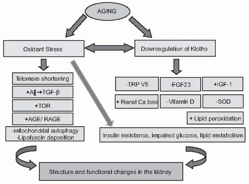
Over the last decade, significant advances have been made in identifying the molecular mechanisms associated with renal senescence. The key mechanisms are discussed in this section (Fig. 65.2).


Telomeres

Telomeres are the nucleoprotein structures constituting the physical ends of linear chromosomes. They prevent chromosomal ends from being recognized as double-strand breaks and protect them from end-to-end fusion and degradation.102 Telomeres in somatic cells shorten with each cell division and this progressive attrition leads to critically short telomeres and cellular senescence, a state characterized by the absence of replication and biochemical changes.102 This phenomenon was observed in earlier in vitro studies showing that cultured fibroblasts could only undergo a limited number of population doublings, the so-called Hayflick limit.103 It is
believed that telomeres act as a mitotic clock, initiating replicative senescence when telomeres become critically short after a certain number of cell divisions. Melk et al.104 have demonstrated that telomere shortening progresses with advancing age in the human kidney, and this phenomenon is more important in the cortical than the medullary area.104


FIGURE 65.2 The pathogenesis of renal aging. As detailed in the chapter, the aging process is associated with oxidant stress and downregulation of the Klotho gene. The downregulation of Klotho begets several downstream events such as the inhibition of FGF23, TRPV5, and impaired vitamin D activation, leading to increased urinary calcium loss. Furthermore, the downregulation of Klotho increases susceptibility to oxidant stress via the stimulation of the IGF-1 pathway and the inhibition of SOD. Increased oxidant stress, in turn, causes the downregulation of the Klotho gene. In addition, oxidative stress leads to the activation of the angiotensin II (Ang-II) pathway, enhancement of AGE formation, shortening of telomeres by the inhibition of telomerase, as well as the accumulation of malignant mitochondria in cells due to the activation of TOR. All these pathways ultimately lead to increased lipid peroxidation, insulin resistance, as well as impaired glucose and lipid metabolism. Final results of the cascade of these intertwined pathways are the age-related structural and functional changes in various organs. In kidneys, there is a progressive regression of renal mass associated with glomerulosclerosis, tubular atrophy, interstitial fibrosis, arterial sclerosis, and hyalinosis. These structural changes are associated with a progressive decline in GFR, myriad tubular abnormalities, and a reduction in renal blood flow. AGE, advanced glycation end product; RAGE, receptor for advanced glycation end product; FGF23, fibroblast growth factor 23; GFR, glomerular filtration rate; IGF-1, insulinlike growth factor 1; SOD, superoxide dismutase; TOR, target for rapamycin; TRPV5, transient receptor potential ion channel. Modified from Zhou XJ, Saxena R, Liu Z, et al. Renal senescence in 2008: progress and challenges. Int Urol Nephrol. 2008;40:823-839.

The enzyme telomerase is required for the maintenance of the length and stability of telomeres. An overexpression of telomerase induces an artificial lengthening of the telomeres and markedly increases the proliferative potential of cells. The antiproliferative effect of aging appears to be governed by two signaling pathways activated by the cellular replication clock: one involves p53, which induces p21 overexpression, and the other stimulates the expression of the cell cycle inhibitor, p16. In vitro studies have shown that senescent cells express p16 and p21, which inhibit cellular proliferation by inhibiting cyclin-dependent kinases.105 In vivo studies have demonstrated low levels of p16 in kidneys from young individuals. There is an overall increase in p16INK4a expression with age, particularly in the renal cortex, although this expression varies from individual to individual.106 Furthermore, in age-related histologic changes, a strong correlation is found between glomerulosclerosis, interstitial fibrosis, tubular atrophy, and p16INK4a and p53 expression.107 In a recent study, Westoff showed that telomerase-deficient mice were more vulnerable to ischemic-reperfusion injury due to reduced tubular regeneration.108

The pharmacologic activation of telomerase could be an appropriate therapy for aging diseases associated with replicative senescence. On the other hand, telomerase inhibitors have been proposed as a new option for chemotherapy, illustrating the trade-off between accelerated biologic aging and increased cancer risk.102,109


The Klotho Gene

The discovery of antiaging gene Klotho has extensively enhanced the understanding of the genetic bases of senescence. Klotho-deficient mice exhibited a syndrome resembling a
premature aging phenotype with soft tissue and vascular calcification, hyperphosphatemia, muscle and skin atrophy, and early death. In contrast, overexpression of the Klotho gene extended the life span in the mouse.110 In addition, several single-nucleotide polymorphisms in the human Klotho gene are associated with a shortened life span, osteoporosis, stroke, and coronary artery diseases, suggesting that Klotho may be involved in the regulation of human aging and age-related diseases.111

The Klotho gene encodes a single-pass transmembrane protein with two homologous extracellular domains, each having a weak homology to the β-glucosidase of bacteria and plants.112 Although the Klotho gene is expressed in limited tissues, notably the kidney, the parathyroid, and the brain, a defect in Klotho gene expression leads to multiple aging-like phenotypes involving almost all organ systems. This effect of the Klotho protein is mediated by its hormonal action via its binding to a cell-surface receptor and repressing the intracellular signals of insulin and insulinlike growth factor 1 (IGF-1). It appears that the antiaging effect of the Klotho-induced inhibition of insulin/IGF-1 signaling is associated with increased resistance to oxidative stress.113 It has been shown that the Klotho protein induces the expression of manganese superoxide dismutase, a mitochondrial antioxidant enzyme that facilitates the removal of superoxide, thereby conferring protection against oxidative stress.114 On the other hand, hydrogen peroxide-induced oxidative stress has been shown to reduce Klotho expression in a mouse inner medullary collecting duct (mIMCD3) cell line.115 Interestingly, the beneficial effects of peroxisome proliferator activated receptor-gamma (PPAR-γ) agonists on age-related glomerulosclerosis are mediated by intrarenal Klotho expression.116

Klotho also plays a central role in calcium and phosphorus homeostasis and the inhibition of active vitamin D synthesis.117 Ang-II has been shown to downregulate Klotho expression, which may, in part, contribute to Ang-II-induced renal damage.117 Klotho polymorphisms are associated with osteopenia in postmenopausal women. Klotho-deficient mice develop calcification of small arteries in the kidney and show increased serum levels of 1,25-dihydroxyvitamin D, which, along with increased calcium phosphate product, may be responsible for severe vascular and soft tissue calcification.117 Interestingly, the abnormalities in bone and phosphate metabolism observed in Klotho-deficient mice are very similar to those observed in fibroblast growth factor (FGF)-23 knockout mice suggesting that Klotho and FGF23 function in a common signal transduction pathway.118 Further studies indicate that Klotho acts as a cofactor by binding to FGF receptors and is essential for the signaling of FGF23 and related FGFs.119 Studies have shown that FGFs play important roles, not only in mitosis and development but also in various metabolic processes including the regulation of insulin sensitivity, glucose/lipid/energy metabolism, and oxidative stress, all of which potentially affect the aging processes.120,121 Klotho may regulate aging processes, partly through controlling the FGF-signaling pathways. The presence of any abnormality in either Klotho or FGF23 leads not only to phosphate retention but to premature aging in mice.122 A recent study by Hu et al.123 suggests that Klotho deficiency may have a direct effect on the rat vascular smooth muscle cell (VSMC) and promote VSMC calcification independent of FGF23 signaling.123

Klotho protein also regulates ion channel activity in renal tubular cells. Recently, Klotho was shown to activate transient receptor potential ion channel (TRPV)5, an epithelial calcium channel expressed on the apical membrane of the distal convoluted tubules and the connecting tubular cells.100 TRPV5 functions as an entry gate for transcellular calcium reabsorption in these cells and participates in renal calcium reabsorption, thus countering the effects of low phosphate on bone.124 Additionally, Klotho decreases cell surface abundance of the TRPC6 channel, the upregulation of which is associated with glomerulosclerosis.125

It has been shown that renal Klotho mRNA is downregulated under sustained circulatory or metabolic stress and in chronic kidney disease.121 Mitani et al.126 have demonstrated that Ang-II, which may be involved in age-related organ damage, plays a pivotal role in reducing renal Klotho gene expression in experimental animals. Conversely, induction of the Klotho gene by an adenovirus vector might protect against Ang-II-induced renal damage, such as tubulointerstitial injury and vascular wall thickening.126 The Ang-II-induced downregulation of the Klotho gene expression may be mediated by promoting intrarenal iron deposition and ROS production.120 In fact, treatment with a free radical scavenger and an iron chelator attenuated Ang-II-induced renal injury. Further investigations are required into other mechanisms involved in the regulation of the Klotho gene expression and senescence.


Oxidative Stress

Cumulative oxidative injury is believed to play a major role in the cellular aging process. Oxidative stress and the generation of free radicals increase with aging.127 Persistent oxidative damage to cytosolic structures leads to the crosslinking of oxidized proteins, the deposition of lipofuscin, and impaired mitochondrial function and structure.128 Lipofuscin (cross-linked proteins), which is insoluble and not degradable by either lysosomal enzymes or the proteasomal system, and giant mitochondria are taken up by lysosomes, wherein they bind additional material and eventually cause lysosomal rupture. The released lipofuscin and lysosomal contents cause cell damage and dysfunction.128 In rats of different ages (2, 11, and 29.5 months) there was up to a 28-fold increase in lipofuscin deposition in kidneys and other organs in older compared to the 2-month-old animals.129 Furthermore, increased oxidant stress (elevated serum lipoperoxides) and a reduced antioxidant activity (erythrocyte superoxide dismutase and glutathione peroxidase) were observed in the elderly in a cross-sectional study involving 249 healthy subjects.127 The magnitude of oxidative stress and lipid peroxidation in the aging kidney correlates with
an elevation of the advanced glycosylation end products and their receptors (AGE and RAGE) that can cross-link adjacent proteins. This, along with the ROS that can activate ubiquitin-proteasome, may degrade hypoxia-inducible factor-1alpha (HIF-1α) and limit the capacity of the aging cells to form HIF-1-DNA hypoxia-responsive recognition element (HRE) complexes (HIF-1-HRE complexes).130,131 In the kidney, the subsequent decrease in the ability of the cells to respond to hypoxia could explain the attenuated anemiainduced secretion of erythropoietin as well as the decreased hypoxia-induced production of vascular endothelial growth factor leading, respectively, to reduced erythropoiesis and angiogenesis.130 In a rat model, renal aging was associated with a 60% decline in GFR, a threefold increase in renal F2 isoprostanes (a marker of oxidative stress), an increase in oxidant-sensitive heme oxygenase, as well as increased AGEs and RAGE. Furthermore, a diet rich in vitamin E attenuated the age-related upregulation of heme oxygenase and RAGE, suppressed the production of F2 isoprostanes, lessened measured markers of oxidative stress, reduced glomerulosclerosis, and improved renal plasma flow and GFR by 50%.132 In cultured rabbit proximal tubular epithelial cells, AGEs and even the early glycosylation end products (Amadori products) directly inhibited NOS activity and AGEs quenched the released NO.133 Immunohistochemical studies of the aging rat kidneys have revealed a reduction of endothelial NOS (eNOS) in the peritubular capillaries and the presence of eNOS immunoreactivity in renal tubular epithelial cells, infiltrating mononuclear cells and foci of tubulointerstitial injury, suggesting that the aging-related renal tubulointerstitial fibrosis may be secondary to the ischemia caused by peritubular capillary injury and impaired eNOS expression.134 Via activation of the angiotensin receptor AT1, the tissue RAS may promote the production of ROS and TGF-β1, events that can promote fibrosis. Indeed, the administration of ACEI and ARB in rats ameliorated the aging-related renal damage and attenuated glomerular sclerosis, mesangial expansion, tubular atrophy, interstitial fibrosis, and mononuclear cell infiltration.135 The salutary effect of RAS blockade may be, in part, mediated by the ability to limit the impact of aging on the structure and function of mitochondria and other cellular organelles involved in energy metabolism and ROS production.136

In addition to the mechanisms described previously, recent data suggest that chronic oxidant stress contributes to the telomere shortening and thus senescent changes.106 Furthermore, by downregulating Klotho gene expression, chronic oxidant stress can mediate the aging-associated changes.137 Oxidative stress also activates the target of rapamycin (TOR) pathway, which plays an important role in the aging process by inhibiting mitochondrial autophagy (see the following).138 Thus, interventions aimed at reducing ROS production, enhancing antioxidant capacity, increasing NO availability, and suppressing fibrogenic pathways may be effective in retarding aging-related renal damage.139 In addition, calorie restriction via mechanisms described elsewhere in this chapter can decrease age-related oxidant stress, mitochondrial lipid peroxidation, and membrane damage.140 Thus, dietary caloric restriction may be another approach to reduce age related renal damage.

May 29, 2016 | Posted by in NEPHROLOGY | Comments Off on Renal Function and Disease in the Aging Kidney

Full access? Get Clinical Tree

Get Clinical Tree app for offline access