Pathology of the Male and Female Urethra


Pathology of the Male and Female Urethra


Jesse K. McKenney

Esther Oliva


ANATOMY AND HISTOLOGY

The urethra is a long muscular tube that carries urine from the urinary bladder to the external urethral orifice for excretion. In males, it also provides the conduit for semen, which enters the urethra through the ejaculatory ducts within the prostate. The urethra consists of an epithelial-lined mucosal surface enveloped by a supporting connective tissue/smooth muscle coat.

The male urethra is 15 to 20 cm in length and is divided descriptively into four regions/segments: preprostatic (intramural or within the bladder neck), prostatic, membranous (intermediate), and penile (spongy or cavernous).1, 2, 3 The prostatic urethra is located in the pelvis, while the membranous and penile regions are in the perineum. The shortest segment (1.0 to 1.5 cm), is the preprostatic or intramural urethra, which runs from the bladder neck to the superior aspect of the prostate. The second segment, the prostatic urethra, is approximately 3 cm in length and begins at the internal urethral orifice at the apex of the bladder trigone. It courses through the prostate, making an anteriorly concave bend, ending where the urethra penetrates the fascia of the urogenital diaphragm, and enters the perineum. The posterior wall of the prostatic urethra has several unique features related to prostatic secretory function. It contains a longitudinal ridge, the urethral crest, lined by two adjacent grooves, the prostatic sinuses.4

The prostatic ducts enter the urethra predominantly in the sinuses with fewer entering along the lateral aspects of the crest. The urethral crest also has a midline protuberance, the seminal colliculus or verumontanum, which contains a 5 mm vestigial opening called the prostatic utricle. The ejaculatory ducts open on each side of the utricle. The prostatic urethra receives blood supply from a branch of the inferior vesical artery, the urethral artery. Venous drainage occurs via the periprostatic plexus, while lymphatic drainage is primarily through the obturator and internal iliac nodes. Sympathetic and parasympathetic innervation of the prostate is derived from the pelvic plexus through the cavernous nerves. The third portion, the membranous urethra, is approximately 2.0 cm in length and extends from the apex of the prostate to the perineal membrane at the bulb of the penis within the deep perineal space. It is surrounded by the striated external urethral sphincter. The small bulbourethral glands, the Cowper glands, are located on each side of the membranous urethra. In the membranous (intermediate) urethra, the striated sphincter corresponds to the location of peak urethral closing pressure. Innervation to this muscle is supplied by the pudendal nerve,5 but a branch of the sacral plexus has also been identified as a second source of innervation.6,7 Blood is supplied via the artery to the bulb and the urethral artery, and lymphatic drainage is also to the obturator and internal iliac nodes. The distal segment, the penile or spongy urethra, is the longest, approximately 15 to 16 cm. It courses through the bulb of the penis and corpus spongiosum to its end at the external urethral orifice (meatus). The ducts of the bulbourethral glands open into the proximal (bulbar) portion of the penile urethra, approximately 2.5 to 3 cm distal to the perineal membrane. The ducts of the urethral (Littre) glands also open into the penile urethra and are concentrated on the dorsal surface of the penile urethra. There are also multiple mucosal recesses, or lacunae, within the penile urethra. The distal region of the penile urethra within the glans penis is a saccular expansion referred to as the fossa navicularis; it contains the largest lacuna, the lacuna magnum. The blood supply to the penile urethra is delivered by the internal pudendal artery and veins, while innervation is provided by branches of the pudendal nerve. The lymphatics of the distal penile urethra drain to the superficial inguinal lymph nodes.

The female urethra is shorter, measuring on average 4 cm in length, and extends from the bladder neck to the external urethral orifice in the vaginal vestibule. It is divided into two regions. The proximal portion corresponds to the prostatic urethra and the distal portion to the membranous urethra. The bulbar and pendulous regions of the penile urethra are androgen dependent and, therefore, are not present in the female. Urethral glands are located along the wall of the urethra, particularly in the superior portion. One group of glands,
the paraurethral (Skene) glands, represent the homolog of the prostate and are located on each side of the urethra. The paraurethral glands share a common duct on each side, the paraurethral ducts, which enter the urethra near the external urethral orifice. The female urethra receives blood circulation from the vaginal and internal pudendal arteries and veins, while it is innervated by the pudendal nerve. Lymphatic drainage courses to the internal iliac and sacral lymph nodes, with some drainage to the inguinal lymph nodes.

In the male, the preprostatic, prostatic, and membranous urethra regions have an epithelial lining identical to that of the urinary bladder with a stratified layer of urothelium and a superficial umbrella cell layer (Fig. 7-1A). The prostatic urethral lining may also contain admixed prostatic secretory cells. The lamina propria within these regions consists of loose collagenous tissue and small thin-walled blood vessels. The deeper structures of the penile urethra (corpus spongiosum, tunica albuginea, and Buck fascia) are described in the penile chapter. The pendulous and bulbar regions of the penile urethra are lined by a stratified layer of small columnar epithelial cells that are distinct from bladder urothelium. This distinctive epithelium is 4 to 15 cells in thickness and lacks an umbrella layer. The fossa navicularis is lined by a stratified nonkeratinizing squamous epithelium that is continuous with the squamous epithelium of the glans penis (Fig. 7-1B). In the female, the urethral lining is urothelial proximally with a transition to squamous distally; however, nonkeratinizing squamous metaplasia of the urothelial regions is common. The posterolateral bulbourethral (Cowper) glands have a lobular arrangement with central ducts lined by a cuboidal layer of epithelial cells and surrounding clusters of tubuloalveolar glands lined by mucin distended columnar cells (Fig. 7-1C). The urethral (Littre and Skene) glands consist of small aggregates of mucous glands (Fig. 7-1D).


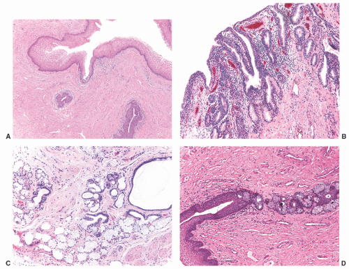
FIGURE 7-1 ▪ Mucinous-type glands are seen in the penile urethra (Littre glands) (D). The penile urethra is lined by glycogenated squamous epithelium (A) while the prostatic urethra shows alternating patches of urothelial and prostatic epithelium (B). Cowper glands show a lobular arrangement with central ducts (lined by urothelium) and glands lined by mucinous cells (C).


EMBRYOLOGY

The epithelium of the female urethra develops from the endodermal urogenital sinus, which is the anterior portion of the cloaca after it becomes separated from the posterior anorectal canal at 4 to 7 weeks’ gestation. The connective tissue and smooth muscle derive from the splanchnic mesoderm. Similarly in the male urethra, the epithelium of the prostatic and membranous portions of the urethra originates from the urogenital sinus and the connective tissue and smooth muscle are also derived from the splanchnic mesoderm.8, 9, 10 In the male, the bulbar and pendulous regions of the penile urethra are derived from the urethral plate on the ventral aspect of the genital tubercle. The epithelium of the fossa navicularis has historically been described as originating distally from ectodermal ingrowth of the glans penis9; however, more recent studies have suggested differentiation from the proximal endoderm.11


CONGENITAL ABNORMALITIES


Posterior Urethral Valves

This is the most common congenital cause of urethral obstruction leading to bilateral renal failure. It typically occurs in boys with an approximate incidence between 1:3,000 and 1:8,000 male births.2 Posterior urethral valves may be associated with hypospadias, ureteropelvic junction stenosis, imperforate anus, dysgenetic kidney, or congenital heart disease and very rarely with the prune belly syndrome.12

The clinical presentation is variable. Newborns with severe obstruction may present with urinary retention, enlarged bladder, hydronephrosis, uni- or bilateral reflux, and a history of prenatal in utero oligohydramnios associated with pulmonary hypoplasia. Older patients usually have a history of urinary tract infections, urinary retention, pyelonephritis, hematuria, and/or urinary incontinence.13 Cystography frequently shows dilatation of the urethra above the valves, bladder hypertrophy and dilation, bladder diverticula, tortuous dilated ureters, and varying degrees of hydronephrosis (Fig. 7-2).12,14,15 Three types of valves have been described. Type I, which is the most common, consists of two posterior folds extending from the verumontanum downward to fuse anteriorly, which has been also described as a complete diaphragm with a central pinhole (depending if a catheter has been put in previously).16 The folds are usually lined by urothelium, and variable degrees of inflammation may be seen associated with it or present in the lamina propria. Type II is the least common and consists of vertical folds between the verumontanum and proximal urethra and bladder neck. Typre III shows concentrick disk within the prostatic urerthra, either below or verumontanum and it is frequently associated with renal impression.

The treatment of posterior urethral valves is surgical. Mortality associated with this congenital abnormality is currently <5%.16 As the prenatal diagnosis of posterior urethral valves may be difficult, most deaths occur in newborns with severe bilateral renal dysplasia, pulmonary hypoplasia, electrolyte imbalances, or sepsis but may also occur in older children secondary to renal insufficiency.14,17


FIGURE 7-2 ▪ Type I posterior urethral valves in a newborn. There is marked dilation of the posterior urethra and severe bladder trabeculation. (Courtesy Dr. R Pieretti.)


Anterior Urethral Diverticulum and Valve

These congenital abnormalities frequently occur together as it has been hypothesized that commonly one of the walls of the diverticulum acts as an obstructive valve.18,19 They are rarer than posterior urethral valves with an approximate incidence of 1 in 5,000 to 8,000 male births,20 are not associated with other defects, and typically occur secondary to a defect of the corpus spongiosum. They occur more frequently in the bulbous region of the penile urethra.21 Clinical manifestations vary with age; neonates (secondary to severe obstruction) may present with hydronephrosis; however, older boys typically present with repetitive infections or voiding symptoms including incontinence, retention, or thin urine stream.22 Surgery is the mainstay of treatment. Complications secondary to obstruction may occur as described in posterior urethral valves including end stage renal disease, but they are much less common.23


Urethral Duplication

This is a rare congenital malformation that typically occurs in males and occasionally may be associated with bladder duplication.24 If both urethra and bladder are affected, patients also have other congenital anomalies, more
frequently involving external genitalia and gastrointestinal tract.25 Most urethral duplications occur in the same sagittal plane (dorsal or ventral to the urethra), and they are classified as complete (with one or two orifices) or incomplete (with a distal or proximal blind end) or may be part of an incomplete or complete caudal duplication (Fig. 7-3).26 This anomaly may be asymptomatic or be associated with infections, incontinence, or even double stream of urine if the duplication is complete.26 Histologically, the anomalous urethra is lined by urothelium that may be associated to variable degrees of inflammation in the lamina propria. In symptomatic cases, surgery is the treatment of choice.


FIGURE 7-3 ▪ Urethral duplication is identified by a catheter and urethral sound. (Courtesy Dr. R Pieretti.)


Megalourethra

This entity is characterized by absence of the corpus spongiosum or absence of both the corpus spongiosum and corpora cavernosa depending on the degree of severity.18,27 It may be seen in association with the VATER syndrome, prune belly syndrome (characterized by congenital absence or hypoplasia of the abdominal wall musculature, bilateral cryptorchidism, and anomalies of the urinary tract), or other isolated genitourinary anomalies.28 An enlarged and deformed penis can be noted in neonates. It is always important to perform a thorough evaluation of the urinary tract due to the high percentage of associated urinary anomalies. Surgical reconstruction depends on the degree of absence of penile tissue.


Hypospadias

This congenital abnormality is one of the most common in the United States with an approximate frequency of 1 in 125 male births.29 It occurs secondarily to arrest of the urethral development with incomplete fusion of the urethral folds resulting in an ectopic urethral opening on the ventral surface of the penis, scrotum, or perineum (Fig. 7-4). It is typically associated with an abnormal ventral curvature of the penis and an abnormal distribution of the foreskin around the area.30 Although the etiology of this condition is unknown, multiple factors have been related to it including maternal (advanced age and primiparity), endocrine (defects in testosterone metabolism or receptors), environmental, and inherited factors.29,31,32 Hypospadias may be an isolated defect or part of an intersex status. The diagnosis is established in most cases on physical examination, except the very minor forms that may require ultrasound. The defect is treated successfully with surgery.30,31


FIGURE 7-4 ▪ Penoscrotal hypospadias. Extreme ectopic urethral opening is present close to the perianal orifice. (Courtesy Dr. R Pieretti.)


Prostatic Utricle Cyst

These are cysts centered in the prostatic midline, around the area of the verumontanum, and arise from the prostatic utricle. They are typically seen in adult males, and in most instances, they represent an incidental finding during screening for other reasons.33 As occurs with the prostatic utricle, these cysts are typically connected to the urethra but not to the prostatic ducts.34 Prostatic utricle cysts are lined by cuboidal to columnar cells histologically similar to those lining the prostatic ducts and acini,35 and they frequently stain for prostatic-specific antigen (PSA).35,36 There seems to be some controversy regarding the embryogenesis of these cysts. In the past, they were thought to derive from müllerian remnants, but recent studies have shown that they may have an origin from both müllerian remnants and the urogenital sinus, hence their histologic and immunohistochemical characteristics.36 Prostatic utricle cyst should be distinguished from the so-called enlarged prostatic utricle, an anomaly frequently seen in the pediatric age group, typically associated with other genitourinary abnormalities, most commonly hypospadias or intersex.37


Other Congenital Abnormalities

Other rare urethral congenital anomalies include narrowing of the bulbar portion of the urethra (Cobb collar),38 Cowper syringocele, and lacuna magna.23,39


INFLAMMATION AND INFECTION


Urethritis


Nonspecific Urethritis

Nonspecific urethritis is defined as the finding of irritative urethral symptoms such as burning micturition without evidence of an infectious etiology. With new molecular assays, many cases that might have been previously diagnosed as nonspecific urethritis are now found to have infectious etiology. Nonspecific urethritis has been reported in association with drug exposure including isotretinoin and alcohol and with contact to chemical irritants such as spermicides.40,42 Nonspecific urethritis may also be part of the spectrum of Reiter syndrome (triad of urethritis, conjunctivitis, and arthritis), typically following dysenteric infections such as Shigella or Salmonella.43


Papillary and Polypoid Urethritis

Papillary and polypoid urethritis is a nonneoplastic lesion that is usually secondary to an inflammatory process; however, unlike the bladder, it is not commonly associated with a catheter.44,45 Irritative urinary symptoms and hematuria are common presenting signs. Characteristically, the exophytic appearance results from edema in the lamina propria (Fig. 7-5A), but variable fibrosis, chronic inflammation, and associated dilated blood vessels are also present.46 Older lesions tend to have less edema with more stromal fibrosis. The papillary component is usually broader at the base, but may also have a bulbous tip. Reactive urothelial changes are frequent and include urothelial hyperplasia, prominent nucleoli, and mitotic activity. The main differential consideration is a papillary urothelial neoplasm, but the inflammatory background and the simple papillary architecture usually aid in this distinction (Fig. 7-5B). These lesions usually regress with resolution of the inciting inflammatory condition.


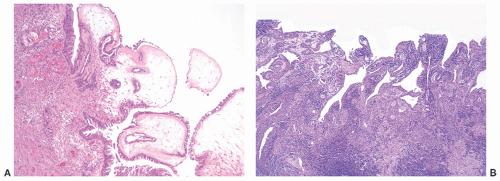
FIGURE 7-5 ▪ Polypoid cystitis. Bulbous and edematous papillae are lined by reactive-appearing urothelium (A). Papillary cystitis. Irregular thin papillae lined by two to three layers of urothelium are associated with prominent inflammatory background in the lamina propria (B).


Infections


Human Papillomavirus Associated Lesions

Human papillomavirus (HPV) is a common source of urethritis.47 HPV-related squamous dysplasia in the urethra is usually present in continuity with lesions of the external genitalia, anus, or perineum. Flat lesions range from low-grade squamous dysplasia with koilocytotic atypia to high-grade squamous dysplasia identical to that seen in the cervix. Flat intraepithelial squamous dysplasia is rarely biopsied de novo because most urethral lesions are subclinical48,49; however, it is common to find squamous dysplasia adjacent to invasive squamous cell carcinoma. Condyloma acuminatum is the most common HPV-related lesion, especially in the male urethra, and it commonly comes to clinical attention because of its intraluminal location with a resultant mass lesion, voiding symptoms, or bleeding.50 These lesions are morphologically identical to the more common perineal counterparts and are characterized by an exophytic squamous proliferation with hyperkeratosis, prominent granular layer, and typical cytologic features of HPV infection (perinuclear halos, binucleation, nuclear hyperchromasia, and irregular nuclear membranes) (Fig. 7-6A and B). HPV types 6 and 11 are most commonly found in association with condyloma, but other types have been also reported.51,52 For condyloma acuminatum, the main differential diagnosis is the rare squamous papilloma of the urothelial tract. The latter occur predominantly in elderly women and are characterized by fibrovascular cores
lined by benign-appearing squamous epithelium without viral cytopathic effect; they are also HPV negative by in situ hybridization studies.53 As in other sites, HPV-associated squamous dysplasia may progress to invasive squamous cell carcinoma, often associated with high-risk HPV types 16, 18, 31, 33, or 35. Therapy for squamous intraepithelial lesions is typically ablative, by surgery, laser, or topical agents.54


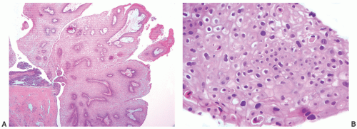
FIGURE 7-6 ▪ Condyloma acuminatum. A striking papillary architecture is seen (A) with the squamous cells showing striking koilocytic changes (B).


Gonococcus

Gonorrhea is a sexually transmitted infection caused by Neisseria gonorrhoeae, which is a Gram-negative diplococcus. Gonococcal urethritis typically affects teenagers or young adults. Patients typically present 2 to 5 days following infection with burning urination or a purulent urethral discharge.55 Gonorrhea may be diagnosed by culture, but ELISA and molecular diagnostic testing are also available.56,57 Tissue biopsy evaluation is not performed routinely. Urethral fluid sampling typically yields neutrophils, and Gram stain may reveal intracellular diplococci. The most significant complication is urethral stricture. Patients with chronic gonorrheal infection also develop urethral fistulae (“watering can perineum”). Ascending infection causing epididymitis in men or pelvic inflammatory disease in women may occur. Chlamydia coinfection is also common. Antibiotic therapy is curative, but drug-resistant strains are emerging.58 Postgonococcal urethritis is defined as persistent signs, symptoms, and/or laboratory evidence of urethritis 4 to 7 days following antibiotic treatment for documented gonorrhea. Most cases result from coinfection with Chlamydia or Ureaplasma infections; thus, the use of prophylactic multidrug treatment is advocated to cover possible coinfections.59


Chlamydia

Urethritis secondary to Chlamydia trachomatis, an obligate intracellular bacteria, is a sexually transmitted disease common in teenagers and young adults. Chlamydia is the most common sexually transmitted disease in the United States with over 1,000,000 infections reported to the Center for Disease Control in 2006.60 Chlamydia is asymptomatic in many patients but may present with painful/burning urination or urethral discharge 1 to 3 weeks after exposure.61 The diagnosis of Chlamydia may be based on culture, antigen detection by ELISA or direct fluorescent antibody (DFA), and/or molecular identification with methods such as nucleic acid amplification. The long-term complications of untreated cases are similar to gonorrhea, including epididymitis and pelvic inflammatory disease. In addition, genital chlamydial infection may be associated with urethritis, conjunctivitis, and arthritis (i.e., Reiter syndrome)62 or with reactive arthritis alone. Chlamydia is curable with antibiotic therapy.


Other

Other bacteria responsible for infectious urethritis include Ureaplasma urealyticum and Mycoplasma genitalium. Evidence suggests that Ureaplasma parvum commonly colonizes the urethra without resultant clinical symptoms.63 Viral etiologies of urethritis include HSV-1 and adenovirus, and being both associated with oral sexual habits.64 Urethral tuberculosis is exceedingly rare (male > female) and occurs secondarily to infection of the upper urinary (most commonly prostate) or female genital tracts. Predisposing factors include previous infection, immunosuppression, or prior BCG treatment for bladder carcinoma. In the urethra, the most common presentation is acute urethritis with or without discharge, or if untreated, stricture and/or fistulae. The microscopic appearance is characteristic and shows caseating granulomas. If chronic, extensive fibrosis may be seen. The standard treatment requires the use of antituberculous drugs for up to 9 months. However, as often patients present with complications, surgery is still a mainstay of treatment.65 Wegener granulomatosis can also rarely affect the urethra. Patients may have a known history but occasionally, this location may be the first manifestation of the disease. In males, the gross appearance can be quite worrisome,
and clinically misdiagnosed as carcinoma, especially in the penile urethra where Wegener granulomatosis may present as painful fungating mass or extensive ulceration (Fig. 7-7A). Histologic examination may not be diagnostic, only showing extensive necrosis as oftentimes the first diagnostic procedure is a biopsy (Fig. 7-7B). However, the classical morphology includes geographic areas of eosinophilic necrosis associated with palisading histiocytes. Vascular involvement typically shows granulomatous vasculitis with giant cells, but it is not always seen. Methotrexate is the treatment of choice, but patients still often require surgery due to local complications.66, 67, 68 The urethra is the least common site of involvement by sarcoidosis in the genitourinary tract. On gross examination, it may grossly simulate cancer.69 The morphology is that of noncaseating loose granulomas, but this histologic appearance is nondiagnostic and can be seen in infections and other processes including malignancy (typically at its periphery). However, these patients often have a well-established history of sarcoidosis.70


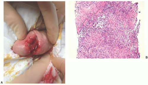
FIGURE 7-7 ▪ Wegener granulomatosis. A deep ulcer is present involving the penile urethra and surrounding tissues (A). (Courtesy Dr. F McGovern.) On microscopic examination, the lesion granulomatous inflammation with microabscesses and eosinophils (B).


METAPLASIA AND HYPERPLASIA


Squamous Metaplasia

Nonkeratinizing squamous metaplasia is a physiologic phenomenon in women. It is common in the trigone of the bladder, but may also involve the proximal urethra. Nonkeratinizing metaplasia is also reported in men following estrogen therapy.71 Urethral keratinizing squamous metaplasia may be seen in association with chronic trauma or chronic inflammatory conditions such as recurring infection, calculi, diverticula, or repeated instrumentation.72 Extensive and multifocal keratinizing squamous metaplasia may be associated with dysplasia and development of penile carcinoma (more frequently verrucous); thus, patients need to be closely followed.73


Glandular Metaplasia

This type of metaplasia is similarly related to a chronic irritative process. It most commonly involves the bladder but may extend into the urethra. Morphologically, glandular metaplasia is typically characterized by columnar epithelium with goblet cells (intestinal metaplasia).


Flat Urothelial Hyperplasia

As in the bladder, the urothelium of the proximal urethra may become markedly thickened. In the absence of cytologic atypia, this is termed flat urothelial hyperplasia.74 In isolation, urothelial hyperplasia is not regarded as a neoplastic precursor lesion, but it may be seen adjacent to low-grade papillary urothelial neoplasms.75


von Brunn Nests, Urethritis Cystica, Urethritis Glandularis

von Brunn nests represent a normal variation of urothelial histology that may result from a prior inflammatory insult. They are characterized by well-circumscribed, evenly spaced
nests of invaginated urothelium in the lamina propria. The nests typically have smooth, rounded contours and are often clustered in groups, but the connection with the overlying urothelium may not be identifiable (Fig. 7-8A). They are usually located in the superficial lamina propria, but they may deeper however, they are never present in the muscularis propria.


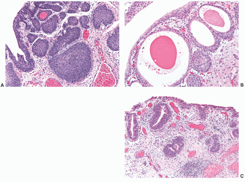
FIGURE 7-8 ▪ von Brunn nests are composed of solid nests of benign-appearing urothelium (A), Urtheritis cystica, shows cystic dilatation of the glands with abundant eosinophilic contents present in the lumens (B). Urtheritis glandularis, conventional type displays glandular metaplasia with a columnar or cuboidal appearance of the luminal cells (C).

The term “urethritis cystica” is used when the von Brunn nests become cystically dilated with an inner luminal cell layer (Fig. 7-8B). When the inner adluminal cells show glandular metaplasia with a columnar or cuboidal appearance and prominent apical cytoplasm, the term urethritis cystica glandularis is used (Fig. 7-8C). Finally, the presence of admixed intestinal-type goblet cells within the von Brunn nests warrants the designation cystitis cystica glandularis with intestinal metaplasia. These lesions are not regarded as obligate precursors of adenocarcinoma.76,77

The main differential diagnosis of von Brunn nests, urethritis cystica, urethritis glandularis are “deceptively benign” urothelial carcinomas that includes nested, microcystic, or tubular variants.78 In general, these carcinomas have architecturally complex, branching and crowded epithelial proliferations, at least focally, but individual cases may be morphologically indistinguishable from florid von Brunn or urtheritis glanduloris on a superficial biopsy. The identification of stromal reaction, cleft-like spaces surrounding the nests, an irregular infiltrative border, or invasion into the muscularis propria are the most helpful findings for recognizing subtle urothelial carcinomas. In contrast, the nests and glands of urethritis cystica, glandularis are superficial and have a sharp border with the lamina propria. Occasionally, the distinction from inverted papilloma (with a cystitis cystica-like pattern) may be somewhat arbitrary.79 Inverted papillomas typically show an anastomosing pattern of the epithelial nests and retain the peripheral palisading of cells even when central lumina are present. Occasionally, urothelial carcinoma in situ may extend into von Brunn nests and mimic invasion. The smooth round contour of the nests, the absence of surrounding retraction spaces, the lobular or linear arrangement of the nests with a noninfiltrative base, and the absence of a stromal reaction should aid in that distinction. Rarely, urothelial (transitional cell) carcinomas may grow as large regular nests with minimal cytologic atypia also mimicking von Brunn nests. However, they often also display irregularly infiltrating nests, some associated with desmoplastic reaction and others with muscularis propria invasion.80


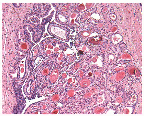
FIGURE 7-9 ▪ Verumontanum hyperplasia. Closely packed and small prostatic acini can cause concern for prostatic carcinoma in a small sample.


Cowper Gland Hyperplasia

One case of extensive Cowper gland hyperplasia presenting as a 6-cm urethral mass has been reported.81 Morphologically, it was characterized by greatly increased number of acini with banal cytologic features maintaining a lobular configuration. In a small biopsy specimen, Cowper glands may raise the differential diagnosis with prostatic carcinoma; however, negative immunohistochemical stains for PSA and prostatic acid phosphatase are helpful to rule out the diagnosis of carcinoma.82


Verumontanum Gland Hyperplasia

The verumontanum is an elevation present in the wall of the distal third of the prostatic urethra containing the prostatic utricle flanked by the ejaculatory ducts. This region of the prostate is infrequently biopsied. Verumontanum hyperplasia can be confused with prostatic adenocarcinoma on a biopsy as it shows an increased number of prostatic acini, which are crowded and small (Fig. 7-9). However, the epithelial cells have frequently cuboidal eosinophilic or, more frequently, pale cytoplasm with small nuclei and tiny to absent nucleoli, and the basal cell layer is preserved. Corpora amylacea with concentric laminations that can be fragmented are commonly seen.83


POLYPS, CYSTS, AND PSEUDOTUMOROUS LESIONS


Polyps


Caruncle

This benign lesion occurs mainly in women between 20 and 60 years of age (Table 7-1). It appears to be reactive and frequently related to trauma.84 Patients may present with dysuria or spotting or be asymptomatic. Caruncles are typically seen at the posterior wall of the urethra near the meatus. Grossly, they are pedunculated or broad-based with a dusky red or gray appearance. From the histologic point of view, three patterns have been recognized in the older literature: (a) granulomatous where the granulation tissue is the most striking component, (b) papillomatous with a prominent lobulated growth of the epithelium, and (c) angiomatous or telangiectatic rich in blood vessels.84 However, this classification is not used in routine practice as it lacks clinical or pathologic significance. Caruncles are typically lined by urothelium or less commonly squamous epithelium that may be hyperplastic (Fig. 7-10A). Metaplastic epithelial changes (intestinal glands) have been reported. The lamina propria is typically rich in acute and chronic inflammatory cells that are associated with abundant small blood vessels and edema (Fig. 7-10A). In some instances, atypical spindled to round mononucleated or rarely binucleated stromal cells arranged either in clusters or diffusely and showing large nuclei and prominent nucleoli may be seen (Fig. 7-10B).85,86 Myeloid metaplasia has been reported.87 The urothelial hyperplasia and striking invaginations of the surface epithelium frequently associated with caruncles may raise the possibility of a low-grade urothelial carcinoma. However, there is a bulbous rather than delicate papillary architecture, the nests of epithelium seen in the superficial stroma are rounded and do not elicit stromal response, and the cells lack any degree of cytologic atypia. When atypical cells are present in the stroma, the differential diagnosis include lymphoma,88,89 malignant melanoma,90 and even more rarely, spindle cell sarcoma.85 The small size of the lesion, scarcity of mitotic activity, as well as immunostaining limited to vimentin, smooth muscle actin, and rarely desmin exclude these possibilities.85 Surgical excision is the treatment of choice.


Prostatic-type Polyp

This benign papillary lesion occurs in adult males. It is typically found in the posterior urethra (more frequently in the verumontanum and prostatic urethra) and less commonly in the bladder or ureteral orifice.91, 92, 93, 94, 95 It often presents with painless gross or microscopic hematuria, voiding symptoms, or hematospermia91,96, 97, 98 although it may be an incidental finding.94 Grossly, it appears as an exophytic papillary to filiform or infrequently sessile lesion <1 cm in size.94 Histologically, prostatic-type polyps are composed of irregular polypoid growths or papillary fronds lined by a double layer of cells identical to that seen in prostatic acini with cuboidal to columnar cells displaying basally located nuclei and abundant clear to slightly eosinophilic cytoplasm overlying flattened basal cells (Fig. 7-11A). Urothelium may be interspersed with the prostatic epithelium on the surface. The underlying stroma may contain prostatic acini that may show luminal corpora amylacea. The prostatic epithelium is positive for PSA and prostatic acid phosphatase (Fig. 7-11B).94,99, 100, 101, 102


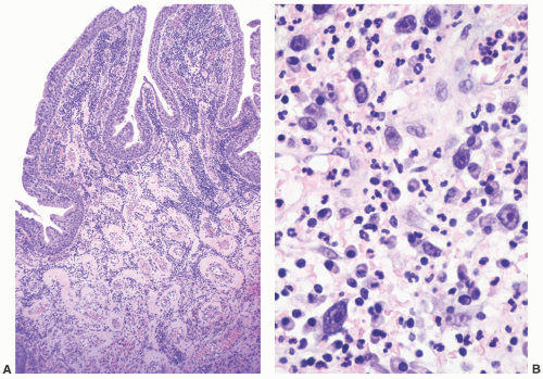
FIGURE 7-10 ▪ Caruncle. A striking papillary growth is seen associated with prominent vessels in the lamina propria (A). Atypical stromal cells with enlarged nuclei and prominent nucleoli are admixed with a prominent inflammatory infiltrate (B).


Table 7-1 ▪ URETHRAL POLYPS AND CYSTS


Urethral polyps:


Caruncle


Frequently related to trauma


Posterior wall


May have atypical stromal cells


Often associated urothelial hyperplasia


Prostatic-type polyp


Verumontanum and prostatic urethra in males


<1 cm


Lined by prostatic epithelium that may alternate with transitional epithelium


Differential diagnosis: prostatic ductal carcinoma


Fibroepithelial polyp


Mostly males, first decade


<4 cm


Club-like projections most common pattern


Lined by unremarkable urothelium


Urethral cysts:


Cowper gland duct cyst


Congenital (children) or acquired (adults)


May be associated with other abnormalities (more frequently urinary tract)


Typically confirmed by urethrography


Skene duct cyst


Most often secondary to infection and obstruction


Lined by stratified squamous epithelium


Differential diagnosis: Urethral diverticulum


This benign lesion should be distinguished from prostatic adenocarcinoma, especially in older that secondary involve the urethra101; however, in the latter, prostatic acini are lined by a single layer of malignant epithelial cells with prominent nucleoli. Ductal carcinoma, the most common variant of prostate carcinoma, if low grade, may enter in the differential diagnosis of a prostatic-type polyp. Grossly, the tumor may be seen as an exophytic mass protruding into the prostatic urethra, although it is typically larger than prostatic-type polyps.103 Cytologically, even though the tumor forms glands that may superficially mimic the low-power appearance seen in a prostatic-type polyp, in contrast to the latter, there is often some degree of cytologic atypia and absence of basal cells. Furthermore, in most cases, a component of acinar carcinoma may be seen. From the immunohistochemical point of view, 34bE12 is helpful to separate ductal carcinoma (except when intraductal) from a prostatic-type polyp, as the former lacks basal cells.103 Rarely, a primary papillary adenocarcinoma of the urethra or the bladder may enter in the differential diagnosis, but these lesions typically display higher degree of cytologic atypia. The histogenesis of this benign polyp is a matter of debate. It has been postulated that it may represent either a metaplastic process (supported by the fact that the prostate normally develops from an anlage of the urothelium of the urethra) or a defect of embryogenesis.94 Surgical excision is the treatment of choice with only rare recurrences being reported.


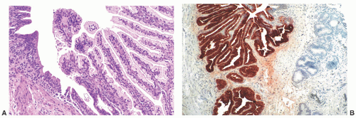
FIGURE 7-11 ▪ Prostatic-type polyp. Irregular fronds of prostatic-type epithelium are intermixed with transitional epithelium (A). The prostatic cells are PSA positive (B).


Fibroepithelial Polyp

This is a nonneoplastic lesion with a marked male predominance occurring typically in the first decade of life92,104; however, it can also be seen in adult and female patients.105, 106, 107 It is more often located in the urethra (verumontanum) and urinary bladder.106,107 The most common clinical manifestations include hematuria and obstructive symptoms, but it may be an incidental finding.104,107,108 On gross examination, fibroepithelial polyps are typically polypoid, soft, and <4 cm in dimension.92,105,107,109 Histologically, at low power magnification, they may display (a) club-like projections (most common pattern) associated with florid cystitis cystica and glandularis in the stalk; (b) abundant small, plump and rounded fibrovascular cores associated with abundant connective tissue; and (c) simple finger-like projections. They are lined by unremarkable urothelium, but rarely columnar epithelium may be seen.107 Surface squamous metaplasia and ulceration may occur. Infrequently, the nonsurface epithelial component may show anastomosing nests of urothelium or back to back glands. The stroma may be edematous and may contain atypical stromal cells and calcifications. Entities that more commonly may cause problems in differential diagnosis include florid cystitis glandularis cystica, polypoid or papillary cystitis, and urothelial papilloma. Distinguishing features include absent polypoid or papillary architecture (florid cystitis glandularis); prominent edema and inflammation; and history of instrumentation/trauma (polypoid or papillary cystitis), more delicate stalks or/and associated connective tissue (urothelial papilloma). As this lesion occurs more frequently in early childhood, the differential diagnosis should include botryoid embryonal rhabdomyosarcoma. However, the latter is characterized by a dense “blue” cambium layer under the surface epithelium composed of small immature cells with high nuclear to cytoplasmic ratio and frequent mitotic activity that may alternate with fully developed rhabdomyoblasts showing prominent eosinophilic cytoplasm and cross-striations.110 After surgical excision, these lesions generally do not recur.


Cowper Gland Duct Cyst

Cowper glands, or bulbourethral glands, are present as paired glands in the bulbomembranous urethra, to which they are connected through small ducts. Their acini form small lobules and are composed of cells with abundant mucin.111 Cowper gland cyst, also known as syringocele, is a cystic dilatation of the main ducts. It is typically detected in children (congenital) or young adult males (acquired) who present with lower urinary tract symptoms including frequency, urgency, dysuria, postvoid incontinence, recurrent infections and/or hematuria, but it may be asymptomatic.112, 113, 114, 115, 116 In children, it may be associated with other congenital abnormalities, most often of the urinary tract.113 The diagnosis is easily confirmed by urethrography,112 and rarely, these specimens undergo pathologic examination. Histologically, the wall of the syringocele may be denuded and associated with prominent inflammation as well as fibrosis of the wall. Treatment consists of marsupialization, and follow-up is uneventful.39,112


Skene Duct Cyst

Skene glands and ducts, the homologue of the male prostate gland, are located in the floor of the distal urethra.117 Skene cysts are rare and frequently occur due to obstruction of the ducts most often secondary to infection. They are seen in female patients who may present with dysuria or obstructive voiding symptoms or be asymptomatic.118 Grossly, the cysts vary in size, and on microscopic examination they are lined by stratified squamous epithelium.119 The most common differential diagnosis is with a urethral diverticulum, a distinction that should be made on cystourethroscopy and pelvic MRI findings. Other entities to consider include cystocele and Gartner duct cyst.118 Surgical excision is the treatment of choice.


Nephrogenic Adenoma

This is a rare pseudoneoplastic lesion that occurs most commonly in adult males, although it can be seen in
children and females, in the latter frequently in association with a urethral diverticulum.120, 121, 122 The urethra is the second most frequent location (15%) following the bladder.123 In most instances, nephrogenic adenoma is an incidental finding at cystoscopy performed for other reasons, but if large it may cause symptoms related to obstruction. In these cases, it may be potentially confused with a malignant tumor, most commonly a low-grade papillary urothelial carcinoma. On microscopic examination, nephrogenic adenoma shows tubular, cystic, papillary, and rarely solid growths, but not infrequently an admixture thereof.122,123 The tubules vary in size and shape and may be solid on rare occasions (Fig. 7-12A). An appreciable basement membrane may be seen surrounding some of the tubules. Of note, the tubules of nephrogenic adenoma may be intermixed with muscle fibers of the muscle fibers present in the wall of the prostatic urethra when the specimen is obtained from transurethral resection (Fig. 7-12B).120,124,125
Recently, a fibromyxoid variant of nephrogenic adenoma has been reported characterized by very small tubules associated with prominent fibromyxoid background that may closely mimic the appearance of an infiltrating carcinoma (Chapter 5).126 Cysts are frequently admixed with tubules, but they are not striking in most cases (Fig. 7-12A). The papillae are simple without branching, and they are rarely seen in the absence of tubules. The solid pattern is very rare and, when present, typically represents a minor component. The cells of nephrogenic adenoma are cuboidal to columnar to flattened with variable eosinophilic to slightly clear and granular cytoplasm, and exceptionally they may be spindled.126 Hobnail cells are typically present, most often lining the cysts, but are rarely conspicuous.123,127 The cells lining tiny tubules have a compressed nucleus with a single vacuole containing basophilic material resembling signet ring cells (Fig. 7-12C). The nuclei are round to oval with minimal cytologic atypia. Nucleoli are inconspicuous, and mitotic figures are rare (<1/10 HPFs) and seen in 5% of the lesions.123 The term “atypical nephrogenic adenoma,” has been used for lesions characterized by nuclear enlargement, nuclear hyperchromasia, and enlarged nucleoli.127 There is no known clinical or biologic significance to this designation. The stroma associated with nephrogenic adenoma is focally edematous and contains variable amounts of inflammatory cells. Stromal calcification may be seen.


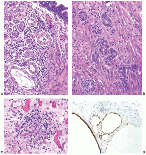
FIGURE 7-12 ▪ Nephrogenic adenoma. Tubules and cysts are lined by one layer of cells (A). Tubules are present between muscle fibers mimicking invasive prostatic carcinoma (B). Signet ring-like cells with basophilic intracellular material are set in an edematous and inflammatory stroma (C). The cells are P504s positive (D).

The immunohistochemical profile of nephrogenic adenoma includes positivity for CK7, epithelial membrane antigen (EMA), vimentin, and PAX2 and PAX8 (the latter two, antigens expressed during embryogenesis),128, 129, 130

Only gold members can continue reading. Log In or Register to continue

Stay updated, free articles. Join our Telegram channel

Jun 10, 2016 | Posted by in UROLOGY | Comments Off on Pathology of the Male and Female Urethra

Full access? Get Clinical Tree

Get Clinical Tree app for offline access