Tumors of the Urinary Bladder


Tumors of the Urinary Bladder


David J. Grignon


CLASSIFICATION OF UROTHELIAL TUMORS

The last decade has seen a tremendous upheaval in this very important area that only recently seems to have settled down to a level of general concurrence.1 For over two decades, the World Health Organization (WHO) classification and grading of urothelial neoplasms2 dominated although several variations and different schemes were published. In the early 1990s, several factors emerged that resulted in the need to reevaluate this approach. First, the controversy of calling grade 1 papillary tumors “carcinoma” arose with several groups led by Dr. William Murphy calling all tumors in the low-grade end as papilloma.3,4 Second, the use of intravesical therapy as a standard practice in the treatment of highrisk noninvasive papillary tumors demanded that high-risk tumors be clearly identified. Third, the WHO (1973) system was criticized for the imprecision of the published criteria (Table 6-1), leading many pathologists to essentially use this three-grade system to create five grade groups (1, 1-2, 2, 2-3, and 3), one result being that only a small percentage of cases were placed in the grade 3 group. For example, in a review of three clinical studies only 25 of 280 (8.9%) newly diagnosed noninvasive papillary tumors (pTa) were called grade 3.5 The effect of the latter was confusion as to how to treat grade 2 Ta tumors, a category that included many high-risk patients who could benefit from intravesical therapy as well as many patients with low-risk disease for whom intravesical therapy may not be appropriate or necessary. Multiple studies have demonstrated that this group includes high-risk patients with progression to invasive carcinoma reported in up to 20% of patients and cancer-specific death in 13% to 20%.6,7

Recognizing the many emerging issues, in 1997 Dr. F. K. Mostofi organized a meeting of a small group of urologic pathologists, urologists, and urologic oncologists to address these concerns. This was followed by a much larger consensus conference that was held under the auspices of the International Society of Urologic Pathology (ISUP) in March of 1998. The results of this consensus were adopted by the WHO and the results published in 1998 as the WHO/ISUP consensus classification (Table 6-2).8 Most controversial was the adoption of the term papillary urothelial neoplasm of low malignant potential. This represented a compromise term where the papilloma and carcinoma advocates could be comfortable and allowed that controversy to be brought to resolution. Most important was the adoption of the grading system that had been described by Malmström et al.9 The value of the latter was viewed as twofold; first, the morphologic criteria for applying the scheme were well defined and, second, it appeared to place the majority of patients with high-risk disease into the high-grade category.

The publication of the 1999 WHO blue book the following year introduced a variation on this system with the splitting of the low- and high-grade categories of the 1998 WHO/ISUP classification into three groups (grades 1, 2, and 3) while retaining the papillary urothelial neoplasm of low malignant potential category.10 This reignited the controversy with some experts urging a return to the 1973 WHO grading system.11 Others criticized the 1998 WHO/ISUP system as simply representing a renaming of the 1973 WHO,12 a clearly incorrect interpretation.13 At a subsequent meeting in Ancona, Italy (2001), a modified version of the 1973 WHO was proposed.13

This issue became the primary focus of discussion at the WHO meetings prior to the release of the 2004 WHO classification. Following extensive debate and discussion, it was agreed overwhelmingly to essentially reproduce the 1998 WHO/ISUP classification as the 2004 WHO recommended classification scheme (Table 6-2).14 The authors of the Fourth Series Armed Forces Institute of Pathology fascicle covering bladder neoplasia also followed this system.15 With this consistent approach adopted by arguably the two most influential references for tumor classification and grading, work can continue on evaluating the biologic and clinical relevance and value of this system.16

The success of the 2004 WHO/ISUP classification in addressing the key issues outlined in the first paragraph has been stressed, in particular the reclassification of high-risk
grade 2 tumors (WHO 1973) into the high-grade papillary carcinoma category.13,17,18 For example, in the study by Yin and Leong,18 13 of 46 WHO (1973) grade 2 tumors (28%) were placed in the WHO (2004) high-grade category resulting in 23% of all cases being considered high-grade in WHO 2004 compared to only 4% being called grade 3 in the 1973 WHO system. Similarly, Samaratunga et al.17 reviewed 134 papillary tumors of which 6 (4%) had been reported as grade 3 (WHO, 1973); on review they considered 29 (22%) to be high grade by WHO/ISUP 1998.


Table 6-1 ▪ 1973 WHO GRADING CRITERIA


Grade 1


Tumors with the least degree of cellular anaplasia compatible with a diagnosis of malignancy


Grade 2


Histologic features between grades 1 and 3


Grade 3


Tumors with the most severe degrees of cellular anaplasia


From Mostofi F, Sobin L. Histologic Typing of Urinary Bladder Tumors. Geneva, Switzerland: World Health Organization; 1973.


This approach has been embraced by many urologic oncologists with interest in bladder cancer. It has important application in the contemporary treatment of Ta tumors. The reclassification of high-risk grade 2 tumors (WHO 1973) into the high-grade category (WHO 2004) has resulted in a large and better-defined group of patients with low-risk Ta tumors. In a series of 215 patients with low-grade (papilloma, papillary urothelial neoplasm of low malignant potential and lowgrade papillary carcinoma) Ta tumors from the Memorial Sloan Kettering Cancer Center treated by transurethral resection only, progression to high-grade Ta or invasive carcinoma occurred in only 3% and 5% of patients, respectively, with a median follow-up of 8 years.19 A prospective study lent further support to these findings.20 This experience has led to the suggestion that patients with low-grade Ta tumors can be followed less frequently.21 Similarly, Nieder and Soloway22 recommended treating patients with papilloma and papillary urothelial neoplasm of low malignant potential by transurethral resection alone, low-grade Ta tumors by transurethral resection with a single dose of mitomycin C, and highgrade Ta tumors by transurethral resection with mitomycin C and bacillus Calmette-Guérin (BCG) immunotherapy. In December 2007, the American Urological Association (AUA) reinforced the current approach in its guidelines for the treatment of non-muscle-invasive bladder cancers by separating noninvasive papillary tumors into two groups— low grade and high grade23—with different treatment recommendations for each group. The use of this system has also been advocated by the European Urology Association.24 The College of American Pathologists (CAP) utilizes WHO 2004 in its recommendations for the reporting of urothelial tumors.25,26 Support for this system was further emphasized in the International Consultation on Bladder Cancer.27


Table 6-2 ▪ 2004 WHO/1998 ISUP CLASSIFICATION


Normal


Hyperplasias


Flat lesions with atypia


Reactive (inflammatory) atypia


Atypia of unknown significance


Dysplasia (low-grade intraurothelial neoplasia)


CIS (high-grade intraurothelial neoplasia)


Papillary neoplasms


Papilloma


Inverted papilloma


Papillary urothelial neoplasm of low malignant potential


Papillary carcinoma, low grade


Papillary carcinoma, high grade


Invasive neoplasms


From Epstein JI, Amin MB, Reuter VR, et al. The World Health Organization/International Society of Urological Pathology consensus classification of urothelial (transitional cell) neoplasms of the urinary bladder. Bladder Consensus Conference Committee. Am J Surg Pathol 1998;22:1435-1448; Eble J, Epstein J, Sauter G, et al. World Health Organization Histologic and Genetic Typing of Tumours of the Kidney, Urinary Bladder, Prostate Gland and Testis. Lyon, France: IARC Press; 2004.


EPITHELIAL TUMORS—BENIGN


Urothelial Papilloma

There has been a long-standing controversy regarding the nature of papillary lesions with minimal cytologic atypia.28 An early definition by Mostofi restricted the term papilloma to noninvasive papillary lesions covered by urothelium that was indistinguishable from normal urothelium.29 This definition was adopted in the WHO (1973) classification.2 The use of this term by some experts for up to one-third of all papillary lesions was a major stimulant to the reevaluation of these lesions that began in 1997.3,4 The current classification retains the restrictive traditional WHO criteria.2,14,15 These tumors do harbor the FGFR3 mutations characteristic of papillary neoplasia.30


Clinical Features

Lesions meeting these restricted criteria occur at a younger age than other urothelial bladder tumors and often present with only one or a few papillary processes. The mean patient age in two large series was 51 years (range, 8 to 87 years).31,32 In one series with individual data presented, 50% of the patients were under 40 years of age.31 They are more common in men (male:female ratio is 2.4:1).31 There does not appear to be a predilection for a specific location in the bladder. Using the restrictive criteria recommended, these lesions account for approximately 1% of papillary tumors.28 In patients with urothelial papilloma and no other urothelial neoplasia, the recurrence rate is <10% and there is a low risk for the subsequent development of higher-grade tumors.28,31, 32, 33, 34 In contemporary studies, the progression to higher-grade tumors has been low, and the subsequent development of
invasive disease has been reported to be <1%.31, 32, 33 In these studies, the one case with progression was on immunosuppressive therapy following renal transplantation.


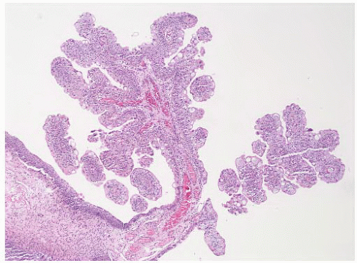
FIGURE 6-1 ▪ Urothelial papilloma showing a small tumor with fine papillary fronds.


Pathology

Grossly, urothelial papilloma consists of a small lesion with a few distinct and separate fronds. Histologically, papilloma is characterized by fine papillary fronds without fusion or complexity (Fig. 6-1). There is minimal if any branching. Individual fronds are covered by an essentially normal urothelium without architectural or cytologic atypia (Fig. 6-2). The absolute number of cell layers is not a criterion for diagnosis, but the urothelium should not be obviously thickened. In some cases, the urothelium can be quite attenuated with only a couple of cell layers. As in normal urothelium, some variability in the histology is allowed. The umbrella cell layer can have enlarged nuclei and cytoplasmic vacuoles, and an apocrine-like appearance of the cytoplasm can be seen. There should be no atypia of the cells other than the umbrella cells. Mitoses are absent. Although immunohistochemistry is not recommended for diagnosis, similar to papillary urothelial neoplasm of low malignant potential, expression of cytokeratin 20 is limited to the umbrella cell layer (Fig. 6-3)35 and proliferation determined by Ki-67 immunohistochemistry is low.36


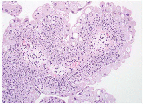
FIGURE 6-2 ▪ Urothelial papilloma with the surface urothelium showing no cytologic or architectural atypia.


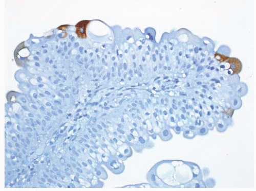
FIGURE 6-3 ▪ Urothelial papilloma with immunohistochemistry for cytokeratin 20 highlighting a few umbrella cells.


Differential Diagnosis

The differential diagnosis includes papillary cystitis, papillary hyperplasia, and low-grade papillary urothelial carcinoma. Papillary cystitis occurs in an inflammatory background and the clinical setting—indwelling catheter, bladder stones, nonfunctioning bladder—may be helpful. The papillae are shorter and broader than typical of papillary neoplasia.37,38 Branching of the papillae is not a feature. The stroma is inflamed and may be edematous (polypoid cystitis). The urothelium often shows a reactive type of atypia. Papillary hyperplasia has more of a pseudopapillary architecture with tenting of the urothelium characterized by a thicker stromal core at the base of the lesion that thins toward the tip of the lesion.39 The uniformly thin fibrovascular core typical of papilloma is absent. The lesion often is associated with thickening of the urothelium of the pseudopapillary projection and the adjacent flat surface. Distinction from papillary urothelial neoplasm of low malignant potential can be difficult. The presence of complex branching, thickening of the epithelium, or increased cell density favors the diagnosis of papillary urothelial neoplasm of low malignant potential, while any fusion of the papillae, architectural disturbance, nuclear enlargement or atypia, and mitoses indicates the diagnosis of low-grade papillary carcinoma.


Inverted Urothelial Papilloma


Clinical Features

Inverted urothelial papilloma accounts for approximately 1% of urothelial neoplasms. It is a distinct clinical and pathologic
entity that occurs over a wide age range but most often after the age of 50.5,40, 41, 42, 43, 44, 45, 46, 47 They are much more common in men than women. These develop throughout the urinary tract but are most common in the urinary bladder, in particular the trigone and bladder neck region. Cases of synchronous inverted papilloma and papillary carcinoma are well described though rare. In rare cases, they can be multifocal.42 Inverted papilloma is treated by transurethral resection and is associated with a low risk of recurrence (<5%) that is distinctly different from low-grade papillary urothelial neoplasms.42,43


Genetic data have confirmed that these represent a neoplastic process with clonality demonstrated by X-linked inactivation studies.42,48 Loss of heterozygosity studies have demonstrated that inverted urothelial papilloma does not have the typical genetic abnormalities of papillary urothelial neoplasms, supporting the concept that these are unrelated.48 There is no association with human papilloma virus (HPV) infection.49


Pathology

Grossly, these lesions typically have an exophytic polypoid growth pattern and can be pedunculated (Box 6-1). Histologically, inverted urothelial papilloma consists of anastomosing trabeculae of urothelium covered by a normal or attenuated urothelium (Fig. 6-4). Multiple sites of origin from the surface urothelium are usually present. The basal layer is often prominent with the basilar nuclei lined up perpendicular to the basement membrane. The cells in the central part of the trabeculae can be spindled with a streaming growth pattern (Fig. 6-5). In general, there is no significant nuclear pleomorphism although occasionally mild atypia can be present.50 Mitotic figures are rare or absent. The cytoplasm can be foamy or vacuolated.51 This can resemble the “glycogenated” squamoid morphology that can be present in some urothelial carcinomas. Squamous or glandular differentiation may be present. In many cases, glands or gland-like spaces that are lined by a cuboidal to columnar epithelium are found (Fig. 6-6). These typically do not appear to be mucin-containing cells on hematoxylin and eosin-stained sections. The lumens can contain eosinophilic secretions that can stain with mucicarmine. In transurethral resection material, the fragmentation of the lesion may result in apparent papillary structures, making diagnosis difficult. There is no stromal desmoplasia and minimal inflammation.


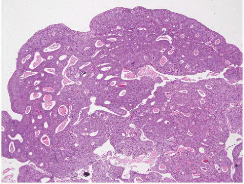
FIGURE 6-4 ▪ Inverted urothelial papilloma with polypoid appearance, flattened surface epithelium, and complex trabecular architecture.


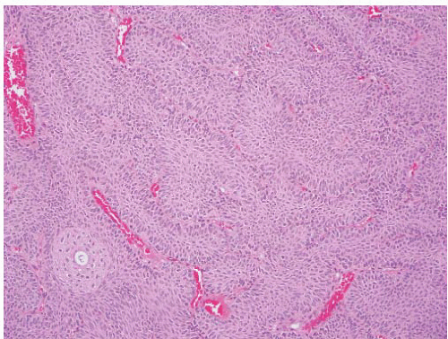
FIGURE 6-5 ▪ Inverted urothelial papilloma with palisading of basilar cells and streaming of centrally located cells.


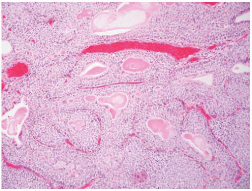
FIGURE 6-6 ▪ Inverted urothelial papilloma with glandular spaces lined by columnar epithelium.


Differential Diagnosis

The major problem is the distinction from papillary carcinoma with an inverted growth pattern.48,50,52 These carcinomas generally have larger pushing borders with more defined nested architecture rather than the complex anastomosing architecture typical of inverted papilloma. The striking peripheral palisading, maturation toward the center of the trabeculae and the spindling growth pattern are also absent.53 In most cases, there is a greater degree of nuclear pleomorphism than is acceptable for inverted papilloma, and mitoses may be frequent. Foci of stromal invasion are often present and would exclude the diagnosis of inverted papilloma.

Florid cystitis glandularis can produce a localized polypoid lesion that can be confused with inverted papilloma. These have well-defined nests without the complex trabecular architecture of inverted papilloma. Similarly, exuberant proliferations of von Brunn nests can mimic inverted papilloma, but the nests are separated and a complex trabecular architecture is absent.54 Finally other tumors that can have a nested architecture such as the nested variant of urothelial carcinoma, paraganglioma, and carcinoid tumor could potentially mimic inverted papilloma in small biopsy specimens.


Villous Adenoma


Clinical Features

Villous adenoma is an uncommon lesion that can arise in the urinary bladder proper or the urachus.55, 56, 57, 58, 59 It is more common in men than women and occurs over a wide age range from young adults to the elderly with most found in the sixth or seventh decade. The most frequent presentation is with hematuria or irritative symptoms. Mucusuria can occur but is infrequent. Lesions in the urinary bladder most often arise in the trigone or bladder dome region. Involvement of bladder diverticula is also described.60 Development following bladder augmentation has been reported.61 At cystoscopy, the lesion has an exophytic appearance.

Villous adenoma is treated by transurethral resection or local resection. If the lesion is a pure villous adenoma, complete local resection is curative. There is, however, a frequent association with adenocarcinoma, and without complete resection and examination of the lesion this possibility cannot be excluded.59 In one series, 50% of cases had either in situ adenocarcinoma (17%) or invasive adenocarcinoma (33%) associated with the villous adenoma.59


Pathology

Villous adenoma in the bladder is morphologically identical to that occurring in the gastrointestinal tract. There are tall villous projections covered by an intestinal-type epithelium with variable numbers of goblet cells (Fig. 6-7). The nuclei are oval to fusiform, enlarged, and frequently pseudostratified with variable degrees of atypia (Fig. 6-8). Nucleoli can be prominent. Mitoses are present but are not frequent. In some cases the degree of atypia is sufficient to warrant the designation of adenocarcinoma in situ.59 Cystitis glandularis can be present.62 Acidic and neutral mucin is demonstrable in most cases.58 The tumors have a similar immunohistochemical profile as in the gastrointestinal tract with cytokeratin 20 (100%) and carcinoembryonic antigen (CEA) positivity (89%).58 Many also express cytokeratin 7 (56%). In one study, expression of prostate-specific membrane antigen (PSMA) was described in one case but without immunoreactivity for prostate-specific antigen (PSA) or prostate-specific acid phosphatase (PSAP).63 There is also a single report of reactivity for PSA in a villous adenoma of the urachus.64


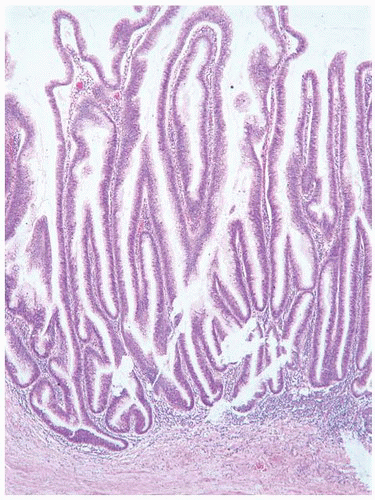
FIGURE 6-7 ▪ Villous adenoma with tall villiform papillae.


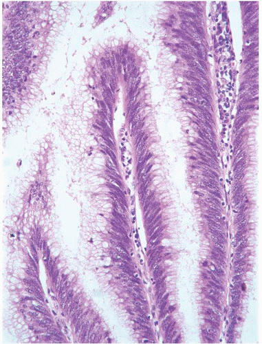
FIGURE 6-8 ▪ Villous adenoma with pseudostratified mucinsecreting epithelium.


Differential Diagnosis

The major consideration is with adenocarcinoma, with either primary or secondary involvement from the gastrointestinal tract. Up to 50% of cases of villous adenoma have associated malignancy, and so, careful examination for areas of in situ and invasive adenocarcinoma is essential (Fig. 6-9). The invasive tumor typically has an enteric pattern with infiltrating glands and reactive stromal desmoplasia. The presence of glands with a wreath-like arrangement with central necrosis also indicates malignant transformation. In reporting a diagnosis of villous adenoma in biopsy or transurethral resection specimens, a note should always be added that the possibility of adenocarcinoma can only be excluded by complete resection and microscopic evaluation of the entire tumor.

Papillary urothelial carcinoma with villoglandular differentiation should also be considered in the differential diagnosis.65 These are papillary urothelial carcinomas where the covering epithelium includes mucin-secreting cells, often with intraepithelial gland formation, mimicking an intestinaltype epithelium. This is almost always a partial change with other areas of the papillae having more a typical urothelial surface. The papillae are branching and irregular in length without the tall villous architecture of villous adenoma.


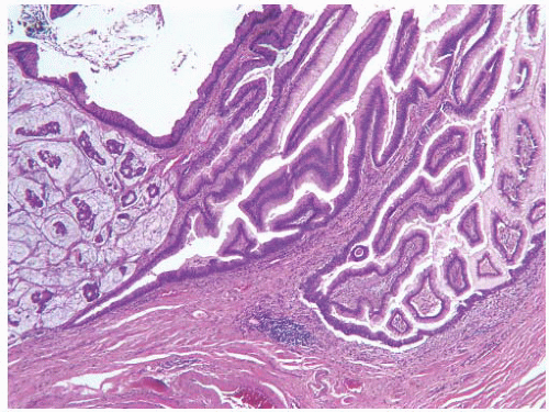
FIGURE 6-9 ▪ Villous adenoma with associated mucinous adenocarcinoma.


Squamous Papilloma


Clinical Features

Squamous papilloma is an uncommon lesion in the urinary bladder.66,67 It is more common in women than men (female:male ratio of 1.5:1). They can occur over a wide age range with a mean age of 62 years (range, 28 to 82 years). There should not be a history of urogenital condylomata. They may be asymptomatic or present with hematuria. At cystoscopy, these may appear as erythematous areas, plaque-like, or as exophytic papillary lesions. In the series reported by Guo et al.,67 one of four patients had a history of papillary urothelial neoplasia and subsequently developed additional urothelial neoplasms. Cheng et al.66 included two patients with a history of urothelial neoplasia in their series of seven cases of squamous papilloma. Cases such as these suggest that some squamous papillomas represent low-grade papillary urothelial tumors with squamous differentiation.


Pathology

The lesion is exophytic with true papillary projections covered entirely by squamous epithelium (Fig. 6-10). The squamous epithelium is essentially normal in appearance. There is no koilocytotic atypia. These do not contain HPV DNA and are diploid.66 There is no overexpression of p53; there is expression of epidermal growth factor receptor (EGFR).66,67


Differential Diagnosis

The differential diagnosis includes verrucous squamous hyperplasia, condyloma acuminatum, papillary urothelial neoplasia with squamous differentiation, and well-differentiated squamous cell carcinoma. Verrucous squamous hyperplasia is characterized by keratinizing squamous epithelium with hyperkeratosis and a spike-like proliferation rather than the well-developed papillae of squamous papilloma.67 Condyloma acuminatum occurs in patients with genital condyloma or in patients who are immunosuppressed.66,68 The lesions tend be more sessile with less well-defined papillae, thickened epithelium, and koilocytic atypia. The lesions contain HPV DNA and many overexpress p53.66 In papillary urothelial neoplasia, there is often a history of prior papillary tumors, and the covering epithelium has areas more typical of urothelium. In the setting of known urothelial neoplasia, the diagnosis of squamous papilloma should be restricted to cases where the surface epithelium is uniformly benign squamous in type. Squamous cell carcinoma is typically a solid, invasive tumor. Infrequently there can be a component with papillary architecture, but the covering epithelium is neoplastic in nature and even in low-grade tumors would not have a benign appearance.


FIGURE 6-10 ▪ Squamous papilloma with fine papillary structures covered by keratinizing squamous epithelium.


EPITHELIAL TUMORS—MALIGNANT


Urothelial Carcinoma


Epidemiology

Bladder cancer is estimated to be the ninth most common cancer worldwide (an estimated 357,000 cases in 2002) and the thirteenth most common cause of death from cancer (145,000 deaths in 2002).69 It is the seventh most common malignancy of men and the seventeenth most common tumor in women worldwide.70 There are significant geographic differences with an approximately 10-fold difference in incidence and death rates between countries. The highest rates of bladder cancer are found in southern European countries (Spain, Italy) and Egypt, closely followed by Israel, the United States, Denmark, and the United Kingdom. The lowest rates are in the Far East, including Japan, China, and Korea. The worldwide age-adjusted mortality rates for bladder cancer are 2 to 10 per 100,000 for males and 0.5 to 4 per 100,000 for females.70 Overall bladder cancer is approximately four times more common in men than in women.71 This has been largely attributed to the higher rate of smoking and more frequent occupational exposure to bladder carcinogens in men.71

In the United States, the most recent data estimate is that there will be 72,570 new cases of bladder cancer diagnosed in 2013, with 54,610 of these in males and 17,960 in females.72 The estimated number of cancer deaths for 2013 is 15,210 with 10,820 and 4,390 in males and females, respectively.72 Bladder cancer is most common in Caucasians (excluding Hispanic Caucasians). Despite the lower incidence of bladder cancer in African Americans the mortality rate is significantly higher.73 This has been partly attributed to tumors being higher stage at diagnosis, but this is not considered to completely explain the observed differences.73

Bladder cancer is generally a disease of older individuals with most patients being diagnosed in the seventh decade or older. The tumor can occur in younger individuals.74 In patients under the age of 20 years, the tumors are almost all low grade with no high-grade tumors identified in the review by Paner et al.74 The rate of recurrence in this age group is significantly lower (3%) than would be expected for papillary tumors with progression being very rare.74


Etiopathogenesis

The most important risk factors for the development of bladder cancer are tobacco smoke and occupational exposure to carcinogens (Table 6-3). Tobacco smoke is estimated to be responsible for 30% to 50% of cases of bladder cancer.75, 76, 77 Smokers have a two- to six-fold increased risk of developing bladder cancer over those who have never smoked.75, 76, 77 Stopping smoking decreases the risk of bladder cancer development, and smoking filtered cigarettes confers a lower risk than smoking nonfiltered types. Cigarette smoke contains numerous carcinogenic compounds including 4-aminophenol (considered the most important), β-naphthylamine, benzene, cadmium, chromium, radon, vinyl chloride, nickel, and many others.71 Changes in worldwide smoking patterns are reflected in the changing incidences in bladder cancer.

Occupational exposure to carcinogens is estimated to account for up to 20% of bladder cancer cases.78 The best documented occupational carcinogens are β-naphthylamine, 4-aminobiphenyl, and benzidine, most often associated with textile dye and rubber industries. With the banning of these compounds in Western countries, these no longer account for a significant percentage of occupation-related bladder cancers. Increased risk for bladder cancer has been reported in a wide range of diverse industries with a varying degree of evidence. In the Western world, even for those that have been most often documented, the relative risks have been low, most often in the range of 1.1 to 1.2.71


Table 6-3 ▪ UROTHELIAL CARCINOMA: IMPLICATED CARCINOGENIC AGENTS


Tobacco (smoking)


4-Aminophenol


β-Naphthylamine


Benzene


Others


Occupational


Benzidine


β-Naphthylamine


Chlorinated aliphatic hydrocarbons


Arylamines


4-Aminobiphenyl


Others


Drugs


Phenacetin


Cyclophosphamide


Infectious


Schistosoma haematobium


Dietary


Nitrites (weak evidence)


Other


There have been extensive analyses of the relationship of drinking water to bladder cancer. Many of these have focused on chlorinated water and the carcinogenic risk of chlorination by products such as trihalomethane.79,80 In a recent review, Wu et al.71 concluded that although exposure to chlorinated drinking water might increase the risk of bladder cancer, this is relatively small. Contamination of drinking water with arsenic has been shown to be associated with an increased risk of bladder cancer.81 There is a dose/effect relationship, and the very low level of arsenic in drinking water in developed countries is not considered to be an important carcinogenic factor. Finally there has been interest in the volume of fluid intake based on the hypothesis that high volumes of liquid would result in dilution of excreted carcinogens and thereby reduce carcinogenic risk. To date, studies of this possible effect have not yielded consistent results.71

Dietary factors have been the subject of intense interest. Although studies have not been entirely consistent, the data have generally supported a protective effect for high consumption of fruit and vegetables.82,83 It has been suggested that this is an effect of antioxidants that detoxify excreted metabolites. In one meta-analysis, diet low in fruit intake was associated with an increased risk of bladder cancer.84 In a prospective analysis of women, however, no association between intake of fruits, vegetables, or vitamins was found.85 In addition, there has been weak evidence linking nitrite and meat-associated nitrates with bladder cancer risk.86 Studies of pesticides have failed to identify a relationship between pesticide exposure and bladder cancer.87 No other dietary factors, including artificial sweeteners and coffee, have been demonstrated to have a relationship to bladder cancer development.

Infectious agents are an important contributor to bladder cancer development. In particular the strong relationship between Schistosoma haematobium and the development of bladder cancer is well known.88,89 This has been most strongly related to the development of squamous cell carcinoma,90 but other histologic types including urothelial carcinoma and adenocarcinoma are also more frequent in areas where Schistosoma infection is endemic. There is no evidence that viral infection plays a role in bladder cancer development.91

Hereditary factors are not considered to be a major contributor to bladder cancer.92 There have been a number of reports of familial clustering of bladder cancer cases, but these have largely been isolated reports. Some of these have been associated with an early age of bladder cancer development supporting a significant hereditary component.92 The reported higher risk in first-degree relatives of bladder cancer patients also suggests a hereditary component, but the importance relative to other environmental factors is unclear.93,94 Urothelial cancers have been found in certain hereditary syndromes such as Costello syndrome,95 Lynch syndrome,96 and the Muir-Torre syndrome.97

Bladder cancer risk has also been associated with a number of other factors including drug exposure (cyclophosphamide, phenacetin)98,99 and radiation therapy.100 Despite considerable interest, exposure to hair dyes has now largely been eliminated as a risk factor.101 Other causes of longstanding irritation of the bladder mucosa such as chronic urinary tract infections, lithiasis, and nonfunctioning bladders of any cause have been linked to the development of bladder cancer.


Genetics

Studies of the genetics of urothelial carcinoma have led to the development of a model of tumorigenesis that includes distinct pathways for papillary and nonpapillary neoplasia (Fig. 6-11). Both pathways can result in the development of invasive high-grade urothelial carcinoma that has the capacity to metastasize and result in the death of the patient. In this section, these pathways and the relevant genetic changes are briefly reviewed.102,103

The major genes implicated in the transformation of normal urothelium into low-grade papillary neoplasia include H-Ras, FGFR3, PI3K, and 9p deletion. Abnormalities in the H-Ras gene are most often the result of mutations, in particular those involving codon 12.104 These are activating mutations and are found in approximately 15% of noninvasive papillary tumors. Mutations in the fibroblast growth factor receptor-3 (FGFR3) gene are present in 60% to 80% of low-grade noninvasive papillary tumors.35,105,106 The rate is much lower in high-grade invasive carcinomas and in urothelial CIS.35 It has been reported that the absence of FGFR3 gene mutations is a predictor of a higher likelihood of progression.36,107 Since the FGFR3 and H-Ras genes are in the same pathway, they are considered to be mutually exclusive events in bladder cancer.108 Mutations in the PI3-kinase gene (PIK3CA) have been described in a subset of approximately 15% of low-grade papillary tumors with the highest frequency in papillary neoplasms of low malignant potential.109 Some tumors harbor more than one of these genetic abnormalities, and it has been suggested that tumors with multiple abnormalities are associated with progression to high-grade tumors.102

Loss of heterozygosity studies have demonstrated abnormalities in both 9p and 9q in over 50% of bladder tumors.103 In many tumors, there is loss of an entire copy of the chromosome. Implicated in the 9p abnormalities are the CDKN2A and CDKN2B genes, the former coding for the p16 protein.110 Loss of heterozygosity studies has demonstrated abnormality of the CDKN2A gene in up to 60% of papillary tumors.111 Mutations in the INK4 gene and deletions of 9q (including the PTCH, DBC1, and TSC1 genes) are frequently present in high-grade papillary tumors and are believed to be important in the progression of low- to highgrade papillary tumors.112,113 Inactivating mutations of the TSC1 gene have been found in approximately 10% of bladder tumors.114 Mutations of the PTCH gene are infrequent.

A second pathway in the development of invasive carcinoma is through urothelial CIS. The major genes implicated in this pathway are the tumor suppressor genes
TP53, retinoblastoma (RB), and PTEN.102 Both the RB and TP53 genes have generated intense interest in bladder cancer. RB gene deletions are common in high-grade bladder tumors and have been associated with an aggressive clinical course.115 The absence of RB protein or inactivation of RB protein affects multiple critical pathways including the E2F family of transcription factors. As in the case of RB, the importance of the TP53 tumor suppressor gene has been extensively studied in bladder cancer. TP53 mutations are present in up to 70% of urothelial carcinomas and are related to high-grade and high-stage disease and also to poor clinical outcome.116 Studies related to loss of chromosome 9 in CIS have had variable results103; in one report chromosome 9 loss was significantly higher in primary CIS than in CIS associated with papillary tumors.117 The TP53 mutation analysis demonstrated a similar relationship suggesting that these might represent two different types of CIS.103 FGFR3 mutations are not present in primary CIS and are infrequent in CIS associated with papillary tumors.106,118


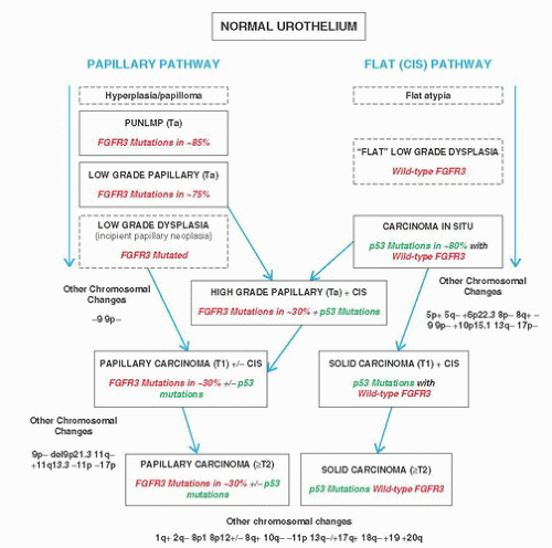
FIGURE 6-11 ▪ Pathways for the development of urothelial tumors.


Clinical Features

The single most frequent presenting symptom of bladder cancer is painless hematuria, present in around 85% of patients. When microscopic hematuria is included, almost all patients have some degree of hematuria. Hematuria can, however, be intermittent, and so a negative examination of the urine for hematuria does not exclude a bladder tumor. In general, most urologists recommend cystoscopy for even a single episode of unexplained hematuria for patients in the right age group for bladder cancer. The latter would include any patient over the age of 60 years or younger patients with a history of smoking or exposure to other significant risk factors.

The second most common presentation is with a group of symptoms that includes urinary frequency, urgency, and irritative voiding symptoms. This group of symptoms is often indicative of an invasive carcinoma or diffuse CIS. Less frequent presentations include those related to ureteral
obstruction, a pelvic mass, or obstruction of pelvic lymphatics. Least common are presentation as a metastasis of unknown origin or general cancer cachexia.


Cystoscopy

Cystourethroscopy is the mainstay of bladder cancer diagnosis and for some tumors, treatment.152 Advances in the optical technology used have resulted in significant improvement in the diagnosis of bladder neoplasia.153 Both rigid and flexible instruments have advantages and disadvantages. The major advantages of the rigid cystourethroscope are the superior optics; the larger working channel that allows for use of a wider range of accessory instruments and increased water flow, improving visualization; and the ease of manipulation. Flexible cystourethroscopes provide greater patient comfort, more flexibility in patient positioning, and ability to inspect the bladder surface from more angles.154 The procedure can be performed in the office setting or in endoscopic suites depending somewhat on the indication. In the outpatient setting, the procedure is well tolerated and does allow for the performance of biopsies and fulguration of small recurrent tumors.155 Intraurethral and intravesical topical anesthesia are sufficient for control of discomfort.

The procedure allows for direct visualization of the bladder mucosa, collection of cytologic specimens, and biopsy of any abnormalities. In the evaluation of hematuria, if the source is in the urinary bladder it can usually be detected or if it is coming from the upper tracts, that can also often be determined. The published sensitivity and specificity for detecting urothelial tumors have shown considerable variation and depend on the indication. For follow-up of patients with known papillary neoplasia, sensitivity and specificity are generally in the 85% to 95% and 90% to 100% range, respectively. The sensitivity is greater for papillary than for flat lesions. Because of the latter, routine biopsy of normalappearing mucosa has been advocated in order to enhance the detection of CIS.156,157 In more contemporary studies, a low yield has been reported for such biopsies.158 Recent advances in the application of photodynamic diagnosis that significantly improve the detection of CIS will also impact the routine performance of random biopsies.153 Experienced urologists have a high degree of reliability in predicting the pathologic features of papillary tumors based on the cystoscopy findings.159 Evaluation of the upper tracts is also possible through this procedure by the performance of retrograde pyelography, selective sampling of material from the upper tracts for cytologic examination, and direct visualization by ureteroscopy.

There have also been advances in the type of light source utilized to evaluate the bladder mucosa. White light cystoscopy has been the standard and remains most widely used. The use of fluorescein labeling with 5-aminolevulinic acid and hexaminolevulinic acid has been extensively studied and in general has resulted in significantly improved detection of CIS.160 The sensitivity for the identification of CIS increases from 56%-68% to 92%-97%.161 The diagnosis of papillary lesions is also improved.162 Studies have also demonstrated an improvement in the transurethral resection of visible tumors with a decrease in residual tumor from 25%-53% to 5%-33%.153

This method does, however, yield a high rate of falsepositive findings, particularly in the presence of inflammation.153,163 This limits the value of this technique following BCG therapy.

More recently narrow band imaging cystoscopy has been evaluated as potentially being superior in the detection of bladder tumors.164,165 Results have been variable, and it remains to be determined whether this or other alternatives being tested will ultimately change the standard use of white light. The application of real-time Raman spectroscopy and optical coherence tomography to enhance the prediction of tissue diagnosis are under investigation and appears promising.153


Biopsy and Transurethral Resection

Methods for sampling of bladder lesions include hot and cold cup biopsy, strip biopsy, and transurethral resection. Evaluation of flat lesions largely relies on the hot and cold cup biopsy methods. The cold cup biopsy does not use electrocautery, and so, thermal tissue artifacts are avoided.166

Transurethral resection of lesions in the bladder is performed to (i) remove the entire visible abnormality as both a diagnostic and therapeutic procedure, (ii) obtain a larger sample of a lesion than is possible by a biopsy technique, and (iii) obtain tissue deeper to the lesion to allow for staging
of the tumor. The general approach to resection of a tumor (TURBT, transurethral resection of bladder tumor) is to first remove the bulk or entirety of the tumor and second to sample the base of the tumor. These may be submitted as a single specimen or as two separately designated specimens. In situations where the complete resection of the lesion is not possible or not indicated, the urologist should be removing sufficient tissue for accurate diagnosis and staging (meaning evaluation of the muscularis propria). The procedure is usually performed with an instrument that cauterizes the remaining surface as the tissue fragments are removed. Depending on instrument settings, this can result in a considerable degree of artifact that can hamper interpretation of tissue specimens removed. The degree of tissue artifact is not related to the use of unipolar or bipolar energy.167


Pathology


Flat Lesions

As discussed above, both our genetic understanding of urothelial carcinoma and the clinical behavior and treatment of these tumors have led to a general classification of urothelial carcinoma into flat and papillary lesions. Flat lesions refer to neoplastic transformation of the urothelium that is not associated with the formation of papillary structures. The following sections discuss the two categories of noninvasive neoplastic change of the urothelium, urothelial dysplasia and urothelial CIS. In the past, there have been many proposed classifications of flat lesions that have included multiple categories such as low-grade dysplasia, moderate dysplasia, severe dysplasia, and CIS as distinct categories. Studies of reproducibility have demonstrated the poor performance of pathologists in applying these types of schemes.168,169 Stratification into a three-tier system (benign, dysplasia, CIS) enhances overall reproducibility particularly in the CIS category; the diagnosis of dysplasia, however, remains problematic.168 Changes in the management of patients with flat lesions have also impacted on the value of stratification of flat lesions into multiple tiers. With the 1998 and 2004 WHO/ISUP classifications, the category of neoplastic flat lesions has been simplified into a two-tier system of lowgrade (dysplasia) and high-grade lesions (CIS).8,14

The current classification of flat urothelial lesions also includes nonneoplastic categories of urothelial hyperplasia, reactive urothelial atypia, and a category for lesions that cannot be placed into a neoplastic or nonneoplastic category with certainty (atypia of unknown significance). Detailed discussions of specific patterns of urothelial atypia are presented in Chapter 5 and will not be repeated here. Rather, a brief review of salient morphologic features important in the distinction from urothelial dysplasia and urothelial CIS is presented.


Urothelial Hyperplasia

Historically, the term “hyperplasia” has been equated with counting cell layers and specifically considering the epithelium to be hyperplastic if there were more than seven cell layers. It is well recognized that the apparent number of cell layers in the normal urothelium is variable and dependent on the state of contraction of the bladder wall. Reactive hyperplasia can result from a wide range of causes of bladder irritation. It can also be present in the setting of known urothelial neoplasia including both flat and papillary lesions. There is no evidence to suggest that flat urothelial hyperplasia is a preneoplastic process when diagnosed in isolation of bladder neoplasia. In patients with known papillary tumors, there have been some data to indicate that flat hyperplasia in this setting contains genetic abnormalities, indicating that this may be an early manifestation of low-grade papillary neoplasia.170 In this study, chromosome 9 abnormalities similar to those in the associated papillary tumor were present in 70% of biopsies with flat hyperplasia and in 50% with histologically normal urothelium.170 The presence of FGFR3 mutations has also been demonstrated in urothelial hyperplasia in the setting of papillary neoplasia.171 These data indicate that urothelial hyperplasia may be an early manifestation of low-grade papillary neoplasms.

The current classification recognizes hyperplasia when there is a “markedly thickened mucosa without atypia.” Counting cell layers is not recommended. The urothelium retains a normal architecture with the epithelial cells having elongated nuclei oriented perpendicular to the basement membrane (Fig. 6-12). The umbrella cell layer is present. The nuclei may be slightly enlarged but otherwise are cytologically normal with nuclear grooves preserved. Mitoses can be found but are largely restricted to the basal layer (Table 6-5). Hyperplasia can have a pseudopapillary architecture with some tenting of the mucosa, but these areas lack the formation of a true central fibrovascular core.


Reactive Urothelial Atypia

In the presence of acute and/or chronic inflammation, the urothelium shows a wide range of reactive changes. There is usually a history of instrumentation, infection, lithiasis, or some other cause of irritation. Urothelial atypia can also result from treatment with intravesical agents (thiotepa, mitomycin-C, BCG), systemic chemotherapeutic drugs (cyclophosphamide), or radiation therapy. Some patterns of atypia are associated with specific etiologies (see Chapter 5).172 Available data in the literature indicate that reactive atypia is not associated with the subsequent development of urothelial carcinoma.173


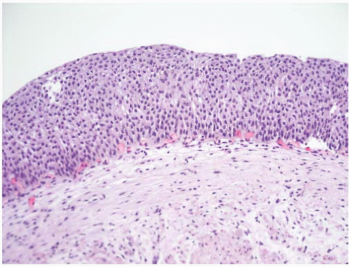
FIGURE 6-12 ▪ Urothelial hyperplasia with thickened epithelium lacking any architectural or cytologic atypia.


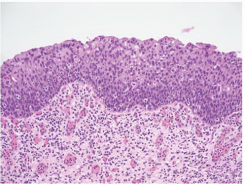
FIGURE 6-13 ▪ Reactive urothelial atypia with thickened epithelium, no architectural atypia, and mildly enlarged nuclei, many containing small nucleoli.

In reactive atypia, the epithelium may or may not be thickened. Although a thickened epithelium is typically associated with a reactive process, CIS can also produce a thicker than normal epithelium. Nuclei are uniformly enlarged, vesicular, and may have a prominent usually centrally located nucleolus (Fig. 6-13). The degree of enlargement is, however, significantly less than is typical of CIS (Table 6-5). The nuclei often have a round shape. Mitoses can be frequent and are located in the lower epithelial layers. The normal architecture is largely preserved though there may be some variation in nuclear orientation. The orientation can also be made unclear by the rounding of the nuclei resulting in no obvious perpendicular orientation to the basement membrane. The cytoplasm can be normal in appearance or have either increased eosinophilia or basophilia. Cytoplasmic clearing or vacuolization can be a feature (Table 6-5). Inflammation is almost always present and often obscures the epithelial stromal interface. Intraepithelial inflammatory cells are a feature of reactive atypia and are infrequently found in urothelial CIS.


Table 6-5 ▪ MORPHOLOGIC FEATURES OF FLAT LESIONS


Feature


Hyperplasia


Reactive Atypia


Dysplasia


Carcinoma In Situ


Thickness


Thickened


Variable


Variable


Variable


Architecture


Normal


Normal


Mildly abnormal


Disorganized


Pagetoid spread


No


No


No


Yes


Denudation


No


Variable


Variable


Commonly present


Nuclear size


Normal or slight enlargement


Normal or slight enlargement


Enlarged


Markedly enlarged


Nuclear shape


Regular


Regular or rounded


Slight irregularities


Pleomorphic


Nuclear membrane


Smooth


Smooth


Some irregularities—notching


Irregular with thickening


Chromatin


Fine


Fine


Variable hyperchromasia


Hyperchromatic


Nucleoli


Inconspicuous


Large


Small or inconspicuous


Large, may be multiple


Mitoses


Infrequent


May be common—basal location


Infrequent


Frequent including abnormal forms


Cytoplasm


Uniform


Uniform or vacuolated


Uniform


Uniform


Atypia of Unknown Significance

One of the gray zones in any consideration of intraepithelial lesions is between reactive atypia and true neoplastic (dysplastic) alterations. Reproducibility studies have clearly demonstrated the lack of consistency in this particular area. This category was created to include those instances where a lesion cannot be confidently placed in the reactive versus dysplastic categories.8

Histologically, there is usually an inflammatory background. The degree of cytologic atypia is judged to be outside of the accepted range for reactive processes although this possibility cannot be excluded. Re-evaluation after inflammation subsides may resolve the problem, particularly in the follow-up of patients with known urothelial neoplasia who have been treated with intravesical therapy. In one study, none of the 35 patients diagnosed with atypia of unknown significance developed urothelial neoplasia with a median 3.5 years of follow-up.173


Urothelial Dysplasia (Low-grade Intraurothelial Neoplasia)

This category also suffers from a significant problem in diagnostic reproducibility.168,169 Previous classification schemes have included grading systems for urothelial dysplasia, but the current approach is to place neoplastic intraepithelial
lesions into only two categories: urothelial dysplasia and urothelial CIS. Urothelial dysplasia has been divided into primary dysplasia for when the lesion is diagnosed de novo and secondary dysplasia when it is found in the setting of known urothelial neoplasia.174 There is evidence, largely genetic, that it shares some abnormalities with CIS and therefore likely represents a precursor lesion.175

The natural history of lesions with dysplastic features of a lesser degree than the moderate to severe categories is unknown.176 Given the difficulties with reproducibility of the diagnosis and changes in the reporting of these lesions over the past several decades, it is difficult to interpret the published data regarding the natural history of this lesion. It is most often diagnosed in the context of known urothelial neoplasia. In the latter group, urothelial dysplasia can be identified in the flat epithelium of 22% to almost 100% of cases.177, 178, 179, 180 Studies that have applied the 1998 WHO/ISUP criteria for primary dysplasia have indicated a 15% to 19% risk of developing cancer with a mean follow-up of 4.9 to 8.2 years.173,181

The limitations of diagnostic criteria and reproducibility also make it difficult to interpret published data on the genetics of urothelial dysplasia. Aberrant expression of cytokeratin 20 in the midlayers of the urothelium has been described.182, 183, 184 Studies have also described overexpression of p53 protein and increased proliferation as indicated by the expression of Ki67.183 Loss of CD44 expression occurs in dysplasia.184 Studies utilizing loss of heterozygosity have demonstrated abnormalities of chromosome 9 in these lesions.185 These findings have supported the hypothesis that urothelial dysplasia is related to CIS and does represent a neoplastic process.

Histologically, the epithelium is of variable thickness, but most often it is within the normal range. The nuclei are irregularly enlarged and tend to be more oval or rounded than normal. In general, the degree of enlargement is significantly less than in CIS. There is some degree of disruption of the normal architecture with variable degrees of loss of polarity and focal nuclear crowding (Fig. 6-14). There is mild nuclear hyperchromasia and pleomorphism but not to the degree seen in CIS. The nuclear membranes are irregularly thickened and notching and sharp angulation can be present. Nucleoli when present are small and inconspicuous. Mitoses can be present but are few and limited to the lower epithelial layers. Cytoplasmic changes may be present with increased eosinophilia. Overall, the features are those of a neoplastic atypia but fall short of the criteria for CIS outlined below. Denudation can be present but is much less frequent than in CIS.


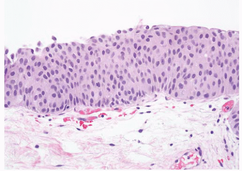
FIGURE 6-14 ▪ Urothelial dysplasia with flat epithelium showing a mild degree of architectural and cytologic atypia.


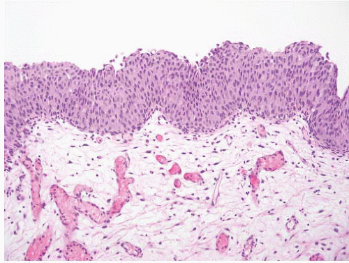
FIGURE 6-15 ▪ Urothelial dysplasia with slight tenting of the surface.

In some instances, the features resemble those of the urothelium present in low-grade papillary carcinoma. In the setting of papillary neoplasia, this may represent the earliest indication of the development of new papillary tumors (Fig. 6-15).185


Carcinoma In Situ (High-grade Intraurothelial Neoplasia)

The current classification recognized the need to expand the category of CIS to include lesions that had been graded in the moderate to severe dysplasia categories in previous systems. This change reflects current practice in major institutions treating bladder cancer. It also recognizes the general trend for underdiagnosis of higher-grade lesions.16 There is further recognition that this is the most reproducible diagnostic category.168,169 CIS is accepted as a precursor of invasive carcinoma.

CIS is most often seen in association with high-grade papillary or invasive urothelial carcinoma. De novo CIS accounts for only 1% to 3% of newly diagnosed cases of bladder cancer.186, 187, 188 These patients are at significant risk for the development of invasive carcinoma. In contemporary series progression to invasive carcinoma occurs in up to 25% of patients and the cancer-specific survival is in the 75% to 85% range with long-term follow-up.189, 190, 191 The presence of
CIS of the bladder in patients with other urothelial tumors is also important.192 The risk of having CIS in the upper tract is three- to four-fold higher and in the prostatic urethra is seven-fold higher than when a bladder tumor does not have associated CIS.193,194 For patients with noninvasive papillary tumors, the presence of CIS is associated with a higher risk of recurrence and progression.195 In cases with lamina propria invasion (T1), the presence of CIS also indicates an increased risk of progression to muscle-invasive disease and has been used as one of the indications for early cystectomy.195,196


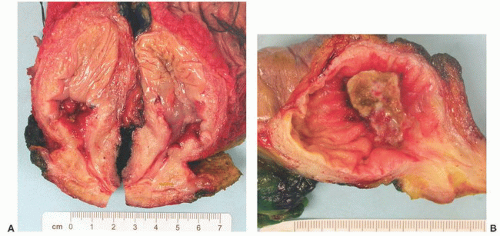
FIGURE 6-16 ▪ Gross appearance of CIS localized to area around an invasive carcinoma (A) and more diffusely involving the mucosa (B).

Grossly, CIS most often appears as an erythematous area (Fig. 6-16). This can be focal, multifocal, or diffuse. In some cases, the mucosa has a velvety or granular character.

The histologic diagnosis of CIS fundamentally requires the recognition of cytologically malignant cells. A variety of descriptive terms have been applied to CIS (Table 6-6).15,172,176,197 These are helpful in highlighting the morphologic variability of the lesion but have no clinical significance. Histologically, CIS is characterized by architectural disorder with haphazard orientation of nuclei and nuclear crowding and clustering (Fig. 6-17). There is nuclear pleomorphism with significant nuclear enlargement, hyperchromasia, and single to multiple nucleoli. The atypical cells need not involve the full thickness of the epithelium, and at the minimum single malignant cells growing in a pagetoid fashion are sufficient for the diagnosis (Fig. 6-18). Individual cells tend to show marked cytologic atypia but an increased nuclear-to-cytoplasmic ratio is not a prerequisite (not present in the large cell type of CIS).


Table 6-6 ▪ MORPHOLOGIC PATTERNS OF UROTHELIAL CIS


Small cell


Large cell


Denuding (denuding cystitis)


Pagetoid


Undermining


CIS with glandular differentiation


CIS with squamous differentiation


In the large cell patterns of CIS, the malignant cells have moderate to abundant cytoplasm that often has increased eosinophilia (Fig. 6-19). These cells can have relatively uniform but markedly enlarged hyperchromatic nuclei or nuclei with a high degree of pleomorphism. The small cell pattern gets its name because the cells have scant cytoplasm that is often basophilic. This gives a similar impression to small cell carcinoma, but the nuclei are actually enlarged and typically have severe hyperchromasia. This is not considered to be related to small cell neuroendocrine carcinoma. Tumor cells in CIS are often discohesive, and in some cases, only a few isolated cells are present clinging to the basement membrane (denuding or clinging CIS) (Fig. 6-20). It has been suggested that this is related to loss of the normal polarity of MUC-1 expression.198 The nuclei in these cases are often intensely hyperchromatic, and it is not possible to appreciate the chromatin pattern. In other cases, the surface may be completed denuded with the CIS present in von Brunn nests only (Fig. 6-21). This pattern should not be overdiagnosed as invasive carcinoma (Fig. 6-22). There is a propensity for the malignant cells to invade the adjacent normal urothelium. This can take the form of single malignant cells resulting in a pagetoid pattern. In other cases the malignant cells burrow beneath and lift up the adjacent benign urothelium. Less frequently the malignant cells will spread within the upper level of the adjacent urothelium. Squamous and glandular differentiation can be seen in CIS (Fig. 6-23).199 When glandular differentiation is present, the term adenocarcinoma in situ should not be used as this implies a precursor lesion of primary adenocarcinoma (see section on adenocarcinoma).


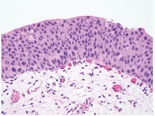
FIGURE 6-17 ▪ Urothelial CIS with haphazardly arranged cells with significant nuclear pleomorphism.


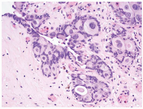
FIGURE 6-18 ▪ Urothelial CIS showing pagetoid spread into von Brunn nests.


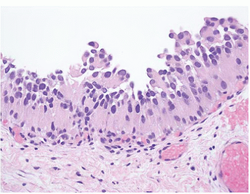
FIGURE 6-19 ▪ Urothelial CIS with cells having abundant eosinophilic cytoplasm.


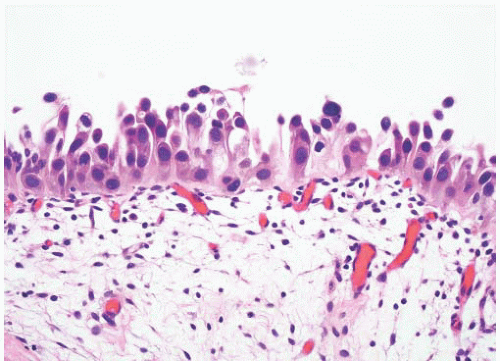
FIGURE 6-20 ▪ Urothelial CIS with a denuding pattern.


Cytologic Features

Urothelial CIS is a high-grade lesion, and urine cytology is highly sensitive for its detection. The lesion is characterized by discohesive cells, and so the tumor cells are often shed singly in the urine (Fig. 6-24). In specimens obtained at cystoscopy, more intact fragments of the epithelium are present. The cells typically are enlarged and have large hyperchromatic nuclei with scant cytoplasm. The nuclear outlines are irregular, and nucleoli are prominent. As described above, there are several histologic patterns to CIS, and in some, the cells can have more abundant cytoplasm. This can result in the malignant cells having an appearance resembling squamous cell carcinoma. It is not possible to distinguish CIS reliably from invasive carcinoma. The presence of necrosis and tissue debris would indicate that invasion is likely present.


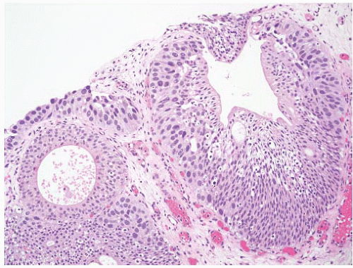
FIGURE 6-21 ▪ Urothelial CIS spreading into von Brunn nest and lifting the overlying benign urothelium.


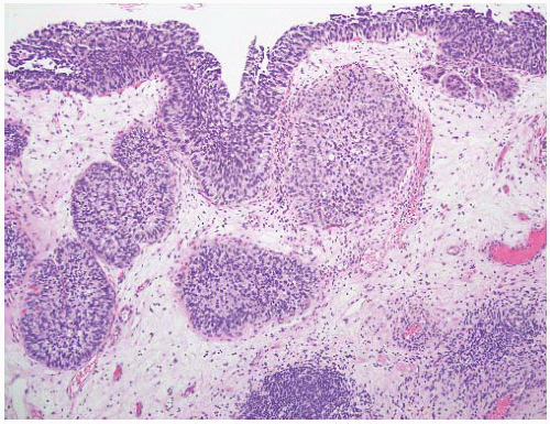
FIGURE 6-22 ▪ Urothelial CIS extending into and expanding von Brunn’s nests simulating an invasive tumor. There is a small focus of early invasion in the upper right.


Immunohistochemistry in Flat Lesions

A variety of immunohistochemical markers have been studied as adjuncts to the diagnosis of flat lesions. The first marker used was cytokeratin 20.182 In normal or reactive urothelium, cytokeratin 20 expression is limited to the umbrella cell layer whereas there is diffuse reactivity for cytokeratin 20 throughout the full thickness of the urothelium in the majority but not all cases of CIS (Fig. 6-25A and B).182, 183, 184,200 In normal or reactive urothelium p53, protein is generally expressed by only a few cells and with weak intensity.184 In some cases, however, 50% or more of the nuclei show variably intense immunoreactivity. Most cases of CIS show diffuse and intense nuclear expression of p53 (Fig. 6-25C).183,184,201 However, not all cases of CIS are p53 positive, and so absence of staining for p53 does not exclude a diagnosis of CIS. Cellular proliferation is significantly higher in CIS than in reactive atypia, and so, Ki67 has been used in this distinction.184,200 There is, however, sufficient overlap in Ki67 scores between CIS and reactive atypia to make this a difficult feature to use in individual cases.200 CD44 is expressed in the basal cells of normal urothelium and shows more diffuse expression throughout all layers in hyperplasia and reactive atypia. In contrast, there is loss of expression of CD44 in dysplasia and CIS.184 Other markers that have been applied to this challenge include p16 and E-cadherin, but there is less experience with these, making their routine use of limited value.202,203 In one study, E-cadherin showed more diffuse and intense immunoreactivity in CIS than in normal epithelium198 while in another loss of E-cadherin staining was observed in 32% of cases.204 The RNA-binding protein IMP3 has been found to be expressed in 48% of cases of CIS compared to 13% in reactive atypia in one study.205


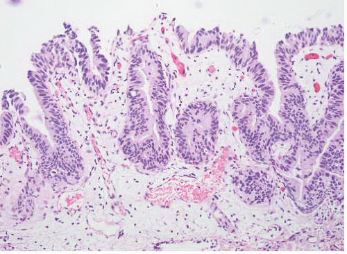
FIGURE 6-23 ▪ Urothelial CIS with cells having fusiform nuclei mimicking adenocarcinoma in situ.


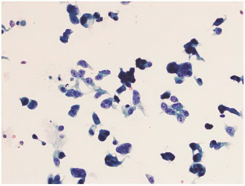
FIGURE 6-24 ▪ Urothelial CIS in a bladder wash specimen with discohesive individual malignant cells.

Because of the variability in results for individual mar kers, it has been recommended that a panel of three markers (cytokeratin 20, p53, and CD44) be applied (i) where the diagnosis of CIS is strongly favored on routine stains but the pathologist is hesitant to make a definitive diagnosis, (ii) when making a diagnosis of de novo CIS in patients without a history of urothelial neoplasia, and (iii) in cases with unusual morphologic patterns of CIS.172

Fluorescence in situ hybridization has also been proposed as an adjunct to histologic evaluation of flat lesions.146 Over 90% of cases of dysplasia and CIS contain polysomies compared with 36% in hyperplasia, 26% in reactive atypia, and
30% in normal epithelium. The high frequency of abnormalities in histologically benign urothelium limits the application of this approach.


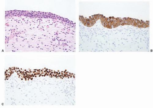
FIGURE 6-25 ▪ Urothelial CIS with malignant cells forming a thin epithelium (A). Immunohistochemistry shows diffuse reactivity for cytokeratin 20 (B) and p53 (C).


Papillary Tumors

In the majority of cases the diagnosis of a lesion as being “papillary” in the urinary bladder is straightforward and the features to be determined are related to proper classification (grading) and evaluation of stage. Occasionally there can be problems in differential diagnosis with lesions that can mimic a papillary process (polypoid or papillary cystitis, fragmented or tangentially cut flat lesions) or papillary lesions that are not strictly speaking urothelial (nephrogenic adenoma, condyloma acuminatum, etc.).206,207

More problematic is the concept of papillary hyperplasia and when to call a small lesion a true papillary neoplasm particularly in the setting of known papillary neoplasia. If a true papillary stalk is evident then the lesion is graded and classified into the categories described below (Fig. 6-26). A papillary stalk is defined by the presence of a central fibrovascular core in an exophytic lesion. In the presence of known neoplasia, I am quite aggressive in diagnosing a lesion as a papillary tumor (Fig. 6-27).

For cases with an undulating surface or tenting of the urothelium where no fibrovascular core is evident, the term papillary hyperplasia has been suggested (Fig. 6-28).39 In some instances, small capillaries are present in the stroma at the base of the pseudopapillae, but these do not extend upward into the stroma. These are therefore more “pseudopapillary” than truly papillary, and that is the terminology I use in my practice. The diagnosis in specimens with a pseudopapillary architecture is dependent on evaluating the surface urothelium as a “flat” lesion; if unequivocal dysplasia or CIS is present the diagnosis should be dysplasia or CIS.


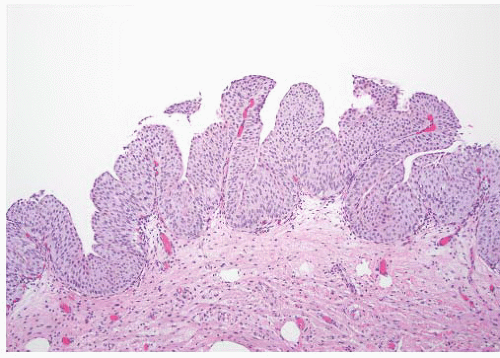
FIGURE 6-26 ▪ Papillary urothelial carcinoma with short papillary fronds.


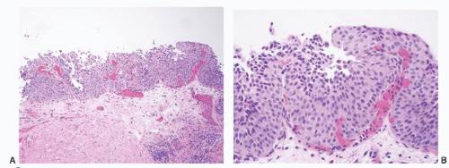
FIGURE 6-27 ▪ Papillary urothelial carcinoma with early formation of identifiable papillae (A,B).

Several studies have looked at a variety of biologic markers in papillary tumors and their relationship to the three groups; for the most part these have demonstrated significant differences of the respective marker in the different categories.18,208

Grossly papillary tumors may be single or multiple (Fig. 6-29). In low-grade tumors, the papillary architecture can often be appreciated grossly. In some cases, the tumors cover much or all of the mucosal surface (Fig. 6-30). In high-grade carcinoma, the tumors often have a more sessile appearance (Fig. 6-31).

Histologically, papillary tumors are defined by the presence of true papillae with central fibrovascular cores covered by the neoplastic epithelium. While the cores tend to be delicate, in some cases they can be quite thickened and hyalinized (Fig. 6-32). Infrequently the stalks are expanded by an inflammatory infiltrate (Fig. 6-33) or by collections of foamy macrophages (Fig. 6-34). The classification (grading) of papillary tumors is based on the features of the covering epithelium and is detailed in the following sections.


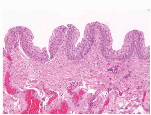
FIGURE 6-28 ▪ Prominent tenting of the mucosal surface producing pseudopapillae (papillary hyperplasia).


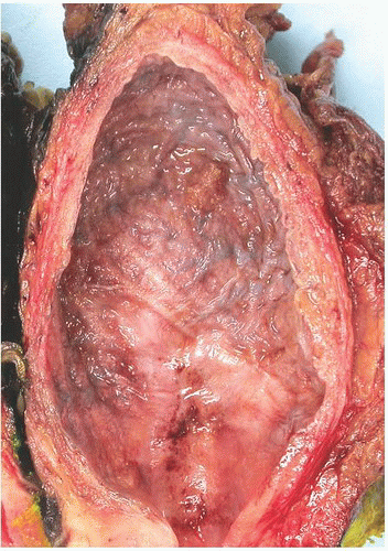
FIGURE 6-29 ▪ Small, single papillary urothelial carcinoma on the posterior wall of the bladder.


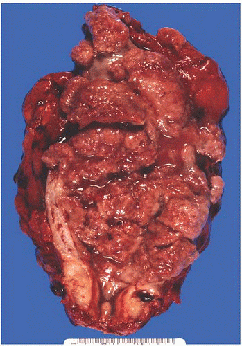
FIGURE 6-30 ▪ Diffuse involvement of the bladder by noninvasive (pTa) low-grade papillary urothelial carcinoma.


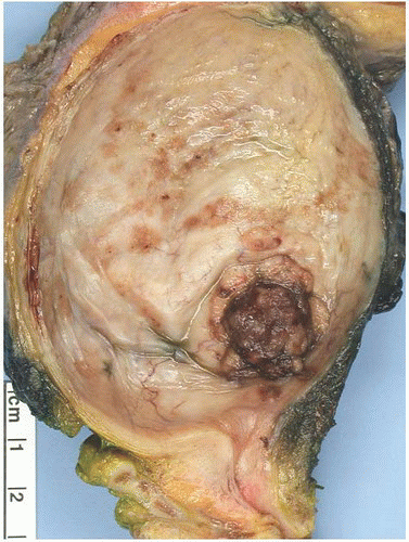
FIGURE 6-31 ▪ Invasive (pT2a), high-grade papillary urothelial carcinoma.


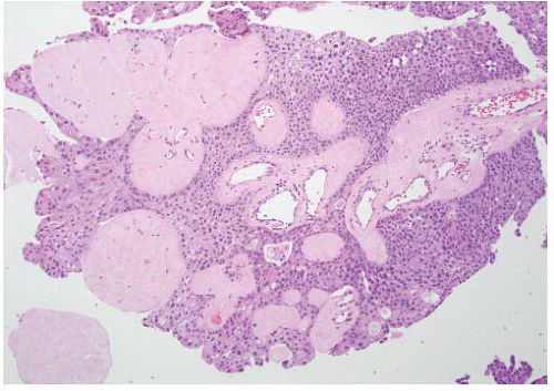
FIGURE 6-32 ▪ Papillary urothelial carcinoma with hyalinization of the papillary cores.


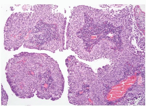
FIGURE 6-33 ▪ Papillary urothelial carcinoma with intense chronic inflammatory infiltrate in the papillary cores.


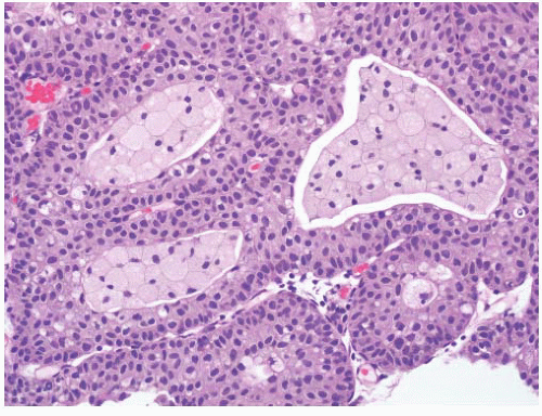
FIGURE 6-34 ▪ Papillary urothelial carcinoma with foamy macrophages in the papillary cores.


FIGURE 6-35 ▪ Papillary urothelial neoplasm of low malignant potential at low (A) and high (B) magnification.


Papillary Urothelial Neoplasm of Low Malignant Potential

The creation of this category represented a compromise between the “papilloma” camp and those insisting on the use of “carcinoma” for all papillary lesions. The 1998 consensus statement acknowledged that the lower-grade papillary neoplasms were not intrinsically malignant but were associated with a significant risk for the development of new papillary tumors (recurrence). These lesions at the lower end of the spectrum were accepted as clinically significant with close clinical follow-up necessary but further intravesical therapy not indicated. These tumors have a significantly lower rate of recurrence than either low- or high-grade papillary carcinomas and have a very low rate of stage progression.9,18,34,209, 210, 211, 212 In a review of published studies, the mean tumor recurrence rate was found to be 36% and the stage progression rate to be 4%.13 The largest single institution experience with 212 cases (representing 21% of the 1,006 noninvasive papillary tumors) reported a recurrence rate of 18% and progression to invasive disease in 2%.213 None of the patients died of bladder cancer.


FIGURE 6-36 ▪ Papillary urothelial neoplasm of low malignant potential at low (A) and high (B) magnification.

Morphologically, this category largely though not completely corresponds to grade 1 papillary carcinoma in the 1973 WHO system.2 The tumor consists of delicate papillae with little or no fusion (Figs. 6-35A and 6-36A). The covering urothelium is usually but not always thickened and shows minimal if any architectural irregularity. Nuclei are normal in size to slightly enlarged and lack significant nuclear hyperchromasia or pleomorphism (Figs. 6-35B and 6-36B). The chromatin is fine, and nucleoli are inconspicuous. Mitoses are infrequent and basally located when present (Tables 6-7 and 6-8).


Papillary Urothelial Carcinoma, Low Grade

This category contains the intermediate group of lesions. In the 1973 WHO system, this would include roughly the lower one-half to two-thirds of grade 2 papillary carcinoma.17,18
These tumors have a significantly higher recurrence rate than for papillary urothelial neoplasm of low malignant potential and similar to high-grade papillary carcinomas.9 They also have a higher rate of stage progression than papillary urothelial neoplasm of low malignant potential but significantly lower than for high-grade papillary carcinoma.9,19,34 Low-grade tumors can be invasive though this is distinctly uncommon. A review of the literature revealed a mean recurrence rate of 50% and mean stage progression rate of 10%.13 In the large series of Pan et al.213 these accounted for 60% of noninvasive papillary tumors and had recurrence and progression rates of 35% and 6.5%, respectively. At last follow-up, 2% of patients presenting with low-grade papillary carcinoma had died of bladder cancer.213 Patients with these tumors require close clinical follow-up though recently it has been suggested that this can be less frequent than for patients with high-grade tumors.21 A single dose of intravesical therapy (most often mitomycin-C) is optional but maintenance intravesical therapy is not recommended.23


Table 6-7 ▪ PAPILLARY UROTHELIAL NEOPLASMS: ARCHITECTURAL FEATURES


PUNLMP*


Low Grade


High Grade


Papillae


Delicate


Delicate


Delicate


Rarely fused


Occasionally fused


Fused and branching


Organization


Polarity normal


Predominantly ordered with minimal crowding and loss of polarity


Predominantly disordered with crowding, overlapping cells and loss of polarity


Any thickness


Any thickness


Any thickness—may be single cells (denuding)


Most increased


Most increased


Cohesive cells


Cohesive cells


Often discohesive


* Papillary urothelial neoplasm of low malignant potential.


The papillae are largely delicate and separate, but some fusion may be seen. At low magnification, there is a generally ordered architectural appearance to the cells within the epithelium (Figs. 6-37A and 6-38A). There is, however, an impression of increased cellularity with increased nuclear density. The nuclei tend to be uniformly enlarged and retain the elongated to oval shape of normal urothelial cells. The chromatin remains fine with small and generally inconspicuous nucleoli. There is often some variability with scattered hyperchromatic nuclei (Figs. 6-37B and 6-38B). Mitoses may be present but are few and generally remain basally located (Tables 6-7 and 6-8).


Table 6-8 ▪ PAPILLARY UROTHELIAL NEOPLASMS: NUCLEAR FEATURES


PUNLMP*


Low Grade


High Grade


Size


Mildly enlarged


Enlarged


Enlarged


Uniform


Some variation


Marked variability


Shape


Elongated


Elongated, oval, or round


Pleomorphic


Uniform


Slight variation


Chromatin


Fine


Fine with some variation


Frequently coarse with marked variation


Nucleoli


Absent to inconspicuous


Usually inconspicuous


Prominent


Single to multiple


Mitoses


Rare


Infrequent


Frequent


Basal if present


Most basal if present


Any level


* Papillary urothelial neoplasm of low malignant potential.


Papillary Urothelial Carcinoma, High Grade

As previously discussed, many WHO grade 2 (1973) tumors (roughly the upper one-half to one-third) have a significant risk of invasion and biologically have more in common with the grade 3 tumors. These tumors not only have a significant risk of recurrence but have a substantial risk of invasion and progression to muscle-invasive disease. For this reason, the consensus was that these were better included in a highgrade category with the WHO grade 3 (1973) neoplasms. The overall progression rate (to invasive carcinoma) ranges from 15% to 40%. Of the patients initially presenting with high-grade papillary carcinoma in the Pan et al.213 series, 22% died of bladder cancer. For high-grade Ta tumors, intravesical BCG therapy with an induction course and maintenance is recommended.23

Heterogeneity of grade is recognized in papillary lesions,214,215 and the consensus is that tumors should be graded on the basis of the worst grade present (Fig. 6-39). There is no consensus on a minimum quantity that should be present to assign a higher grade in cases such as this. Some authors have used a 5% cutoff for recognition of a higher-grade component.215,216 After reviewing this topic, the recent International Consultation on Urologic Diseases concluded that there is some evidence to indicate that pure high-grade tumors are more aggressive than mixed low- and high-grade ones.27 This is a subject that is in need of further study.8,14,172 Our current approach is to assign the worst grade present, irrespective of quantity, but to include a note indicating that there is grade heterogeneity and which grade is predominant.


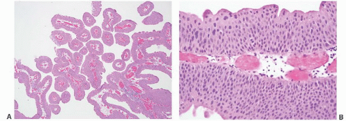
FIGURE 6-37 ▪ Low-grade papillary urothelial carcinoma at low (A) and high (B) magnification.


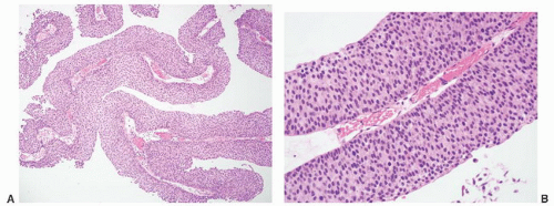
FIGURE 6-38 ▪ Low-grade papillary urothelial carcinoma at medium (A) and high (B) magnification.


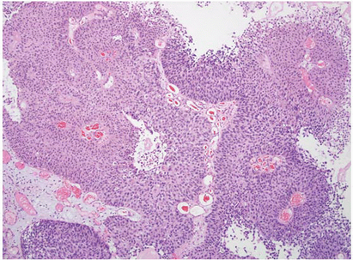
FIGURE 6-39 ▪ Papillary urothelial carcinoma with low-grade (left) and high-grade (right) areas.


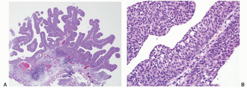
FIGURE 6-40 ▪ High-grade papillary urothelial carcinoma at low (A) and high (B) magnification.

The papillae are frequently fused forming apparently solid masses. The overall impression is one of disordered growth (Figs. 6-40A and 6-41A). The epithelium is of variable thickness and may resemble “denuding carcinoma in situ” in some instances. Individual cells are haphazardly arranged within the epithelium and have a generally discohesive nature. Nuclei are hyperchromatic and pleomorphic. The chromatin is dense, irregularly distributed, and often clumped. Nucleoli may be single or multiple and are often prominent (Figs. 6-40B and 6-41B). Mitoses are generally frequent and may be seen at any level of the epithelium. Abnormal mitoses are often present (Tables 6-7 and 6-8). CIS can be present in the adjacent mucosa.


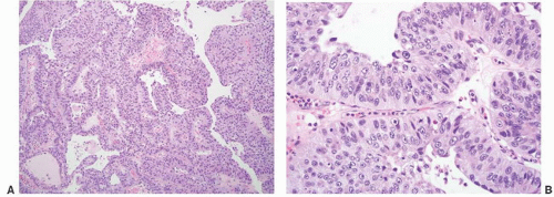
FIGURE 6-41 ▪ High-grade papillary urothelial carcinoma at low (A) and high (B) magnification.


Cytologic Features

The sensitivity of cytology for detecting papillary tumors is strongly correlated with the tumor grade. There is a low degree of sensitivity for papillary urothelial neoplasms of low malignant potential and low-grade papillary urothelial carcinoma. Particularly in bladder washings, fragments of papillary tumors can be obtained and these are very helpful in making the diagnosis. The presence of papillary fragments in voided urine in the absence of inflammation or lithiasis is suspicious for papillary urothelial neoplasia. In these fragments an increased cellularity and crowding of nuclei may be evident (Fig. 6-42). The umbrella cells are variably preserved. The nuclei are uniformly enlarged with an increased nuclear to cytoplasmic ratio. The chromatin tends to be uniformly distributed with inconspicuous or small nucleoli. The nuclear membranes show variable thickening and there is irregularity of nuclear shape. Nuclear membrane features have been reported to be most helpful in identifying low-grade lesions.217 The cytoplasm
is homogeneous and lacks vacuolization. Mitoses are absent or infrequent.


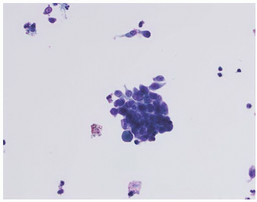
FIGURE 6-42 ▪ Bladder washing specimen from a low-grade papillary urothelial carcinoma.

In high-grade papillary tumors, the most identifying feature to distinguish from low-grade tumors is the presence of nuclear pleomorphism. The cells are markedly enlarged and are present as cohesive groups and as single cells (Fig. 6-43). There is an increased nuclear to cytoplasmic ratio. The nuclei are hyperchromatic with irregular clumped chromatin and prominent nucleoli. The nuclear membranes are thick and irregular. Mitoses are frequent. Umbrella cells are absent. The cytoplasm is not homogeneous, and cytoplasmic features may indicate squamous or glandular differentiation.


Differential Diagnosis

There are several benign processes in the urinary bladder that can mimic papillary urothelial neoplasms (Table 6-9). Papillary hyperplasia is discussed above. Nephrogenic adenoma is covered in the section on clear cell adenocarcinoma. Condyloma acuminatum is discussed in the section on squamous papilloma.

Papillary/polypoid cystitis is a condition characteristically associated with indwelling catheter and is reported to occur in up to 80% of patients with catheters.37,38,207,218,219 These lesions may also occur in other settings related to chronic irritation, such as fistula and long-standing urinary tract obstruction. By cystoscopy, polypoid cystitis appears as exophytic lesions that may be large and polypoid, or somewhat narrower mimicking papillary structures. Histologically, the typical finding is that of marked edema of the lamina propria resulting in exophytic polypoid projections. The overlying urothelium ranges from flattened and attenuated to normal to hyperplastic, but shows no significant cytologic or architectural atypia. In some cases, the stroma may be less edematous resulting in a more papillary appearance (so-called papillary cystitis). There always continues to be some stroma present frequently with interspersed inflammatory cells and small capillaries.


Table 6-9 ▪ MIMICS OF PAPILLARY NEOPLASMS


Papillary urothelial hyperplasia


Nephrogenic adenoma


Polypoid cystitis


Papillary cystitis


Fibroepithelial polyp


Condyloma acuminatum


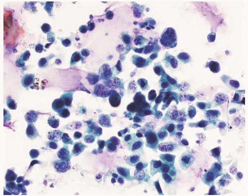
FIGURE 6-43 ▪ Bladder washing specimen from a high-grade papillary urothelial carcinoma.

Fibroepithelial polyp can occur throughout the urinary tract and over a wide age range from children to adults.220, 221, 222 In the pediatric population, fibroepithelial polyp is the second most common mass in the bladder (after rhabdomyosarcoma).222 These are considered to be nonneoplastic lesions that can be congenital or acquired and may represent a hamartoma. There is a male predominance. Presentation is variable but includes obstruction (particularly in urethral and ureteral lesions), hematuria, and pain. Grossly these are polypoid structures that can have multiple finger-like projections (Box 6-2). The overall polypoid appearance distinguishes these from papillary urothelial neoplasms. The finger-like projections have central fibrous cores covered by attenuated to hyperplastic urothelium with a scattered inflammatory infiltrate. These can simulate papillary neoplasia; however, the fibrous cores tend to be wider and are more dense and fibrotic in nature than papillary neoplasms. Atypical stromal cells can be present.223 In a minority of cases, the urothelium proliferates producing complex anastomosing nests. These can mimic inverted papilloma. In fibroepithelial polyp the anastomosing nests of urothelium are embedded in a fibrous stroma and are more irregular and less “solid” than is seen in inverted papilloma.


Invasive Urothelial Carcinoma


Gross Features

Invasive urothelial carcinoma can be unifocal or multifocal. In most cases the invasive carcinoma forms a single mass. Additional foci, when present, are often noninvasive papillary tumors, papillary tumors with invasion, or areas of CIS. The latter may be appreciated as hemorrhagic or red mucosa. Invasive carcinoma can be polypoid, sessile, or ulceroinfiltrative (Figs. 6-44, 6-45, and 6-46). Polypoid growth is most often associated with the sarcomatoid variant and these can be attached to the bladder wall by a narrow pedicle. Invasive tumors are solid, gray-white, and poorly circumscribed with infiltrative borders. In cases with extensive squamous differentiation there can be abundant keratinous debris on the surface. Invasive carcinomas developing from papillary tumors often have a sessile appearance and there may be papillary structures evident on the surface or at the lateral mucosal margins. In some cases there is diffuse thickening of the bladder wall resembling linitis plastica, a pattern most often associated with the plasmacytoid and micropapillary variants. An ulcero-infiltrative pattern of growth may grossly resemble a site of prior TURBT. These areas can be deceiving with the poorly defined lesion representing scarring and secondary changes rather than solid tumor.


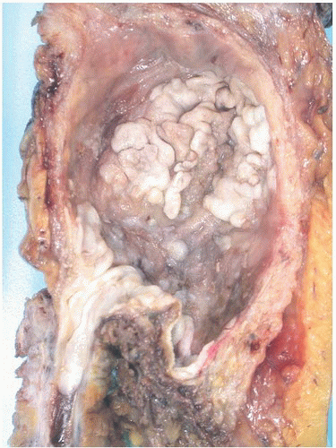
FIGURE 6-44 ▪ Invasive (pT2b) high-grade urothelial carcinoma with a somewhat polypoid appearance.


Microscopic Features

Invasive urothelial carcinoma is most remarkable for the diversity of histologic patterns that can be found. Many of these are so distinctive that they are now recognized as specific variants, some of which have significant clinical characteristics. These are discussed in detail in a following section. The architectural patterns of invasive carcinoma include solid sheets, large and small nests, trabeculae, cords, and single cells (Box 6-3) (Figs. 6-47, 6-48, 6-49, 6-50 and 6-51). In larger nests, the cells at the periphery may have palisading of the nuclei with a pattern of maturation toward the center reminiscent of normal urothelium. In most cases, however, the urothelial nature of the tumor is assumed based on the absence of another specific histology. The pattern of invasion has been linked to prognosis with tumors having well-defined nests, trabeculae, and pushing borders behaving better outcome than those with infiltrating nests, cords, and single cells.224,225


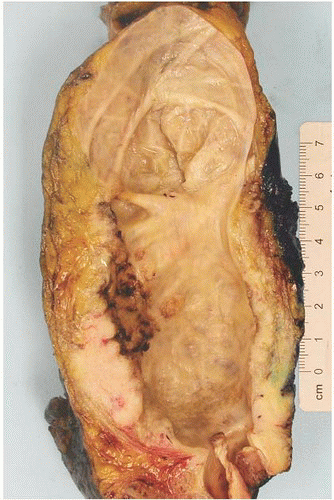
FIGURE 6-45 ▪ Invasive (pT3b) high-grade urothelial carcinoma.


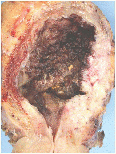
FIGURE 6-46 ▪ Invasive (pT3b) high-grade urothelial carcinoma.


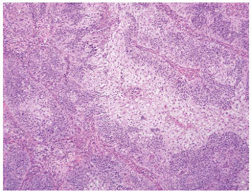
FIGURE 6-47 ▪ Invasive high-grade urothelial carcinoma growing in large nests. Note the squamoid appearance.


FIGURE 6-48 ▪ Invasive high-grade urothelial carcinoma with small variably shaped nests.


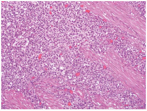
FIGURE 6-49 ▪ Invasive high-grade urothelial carcinoma growing as a solid sheet of cells.


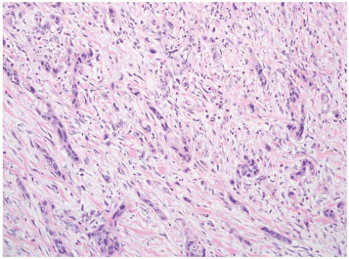
FIGURE 6-50 ▪ Invasive high-grade urothelial carcinoma forming small cords in a desmoplastic stromal background.


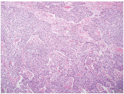
FIGURE 6-51 ▪ Invasive high-grade urothelial carcinoma with a trabecular architecture.

The majority of invasive carcinomas are high grade and this is reflected in striking nuclear pleomorphism manifest by variability in nuclear size and shape, nuclear chromatin pattern, and presence or absence of single or multiple nucleoli (Fig. 6-52). Nuclear contours are often irregular with sharp angles. Bizarre giant tumor nuclei and multinucleated tumor giant cells can be present (Fig. 6-53). Mitotic activity is variable but in most cases mitoses are frequent and abnormal mitotic figures are identifiable. There is generally moderate to abundant cytoplasm that is eosinophilic. In many cases this results in a squamoid appearance to the tumor. In other tumors the cytoplasm can be basophilic. Cytoplasmic vacuoles can occur and cytoplasmic mucin may be visible. Mucin stains are positive for cytoplasmic mucin in up to 60% of invasive high-grade tumors (Fig. 6-54).226 The presence of mucin does not indicate glandular differentiation; the latter diagnosis is reserved for tumors with the presence of well-formed glandular structures. Cytoplasmic clearing due to the accumulation of glycogen is another feature that can be present. This is usually focal but when more extensive has been described as the clear cell variant of urothelial carcinoma.227


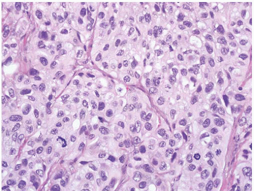
FIGURE 6-52 ▪ Invasive high-grade urothelial carcinoma with nuclear pleomorphism.


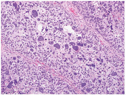
FIGURE 6-53 ▪ Invasive high-grade urothelial carcinoma with bizarre pleomorphic nuclei.

In most cases of invasive carcinoma, there is a desmoplastic stromal response (Fig. 6-55). This can be quite prominent and in some cases consists of a cellular pseudosarcomatous spindle cell stroma mimicking sarcomatoid carcinoma.228 The stroma can also be myxoid, and in some tumors, this myxoid background becomes a predominant part of the tumor (Fig. 6-56).229 Benign osseous metaplasia can develop and should not be confused with a heterologous element of sarcomatoid carcinoma.230,231 Retraction of the stroma away from invasive tumor nests is also a characteristic feature of urothelial carcinoma. This can be a particularly helpful feature in foci of early invasion. In most tumors this is a focal finding, when prominent a diagnosis of the micropapillary variant of urothelial carcinoma should be considered. It also creates a problem in distinguishing retraction artifact from lymph-vascular invasion.232 Invasion of the stroma by tumor can be accompanied by an associated inflammatory infiltrate that can be polymorphous or predominantly lympho-plasmacytic (Fig. 6-57). In some cases, this is particularly intense and obscures the tumor resulting in a lymphoepithelioma-like morphology.233


Cytologic Features

Cytology is highly sensitive in cases of invasive urothelial carcinoma. These tumors are almost always high grade. Cells are shed singly and in cohesive groups; the latter are most apparent in specimens obtained at cystoscopy (Fig. 6-58). The cells are markedly enlarged and have increased nuclear to cytoplasmic ratios. Nuclei are large, hyperchromatic, and have coarsely clumped chromatin and single to multiple nucleoli. The nuclear membranes are thick and irregular. There is striking nuclear pleomorphism (Fig. 6-59). Mitotic figures are common. The background is typically bloody with inflammation and necrotic tissue fragments (Fig. 6-60). Cytoplasmic features are variable and can reflect squamous or glandular differentiation. Features of specific histologic variants can be present.


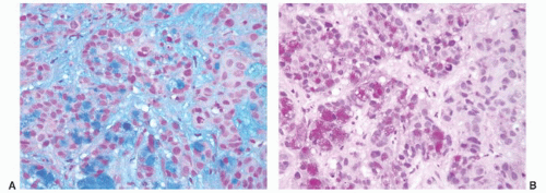
FIGURE 6-54 ▪ Mucin positivity (A, Alcian blue pH 2.4; B, mucicarmine) in an otherwise typical high-grade urothelial carcinoma.


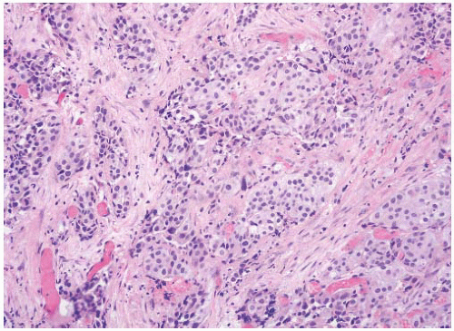
FIGURE 6-55 ▪ Invasive high-grade urothelial carcinoma with desmoplastic stroma.


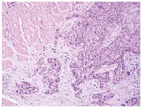
FIGURE 6-56 ▪ Invasive high-grade urothelial carcinoma with myxoid stroma.


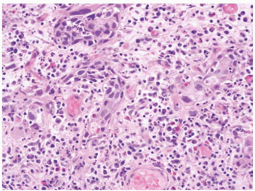
FIGURE 6-57 ▪ Invasive high-grade urothelial carcinoma with inflammation including lymphocytes, plasma cells, and eosinophils.


FIGURE 6-58 ▪ High-grade urothelial carcinoma in a voided urine specimen.


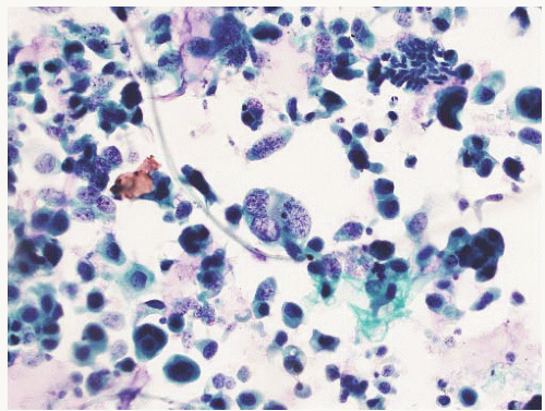
FIGURE 6-59 ▪ High-grade urothelial carcinoma in a bladder wash specimen.


Differential Diagnosis

The differential diagnosis of invasive urothelial carcinoma includes benign conditions that can mimic invasive carcinoma, nonurothelial carcinomas, other primary tumors, and secondary tumors (either direct invasion from another site or metastases). Benign proliferations that can mimic urothelial carcinoma such as von Brunn’s nest hyperplasia and proliferative cystitis are discussed in the section on the nested variant of urothelial carcinoma. Inverted papilloma is dealt with in the section on urothelial carcinoma with inverted papilloma-like architecture. Nephrogenic adenoma is reviewed in the section on clear cell adenocarcinoma. Other benign glandular lesions are discussed in the nonurachal adenocarcinoma section. Other tumors such as paraganglioma are covered in the relevant section. Secondary involvement of the bladder is covered in detail in the section on secondary neoplasms.


Pseudocarcinomatous Proliferations.

Baker and Young234 first described the occurrence of benign reactive epithelial proliferations that mimic invasive urothelial carcinoma in the setting of radiation therapy. Subsequently similar lesions have been described following chemotherapy or without any evident pathogenic factor.235,236

These lesions are characterized by irregular nests of urothelium extending into the lamina propria simulating invasive carcinoma. This is often accompanied by cytologic atypia and mitotic figures as well as cytoplasmic eosinophilia, all typical of invasive carcinoma. A key clue to recognition is the impression of an inflammatory background with stromal hemorrhage, fibrin, edema, hemosiderin deposition, and inflammation. There are often prominent ectatic vascular structures. In the setting of radiation the typical vascular changes may be evident as well as stromal cell atypia. There may be overlying mucosal ulceration. If a lesion reminds you of a urethral caruncle, think of this as a possibility.


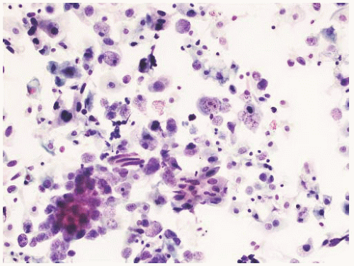
FIGURE 6-60 ▪ High-grade urothelial carcinoma in a bladder wash specimen.


Immunohistochemistry

Immunohistochemistry can play a vital role in the diagnosis and differential diagnosis of urothelial carcinoma.237,238 Given the diversity of the morphologic manifestations of urothelial carcinoma, not surprisingly a large number of immunohistochemical markers can be expressed by these tumors (Table 6-10). It is important to recognize that the reported frequency of expression is often dependent on the type of cases studied. For example, in one study thrombomodulin was expressed by 86% of papillary urothelial neoplasms of low malignant potential but only 39% of invasive urothelial carcinomas.244 The immunohistochemical profiles of urothelial carcinoma variants are discussed in the specific sections below and are not repeated here. Immunohistochemistry is also included in the differential diagnosis sections throughout this chapter and the reader is referred to those discussions for specific questions.

There are markers that are used to assist in identifying a tumor as being of urothelial origin. Uroplakin III has been reported to be 100% specific in some studies but its usefulness is somewhat limited by low sensitivity (57% to 60%).241,244,255 Expression decreases with increasing grade and so in the types of tumors most often causing difficulty, it is least often expressed.244 Thrombomodulin has also been used as a specific marker of urothelial carcinomas. It is expressed by 49% to 91% of tumors with expression decreasing in invasive high-grade tumors.241,244,254 It is not as specific as uroplakin III and is also expressed in some cases of squamous cell carcinoma, mesothelioma, and adenocarcinoma
of the bladder, lung, ovary, pancreas, and breast among others.244,254 More recently, GATA3 has been shown to be expressed in 67% of urothelial carcinomas.256 In an extensive tissue microarray study, Higgins et al.256 found only ductal adenocarcinoma of the breast to also express GATA3 indicating a high degree of specificity for this marker. These authors also reported expression of S100P in 78% of bladder urothelial carcinomas but this marker was less specific with gastrointestinal tract adenocarcinoma, hepatocellular carcinoma, ovarian carcinoma, and rarely (2%) prostatic adenocarcinoma also being positive.256


Table 6-10 ▪ IMMUNOHISTOCHEMISTRY OF UROTHELIAL CARCINOMA (USUAL TYPE)


Marker


Positive (%)


Comments


Cytokeratin 5/6239,240


6-75


Cytokeratin 7241,242


87-94


Cytokeratin 8/18239,243


83-87


Cytokeratin 19239


92


Cytokeratin 20239,241,244


25-67


Cytokeratin 34βE12244,245


65-97


EMA246


80-94


Vimentin239,247


11-33


P63242,247,248


81-100


P16249,250


30-50


P53208,251


34-68


Higher percent positive with higher-grade and nonpapillary type


CD10252,253


54-67


Predominantly cytoplasmic


Thrombomodulin241,244,254


49-91


Lower % positive with invasive carcinomas


Uroplakin III241,244,255


57-60


Lower % positive with higher grade


GATA3256


67


CD117246


30


AMACR (racemase)242,257


31-36


S100P256,258


71-78


TTF-1259


5


Calretinin260


5


Hepatocyte nuclear factor-1β261


3


PAX-2247,262


0-6


PAX-8247,263,264


0-17


Results appear antibody-dependent


PSA241,242


0


PAP241,242


0-11


PSMA265,266


0-13


Weak in one case of urothelial carcinoma; may be + with glandular differentiation


Prostein (P501S)258,267


0-6


NKX3.1258


0


CDX-2268


0


Positive with enteric-type glandular differentiation


Estrogen receptor-β269,270


63-80


Estrogen receptor-α270,271


1.4-4.5


Progesterone receptor271


0


HMB-45272


0


Melan-A273


0


Synaptophysin248


6


Rare cells


CD56248


0


Chromogranin-A248


0


The expression of cytokeratins is also of interest in urothelial carcinoma. The urothelium is a stratified epithelium and as such has some similarities with squamous cell carcinoma. One characteristic is the expression of high molecular weight cytokeratins. One of the more frequently studied high molecular weight cytokeratin clones, 34βE12, is expressed in 65% to 97% of cases (Fig. 6-61A and B).242,244,245,274 Urothelial carcinoma is also one of the tumors that coexpress cytokeratins 7 and 20 (Fig. 6-61C and D). There has, however, been wide variation in the literature with the reported frequency of this ranging from
50% to 62%.241,242 Up to 14% of high-grade urothelial carcinomas do not express either cytokeratin 7 or 20.241,275 The results are highly dependent on case selection as cytokeratin 20 expression is much less often present in invasive and metastatic urothelial carcinomas (44%).244


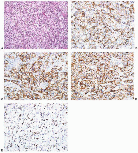
FIGURE 6-61 ▪ High-grade urothelial carcinoma with an unusual architecture (A) that has positive immunoreactivity for high molecular weight cytokeratin 34βE12 (B), cytokeratin 7 (C), cytokeratin 20 (D), and p63 (E).

Lastly p63 has become a useful marker of urothelial carcinoma. It is expressed in 81% to 92% of high-grade urothelial carcinomas (Fig. 6-61E).242,248 This marker is also consistently expressed in squamous and basal cell carcinomas. Its value in the urinary bladder has been primarily
related to assisting in the differential diagnosis with prostatic adenocarcinoma242 and its expression in the spindle cell component of sarcomatoid carcinomas.


Histologic Variants of Urothelial Carcinoma

The morphologic diversity of tumors arising from the urothelium is well recognized and is reflected in the current classification system.14,15,53,276 In this section, the focus will be on those tumors that are considered variants of urothelial carcinoma. Other epithelial tumors such as squamous cell carcinoma, adenocarcinoma, and small cell carcinoma that can develop in the urinary tract are not considered. Table 6-11 provides a modified listing of these variants as described within the WHO classification scheme14 as well as in more recent publications.53,276


Urothelial Carcinoma with Mixed Differentiation

Urothelial tumors have a great capacity for divergent differentiation.277 Squamous differentiation, defined by the presence of intercellular bridges and/or keratinization, occurs in up to 40% of urothelial carcinomas (Fig. 6-62).53,276,278, 279, 280 For this reason, the diagnosis of squamous cell carcinoma is reserved for pure lesions without any associated urothelial component, including urothelial CIS.14,15,280 Tumors with any identifiable urothelial element are classified as urothelial carcinoma with squamous differentiation. Some authors will classify urothelial carcinoma with extensive squamous differentiation as squamous cell carcinoma (but we prefer to continue to follow the WHO guidelines).281 The clinical significance of squamous differentiation remains uncertain; it has been reported to be an unfavorable prognostic feature in patients undergoing radical cystectomy, possibly because of its association with high-grade urothelial carcinoma.282 Mixed differentiation in transurethral resection specimens has been associated with a higher frequency of muscle invasion and extravesical extension, but not decreased survival.279 Squamous differentiation has been reported in a limited number of reports to be predictive of a poor response to radiation therapy and has been associated with a poor response to systemic chemotherapy.282, 283, 284 There is no association with HPV infection.285 Immunohistochemical markers of squamous differentiation such as caveolin-1 and -2, MAC387, and desmocolin-2 can identify this component in urothelial carcinoma.278,286,287 We currently report the percentage of the squamous component in cases with mixed differentiation.


Table 6-11 ▪ VARIANTS OF UROTHELIAL CARCINOMA


Mixed differentiation


With squamous differentiation


With glandular differentiation


With small cell differentiation


Nested


Microcystic


Micropapillary


Lymphoepithelioma-like


Plasmacytoid


Inverted papilloma-like carcinoma


Giant cell carcinoma


Urothelial carcinoma with trophoblastic differentiation


Clear cell (glycogen-rich) urothelial carcinoma


Lipid-rich (lipoid) urothelial carcinoma


Sarcomatoid carcinoma (carcinosarcoma)


Undifferentiated carcinoma


Adapted from Eble J, Epstein J, Sauter G, et al. World Health Organization Histologic and Genetic Typing of Tumours of the Kidney, Urinary Bladder, Prostate Gland and Testis. Lyon, France: IARC Press; 2004.


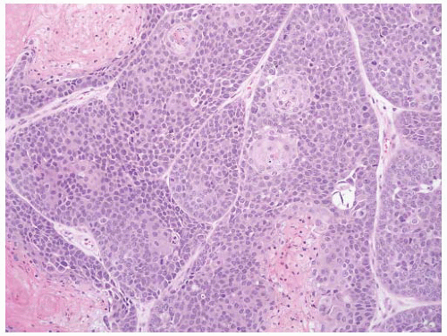
FIGURE 6-62 ▪ Urothelial carcinoma with squamous differentiation.

Glandular differentiation is less common than squamous differentiation, being present in up to 18% of cases.14,279 Glandular differentiation is defined as the presence of true glandular spaces within the tumor.14,280 In most cases, these glands have an enteric morphology (Fig. 6-63). Tumors with a mucinous (colloid) carcinoma or signet ring cell adenocarcinoma component are also included. Pseudoglandular spaces caused by necrosis or artifact should not be considered evidence of glandular differentiation. Mucin-containing cells are common in high-grade urothelial carcinoma. Donhuijsen et al.226 found mucin-positive cells in 14% of grade 1, 49% of grade 2, and 63% of grade 3 urothelial carcinomas. The presence of cytoplasmic mucin is not considered evidence of glandular differentiation. The diagnosis of adenocarcinoma is reserved for pure tumors14,288 and a tumor with mixed glandular and urothelial differentiation is classified as urothelial carcinoma with glandular differentiation regardless of the extent of the glandular differentiation. The clinical significance of glandular differentiation and mucin positivity in urothelial carcinoma remain uncertain. In one study, the presence of glandular differentiation was associated with a poorer response to systemic chemotherapy.283 In another report, mixed differentiation in transurethral resection specimens was associated with a higher frequency of muscle invasion and extravesical extension, but not survival.279 Immunohistochemistry for MUC5AC-apomucin may be an indicator of glandular differentiation.41 In cases
with glandular differentiation, we report the percentage of the glandular component.


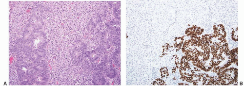
FIGURE 6-63 ▪ Urothelial carcinoma with glandular differentiation (A). The glandular component has positive immunoreactivity for CDX2 (B).

Small cell differentiation histologically identical to that occurring in the lung is estimated to account for approximately 0.5% of bladder tumors.289,290 In about 50% of cases it is pure and in the others mixed with other histologic types.291 In contrast to the way squamous and glandular differentiation is reported, given the clinical significance of this diagnosis, we report these mixed tumors as small cell carcinoma and then indicate the percentage of the small cell and urothelial components. This topic is covered in detail in the section on small cell carcinoma later in the chapter.


Nested Variant

In 1992, Murphy and Deana292 described four cases of invasive urothelial carcinoma with a distinctive growth pattern of small nests, a pattern that had also been described by Talbert and Young.293 There are no specific clinical or epidemiologic characteristics. These are invasive carcinomas. Larger series have confirmed the aggressive nature of this tumor despite its deceptively benign morphology.294, 295, 296, 297 Some studies have suggested these are more aggressive than usual urothelial carcinoma.295, 296, 297 In the largest series to date, Wasco et al.297 reported lymph node metastases in 67% of the 30 cases undergoing cystectomy.

This variant of urothelial carcinoma is characterized by infiltrating discrete nests made up of uniform benign-appearing urothelial cells, closely resembling von Brunn nests (Box 6-4) (Fig. 6-64). The nests can be closely packed or more scattered and haphazard and can show considerable size variation (Fig. 6-65). There is often no associated stromal response. Some nests have small tubular lumens. This can result in focal areas resembling cystitis cystica or glandularis. In some areas the nests become more complex or confluent, features that are helpful when present (Fig. 6-66). Nuclei generally show little or no atypia but are often considerably enlarged when compared to normal urothelial cells (Fig. 6-67). Invariably the tumor also contains foci of unequivocal cancer with more variable nuclear size, enlarged nucleoli, and a coarse chromatin pattern. Increasing levels of atypia are often identified in the deeper portions of the tumor. Invasion of the muscular propria is common and when identified is diagnostic of carcinoma. There may be an associated typical urothelial carcinoma component and/ or urothelial CIS.


FIGURE 6-64 ▪ Nested variant of urothelial carcinoma with infiltrating small nests of cells.


FIGURE 6-65 ▪ Nested variant of urothelial carcinoma with a more obvious high-grade component.

The immunohistochemical profile is typical of urothelial carcinoma with cytokeratin 7 (93%), cytokeratin 20 (68%), high molecular weight cytokeratin (34βE12; 92%), and p63 (92%) positivity.297 Overexpression of p53 was found in 25% of cases in one series.298

The major differential diagnosis is with benign mimics such as prominent von Brunn nest or von Brunn nest hyperplasia (proliferative cystitis).54 Useful features in recognizing this lesion as malignant are the closely packed and haphazard arrangement of the nests, tendency for increasing cellular anaplasia in the deeper portions of the lesion, the infiltrative nature, and the presence of deep lamina propria and muscle invasion. Von Brunn nests tend to extend downward to a consistent level forming almost a linear edge to the proliferation with the cells maintaining the features of urothelium and with minimal cytologic atypia. In von Brunn nests, the cells mature toward the center, a feature not seen in the nested variant. Although Ki-67 (MIB-1) staining generally shows a higher rate of positive cells in carcinoma than in proliferative cystitis, there is sufficient overlap such that this is not a reliable criteria to base the diagnosis on.54,298 Similarly, overexpression of p53 protein does not seem to have value in this distinction.54


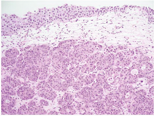
FIGURE 6-66 ▪ Nested variant of urothelial carcinoma with the nests focally becoming confluent.


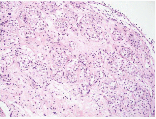
FIGURE 6-67 ▪ Nested variant of urothelial carcinoma with uniform nests and uniform round nuclei.

The nested variant of urothelial carcinoma may mimic paraganglioma or paraganglionic tissue; the prominent vascular network of paraganglioma that surrounds individual nests is different than in carcinoma.299 Rare carcinoid tumors in the bladder can have a nested architecture and bland cytologic features. Immunohistochemistry can distinguish carcinoma from paraganglionic tissue, paraganglioma, and carcinoid tumor in difficult cases. Another mimic is nephrogenic adenoma that can have solid nests and an apparent infiltrative growth; recognition of other architectural patterns including tubules, microcysts, and surface papillary component lead to the correct diagnosis. Prominent basement membranes around the nests and hobnail-type cells further indicate a diagnosis of nephrogenic adenoma.300,301


Microcystic Variant

Young and Zukerberg302 described four cases of invasive urothelial carcinoma characterized by the formation of microcysts. The pattern was similar to some foci of tubular differentiation included in the “nested variant” of Murphy and Deana and “deceptively benign-appearing” bladder cancer of Talbert and Young.293 The cases included lesions with intermediate- to high-grade urothelial carcinoma having areas of microcystic and/or macrocystic change or tubular (glandular) differentiation. Microcysts can be identified in up to 1% of urothelial carcinomas, most of which are high grade and high stage.303,304 The cysts and tubules may be empty, or contain necrotic debris or mucin (Fig. 6-68). In my experience the nested and microcystic patterns often coexist. If the lining cells become flattened with scant cytoplasm, the spaces can mimic lymph-vascular channels (Fig. 6-69).


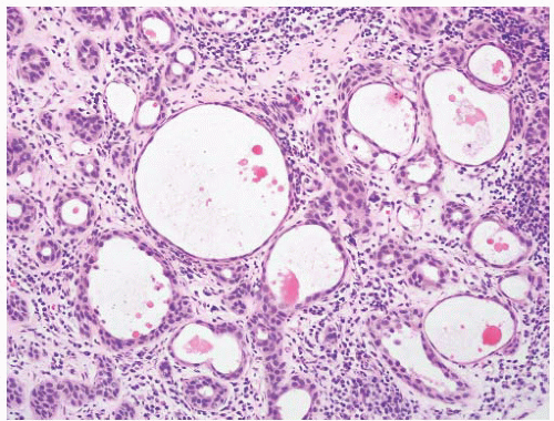
FIGURE 6-68 ▪ Microcystic variant of urothelial carcinoma with variably sized cystic spaces and some solid nests.

It is important to distinguish cystic change in urothelial carcinoma from benign and malignant mimics. It may be confused with benign proliferations such as cystitis cystica and glandularis and nephrogenic adenoma. Clues to nephrogenic adenoma include the presence of a papillary component (papillae covered by a single layer of cells), prominent basement membrane around the tubules, and a heterogeneous lining with attenuated, cuboidal, and hobnail cells.300,301 There can be a fibromyxoid background that could be confused with a stromal response to tumor invasion.305 The presence of significant nuclear atypia at least focally and areas of typical invasive urothelial carcinoma allow accurate separation. More problematic is the separation of the microcystic pattern of urothelial carcinoma and adenocarcinoma of the bladder. The diagnosis of adenocarcinoma should be restricted to pure tumors with true gland formation.14,15 In microcystic urothelial cancer, the lining cells are urothelial and the spaces formed are pseudoglandular and not true glands.


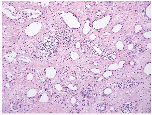
FIGURE 6-69 ▪ Microcystic variant of urothelial carcinoma with the tumor cells having scant cytoplasm producing structures that mimic lymph-vascular spaces.


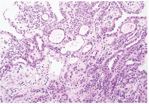
FIGURE 6-70 ▪ Urothelial carcinoma with tubule formation. This case mimics nephrogenic adenoma.


Urothelial Carcinoma with Small Tubules

In some cases of urothelial carcinoma there are areas of the tumor with the formation of small- to medium-sized tubular structures, some of which can be elongated (Fig. 6-70).172,306 The tubules are lined by urothelial cells and show some degree of nuclear pleomorphism. This generally occurs in the setting of a usual type of urothelial carcinoma but in limited biopsy material it is possible that this could be diagnostically challenging. There is considerable overlap between this feature and the microcystic pattern described above. The major differential diagnosis is with cystitis glandularis and nephrogenic adenoma. These tumors are distinguished from benign conditions using the same features already described in the preceding sections on the nested and microcystic variants. In one reported example the tubular pattern closely resembled prostatic adenocarcinoma.307


Micropapillary Variant

The occurrence of this distinctive morphologic variant of carcinoma in the urinary bladder was first presented in detail by Amin et al.308 in a description of 18 patients. A recent review article summarized 268 cases of micropapillary urothelial carcinoma (including those originating in the ureter and renal pelvis) culled from the literature.309 There are no distinctive epidemiologic features. Although one series suggests that the greater the proportion of the micropapillary component the more aggressive the tumor,310 most recommend diagnosing this variant irrespective of the proportion present.311 It is estimated to constitute 0.6% to 1.0% of urothelial carcinomas53 and is clinically significant for its aggressive clinical course and propensity for extensive and deep invasion.308, 309, 310, 311, 312, 313, 314 Cases with as little as 10% micropapillary morphology have been shown to have this aggressive behavior.315 The tumor has been described elsewhere in the urothelial tract.309,316,317 When seen in biopsy or transurethral
resection specimens, a repeat resection of the area is indicated if muscle invasion is not demonstrable in the original material. Occult lymph node metastases were present in 27% of cases at the time of cystectomy in one series.311 Some authors have advocated immediate cystectomy in T1 cases.318


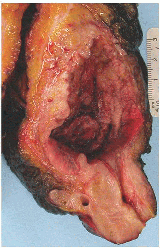
FIGURE 6-71 ▪ Micropapillary variant of urothelial carcinoma. The tumor in this case was 90% micropapillary.

There are no unique gross pathologic features although these can produce a diffuse thickening of the bladder wall similar to linitis plastica (Fig. 6-71). The tumor is composed of cytologically malignant cells arranged in small pseudopapillary clusters. A similar pattern is identifiable in urine cytology specimens.317 At low magnification, the striking pattern of small tumor cell nests with prominent tissue retraction is immediately evident (Box 6-5) (Figs. 6-72 and 6-73). The cells often have the nuclei toward the outer aspect of the nests with a tapering of cytoplasm centrally (Fig. 6-74). This mimics a papillary structure; however, a true fibrovascular core is absent (Fig. 6-75). It has been hypothesized that this architectural growth is related to an inversion (reverse apical) of the location of the MUC1 protein.319 These clusters are usually floating free in an empty space suggesting lymph-vascular invasion. Although most of these spaces appear related to tissue retraction artifact, extensive lymphvascular invasion is generally present (Fig. 6-76). The tumor is often associated with overlying CIS. In some cases the pattern mimics adenocarcinoma and combined with the frequent expression of CA125, it has been suggested that this may represent a type of glandular differentiation.320 A surface micropapillary pattern has been described in some cases with slender delicate filiform projections that lack a central fibrovascular core. These tumors are often quite extensive at the time of resection and can spread widely. Care should be taken when evaluating ureter resection margins at frozen section as the tumor can be found in the periureteral soft tissue (Fig. 6-77). The tumor retains this distinctive morphology in metastases. This is important when a biopsy from a lymph node or other presumed metastasis shows this morphology (Fig. 6-78). In cases of an unknown primary, bladder origin should be considered and secondly in cases with known bladder cancer this could represent a metastasis from the known primary.


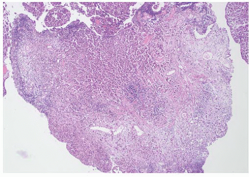
FIGURE 6-72 ▪ Micropapillary variant of urothelial carcinoma in a TURBT chip.


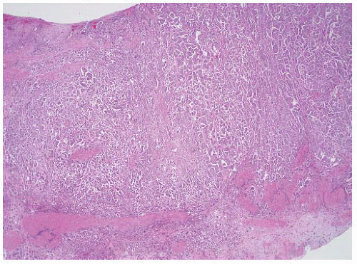
FIGURE 6-73 ▪ Micropapillary variant of urothelial carcinoma in a section from a cystectomy specimen.


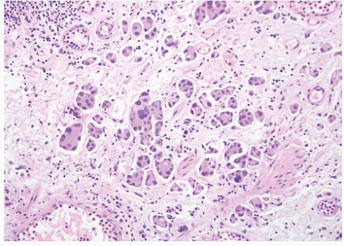
FIGURE 6-74 ▪ Micropapillary variant of urothelial carcinoma in the lamina propria.


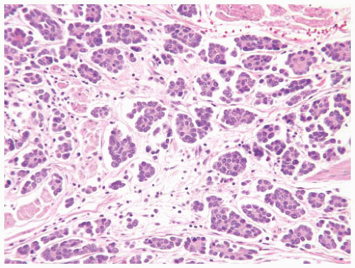
FIGURE 6-75 ▪ Micropapillary variant of urothelial carcinoma with prominent peripheral location of nuclei.


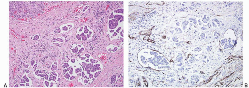
FIGURE 6-76 ▪ Micropapillary variant of urothelial carcinoma (A) with some of the nests in spaces lined by CD34-positive cells (B). Note the usual urothelial carcinoma component in the upper left.


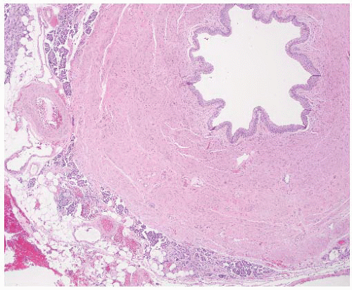
FIGURE 6-77 ▪ Micropapillary variant of urothelial carcinoma involving a ureter resection margin (the surgeon could not get a clear margin in this case).


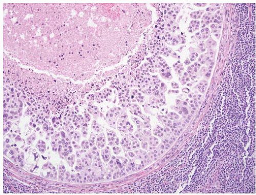
FIGURE 6-78 ▪ Micropapillary variant of urothelial carcinoma metastatic to a pelvic lymph node.


The immunophenotype is similar to usual urothelial carcinoma with most expressing cytokeratin 7 (100%), cytokeratin 20 (54% to 90%), high molecular weight cytokeratin (34βE12, 15% to 54%), p63 (27%), epithelial membrane antigen (EMA) (100%), CEA (65%), thrombomodulin (23%), and uroplakin III (77% to 92%).53,244,310,312,314,321,322 The vast majority are MUC1 and MUC2 positive314,319,321 although this does not distinguish the micropapillary variant from usual urothelial carcinoma with retraction artifact. There is no expression of MUC5A, MUC6, or CDX2.314 P53 overexpression is present in up to 100% of cases.314

Morphologically, the tumor bears a striking resemblance to serous papillary carcinoma of the ovary, a differential diagnosis that may require clinical correlation for exclusion in female patients. One feature of note is that psammoma bodies are extremely rare in the bladder tumor. The presence of a typical urothelial carcinoma component in the invasive carcinoma and/or the presence of a surface component would support interpretation as a bladder primary. Immunohistochemistry could also be of assistance with cytokeratin 20 and uroplakin positivity supporting a bladder origin and WT-1 positivity supporting a serous ovarian carcinoma.322 Metastatic micropapillary carcinoma from other organs where this histology occurs such as lung and breast should also be considered. Clinical correlation may be necessary in some cases; immunohistochemistry for TTF-1 (lung) and mammaglobin (breast) may be of value.322 A small percentage (5%) of urothelial carcinomas will express TTF-1.259

The micropapillary variant must also be distinguished from retraction artifact in usual urothelial carcinoma. In the latter, this is typically focal and scattered, whereas in the micropapillary variant, the clefts surround virtually all of the tumor nests. The features that best distinguish this from usual urothelial carcinoma with retraction artifact are multiple nests in single spaces, peripherally located nuclei, intracytoplasmic vacuolization, back-to-back spaces, epithelial ring forms, small nests (<4 cells across), and extensiveness of the spaces.323


FIGURE 6-79 ▪ Plasmacytoid variant of urothelial carcinoma in a TURBT specimen. The mucosa is edematous (A) with the tumor cells in the deep lamina propria (B).


Plasmacytoid Variant

Rare carcinomas of the urinary bladder can mimic malignant lymphoma. Small cell carcinoma and lymphoepitheliomalike carcinoma are discussed elsewhere. Zukerberg et al.324 described two cases of bladder carcinoma that diffusely permeated the bladder wall and were composed of cells with a monotonous appearance mimicking lymphoma. Similar tumors have been referred to as plasmacytoid or lobular carcinoma-like and have been included in some series as signet ring carcinomas.325, 326, 327, 328, 329, 330 In the original description of signet ring cell carcinoma of the bladder, Saphir331 reported two cases of signet ring cell carcinoma that contained “monocyte-like” cells. The resemblance to lobular carcinoma of the breast has also been noted.328 These are aggressive tumors with over 95% of cases locally advanced (T3/4) and/or with lymph node metastases at the time of presentation.325, 326, 327,329,332 In the 16 cases with follow-up reported by Nigwekar et al.,329 11 patients were dead from disease (1 to 43 months; median, 6 months) and the remaining 5 were alive but with known disease. These also have an unusual predilection for involving peritoneal surfaces.333

These tumors may not produce a discrete mass but rather produce thickening of the bladder wall with a linitis plasticalike appearance. Edema of the mucosa can be present. This feature is important; in a bladder biopsy or transurethral resection with a clinical impression of a tumor, the presence of mucosal edema should raise this possibility and a careful examination for rare tumor cells should be undertaken (Fig. 6-79). In some instances, it may be appropriate to use immunohistochemistry with a pancytokeratin antibody to be certain not to miss a few of these cells. The tumor
cells are medium sized with pale eosinophilic cytoplasm and eccentric nuclei producing the plasmacytoid appearance (Fig. 6-80). The appearance can be strikingly plasma cell-like such that plasmacytoma is strongly considered in the differential diagnosis.334 In some cases, the cytoplasm is more basophilic. Many cells have an area of perinuclear clearing that may be mucin positive. In all cases, a minority of cells have true signet ring morphology. The nuclei tend to be round or indented with prominent nucleoli in a minority. Overall there is a more uniform nuclear appearance than typical of urothelial carcinoma (Fig. 6-81). The cells tend to infiltrate as single cells (Fig. 6-82) or cords of cells but can also form nests and sheets (Fig. 6-83). The diffusely permeative nature results in the tumor being more extensive than appreciated clinically or on gross examination. These can be extremely challenging at frozen section evaluation of ureter margins, and knowledge of the histology is very helpful in avoiding false-negative interpretations. Typical urothelial carcinoma, often a surface papillary carcinoma, is present in the majority of cases. The diagnosis of carcinoma can be confirmed by positive immunoreactivity for cytokeratin, EMA, and CEA with negative reactivity for lymphoid markers.


FIGURE 6-80 ▪ Plasmacytoid variant of urothelial carcinoma in the lamina propria.


FIGURE 6-81 ▪ Plasmacytoid variant of urothelial carcinoma illustrating the morphologic range of the individual cells. A few tumor cells have cytoplasmic vacuoles.


FIGURE 6-82 ▪ Plasmacytoid variant of urothelial carcinoma infiltrating between smooth muscle bundles of the muscularis propria.

The differential diagnostic considerations are plasmacytosis, lymphoma, and multiple myeloma. Identification of an epithelial component confirms the diagnosis. If immunohistochemistry is needed, a pancytokeratin is most useful. These tumors have been reported to be immunoreactive for CD138, a plasma cell marker, in over 90% of cases (Fig. 6-84).329,335 The possibility of metastasis from another site can also be considered in pure tumors. These tumors have an immunohistochemical profile similar to usual urothelial carcinoma with expression of cytokeratins 7 (70% to 100%) and 20 (31% to 100%), p63, and uroplakin III (11%) (Fig. 6-85).326, 327, 328, 329 In one study, all 5 cases showed loss of E-cadherin expression,327 and in another, 7 of 10 lacked E-cadherin expression (Fig. 6-86).328 Like urothelial carcinoma, these tumors can express estrogen and progesterone receptors in a minority of cases,328 an important feature to recognize if the possibility of metastatic carcinoma of the breast is considered.


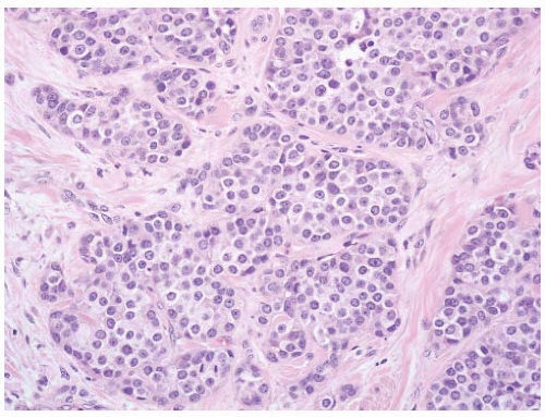
FIGURE 6-83 ▪ Plasmacytoid variant of urothelial carcinoma with the cells forming more defined nests.


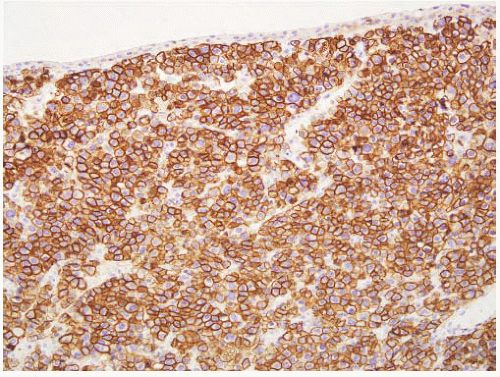
FIGURE 6-84 ▪ Plasmacytoid variant of urothelial carcinoma with strong immunoreactivity for CD138.


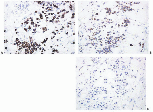
FIGURE 6-85 ▪ Plasmacytoid variant of urothelial carcinoma with positive immunoreactivity for cytokeratins 7 (A) and 20 (B) but not for p63 (C).


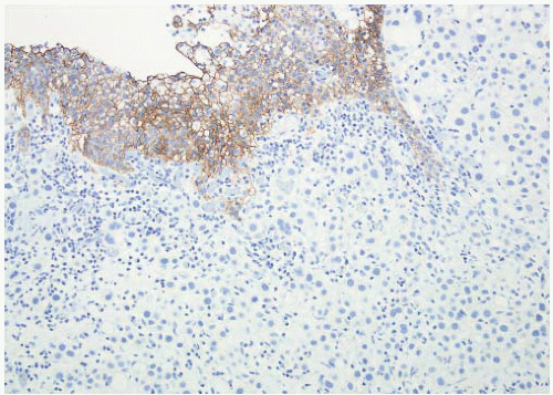
FIGURE 6-86 ▪ Plasmacytoid variant of urothelial carcinoma with loss of expression of E-cadherin.

Jun 10, 2016 | Posted by in UROLOGY | Comments Off on Tumors of the Urinary Bladder

Full access? Get Clinical Tree

Get Clinical Tree app for offline access