Nonneoplastic Lesions of the Urinary Bladder


Nonneoplastic Lesions of the Urinary Bladder


Jesse K. Mckenney

John N. Eble

Robert H. Young


A broad spectrum of nonneoplastic lesions occurs in the urinary bladder. These range from congenital abnormalities to infectious/inflammatory conditions of only microscopic dimension to mass forming pseudotumors. As background, this chapter first presents a brief review of the embryology, normal anatomy, and histology of the urinary bladder and then details the full spectrum of nonneoplastic conditions that may be encountered in diagnostic surgical pathology practice.


NORMAL EMBRYOLOGY, ANATOMY, AND HISTOLOGY


Embryology

The urinary bladder develops during the first 12 weeks of gestation. It derives predominantly from the vesical (rostral) part of the urogenital sinus; the trigone originates from the caudal end of the mesonephric ducts. The urothelium is derived from the endoderm, while all other layers are from the splanchnic mesenchyme. The formation of the bladder and trigone is regulated by complex epithelial-mesenchymal signaling events.1,2 Initially in development, the bladder is continuous with the allantois; however, neither the urachus nor the allantois is involved in the formation of the bladder. The allantois typically regresses to a fibrous cord in adults, contained within the urachus, which is also called the median umbilical ligament. As the bladder increases in size, the distal parts of the mesonephric ducts are incorporated into the dorsal wall forming the connective tissue of the trigone, where the developing ureters implant. The anterior bladder wall is formed with the caudal migration of the cloacal membrane. In the 7th week of gestation, the urorectal septum separates the rectum from the part of urogenital sinus that forms the dome and the posterior wall of the bladder.


Anatomy

The urinary bladder is located in the pelvis but, when filled, may extend up to the level of the umbilicus. The bladder is wider at the superior aspect and narrows toward the inferior region, creating the shape of an inverted pyramid. The anatomic regions of the bladder are the bladder neck (inferior), the dome (superior), the apex (most anterior and superior point), the fundus (posterior wall), and the trigone, the last defined as the triangular region at the base of the bladder, bounded posterolaterally by the ureteric orifices and inferiorly by the internal urethral orifice.3, 4

In the male, the bladder is held in place at the bladder neck by the puboprostatic ligaments that attach to the prostate gland and directly in the female by the pubovesical ligament. The remainder of the bladder is surrounded by the loose fibrous connective tissue and adipose tissue of the pelvis, which allows it to expand with filling. Other points of attachment include the rectovesical ligaments that attach to the rectum and sacrum, and the median umbilical ligament that extends from the apex to the anterior abdominal wall.

In the male, the anatomic relation to surrounding structures is (a) fundus: the rectovesical septum, seminal vesicles, and vas deferens; (b) apex: median umbilical ligament; (c) superior: peritoneal surface; (d) inferolateral: space of Retzius; and (e) neck: prostate. In the female, the fundus is separated from the anterior aspect of the uterus by the vesicouterine pouch and more inferiorly by the cervix and upper vaginal wall, while the inferior surface rests on the pelvic and urogenital diaphragms. The uterus also rests on the superior surface of the bladder in the emptied state.

The ureters course obliquely through the bladder wall and are surrounded by smooth muscle and fibrous tissue called Waldeyer sheath. The ureters enter the lumen at the trigone, where their muscle fibers are admixed with the muscularis propria. The urethra begins at the neck where the walls of the bladder converge. The smooth muscle fibers of the muscularis propria and urethra intermix at the bladder neck, but the internal sphincter consists predominantly of muscularis propria.

The blood supply for urinary bladder is derived from the superior and inferior vesical arteries, which arise from the internal iliac artery. Blood drains through the vesical venous plexus, which empties into the internal iliac veins. Most of the lymphatics drain to the internal and external iliac nodes; however, the bladder neck drains to the sacral or common
iliac nodes. The sympathetic nerves that innervate the bladder are derived from the T11-L2 nerve roots; these sympathetic nerves play no role in micturition. The parasympathetic nerves originate from the S2-S4 roots and travel to the bladder via the pelvic nerve and inferior hypogastric plexus. These peripheral nerves cause contraction of the muscularis propria fibers, which leads to traction of the bladder, opening of the internal sphincter, and emptying of urine into the urethra.
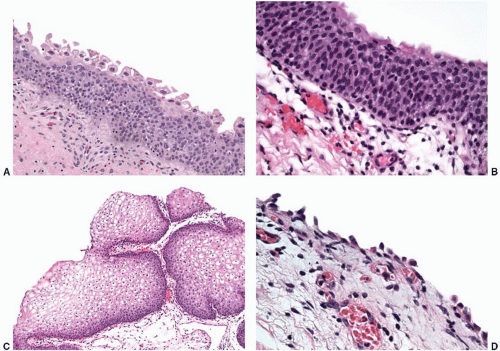


FIGURE 5-1A: Normal urothelium may have a prominent umbrella cell layer. These cells may have enlarged nuclei with multilobulation and cytoplasmic vacuolization. These features should not be regarded as neoplastic. B: In general, normal urothelium shows a streaming arrangement of the urothelial cells arranged perpendicular to the basement membrane. The urothelial cells often have nuclear grooves along the long axis of the cell. C: In the trigone of women, the surface lining is often composed of a glycogenated nonkeratinizing squamous epithelium. D: In biopsy specimens, particularly those performed with “hot” loops, denudation of the surface urothelium is not uncommon. Scattered residual benign basal cells may be present.


HISTOLOGY


Urothelium

Urothelium is a multilayered epithelium comprised of oval to fusiform cells that typically have pale nuclei containing longitudinal nuclear grooves in many of the cells. These urothelial cells mature to form very large surface cells known as “umbrella” or superficial cells.5,6 Umbrella cells may have binucleation, prominent eosinophilic cytoplasm, and nuclear atypia characterized by nucleomegaly, nuclear multilobation, and smudgy nuclear hyperchromasia, which should not be misconstrued to be dysplastic or preneoplastic (Fig. 5-1A). The number of cell layers varies considerably depending on distention of the bladder, but typically is three to six cell layers. The urothelial cells (previously referred to as transitional cells) typically have a somewhat linear organization streaming upward, perpendicular to the basement membrane (Fig. 5-1B). Nonkeratinizing, glycogenated squamous metaplasia is relatively common in the trigone of women and is generally considered a variation of normal histology (Fig. 5-1C).6 In routine diagnostic biopsies, the surface urothelium may be denuded due to biopsy technique (i.e., thermal effect), but the retained basal cells that may be present are cytologically benign (Fig. 5-1D).

Heterogeneity of the thickness, the relation of the individual urothelial cells to the basement membrane, and the nuclear size of the urothelium are common, even within the same biopsy. Historically, there has been overuse of the term “mild dysplasia” for urothelium with a minimally disordered architecture and mild nuclear variation.7


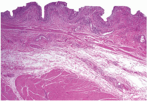
FIGURE 5-2 ▪ The lamina propria is defined as the tissue between the urothelium and the muscularis propria. It often contains disorganized wispy fascicles of smooth muscle called the muscularis mucosae, as well as loose stroma and varying caliber blood vessels.
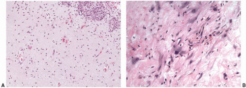

Lamina Propria

The lamina propria lies between the basement membrane of the urothelium and the muscularis propria (Fig. 5-2).5,6 It consists predominantly of hypocellular, loosely collagenized stroma (Fig. 5-3A). Rare, scattered stromal cells that are hyperchromatic and often multinucleated are common in the lamina propria (Fig. 5-3B) (see giant-cell cystitis).8 These atypical stromal cells may occasionally be numerous (Fig. 5-3C). In addition, prominent medium-sized blood vessels are common (Fig. 5-3D) and may be associated with smooth muscle that comprises the muscularis mucosae. Normal adipose tissue is also present within the lamina propria.9 The thickness of the lamina propria varies; it is usually thinner in the trigone and bladder neck.


FIGURE 5-3A: The stroma within the lamina propria may be edematous or finely collagenized, as in this example. The majority of the stroma cells are small and inconspicuous. B: However, scattered stromal cells with enlarged hyperchromatic or multilobated nuclei are not uncommon, and (C) these stromal cells may be numerous. D: Varying caliber blood vessels set in a fine stroma are also characteristic of the lamina propria.


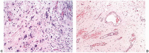
FIGURE 5-3 ▪ (Continued)

The muscularis mucosae is generally composed of irregular, usually isolated, thin wispy fascicles of smooth muscle within the lamina propria (Fig. 5-4A).10 The muscularis mucosae varies considerably between individuals, but it is typically discontinuous; a continuous layer of smooth muscle layer is only rarely seen in the lamina propria. Recent detailed studies of the muscularis mucosae suggest greater heterogeneity of appearance than previously reported.11,12 Particularly in the dome, the muscularis mucosae may be characterized by individual thick bundles of compact “hypertrophic” smooth muscle, even in women (Fig. 5-4B and C). These newly described round contoured smooth muscle bundles of the muscularis mucosae are separated by the stroma of the lamina propria, in contrast to the more typical larger aggregates of smooth muscle that comprise the muscularis propria. In men with benign prostatic hyperplasia, the muscularis mucosae may become more prominent throughout the entire bladder due to compensatory hypertrophy (Fig. 5-4D). These compensatory hypertrophic fibers are generally irregular, haphazard aggregates of smooth muscle fibers splayed in multiple direction; however, there is morphologic variation and overlap with muscularis propria may make distinction very difficult in a subset of biopsy specimens. For cancer staging purposes, we recommend defining the border between the muscularis propria and lamina propria as the line of demarcation where the dense smooth muscle bundles become organized into large aggregates (Table 5-1).

The lamina propria also contains a continuous band of ill-defined haphazardly oriented compact spindle cells that are immediately subjacent to the urothelium that has been termed the suburothelial band of myofibroblasts (Fig. 5-5). These spindle cells blend with the thin slender fascicles of the muscularis mucosae and show immunoreactivity for smooth muscle actin, but not desmin.13


Muscularis Propria

The muscularis propria, or detrusor muscle, consists of aggregates of large, thick, compact bundles of smooth muscle with variable amounts of interspersed collagen
and adipose tissue (Fig. 5-6A).5,6 Although the muscularis mucosae may have individual thick muscle bundles (i.e., hyperplastic muscularis mucosae), the muscularis propria has distinct tight aggregates of several discernable compact smooth muscle bundles that aid in their recognition (Fig. 5-6B). Because of variation in thickness of the lamina propria, the muscularis propria may be surprisingly superficial in some biopsies, particularly in the trigone (Fig. 5-6C). In this location, typical deeper muscularis propria bundles become smaller in caliber as they reach toward the surface and are found in almost a suburothelial location.
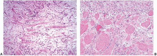


FIGURE 5-4A: The histology of the muscularis mucosae is classically described as disorganized wispy fascicles of smooth muscle within the lamina propria. B: However, recent studies have described more histologic variability. In the dome, individual rounded bundles of smooth muscle are common. C: Unlike muscularis propria, these individual rounded bundles of muscularis mucosae are separated by stroma. D: In patients with urinary outlet obstruction, typically due to prostatic hyperplasia, the muscularis mucosae may become hypertrophic. The disarray of the smooth muscle fibers characterizes hyperplastic muscularis mucosae.


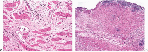
FIGURE 5-4 ▪ (Continued)

Recent studies have described distinct patterns of immunoreactivity between the muscularis mucosae and muscularis propria with the antibody to smoothelin, a monoclonal antibody to contractile smooth muscle.13, 14, 15, 16, 17, 18, 19 Muscularis propria reportedly shows strong and diffuse immunoreactivity to smoothelin, while the muscularis mucosae is negative or have only weak, focal staining (Fig. 5-7). Some studies, however, have reported more overlap in intensity between the muscularis mucosae and muscularis propria; therefore, marked caution should be maintained while using this antibody in a diagnostic setting for the recognition of muscularis propria.20, 21, 22


Adventitia and Perivesical Adipose Tissue

These tissues are deep to the muscularis propria and consist of loose fibroconnective and adipose tissue with interspersed small peripheral nerves. The demarcation between the muscularis propria and perivesical fat is not well delineated, which may create problems in staging some bladder cancers that extend to this irregular border (i.e., pT2 vs. pT3 disease) (Fig. 5-8).


Paraganglia

Paraganglia may be present at any level within the bladder wall, but are more commonly deep seated (Fig. 5-9). They are generally not diagnostically important, but their existence explains the occasional occurrence of paraganglioma in the bladder. Occasionally, they may be mistaken as nests of carcinoma in resections for prostatic or urothelial carcinoma. The typically prominent nucleoli in the endocrine cell population of paraganglia may lead to the consideration of prostatic carcinoma. The presence of coarse intracytoplasmic granules or prominent interspersed capillary-sized blood vessels may aid in recognition as paraganglia. The endocrine cells of paraganglia express synaptophysin and other neuroendocrine markers, but are nonreactive for cytokeratins.


Table 5-1 ▪ MUSCULARIS MUCOSAE VERSUS MUSCULARIS PROPRIA


Muscularis Propria


Muscularis Mucosae


  • Typical: Dense compact smooth muscle bundles arranged into large solid aggregates.


  • Dispersed: In some examples, there may be dispersion of thin smooth muscle bundles from the muscularis propria into the deep lamina propria. In this setting, we use the point at which the muscle is comprised of a solid compact layer as the line of demarcation between muscularis propria and lamina propria.


  • Typical: Thin, irregular, wispy fascicles of smooth muscle of variable caliber.


  • Dome/trigone variation: The muscularis mucosae in the dome/trigone may consist of small individual rounded dense bundles of smooth muscle separated by stroma.


  • Compensatory hyperplasia: With obstruction, the number of fascicles of muscularis mucosae may be markedly increased; however, they remain disorganized and are often splayed in multiple directions.


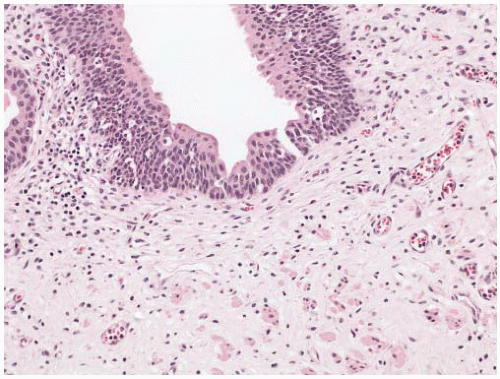
FIGURE 5-5 ▪ There is a variably cellular layer of spindled myofibroblasts beneath the urothelium. These cells often show elongated nuclei and long cytoplasmic processes.
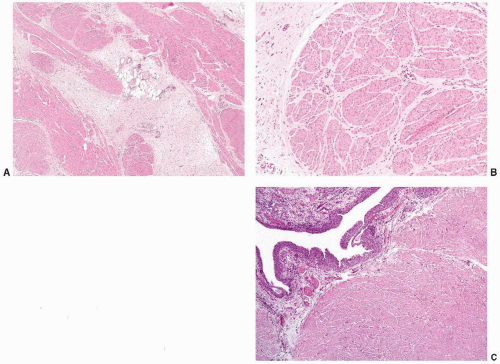


FIGURE 5-6A: Adipose tissue may be present at any level within the bladder wall. In this example, a lobule of adipose tissue is seen within the muscularis propria. B: The muscularis propria is comprised of compact well-delineated aggregates of smooth muscle. C: In some areas, such as the trigone, the muscularis propria is located very superficially.


MALFORMATIONS


Agenesis and Hypoplasia

Congenital absence of the urinary bladder is extremely rare, and <70 cases are reported. By definition, the ureters empty into a structure other than the bladder such as the vagina,23,24 uterus,25 rectum,26 or skin.27 Accompanying hydronephrosis secondary to ureteral obstruction is common. In a subset of cases, a cloaca persists. Agenesis has been associated with a variety of other malformations28 and is reportedly common in sirenomelia.29 Agenesis is most often associated with stillbirth, but viable live births with agenesis are reported, most commonly female neonates.

Hypoplasia of the urinary bladder is defined as a small bladder with a thin wall and an abnormally formed muscularis propria. Hypoplasia is almost always found in the setting of congenital renal malformations such as bilateral renal
agenesis or other abnormalities that lead to an absence of urine entering the bladder (e.g., bilateral ureteral ectopia).30 Rarely, bladder hypoplasia may be due to an absence of urine collection in the bladder due to low outlet resistance from causes such as epispadias.


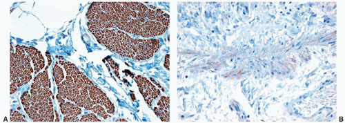
FIGURE 5-7 ▪ Antismoothelin antibodies highlight the muscularis propria (A) with a strong and diffuse pattern of immunoreactivity. In contrast, the muscularis mucosae (B) are typically negative or show only weak, focal staining.


Megacystis

This is defined as massive congenital dilatation of the bladder. Some authors have restricted the term to cases with thin and untrabeculated walls (i.e., functional obstruction), while others have included cases with bladder neck or urethral obstruction showing classic hypertrophy of the bladder musculature. Severe vesicoureteral reflux is common with associated hydroureter, hydronephrosis, and cystic renal dysplasia.

In rare cases, megacystitis is associated with the intestinal pseudoobstructive, autosomal recessive disorder termed megacystitis-microcolon-intestinal hypoperistalsis syndrome. One report has suggested a loss of interstitial cells of Cajal as part of the pathogenesis,31 but a consensus has not been reached.32


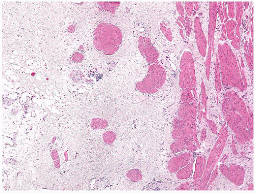
FIGURE 5-8 ▪ The interface between the muscularis propria and the perivesical soft tissue is often irregular, which may cause difficulties in assessing microscopic invasion for staging purposes.


Duplication and Septation

Congenital division of the urinary bladder refers to the presence of more than one vesical lumen. This may result from a spectrum of abnormalities that range from complete duplication with separate bladders to the presence of an intravesical septum that divides the lumen of a single bladder. Although many patients are asymptomatic, incomplete bladder emptying may predispose to urinary tract infections. The clinical treatment is extremely variable, depending on the extent of anomalies and the presence of any functional impairment in an individual patient.33

Congenital division of the bladder may occur by complete or partial duplication.34,35 In complete duplication, there are
two bladders with fully formed mucosal and muscular walls. Each side receives a single ureter and drains into separate duplicate urethras. Complete duplication is typically associated with duplication of the internal and external genital organs, hindgut, and caudal vertebral column.36 Partial duplication, which is less common than the complete form, is defined as two bladders that share a common bladder neck and drain into a common urethra.


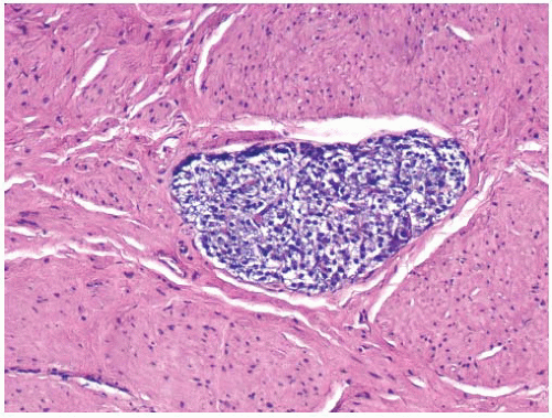
FIGURE 5-9 ▪ Incidental paraganglia may rarely be found in the bladder wall. These are likely the origin of rare bladder paragangliomas.

Septation is most commonly characterized by a complete sagittal septum that divides the bladder into two compartments. The bladder may appear grossly normal from the external surface. In contrast to partial duplication, only one side drains to the urethra. If a ureter empties into the blind-ending, obstructed side, then resultant unilateral dilatation with hydroureter, hydronephrosis, and cystic renal dysplasia are expected. Microscopically, the septum consists of two mucosal surfaces divided by fibroconnective tissue, with or without smooth muscle. Coronal septations are very rare, as are incomplete, nonobstructing septations. Finally, the “hourglass” bladder has a horizontal narrowing near the middle secondary to a horizontal band of smooth muscle, giving it its distinctive shape.


Diverticula

Bladder diverticula may be congenital or acquired. The majority occur in men >50 years of age with urinary outflow obstruction secondary to benign prostatic hyperplasia. In children, causes include localized alterations of the muscularis propria, neurogenic bladder, and a variety of syndromes including Menkes,37 Williams,38 prune-belly, and Ehlers-Danlos syndromes.39 Diverticula typically occur in the region of the ureterovesical junction, likely secondary to the normal disruption of the muscularis propria by the adjoining ureter. While most diverticula are small and asymptomatic, larger lesions may cause ureteral obstruction, recurring infections, or stones. Diverticula also are at risk for the development of neoplasia secondary to urinary stasis.40, 41, 42


Exstrophy

Exstrophy is a congenital defect in the anterior bladder wall and ventral body wall, which results in external protrusion of exposed bladder mucosa (Fig. 5-10). At the margins of the defect, the urothelial lining is contiguous with the epidermis. With exstrophy, urine drains from the ureteric orifices onto this exposed surface of the bladder, which has a hyperemic appearance and may contain polyps.43 The prevalence of exstrophy at birth is approximately 1 per 30,000 (ranging from 1:10,000 to 1:50,000 in published reports), and there is a male predominance up to 6:1; it is usually diagnosed with routine prenatal imaging. The recurrence risk for an individual family is approximately 1 in 100, which is approximately 500 times the expected risk. It is usually associated with other defects in the bony pelvis and external genitalia. In males, an open (epispadic) urethral plate covers the whole dorsum of the penis from the open bladder to the glandular groove. Females typically have a split clitoris next to the open urethral plate with associated narrowed vagina and shortened perineum. The frequent association with epispadias has led to the use of the term exstrophy-epispadias complex. Associated bilateral inguinal hernias are also common, and spina bifida is present in 18% of cases.


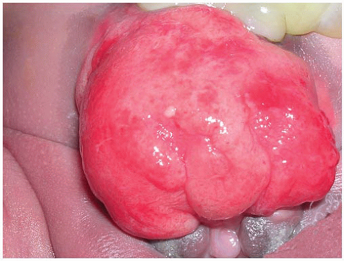
FIGURE 5-10 ▪ Bladder exstrophy is clinically striking with the bladder mucosa exposed on the surface of the abdominal wall. (Courtesy: Dr. Michael Hsieh, Stanford University.)

The etiology of exstrophy is not fully understood. The defect in the urinary bladder and anterior body walls results from incomplete closure of the mesoderm. There are varying theories about the mechanism of this incomplete closure that include (a) overdevelopment of the cloacal membrane, (b) premature rupture of the cloacal membrane, and (c) caudal maldevelopment of the genital tubercles.44

Histologically, the mucosa of the extruded bladder is comprised of metaplastic epithelium with acute and chronic inflammation and ulceration. Squamous metaplasia and cystitis glandularis with intestinal metaplasia are minimal at birth, but develop with time.45 After closure, squamous metaplasia and inflammation persist, but glandular changes become less prominent.46 Unusual “hyperplastic”-appearing benign mucosal polyps are also commonly seen.

Patients with exstrophy have a well-described risk of developing cancer, most commonly adenocarcinoma.47,48 Squamous cell carcinoma may also arise, and urothelial carcinoma and even rhabdomyosarcoma are rarely reported.49, 50, 51, 52 Although this cancer risk seems greatest in bladders without complete repair or those surgical repairs in which fecal exposure to the urothelium is present,53 the risk following complete surgical repair is likely low, but remains uncertain.54 These neoplasms may occur in the bladder proper or in urinary-intestinal anastomoses.


Vascular Malformations and Other Benign Vascular Abnormalities

Vascular malformations may rarely involve the urinary bladder.55, 56, 57 Patients may present with hematuria, or the lesions may be discovered incidentally during cystoscopic evaluation. They may be small hemorrhagic submucosal lesions to large broad-based polypoid masses measuring up to 6 cm.57
Some are associated with syndromes such as Klippel-Trenaunay-Weber syndrome.58,59 Preoperative imaging often suggests a vascular lesion, and in large masses Doppler flow studies may show a shunt in lesions with an arterial component. Morphologically, these lesions consist of large abnormally dilated vascular channels with varying components of arterial, venous, and lymphatic vessels (Fig. 5-11). The vessel walls vary in thickness with varying amounts of medial and elastic layers. Vascular malformations may have overlying ulceration and associated reactive urothelial atypia, including pseudosarcomatous epithelial hyperplasia.60


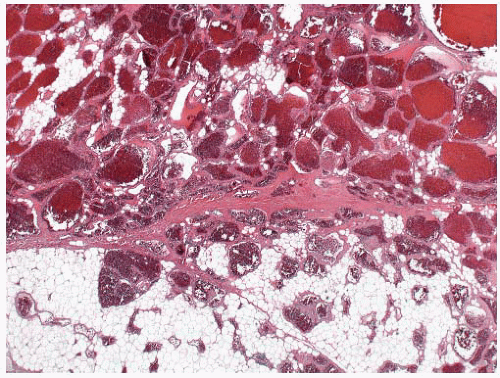
FIGURE 5-11 ▪ Vascular malformations are typically comprised of dilated vascular spaces admixed with fat or normal bladder tissues.
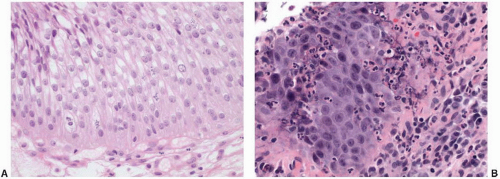
Some cases may show rather florid papillary endothelial hyperplasia, either with or without an associated vascular malformation, and this hyperplasia can mimic angiosarcoma. The overall circumscription of this endothelial hyperplasia within the confines of a vascular space and the absence of significant cytologic atypia generally allow distinction from an angiosarcoma. Papillary endothelial hyperplasia is well described in the setting of prior local radiation.57 A component of adipose tissue is often admixed with large vascular malformations and may cause confusion with adipocytic tumors; however, the presence of the large dilated vascular channels excludes the possibility of an adipocytic tumor. Angiomyolipoma is distinguished by the presence of neoplastic cells within the walls of the vessels that have eosinophilic to clear cytoplasm and coexpress actin and HMB-45 or other melanocytic markers.61


Persistent Cloaca

Persistent cloaca, also called cloacogenic bladder, is defined as a retained connection between the urinary bladder, rectum, and/or vagina. The cloaca is a single embryonic canal from which the urinary, genital, and intestinal tracts arise at gestational weeks 5 to 6. Although the diagnosis has historically been restricted to females due to relatively arbitrary distinctions, many now accept that it occurs in both sexes when the diagnosis is based on contemporary embryologic criteria. The literature has referred to these abnormalities in the male as “partial urorectal septum malformation sequence” and “cloacal dysgenesis sequence.” Determining its frequency is difficult because of variable definitions employed in the literature and the exclusion of male defects. By traditional definitions based on malformations in females, it occurs in 1 per 20,000 births. In females, the defect is characterized by the terminal rectum, vagina, and urinary tracts all opening into a shared cloacal pouch. The persistent cloacal pouch is often connected to a single perineal opening, but this is occasionally absent. In males, the lumen of the rectum and urinary bladder are connected by a fistula, and the anus is imperforate. Associations with a variety of other complex malformations of the genital tract are common. Some of the associated malformations may actually be due to the resultant oligohydramnios. Persistent cloaca is presumably due to incomplete septation of the embryonic cloaca by the urorectal septum, but the underlying cause of this has not been determined and may be multifactorial.44 Some animal models suggest the role of B-class Eph/ephrin signaling.62 Complex surgical reconstruction is the standard treatment.63


INFLAMMATION AND INFECTION


Reactive Urothelial Atypia

Reactive urothelial changes, sometimes florid, may be associated with instrumentation, indwelling catheters, and any inflammatory condition. Morphologically, the reactive urothelial cells may show some degree of nucleomegaly and hyperchromasia, but the overall architecture of the urothelial cells (i.e., even spacing and alignment perpendicular to the basement membrane) is generally maintained. Intercellular edema may be conspicuous, and an associated intraurothelial inflammatory infiltrate is common.7,64 Despite the prominence of one or multiple nucleoli and increased mitotic activity (which may be striking), the chromatin remains finely dispersed throughout the nucleus (Fig. 5-12A and B). These nuclear changes suggest urothelial carcinoma in situ (CIS), but the nuclear pleomorphism, irregular distribution of nuclear chromatin (i.e., striking nuclear hyperchromasia), and the loss of orderly alignment of the individual cells in CIS are distinctive (Table 5-2). The use of adjunctive immunohistochemistry to aid in this distinction has been studied, and a panel of antibodies to cytokeratin 20, p53, and standard isoform CD44 may be useful in some settings (Fig. 5-13).64 CIS shows strong diffuse cytoplasmic immunoreactivity for CK20 in approximately 80% of cases, while diffuse nuclear p53 reactivity is seen in up to 57%. CD44s expression is either limited to the basal layer or absent in CIS. In contrast, reactive atypia is characterized by full-thickness membranous reactivity for CD44s with CK20 expression limited to superficial umbrella cells. p53 may show patchy nuclear staining in benign and reactive urothelium, but it does not have the intensity or the diffuse immunoreactivity pattern typical of CIS. Reactive urothelial atypia due to radiation and chemotherapy is discussed in detail below (see Box 5-1).


FIGURE 5-12A: This example of reactive urothelial atypia is associated with an indwelling catheter. Nuclear enlargement and small nucleoli are typical in reactive urothelial changes. In contrast to flat neoplasia, the nuclear contours are sharp and the chromatin remains fine and evenly distributed. Mitotic figures may be increased and may extend into the upper layers of the urothelium. B: In this example of reactive atypia, the nucleoli are more prominent, but the chromatin remains fine. Neutrophilic infiltrates are also common.


Papillary-Polypoid Cystitis

Papillary/polypoid cystitis is a clinically benign pattern of urothelial injury often secondary to an indwelling catheter or vesical fistula, but of diverse potential etiologies.65, 66, 67, 68, 69 This lesion affects a broad age range, but the mean patient age is 49 years, and the majority of patients are male. It is relatively rare, but polypoid cystitis may be seen in up to 80% of the patients with an indwelling catheter. It may also be seen after radiation therapy. Although usually of microscopic size, grossly visible polypoid lesions may be seen. The entire bladder may be involved when a catheter has been present for a prolonged period of time, usually 6 months or more. With vesical fistula, extravesical symptoms may be initially absent in approximately half the cases, making diagnosis difficult. The cystoscopic appearance may also closely mimic a neoplasm.

Morphologically, the papillary and polypoid patterns may be intermixed, but lesions with relatively slender, nonbranching exophytic projections are termed papillary cystitis, while broad-based, edematous lesions have been termed polypoid cystitis (Fig. 5-14A and B). Characteristically, the exophytic appearance results from edema in the lamina propria, but variable fibrosis, chronic inflammation, and associated dilated blood vessels are also present. These lesions exist along a morphologic continuum with bullous cystitis depending on the degree of edema; some authors have used the following convention: Lesions that are taller than they are wide are termed papillary/polypoid cystitis and vice versa for bullous cystitis. Older lesions tend to have less edema with more stromal fibrosis. As papillary and polypoid cystitis are usually associated with inflammation, there may be metaplastic changes in the lesional or adjacent urothelium. In both types of cystitis, the urothelium may be hyperplastic and reactive appearing, but usually is not as stratified as in a neoplasm. Some authors have suggested that the lesion described as “fibroepithelial polyp” of the bladder has many similar histologic features and may represent an end-stage phase of papillary-polypoid cystitis.

The main diagnostic consideration is a low-grade papillary urothelial neoplasm, particularly urothelial papilloma or papillary urothelial neoplasm of low malignant potential. In
some cases, usually with recent catheterization, papillary/polypoid cystitis shows marked reactive epithelial changes with small, prominent nucleoli, urothelial hyperplasia, and mitotic activity mimicking a high-grade lesion.70 In general, papillomas have more slender papillae; the papillae of papillary cystitis often have a bulbous tip with prominent stromal edema. In addition, urothelial papillomas have other features that, at least in aggregate, may aid in the distinction when present: a very prominent umbrella cell layer or marked cytoplasmic vacuolization, a gland-in-gland pattern within the papillae, a dilated lymphatic space filling the papillae, a more complex papillary pattern with secondary and tertiary branching, and an admixed endophytic (inverted) component.71,72 Significant cytologic atypia within a papillary lesion or the adjacent urothelium favors a diagnosis of neoplasia.


Table 5-2 ▪ IMMUNOPHENOTYPE OF FLAT LESIONS


CIS


Reactive Atypia


CK20


  • Strong cytoplasmic reactivity in neoplastic cell population


  • May be full thickness or individual cell staining depending on pattern of CIS (e.g., pagetoid CIS)


  • Strong cytoplasmic reactivity in the umbrella cell layer only


  • Typically no staining in basal and intermediate cell population


CD44


  • Strong membranous reactivity may be seen in residual basal cells, if present


  • Membranous reactivity may also be seen in surrounding benign cells in pagetoid CIS


  • Strong membranous immunoreactivity typically seen in the full thickness of the urothelium


p53


  • Strong diffuse nuclear reactivity in the neoplastic cell population (requires high threshold for positive staining)


  • Various levels of patchy nuclear staining may be seen depending on individual lab


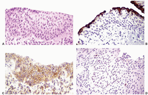
FIGURE 5-13 ▪ Reactive urothelial atypia (A, H&E) has a characteristic immunophenotype with CK20, CD44, and p53 immunohistochemistry. CK20 (B) highlights the umbrella cell layer, while CD44 (C) shows strong membranous reactivity in the full thickness of the urothelial cells. p53 (D) shows weak and isolated nuclear staining, but strong and diffuse reactivity is not seen.
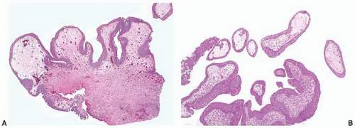


Giant-Cell Cystitis

Atypical, mononucleated or multinucleated mesenchymal cells are a relatively frequent finding in the lamina propria of the bladder (Fig. 5-15). Wells found them in one-third of cases of cystitis at autopsy and applied the term giant-cell cystitis to those cases.8 Cells of this type are relatively common in the lamina propria of routine biopsies without significant cystitis. Their nuclei are frequently hyperchromatic and multilobated, but mitotic figures are typically absent. The cells resemble those that may be seen in various benign mesenchymal tumors and in the stroma of the female genital tract; therefore, when clustered closely, they may mimic a mesenchymal neoplasm. Similar cells may be seen in patients treated with chemotherapeutic agents and radiation.73


FIGURE 5-14A: In papillary/polypoid cystitis, the exophytic projections have a broad base due to underlying edema. In addition, the complex hierarchical branching of papillary neoplasia is absent. B: Biopsies taken near the tips of the papillary component may easily be mistaken for papillary neoplasia.


Follicular Cystitis

Follicular cystitis, which is also called cystitis follicularis or lymphofollicular cystitis, is reportedly more prevalent in children, but occurs across a wide age range and may have multiple etiologies.74, 75, 76 It is often associated with repeated bacterial urinary tract infections and factors that contribute to prolonged infection such as paraplegia and indwelling catheters.77 Other associations include carcinomas of the urinary bladder (with sterile urine), intravesical Bacillus Calmette-Guérin (BCG) or interferon therapy, and Salmonella infection.78 Grossly, the urothelial mucosa is edematous with small pale to white nodules. Microscopically, the nodules are composed of lymphoid follicles in the lamina propria with or without germinal center formation (Fig. 5-16). When due to infection, the lymphoid follicles often disappear when abacteriuria is achieved.74 Although rare, the main differential diagnosis is a low-grade malignant lymphoma, such as follicular lymphoma, involving the urinary bladder. Lymphoma may be excluded by a routine immunohistochemistry evaluation to distinguish reactive lymphoid from neoplastic proliferations.79,80 Clinically, the nodular lesions seen on cystoscopy may mimic other infections such as tuberculosis, but the absence of granulomatous inflammation excludes that possibility.


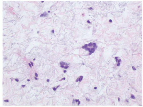
FIGURE 5-15 ▪ Increased numbers of multinucleated atypical stromal cells within the lamina propria have been termed giant-cell cystitis.


Painful Bladder Syndrome or Interstitial Cystitis

Interstitial cystitis, now commonly referred to as “painful bladder syndrome,” is a poorly defined chronic inflammatory process of unknown etiology that affects the urinary bladder.81, 82, 83 The clinical symptoms include urinary frequency, urgency, nocturia, suprapubic pressure, and pain with either bladder distention or voiding. By definition, patients experience the clinical urinary symptoms despite sterile urine cultures by routine laboratory techniques. Urodynamic studies typically reveal decreased bladder filling capacity. Prior therapies with
known bladder irritants, concomitant urothelial neoplasia, and infections would exclude the diagnosis of interstitial cystitis.81 The American Urologic Association guidelines have provided the following modified definition: “An unpleasant sensation (pain, pressure, discomfort) perceived to be related to the urinary bladder, associated with lower urinary tract symptoms of more than 6 weeks duration, in the absence of infection or other identifiable causes.”83


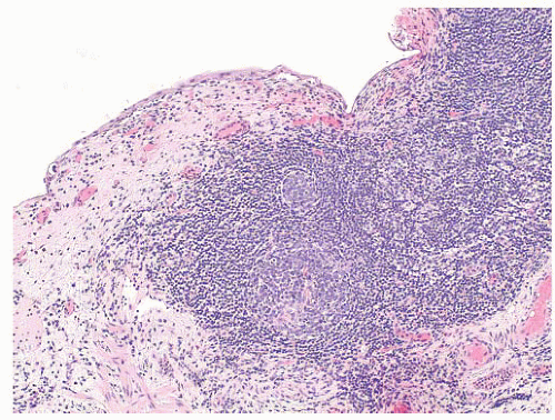
FIGURE 5-16 ▪ Follicular cystitis is characterized by mature lymphocytic infiltrates with germinal center formation.

The reported incidence of painful bladder syndrome/interstitial cystitis varies widely, ranging from 10 to 510 per 100,000. This variation is likely due to the varied criteria that have been utilized for diagnosis between different studies. Despite being described almost 100 years ago, the disease remains a clinical and therapeutic enigma. Some research has suggested the possibility of an autoimmune disorder, but there is no general agreement about the pathophysiology of the disease.

Cystoscopic evaluation of the disease may be categorized as “nonulcer or early disease” and the classic type. The nonulcer disease is characterized by normal mucosa at initial evaluation with the development of small submucosal hemorrhagic foci, called glomerulations, and linear cracks during/after distention (Fig. 5-17). In the classic pattern, there are single or multiple patches of reddened mucosa with small blood vessels radiating from a central mucosal scar. Classically, the mucosa ruptures under hydrodistention creating oozing of blood, which is the prototypical Hunner ulcer. In well-developed cases, the entire wall may be fibrotic and contracted. The trigone, in general, is not involved.

The role of morphology in the diagnosis of interstitial cystitis remains controversial. In our opinion, the role of the surgical pathologist is twofold: (a) most importantly, to exclude other specific forms of cystitis and urothelial CIS and (b) to detail histologic features for correlation with cystoscopy. There are no pathognomonic histologic features of interstitial cystitis, but common findings include ulceration with variably admixed fibrin, erythrocytes, and inflammatory cells, especially neutrophils. Associated granulation tissue and perineural lymphocytic infiltrates are common, and urothelial denudation is frequent. Ulcers typically extend deep into the lamina propria with surrounding edema and congestion. In patients without ulcers, the morphologic changes may be subtle and include suburothelial hemorrhage, edema, and possibly mucosal tears (Fig. 5-18). In long-standing disease, fibrosis of the muscularis propria may be present. There is considerable debate regarding the utility (i.e., specificity) of mast cell counts in the distinction from other inflammatory processes. However, there are reports of increased intravesical mast cell infiltrates in patients with interstitial cystitis (Fig. 5-19A and B).84, 85, 86 One of the major roles of biopsy evaluation is to exclude other lesions in the clinical differential diagnosis, particularly urothelial CIS. When the urothelium is extensively denuded, additional levels may be necessary to exclude this possibility. Since the morphologic features are not entirely specific, the final diagnosis of interstitial cystitis requires close clinical (i.e., history, cystoscopy, and voiding studies) and pathologic correlation.


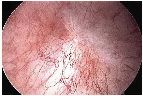
FIGURE 5-17 ▪ Interstitial cystitis is often characterized cystoscopically by small foci of mucosal hemorrhage called glomerulations.


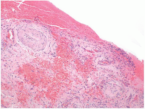
FIGURE 5-18 ▪ In interstitial cystitis, variable surface erosion with hemorrhage is common.

A recent review of patient management identified >180 published treatment modalities.82 These modalities include variations of behavioral, dietary, pharmacologic, and surgical interventions. There is general agreement on the use of some oral and intravesical agents, such as amitriptyline, hydroxyzine, and pentosan polysulfate sodium; however, there is a lack of definitive conclusions regarding optimal therapy.


Radiation and Chemotherapy Cystitis

In radiation or chemotherapy cystitis, patients often present with hematuria or voiding symptoms. Biopsy evaluation of the urothelium may show striking cytologic atypia.73 Cytoplasmic and nuclear vacuolation, karyorrhexis, and a normal nuclear-cytoplasmic ratio are features suggestive of radiation injury (Fig. 5-20A and B). In general, robust mitotic activity is absent. These histologic and clinical changes are both time and dose dependent; however, histologic changes may be seen for years. In addition, the
toxicity may be potentiated by concomitant therapy with cyclophosphamide. Radiation-induced histologic changes are similar to those seen with intravesical chemotherapy, but with intravesical chemotherapy the histologic changes are often more restricted to the superficial urothelial cell layer. Atypical mesenchymal cells similar to those seen in giantcell cystitis are also typically present in the lamina propria (Fig. 5-21). Other characteristic changes of radiation injury including marked stromal edema or fibrosis; prominent telangiectatic change, hyalinization, and thrombosis of the vessels are also helpful. Pseudocarcinomatous hyperplasia of the epithelium (discussed later) may be striking.
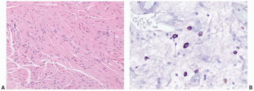


FIGURE 5-19A: Although its specificity has been debated, increased mast cells within the muscularis propria are reported in interstitial cystitis. B: The mast cells may be highlighted by a toluidine blue stain.

In the posttherapy setting, one should have a high threshold for the diagnosis of urothelial CIS. In difficult cases with uncertainty as to the appropriate diagnosis, use of the diagnostic term “atypia of unknown significance” is warranted. This allows repeat cystoscopy and biopsy evaluation after inflammation resides and avoids the potential overdiagnosis of reactive urothelial neoplasia.

Marked urothelial changes may be seen following radiation or chemotherapy. In general, radiation atypia is characterized by changes in the umbrella cell layer that include cytoplasmic vacuolization, nucleomegaly, and multinucleation. A reactive immunophenotype is maintained in reactive urothelial changes secondary to radiation therapy.87 Intravesical chemotherapy with agents such as mitomycin may also cause marked epithelial atypia. There are very little data available in the diagnostic pathology literature on the specific urothelial alterations after intravesical chemotherapy,88 but in our experience the cytoplasm of the urothelial cells becomes more
uniformly eosinophilic, and nuclear changes similar to those seen with radiation are common (Fig. 5-22A and B).
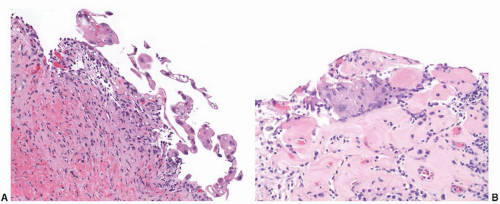


FIGURE 5-20A: Urothelial atypia secondary to radiation therapy is generally characterized by nuclear enlargement, prominent cytoplasmic vacuolization, and (B) multinucleation. The underlying tissue, including blood vessels, commonly shows hyalinization.


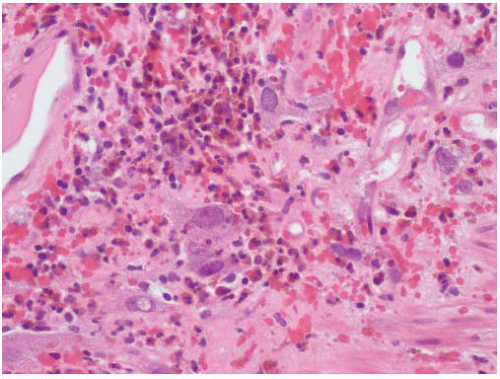
FIGURE 5-21 ▪ With radiation therapy, the stromal cells of the bladder may suggest prior radiation exposure given the degree of atypia with marked variation in size.


Hemorrhagic Cystitis

Hemorrhagic cystitis is caused predominantly by chemotherapeutic agents, most commonly cyclophosphamide, methotrexate, and radiation therapy,89 but viral etiologies are also well described (see “Viral Cystitis”). Less common causes include other chemotherapeutic agents such as busulfan and thiotepa, aniline and toluidine derivatives used in dyes and insecticides, and many other compounds.90 The clinical presentation is typically onset of severe hematuria shortly after exposure to the inciting agent (Fig. 5-23). Morphologically, hemorrhagic cystitis is characterized by extensive hemorrhage into the lamina propria with vascular congestion and edema. The overlying epithelium may show striking nuclear atypia similar to that seen in association with radiation or intravesical chemotherapy.


Eosinophilic Cystitis

“Eosinophilic cystitis” is a descriptive term that has been applied to mixed inflammatory infiltrates of the lamina propria rich in eosinophils (Fig. 5-19).91,92 This type of inflammatory infiltrate is seen in a broad age range, but approximately 20% of cases occur in children. There is an equal sex distribution. Patients often present with dysuria, frequency, hematuria, and/or flank pain. Rarely, patients can present with an infiltrative mass lesion that may induce urinary obstruction. On cystoscopy, the lesions often have a polypoid appearance that may mimic polypoid cystitis or urothelial carcinoma in adults or a botryoid rhabdomyosarcoma in children.

Eosinophilic cystitis is best regarded as a pattern of inflammation associated with a variety of causes and not a single diagnostic entity; however, there are two general settings: (a) allergy related and (b) injury related. Bladder eosinophilia has been reported in association with allergic gastroenteritis, asthma, or other allergic disorders, especially in women and children.91 This likely represents a systemic eosinophilic process that is distinct from the other nonspecific infiltrates. In adults, most commonly older males, eosinophilic infiltrates are more frequently associated with bladder injury from underlying prostatic hyperplasia, bladder carcinoma, or prior biopsy. Rarely, eosinophilic cystitis may be seen secondary to a parasitic infection.93 The word “eosinophilic” occasionally causes some confusion with neoplastic entities in children and
young adults; however, it should be emphasized that there is no relationship to Langerhans cell histiocytosis (previously called eosinophilic granuloma).
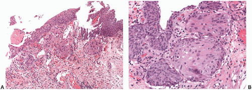


FIGURE 5-22A: After intravesical chemotherapy, nests of urothelial cells with atypical features may underlie a relatively normal appearing re-epithelialized urothelium. These reactive cells have enlarged nuclei and commonly have more eosinophilic cytoplasm imparting a squamoid appearance. B: At higher magnification, the fine chromatin of the reactive urothelial cells with enlarged nuclei can be seen. Compared to the overlying urothelium, these nests of cells with prior exposure to intravesical chemotherapy have a more pronounced cytoplasmic eosinophilia.


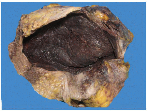
FIGURE 5-23 ▪ In this postmortem bladder specimen, the diffuse mucosal hemorrhage characteristic of hemorrhagic cystitis due to prior systemic chemotherapy is seen. (Courtesy: Dr. Don Regula, Stanford University.)

Histologically, early lesions of eosinophilic cystitis are characterized by inflammatory infiltrates rich in eosinophils (Fig. 5-24). In some cases with extensive infiltrates, necrosis of the muscularis propria may be seen. As lesions mature, the eosinophilic infiltrate becomes less prominent with an admixture of mature lymphocytes and plasma cells.

Patient management depends on the clinical setting. With allergy-associated lesions, removal of the inciting agent may alleviate the symptoms in the bladder. In idiopathic cases, a course of antihistamines and nonsteroidal anti-inflammatory agents are often tried. Other intravesical therapies, such as mitomycin C, dimethylsulfoxide, cyclosporine A, and oral corticosteroids, have also been utilized. In severe cases with unremitting symptoms, transurethral surgical resection and rarely cystectomy may be required.


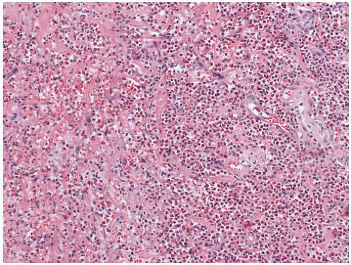
FIGURE 5-24 ▪ Although nonspecific, the extensive infiltrates of eosinophils typical of eosinophilic cystitis are seen in this biopsy specimen from a bladder mass in a child.


Nonspecific Acute and Chronic Cystitis

In some patients with symptomatic inflammatory conditions of the urinary bladder, no identifiable etiology is determined after workup for an infectious etiology, and the patients do not have clinical features of interstitial cystitis. When biopsied, nonspecific histologic changes may be seen. Recent changes may include edema, hyperemia, neutrophilic infiltrate, and histiocytes. Surface ulceration as well as associated reactive urothelial atypia may also be present. Chronic changes include fibrosis and mixed chronic inflammatory infiltrates.


Bacterial Cystitis

Bacterial cystitis is typically secondary to coliform bacteria such as Escherichia coli, Klebsiella pneumoniae, and Streptococcus faecalis.94, 95, 96 Another important cause is Proteus mirabilis. These infections are typically diagnosed by urine culture, so tissue biopsy plays little role in management.


Encrusted Cystitis

Encrusted cystitis refers to the deposition of inorganic salts in the bladder mucosa and is caused by urea-splitting bacteria that alkalinize the urine.97, 98, 99 It is most common in women, and presenting symptoms are similar to other urinary tract infections. Cystoscopically, the lesions may be diffuse and have a gritty appearance. Morphologically, encrusted cystitis is characterized by a fibrinous exudate with admixed calcified, necrotic debris and inflammatory infiltrates (Fig. 5-25A and B). The main differential consideration is urothelial carcinoma associated calcification, which may be seen in areas of necrosis or prior biopsy/resection.100 Accordingly, in a limited sample the carcinoma may be missed, and careful correlation with the cystoscopic impression is important to avoid a delay in diagnosis.


Gangrenous Cystitis

Gangrenous necrosis of the bladder is most commonly a complication of infection, but may also be due to systemic disorders (e.g., severe diabetes mellitus, metastatic carcinoma, sepsis, vascular disease) or corrosive chemical injury.101, 102, 103 The necrosis generally begins in the mucosa and may progress to involve the entire wall to the serosa.


Emphysematous Cystitis

Emphysematous cystitis, which is more common in women than men, is defined as the presence of gas-filled vesicles that are visible by cystoscopy or gross examination.104,105 A clinical diagnosis is typically made based on plain film and CT imaging, which show air within the wall of the
bladder. This lesion is typically associated with bacterial infections such as E. coli or Aerobacter zaerogenes. In adults, an association with diabetes/hyperglycemia is described in up to 50% of cases, but it may also be seen in association with neurogenic bladder, chronic cystitis, or immunosuppressed states.105,106

Only gold members can continue reading. Log In or Register to continue

Stay updated, free articles. Join our Telegram channel

Jun 10, 2016 | Posted by in UROLOGY | Comments Off on Nonneoplastic Lesions of the Urinary Bladder

Full access? Get Clinical Tree

Get Clinical Tree app for offline access