Tumors of the Testis


Tumors of the Testis


VICTOR E. REUTER

SATISH K. TICKOO


ANATOMY OF THE TESTIS

The adult testes are suspended by the spermatic cord and located within the scrotum. The testis proper is surrounded by a thick connective tissue layer called the tunica albuginea that itself is lined by the visceral tunica vaginalis (Fig. 12-1). In the posterior aspect of the gonad is the mediastinum testis that contains blood vessels, lymphatics, nerves, and portions of the rete testis. The testis contains multiple fibrous septa that radiate from the mediastinum testis to the tunica albuginea, and these divide the organ into approximately 250 compartments that contain the seminiferous tubules.1,2 Surrounding the seminiferous tubules is the interstitium, which contains Leydig cells, blood vessels, lymphatics, and nerves. Each compartment of the testis contains a maximum of four seminiferous tubules that are very convoluted and that usually empty into the straight portion of the rete (tubuli recti). Each seminiferous tubule is lined by a basement membrane and a thin lamina propria. Within the seminiferous tubule are Sertoli cells and germ cells at different stages of differentiation (Fig. 12-2).

Sertoli cells comprise 10% to 15% of cells within the tubule. They are columnar to pyramidal in shape with their long axis is perpendicular to the basement membrane. The cytoplasm is granular-eosinophilic and may contain fine vacuoles. The nuclei are round to oval with finely granular chromatin and are commonly located within a cell or two of the basement membrane. They contain a prominent nucleolus, the only normal cell within the tubule to do so. Intracytoplasmic Charcot-Böttcher crystalloids are best seen preferentially by electron microscopy.

Sertoli cells have phagocytic capacity but also play an important role in regulating spermatogenesis. By immunohistochemistry, they have been shown to express vimentin; cytokeratins 8, 18, and 19; as well as inhibin.3,4 Cytokeratin positivity is routinely observed in immature but not mature Sertoli cells; however, expression of this intermediate filament may be seen in adults in association with various conditions including testicular atrophy, Sertoli cell tumors (SCTs) and Sertoli cells adjacent to germ cell tumors (GCTs), orchitis, and infarct. In these settings, it is not unusual to see a gradient of immunoreactivity from positive to negative the farther away the tubules are located from the lesion.

Germ cells originate in the yolk sac and migrate to the genital ridge during the first 7 weeks of gestation.5,6 They comprise 85% to 90% of cells within the seminiferous tubule and have the capacity to differentiate (mature). Spermatogonia are undifferentiated cells located adjacent to the basement membrane. They have clear or basophilic cytoplasm, distinct cytoplasmic membranes, small round nuclei with dark chromatin, and no nucleoli. They have the capacity to proliferate and give rise to primary spermatocytes. The latter cells are subclassified into preleptotene, leptotene, zygotene, pachytene, and diplotene spermatocytes based on their nuclear chromatin pattern. These subtle differences are difficult, if not impossible, to discern on routine histologic preparations and irrelevant when evaluating tumor-bearing gonads. In general, primary spermatocytes are larger than spermatogonia, have basophilic cytoplasm, indistinct nuclear borders, round nuclei with distinct chromatin patterns, and absent nucleoli. Completion of the first meiotic division gives rise to secondary spermatocytes, which have a short half-life and undergo a second meiotic division to form spermatids. Secondary spermatocytes are smaller than their progenitor cells and have denser chromatin. Spermatids are located toward the lumen of the tubule and have small nuclei with dense chromatin. They transform into spermatozoa through metamorphosis.

Leydig cells are present in the interstitium as single cells or in clusters. Interestingly, they may also be observed in the tunica albuginea, mediastinum testis, epididymis, and even along the spermatic cord, usually intimately associated to nerve bundles.5,7 Leydig cells have abundant eosinophilic cytoplasm and round, regular nuclei with prominent nucleoli. Intracytoplasmic lipofuscin pigment may be seen, more commonly in older males. Intracytoplasmic Reinke crystals are characteristic of Leydig cells, rarely seen in normal cells, and more commonly observed by electron microscopy where they appear as a hexagonal prism.8,9


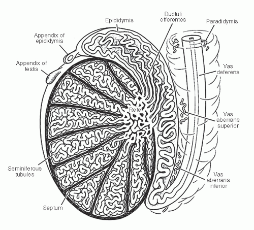
FIGURE 12-1 ▪ Schematic representation of the testis and its adnexa.

Leydig cells have the capacity to produce testosterone and share an important paracrine function with Sertoli cells.10 They express inhibin but not cytokeratins or vimentin by immunohistochemistry.11

The rete testis collects the effluent from the seminiferous tubules. It is located within the hilus of the testis and includes the tubuli recti, the mediastinal rete, and the extratesticular rete. The tubuli recti are short segments within the septa that connect the seminiferous tubules to the mediastinal rete. The mediastinum rete forms a series of epithelium-lined, interconnecting channels that lead to several dilated vesicular channels constituting the extratesticular rete, which anastomose to give rise to the efferent ducts or tubuli efferentia. The epithelium of the rete is low columnar and exhibits luminal microvilli. Every cell contains a flagellum that is not visible by routine light microscopy. The cells are immunoreactive for cytokeratins as well as vimentin.12


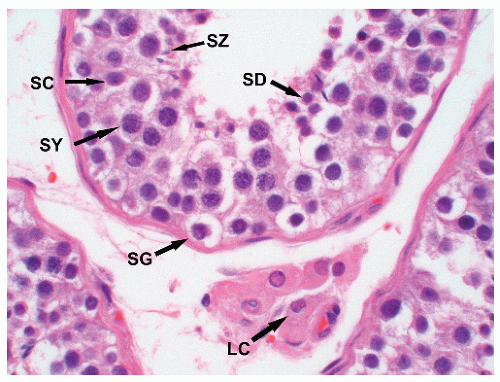
FIGURE 12-2 ▪ Normal seminiferous tubules in an adult containing spermatogonia (SG), spermatocytes (SY), spermatids (SD), spermatozoa (SZ), and Sertoli cells (SC). A cluster of Leydig cells (LC) is present in the interstitium.

The efferent tubules have an irregular (undulating) luminal contour. They receive the luminal content from the rete testis and are responsible for resorption of fluid. The epithelial lining is a mixture of ciliated and nonciliated columnar pseudostratified cells that express cytokeratin and variably vimentin by immunohistochemistry. The epithelial cells are surrounded by a thick basement membrane, which in turn is surrounded by a layer of smooth muscle. These tubules lead into the epididymis, a convoluted tubular structure that plays a role in the transport, maturation, and storage of sperm.7 Transport is aided by a thick smooth muscle layer that surrounds the epididymis. The epididymis is lined by a thick basement membrane and can be divided anatomically into three sections: head, body, and tail, the latter where sperm storage and maturation occurs. The epithelial lining of the epididymis is predominantly tall columnar cells (principal cells), many of which exhibit stereocilia, but basal cells, clear cells, and luminal cells are also present. The luminal contour of the epididymis is rigid rather than undulating.

The vas deferens arises from the caudal portion of the epididymis, which proximally joins the excretory duct of the seminal vesicles to form the ejaculatory duct. The vas deferens is lined by pseudostratified, columnar epithelium
and basal cells, the former containing long stereocilia. The luminal contour of the vas is variably folded, and the epithelium is surrounded by loose connective tissue and a very thick smooth muscle layer.

Several appendages may be encountered on the testis, testicular adnexa, or spermatic cord, the most common being the appendix testis and appendix epididymis.13,14 The appendix testis is a vestige of the müllerian duct attached to the tunica vaginalis along the anterosuperior surface of the testis adjacent to the head of the epididymis. It is a small pedunculated structure lined by columnar nonciliated epithelium. The epithelium covers richly vascular connective tissue. The appendix epididymis is a vestige of the mesonephric duct. It is a cystic, pedunculated structure attached to the head of the epididymis. The cyst is lined by low columnar epithelial cells, and the external surface is lined by mesothelium. Two other types of appendages are present as incidental findings and represent remnants of mesonephric tubules. They appear as epithelial-lined tubular or cystic structures, which are seen along the testicular adnexa or spermatic cord. Depending on their location, they are called vas aberrans or paradidymis.7,14


TUMORS OF THE TESTIS


Introduction

Primary testicular neoplasms are rare with <10,000 new cases reported each year in the United States. It is not unusual for there to be a delay in diagnosis since patients can present with testicular pain and be treated with antibiotics for a presumed orchitis or epididymitis. Metastases usually are first seen in retroperitoneal lymph nodes and then to lungs, consistent with the fact that metastases are usually through lymphatic spread except in the case of choriocarcinoma where dissemination is usually vascular. In more advanced cases, metastases are to the mediastinum and viscera, particularly the liver, and brain. The overwhelming majority of primary tumors are unilateral, whether of germ cell or sex cord-stromal origin. Because of the rarity of these tumors, it is prudent to always consider the possibility of secondary (metastatic) disease, even if the tumor is unilateral and particularly in older patients. However, as we discuss later, it is rare for a metastasis to the testis to be the first site of diagnosis since it is usually part of systemic disease. Primary lymphoma of the testis may present as unilateral disease and may mimic seminoma in its gross appearance as well as superficially at the microscopic level.


Gross Examination and Sampling of Testicular Tumors

In general, testicular neoplasms are managed surgically by a radical orchiectomy. This entails an inguinal incision and evacuation of the testis, surrounding tunica vaginalis, and extraperitoneal spermatic cord. Gross examination and proper prosecting of the orchiectomy specimen are crucial in establishing a proper diagnosis and pathologic staging of the tumor.15 The tunica vaginalis should be opened and any adhesions to the tunica albuginea noted since these may be secondary to invasion by tumor. The tunica albuginea should be carefully examined for any areas of bulging, discoloration, or disruption. Palpation of the gonad will allow us to locate any mass within the testicular parenchyma and its relation to the overlying tunica albuginea, rete testis, mediastinum testis, and testicular adnexa. In general, it is best to incise the testis along its long axis from the lateral aspect of the gonad toward the mediastinum testis.

Sampling of the mass should include a minimum of one section per centimeter of lesional tissue unless the entire lesion can be submitted in 10 cassettes or less, in which case the entire tumor should be submitted. Tumors that have a variegated gross appearance should be sampled in a manner that will allow examination of these areas microscopically; this need may require more through sampling. In cases where the morphology of the tumor is not straightforward, for example, in cases of atypical-looking seminomas or undifferentiated sex cord-stromal tumors, it may be necessary to go back to the specimen to take additional sections. The importance of serum tumor markers cannot be overstated. In general, seminomas are associated with normal serum levels of alfa fetoprotein (AFP) and either normal or mildly elevated levels of beta-human chorionic gonadotropin (β-HCG). Sex cord-stromal tumors are not associated with elevation of either of these markers. Cases in which there is a discrepancy between the pathologic diagnosis and serum levels of these oncoproteins should be reexamined and, if needed, additional sections taken.


Staging and Morphologic Prognostic Factors in Testicular Tumors

It is important to note that proper sampling is required not only to classify the tumor but also to stage it pathologically and evaluate risk factors for clinical progression. In the case of GCTs, staging is quite complicated and takes into consideration pathologic factors, size and sites of metastases, and serum levels of oncofetal proteins (AFP and β-HCG) as well as lactic dehydrogenase [LDH]) (Tables 12-1A, 12-1B and 12-1C). In the case of GCTs, the percentage of the embryonal carcinoma (EC) component as well as the presence of lymphovascular invasion has been associated with the development of metastatic disease in clinical stage 1 tumors. Lymphovascular invasion is particularly important since it is included in the AJCC-UICC staging system (Table 12-1B). A tumor that is otherwise pathologic stage 1 but with lymphovascular invasion is classified as pathologic stage 2 (pT2). Since vascular invasion (VI) is commonly seen adjacent to the mass, sections must include the tumor-parenchymal interface; sections that only include tumor are of no utility other than to classify the tumor and are discouraged. As mentioned earlier, lymphatic drainage
of the gonad flows toward the rete testis/mediastinum testis as well as toward the tunica albuginea (Figs. 12-3A and B). For this reason, sections that include tumor and overlying tunica as well as tumor with adjacent rete testis/mediastinum testis are particularly informative. Sections of the testicular adnexa (efferent tubules and epididymis) should be taken although involvement of these structures by tumor does not affect pathologic stage and does not seem to affect prognosis. This also applies to tumor involvement of soft tissue in the mediastinum, in the absence of lymphovascular invasion. A recent publication suggests that both rete testis involvement and hilar fat involvement are independently associated with advanced disease at presentation, but the study does not address the issue of association with relapse in clinical stage 1 disease17 (Fig. 12-4). We suggest that at
least three sections of the spermatic cord be taken: proximal (margin of resection), mid, and distal (near the adnexa). Involvement of the soft tissue of the spermatic cord is considered pathologic stage 3 and clinical stage 2 disease and is commonly associated with the presence of metastatic disease at the time of orchiectomy. Of note, intravascular or intralymphatic involvement within any part of the spermatic cord is still considered pT2 (Fig. 12-5).


Table 12-1A ▪ TNM CLASSIFICATION OF GCTs OF THE TESTIS


pTNM Pathologic Classification of the Primary Tumor (pT)


pTX


Primary tumor cannot be assessed


pT0


No evidence of primary tumor (e.g., histologic scar in testis)


pTis


Intratubular germ cell neoplasia


pT1


Tumor limited to testis and epididymis without vascular/lymphatic invasion; tumor may invade the tunica albuginea but not the tunica vaginalis


pT2


Tumor limited to testis and epididymis with vascular/lymphatic invasion, or tumor extending through the tunica albuginea with involvement of the tunica vaginalis


pT3


Tumor invades spermatic cord with or without vascular/lymphatic invasion


pT4


Tumor invades scrotum with or without vascular/lymphatic invasion


pTNM Pathologic Classification of the Regional Lymph Nodes (pN)


pNX


Regional lymph nodes cannot be assessed


pN0


No regional lymph node metastasis


pN1


Metastasis with a lymph node mass 2 cm or less in greatest dimension; or multiple lymph nodes, none more than 2 cm in greatest dimension


pN2


Metastasis with a lymph node mass more than 2 cm but not more than 5 cm in greatest dimension; or multiple lymph nodes, any one mass >2 cm but not more than 5 cm in greatest dimension


pN3


Metastasis with a lymph node mass more than 5 cm in greatest dimension


From Edge SB, Byrd DR, Compton CC, et al., eds. AJCC Cancer Staging Manual. 7th ed. New York: Springer; 2010, with permission, Ref.16


Table 12-1B ▪ CLINICAL STAGING OF GCTs OF THE TESTIS


Anatomic Stage/Prognostic Groups


Group


T


N


M


S*


Stage 0


pTis


N0


M0


S0


Stage I


pT1-4


N0


M0


SX


IA


pT1


N0


M0


S0


IB


pT2


N0


M0


S0


pT3


N0


M0


S0


pT4


N0


M0


S0


IS


Any pT/Tx


N0


M0


S1-3


Stage II


Any pT/Tx


N1-3


M0


SX


IIA


Any pT/Tx


N1


M0


S0


Any pT/Tx


N1


M0


S1


IIB


Any pT/Tx


N2


M0


S0


Any pT/Tx


N2


M0


S1


IIC


Any pT/Tx


N3


M0


S0


Any pT/Tx


N3


M0


S1


Stage III


Any pT/Tx


Any N


M1


SX


IIIA


Any pT/Tx


Any N


M1


S0


Any pT/Tx


Any N


M1


S1


IIIB


Any pT/Tx


N1-3


M0


S2


Any pT/Tx


Any N


M1


S2


IIIC


Any pT/Tx


N1-3


M0


S3


Any pT/Tx


Any N


M1


S3


Any pT/Tx


Any N


M1*


Any S


* Serum tumor markers.

Measured after orchiectomy.

N indicates upper limit of normal for the LDH assay.


From Edge SB, Byrd DR, Compton CC, et al., eds. AJCC Cancer Staging Manual. 7th ed. New York: Springer; 2010, with permission, Ref.16


Table 12-1C ▪ SERUM TUMOR MARKERS IN TESTICULAR GCTs*
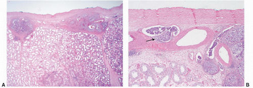


SX


Marker studies not available or not performed


S0


Marker study levels within normal limits


S1


LDH <1.5 × N and hCG (mIU/mL) <5,000 and AFP (ng/mL) <1,000


S2


LDH 1.5-10 × N or hCG (mIU/mL) 5,000-50,000 or AFP (ng/mL) 1,000-10,000


S3


LDH >10 × N or hCG (mIU/mL) >50,000 or AFP (ng/mL) >10,000


* Measured after orchiectomy.

N indicates the upper limit of normal for the LDH assay.


From Edge SB, Byrd DR, Compton CC, et al., eds. AJCC Cancer Staging Manual. 7th ed. New York: Springer; 2010, with permission, Ref. 16


FIGURE 12-3A: Extensive lymphovascular invasion by EC. Notice that the tumor involves lymphatics beneath the tunica albuginea, away from the main tumor mass. For this reason, sections should always include the interface between the tumor and the tunica albuginea. B: Lymphovascular invasion. The invasive tumor cells conform to the contours of the lymphatic channel and are focally attached to the vascular wall. Focal stromal response to the tumor emboli is present (arrow).

Criteria for establishing VI are similar to those used in other organ systems but may present a greater challenge at this site due to the fact that the tumor cells are very discohesive and commonly contaminate the adjacent parenchyma and vascular structures at the time of sectioning. It is imperative to restrict the diagnosis of VI to vessels, usually lymphatics, at the periphery of (not within) the tumor; in which the tumor cells conform to the shape of the vessel and preferably are attached to the wall with associated red blood cells or fibrin. While the use of immunohistochemistry is rarely necessary and not encouraged, we have recently found that ERG, a nuclear protein expressed in endothelial cells, may be of some utility when lymphovascular invasion is suspected but the presumed vascular space is entirely filled with tumor, as commonly happens with EC. In pure seminoma we should also mention if VI is present as it affects pathologic stage. However, its importance in establishing a higher risk of disease progression has not been proven. While most investigators suggest that tumor size (>4 cm) and rete testis involvement by seminoma predicts disease progression
on multivariate analysis, at least one recent large study that included 687 stage 1 tumors puts this belief into question18 (Fig. 12-6).


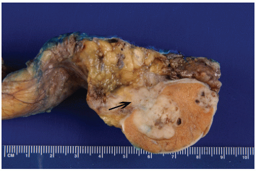
FIGURE 12-4 ▪ Gross appearance of mixed GCT. The large lesion can be seen invading into the testicular hilum (mediastinum) and hilar soft tissue. The rete testis and hilum is the most common site where tumors extend outside the testicular parenchyma. It has also been associated with metastatic disease at presentation. The smaller lesion involves the overlying tunica albuginea.


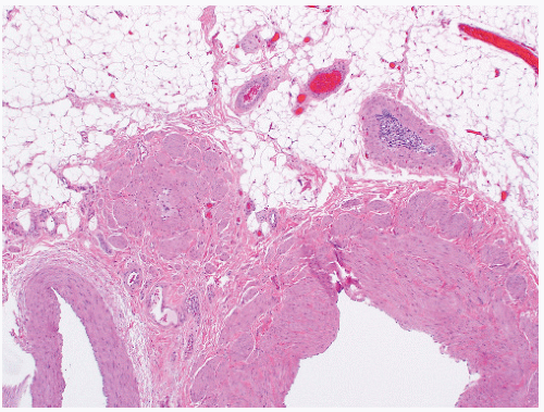
FIGURE 12-5 ▪ VI in a blood vessel within the spermatic cord. This finding is commonly seen in patients with advanced disease at presentation. However, in the absence of stromal invasion at this site, it is still regarded as pT2 disease.


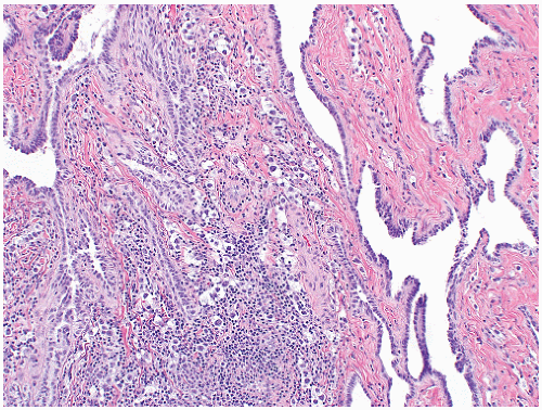
FIGURE 12-6 ▪ Seminoma invading the rete testis. While this finding has been associated with tumor relapse on multivariate analysis, this finding has been recently challenged. However, rete testis involvement should be reported.

Sampling of primary (chemotherapy naïve) retroperitoneal resections requires a thorough gross examination of the sample and submission of all suspected lymph nodes for microscopic examination. When this surgical procedure is performed, it is in a setting of nonseminomatous primary tumor with a high risk for relapse or when imaging of the retroperitoneum is questionable. In other words, the presence of metastatic disease is likely to be minimal and microscopic. The average number of lymph nodes found at our institution in this setting is more than 50.19 In fact, cases in which <40 lymph nodes are identified are submitted to regrossing of the residual tissue, with many additional lymph nodes identified. When reporting metastatic disease, the histologic type, number, and size of involved lymph nodes should be reported. Extranodal extension by tumor should be mentioned although the clinical significance of this finding has recently come into question.20

Great care should also be placed in sampling postchemotherapy resections. These resections should be carefully described grossly and sampled thoroughly, particularly those that are not obviously necrotic. A minimum of one section for each centimeter of lesional tissue should be submitted, making sure that all variegated areas are sampled. The sampling of an apparent mass does not mean that a thorough examination and sampling of the remaining soft tissue can be bypassed. It is common to find multiple lymph nodes in these areas, some with microscopic areas of treatment effect or even viable tumor. Once again, the size, number of lymph nodes involved, and the histology of the residual viable disease and the presence of extranodal extension should be reported.

In the case of sex cord-stromal tumors, similar care should be taken in the gross examination and prosecting of the specimen. Sampling of the mass is similar to germ cells tumors, with special attention paid to variegated areas, tumor-parenchymal interface, rete testis, adnexa, and cord. While no single morphologic factor can predict disease progression, a combination of factors has been associated with the development of metastatic disease and should be included in the pathology report. The report should include size, presence or absence of infiltrative borders, coagulative necrosis, nuclear atypia, mitotic activity, and lymphovascular invasion; more details are provided in the subsequent sections of this chapter. Because there is no effective chemotherapy for these tumors, it is customary for patients with clinical stage 1 disease with multiple high-risk factors to undergo retroperitoneal lymph node dissection (RPLND) since surgery is their best chance for cure.


Clinical Management of Testicular Tumors

While all agree that radical orchiectomy is the treatment of choice for primary testicular GCTs, there is disagreement on how best to manage cases with a high risk of progression.21, 22, 23, 24 Many investigators advocate clinical surveillance since salvage chemotherapy at recurrence is associated with a high cure rate. Some advocate for a primary RPLND in this setting because the presence of minimal metastatic disease will allow the oncologist to give fewer cycles of chemotherapy. All agree that patients with bulky retroperitoneal disease or metastasis above the diaphragm should receive a full course of systemic chemotherapy although the exact regimen to follow will depend on the sites, the number of metastases, the levels of AFP, β-HCG, and LDH in serum.25 Postchemotherapy retroperitoneal node dissection is controversial but advocated by some investigators as a means to diminish the incidence of late recurrences.


CLASSIFICATION

The 2004 World Health Organization classification of testicular tumors is presented in Table 12-2. This classification varies very little from the one described in the third edition of the Armed Forces Institute of Pathology Fascicle.26 It builds upon previous classifications proposed by pioneers in this field, including Friedman and Moore,27,28 Dixon and Moore,29 Melicow,30 and Mostofi and Price.31 Practically speaking, these efforts at classifying testicular tumors are morphology based and reproducible. However, the present classification scheme described in Table 12-1A correlates with serum tumor marker abnormalities and has superior clinical application.


Germ Cell Tumors


Epidemiology and Pathogenesis

Testicular GCTs constitute approximately 98% of all testicular neoplasms and are the most common malignancy in males between the ages of 15 and 35 years.32 They are relatively uncommon; approximately 7,920 new cases will have
been diagnosed in the United States in 2013 with only 370 dying of disease33 (Table 12-3). Because of their relative rarity, they present a diagnostic challenge to most practicing pathologist (Box 12-1).


Table 12-2 ▪ WHO HISTOLOGIC CLASSIFICATION OF TESTIS TUMORS*


GCTs


Intratubular germ cell neoplasia, unclassified


Other types


Tumors of One Histologic Type (Pure Forms)


Seminoma


Seminoma with syncytiotrophoblastic cells


Spermatocytic Seminoma (SS)


SS with sarcoma


EC


YST


Trophoblastic tumors


Choriocarcinoma


Trophoblastic neoplasms other than choriocarcinoma


Monophasic choriocarcinoma


Placental site trophoblastic tumor


Teratoma


Dermoid cyst


Monodermal teratoma


Teratoma with somatic-type malignancies


Tumors of More Than One Histologic Type (Mixed Forms)


Mixed EC and teratoma


Mixed teratoma and seminoma


Choriocarcinoma and teratoma/EC


Others


Sex Cord/Gonadal Stromal Tumors


Pure forms


LCT


SCT


SCT lipid-rich variant


Sclerosing SCT


LCCSCT


Granulosa cell tumor


Adult-type granulosa cell tumor


Juvenile-type granulosa cell tumor


Tumors of the thecoma/fibroma group


Fibroma


Thecoma


Sex cord/gonadal stromal tumor, incompletely differentiated


Sex cord/gonadal stromal tumors, mixed forms


Tumors containing both germ cell and sex cord/gonadal stromal elements


Gonadoblastoma


Germ cell/sex cord/gonadal stromal tumor, unclassified


Miscellaneous Tumors


Carcinoid tumor


Nephroblastoma


Paraganglioma


Hematopoietic Tumors


Secondary Tumors of the Testis


*Modified from the World Health Organization Classification of Tumours, Pathology & Genetics: Tumours of the Urinary System and Male Genital Organs. In: Eble JN, Sauter G, Epstein JI, et al., eds. WHO Histological Classification of Testis Tumours. Lyon, France: IARC Press; 2004:218.


Testicular GCTs can be divided into three groups (infantile/prepubertal, adolescent/young adult, and spermatocytic seminoma [SS]), each with its own constellation of clinical histology, molecular, and clinical features.34,35 (Table 12-4) They originate from germ cells at different stages of development. Tumors arising in prepubertal gonads are either teratomas or yolk sac tumors (YSTs), tend to be diploid, and are not associated with i(12p) or intratubular germ cell neoplasia unclassified (IGCNU). The annual incidence is approximately 0.12 per 100,000. SS arises in older patients. These benign tumors may be either diploid or aneuploid and have losses of chromosome 9 rather than i(12p). Intratubular SS is commonly encountered but IGCNU is not. Their annual incidence is approximately 0.2 per 100,000. The pathogenesis of prepubertal GCT and SS is poorly understood.

The most common testicular cancers arise in postpubertal men; they are characterized genetically by the presence of excess genetic material of the short arm chromosome 12, usually due to one or more copies of i(12p), or other forms of 12p amplification and aneuploidy36 (Fig. 12-7). The consistent gain of genetic material from chromosome 12 seen in these tumors suggests that it has a crucial role in their development. IGCNU is the precursor to these invasive tumors.

While IGCNU is considered to be the precursor of all GCTs, the stage in GC development at which transformation occurs is not known. One model proposed by Skakkebaek et al.37 suggests that fetal gonocytes (primordial germ cells) undergo abnormal cell division (polyploidization) in utero, primarily due to environmental factors. These cells undergo abnormal cell division mediated by a kit receptor/kit ligand (stem cell factor) paracrine loop, leading to uncontrolled proliferation of gonocytes. Subsequent invasive growth may be mediated by postnatal and pubertal gonadotrophin stimulation. In this model 1(12p) is seen only after there is stromal invasion. A second model proposed by Chaganti et al. suggests that aberrant chromatid exchange events during meiotic crossing-over may lead to increased 12p copy number and overexpression of cyclin D2 (CCND2). In a cell containing unrepaired DNA breaks (recombination-associated), overexpressed cyclin D2 may block a p53-dependent apoptotic response and lead to reinitiation of cell cycle and genomic instability. This aberrant, genomically unstable cell is now able to escape the apoptotic effects of p53 and may reenter the cell cycle, now as a neoplastic cell. In this model, i(12p) is present in IGCNU.38

The incidence of adult-onset GCTs is approximately 6.0 per 100,000 per year with the majority being discovered between 15 and 40 years of age. The incidence of GCTs has increased over the last five decades while the death rate has decreased.39,40 The increase in incidence is seen across all histologies but mostly in seminomas. While more effective therapy is responsible for the dramatic increase in the cure rate, it is less evident why the incidence continues to climb.
There appears to be a geographic and racial predisposition to the development of testicular GCTs with a twofold increase in Scandinavian countries as compared to the United States and a lower incidence in other countries such as Africa and some Latin American countries such as Puerto Rico.39 Patients with prior testicular GCT are more likely to develop a contralateral tumor than does the general population.


Table 12-3 ▪ IMMUNOHISTOCHEMICAL PROFILE OF TESTICULAR TUMORS


Marker


IGCNU


Seminoma


Spermatocytic Seminoma


Embryonal Carcinoma


YST


Trophoblastic Tumors


Sex Cord/Gonadal Stromal Tumors


PLAP


+


+


+


+/−


Diffuse cytoplasmic/membranous


Diffuse cytoplasmic/membranous


Diffuse cytoplasmic/membranous


Focal cytoplasmic/membranous


CD117 (c-kit)


+


+


−/+


Diffuse membranous


Diffuse membranous


Focal weak


Oct3/4


+


+


+


Diffuse nuclear


Diffuse nuclear


Diffuse Nuclear


AFP


+


Variable cytoplasmic


SALL4


+


+


+/−


+


+


+


Diffuse nuclear


Diffuse nuclear


Weak


Diffuse nuclear


Diffuse nuclear


Mononucleated trophoblastic cells


Glypican 3


+


+/−


Cytokeratin


+


+


+


+/−


Focal dot-like


Membranous/cytoplasmic, may be focal and weak


SCTs


HCG


+


syncytiotrophoblasts


Inhibin


+


Calretinin


+


SF-1


+


WT1


+


The overwhelming majority of GCTs are sporadic with approximately 2% having a familial basis in which first-degree male relatives of patients with testicular GCTs will develop the disease.41, 42, 43, 44 Several other factors have been associated with their pathogenesis, including cryptorchidism, gonadal dysgenesis, and infertility. Testicular microlithiasis has been associated with the presence of GCT, but it may be seen in other conditions as well.


Cryptorchidism is one of the best-established risk factors,43 with approximately 3.5 to 5 times elevated risk of development of a testicular GCT compared with control populations.45, 46, 47 Testicular biopsies are commonly performed
in these high-risk patients to detect incipient neoplasia. In a large series, only one of more than 1,500 cryptorchid patients with testicular biopsy specimens negative for IGCNU developed testicular cancer over a follow-up period of 8 years,48 in contrast to 50% of patients with IGCNU who developed invasive tumors over a 5-year period.49


Table 12-4 ▪ TESTICULAR GERM CELL TUMORS


Histology


Age


Ploidy


Chromosomes


Teratoma/YST


Infantile


Diploid aneuploid


−1p, −6q


Seminoma/NS


Postpubertal


CIS: hypertriploid


1(12p) or +12p


SEM: hypertriploid


1(12p)


NS: hypotriploid


1(12p)


SS


Older


Diploid or tetraploid


+9


Modified from Oosterhuis JW, Looijenga LH. Current views on the pathogenesis of testicular germ cell tumours and perspectives for future research: highlights of the 5th copenhagen workshop on carcinoma in situ and cancer of the testis. APMIS 2003;111:280-289.


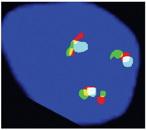
FIGURE 12-7 ▪ Three color FISH probe demonstrating two copies of i(12p). The aqua is a cep12 probe while the red and green are ETV6(12p13). There is one normal chromosome (aqua adjacent to one red and one green signal) and two isochromosomes in which the cep12 signal is flanked by two red-green signals.

There is an approximately 30% risk of GCTs observed in men with gonadal dysgenesis who carry a Y chromosome.48 Gonadoblastoma is the most frequent tumor type arising in this setting, and its presence predisposes to the development of invasive GCTs, most commonly seminoma. Of patients with androgen insensitivity syndrome, 5% to 10% develop GCTs. These tumors generally are diagnosed after the complete development of female secondary sexual characteristics.50,51

The frequency of IGCNU in subfertile men has been reported to be 0.4% to 1.1%.52,53 However, the relative risk of infertile men developing GCTs is not clear. Since many cases are associated with cryptorchidism or gonadal dysgenesis, it is difficult to judge whether infertility itself is an independent risk factor.

It is remarkable that GCTs, having such diverse morphology and clinical behavior, should be considered as variants of one entity. Nevertheless, there is circumstantial and laboratory evidence to support this practice. First, these tumors, in the postpubertal setting, tend to arise along the axial skeleton, be it the pineal gland, anterior mediastinum, retroperitoneum, or gonads. Second, mixed histologic patterns predominate over tumors with one histologic type. A third compelling piece of evidence relates to the so-called precursor lesion. When these tumors arise in the male gonad, irrespective of the morphology, one is likely to identify IGCNU in adjacent seminiferous tubules, originally described as in situ carcinoma by Neils Skakkebaek. A fourth important piece of evidence linking these tumors comes to us from genetics, since approximately 80% of tumors regardless of the primary site and histology will have at least one isochromosome of the short arm of chromosome 12, which is known as i(12p) (Fig. 12-3). This genetic abnormality is not absolutely pathognomonic of germ cell neoplasia, yet it is a very useful diagnostic tool in selected circumstances due to its rare occurrence in other solid tumors.54, 55, 56


Intratubular Germ Cell Neoplasia

This term refers to the lesion initially described by Skakkebaek as “carcinoma in situ” as well as to other “differentiated” forms of intratubular germ cell neoplasia.57, 58, 59, 60 Strictly speaking, the lesion originally described by Skakkebaek is now called “intratubular germ cell neoplasia, unclassified” by most, at least in the Western hemisphere; with increasing frequency in other parts of the world as well. It is the precursor lesion of all invasive GCTs with the exceptions of SS, prepubertal YST, and teratoma. It is found in virtually all cases in which residual seminiferous tubules are identified around an invasive GCT. It also is present in 5% to 8% of cases of cryptorchidism,61 5% of patients with an invasive GCT in the contralateral testis,61 and frequently in dysgenetic gonads.

The story of testicular “carcinoma in situ” (intratubular germ cell neoplasia) is fascinating and serves as a paradigm for the concept of progression from incipient or preinvasive neoplasia to invasive disease.59,62,63 In 1972, Skakkebaek reported “atypical spermatogonia” in two men undergoing testicular biopsies during a workup for infertility who subsequently developed invasive testicular GCTs. He hypothesized that these cells constituted “carcinoma in situ.” Two subsequent seminal studies by his group proved that this was indeed the case. In 1978, he reported a series of 555 men who underwent testicular biopsies for infertility.62,63 They identified six patients with evidence of “carcinoma in situ.” With a median follow-up period of approximately 3 years, three of these patients developed evidence of an invasive GCT; one of them with bilateral disease. The remaining 449 patients were tumor free during the same follow-up period (Box 12-2).

In 1986, the Skakkebaek group reported their experience with contralateral biopsies in 500 patients with unilateral GCT.64 Twenty-seven patients (5.4%) were found to have “CIS.” Eight patients received systemic chemotherapy for advanced disease. Of the remaining 19 patients, 7 (37%)
developed invasive GCT at this site within the follow-up period. Mathematical modeling suggested that 50% of biopsy-positive cases would develop disease within 5 years. Remarkably, not a single case of contralateral GCT developed in the remaining 463 biopsy-negative patients during the same follow-up period. In a subsequent report the authors revealed that at least two of the biopsy-positive cases that received systemic therapy subsequently developed contralateral tumors, suggesting that systemic therapy is not always effective against preinvasive disease.65


It is clear that the original lesion described by Skakkebaek is the precursor to all types of GCTs, at least for those that originate in postpubertal gonads other than SS. In early 1980, a group of distinguished pathologists including Drs. Robert Scully, Juan Rosai, F.K. Mostofi, and Robert Kurman met in Minnesota to discuss the nomenclature of incipient germ cell neoplasia. They agreed that “carcinoma in situ” was a poor choice to describe this lesion since it had no features of epithelial differentiation. They suggested the term “intratubular germ cell neoplasia, unclassified” (IGCNU) because it was associated with all morphologic types of GCT with the exception of SS. It also underscores the fact that differentiated forms of intratubular germ cell neoplasia may occur, including intratubular EC.

IGCNU can be seen adjacent to invasive GCTs in virtually all cases in which residual testicular parenchyma is present.57,66 As previously mentioned, it is present in up to 4% of cryptorchid patients, up to 5% of contralateral gonads in patients with unilateral GCT, and up to 1% of patients biopsied for oligospermic infertility. Its association with testicular GCTs arising in prepubertal patients is still a source of controversy.34,67,68 While some authors suggest that it does not occur, others state that it does. In either case, we can state with reasonable certainty that, if IGCNU does occur in childhood tumors, it is certainly less apparent.


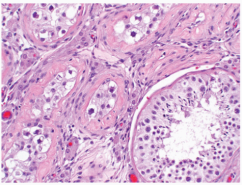
FIGURE 12-8 ▪ Intratubular germ cell neoplasia. Notice the seminiferous tubules with irregular thickening of the basement membrane that contain cells with perinuclear clearing, irregular hyperchromatic nuclei with prominent nucleoli. An adjacent uninvolved tubule is present.
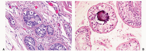


FIGURE 12-9A: Intratubular germ cell neoplasia. The surrounding Sertoli cells are small, fusiform, and lack nucleoli, similar to what is seen in prepubertal gonads. In adults the presence of seminiferous tubules with immature Sertoli cells is believed to be a sign of testicular dysgenesis. The combination of IGCNU and immature Sertoli cells is also a feature of gonadoblastoma. B: Testicular microlithiasis associated with IGCNU. While this finding can be associated with the presence of a germ cell neoplasm, it is in no way specific as it can be seen in many other conditions.


Microscopic Features

IGCNU is characterized morphologically by the presence of enlarged, atypical germ cells located adjacent to a usually irregularly thickened basement membrane (Figs. 12-8, 12-9A and B). The atypical cells are either isolated or form a single row along the basement membrane. They are typically larger than spermatogonia, the other cell that usually resides near the basement membrane. IGCNU cells have clear cytoplasm, irregular nuclear contours, coarse chromatin, and enlarged nucleoli, which may be single or multiple. Spermatogonia may also have clear cytoplasm but the cells are small, have round and regular nuclear contours, densely packed
chromatin, and absent nucleoli. In most cases, tumor-bearing tubules do not have active spermatogenesis and contain mostly Sertoli cells. Sertoli cells may be displaced toward the tubular lumen. Characteristically, they contain a single nucleolus that is small and regular. The nuclei are oval or round with regular borders and the chromatin is fine. The cytoplasm is amphophilic/eosinophilic and not vacuolated.


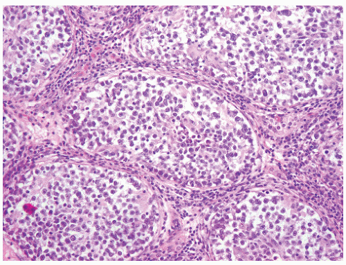
FIGURE 12-10 ▪ Seminoma cells fill the lumen of a seminiferous tubule. This should not be called intratubular germ cell neoplasia.

In essence, the cytologic features of classic IGCNU are those of seminoma. The relationship is supported by the coexpression of a host of histochemical and immunohistochemical markers among both cell types. Further evidence comes from electron microscopy; which has shown that both share common ultrastructural features: including the absence of well-developed cytoplasmic intermediate filaments, inconspicuous organelles, glycogen particles, lack of mature desmosomes and cell junctions, and nucleoli with ropy nucleolonema. Tubules whose lumens are filled with these cells may be regarded as “intratubular seminoma” (Fig. 12-10). The presence of i(12p) in IGCNU remains controversial with some investigators suggesting it is an early event and present in this stage of the disease while others suggest that it occurs only after the tumor cells invade into the parenchyma.37 This debate is interesting in understanding the pathogenesis and progression of testicular GCTs but is of little or no clinical and diagnostic importance. Given the availability of excellent immunohistochemical markers, it is never necessary to resort to fluorescence in situ hybridization (FISH) or other molecular tests to document its presence. It is said that ICGNU is triploid or hypertetraploid. However, once again, this finding is of little diagnostic consequence.38

IGCNU may extend into the rete testis, usually undermining the epithelium in a “pagetoid” pattern (Fig. 12-11). At times, the epithelium of the rete may become hyperplastic, and in this setting, it is important not to confuse this finding with the presence of nonseminomatous GCT. At times, the rete testis epithelium adjacent to the tumor may contain intracytoplasmic eosinophilic droplets that may be confused with YST (Fig. 12-12).69


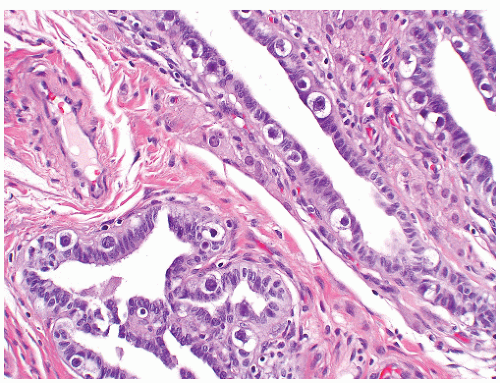
FIGURE 12-11 ▪ Intratubular germ cell neoplasia extends into the rete testis in a pagetoid fashion.


Histochemistry and Immunohistochemistry

IGCNU cells contain glycogen and thus are PAS-positive, diastase-sensitive. Rarely will other intratubular cells, whether spermatogonia, spermatocytes, or Sertoli cells, show similar positivity. Placental-like alkaline phosphatase (PLAP) is one of the isoforms of alkaline phosphatase (Table 12-5). PLAP antibodies will stain IGCNU, the majority of seminomas and ECs, as well as a smaller percentage of YSTs. Immunoreactivity is seen in virtually all cases of IGCNU and the staining pattern is usually membranous or cytoplasmic. No other nonneoplastic intratubular cells are immunoreactive for PLAP, but immunoreactivity may be
seen in other types of non-germ cell malignancies.70, 71, 72, 73 C-kit (CD 117) is expressed in a large percentage of IGCNU as well as seminomas, but not in other GCTs.74 Once again, the staining pattern is cytoplasmic/membranous (Fig. 12-13A). Despite the overexpression of this antigen, C-kit is rarely mutated in these tumors. Other antibodies that immunoreact with IGCNU but are rarely used in clinical practice include M2A and 43-F.73,75,76 POU5F1 (Oct3/4) is a very interesting marker with great clinical utility.77 The gene serves as a transcription factor and its product is expressed in pluripotent mouse and human embryonic stem cells and is downregulated during differentiation. Since the gene is also required for self-renewal of embryonic stem cells, knocking out the gene is lethal. This antigen is expressed solely in IGCNU, seminoma, and EC, suggesting that these are the types of GCT cells with pluripotency, that is, with the capacity to differentiate. As a transcription factor, staining is localized to the nucleus (Fig. 12-13B).


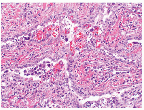
FIGURE 12-12 ▪ Rete testis epithelium with reactive changes and intracytoplasmic eosinophilic globules, mimicking YST. This pitfall can be avoided by paying close attention to the location of the findings, since transition to more typical rete testis epithelium is usually seen.


Table 12-5 ▪ GCTs RISK CLASSIFICATION: INTERNATIONAL CONSENSUS
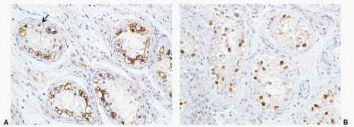


Risk Group


Seminoma


Nonseminoma


Good


Any HCG


AFP < 1,000 ng/mL


Any LDH


HCG < 5,000 mIU/mL


Nonpulmonary visceral metastases absent


LDH < 1.5 × ULN


Any primary site


Nonpulmonary visceral metastases absent


Gonadal or retroperitoneal primary tumor


Intermediate


Nonpulmonary visceral metastases present


AFP 1,000-10,000 ng/mL


Any HCG


HCG 5,000-50,000 mIU/mL


Any LDH


LDH 1.5-10.0 × ULN


Any primary site


Nonpulmonary visceral metastases absent


Gonadal or retroperitoneal primary site


Poor


Does not exist


Mediastinal primary site


Nonpulmonary visceral metastases present (e.g., bone, liver, brain)


AFP 10,000 ng/mL


HCG 50,000 mIU/mL


LDH 10 × ULN


AFP, α-fetoprotein; HCG, human chorionic gonadotropin; LDH, lactate dehydrogenase; ULN, upper limit of normal.


Manivel JC, Jessurun J, Wick MR, et al. Placental alkaline phosphatase immunoreactivity in testicular germ-cell neoplasms. Am J Surg Pathol 1987;11:21-29.


FIGURE 12-13A: Intratubular germ cell neoplasia. C-kit is positive in a membranous distribution. Notice that a few spermatogonia may be immunoreactive (arrow). B: Intratubular germ cell neoplasia. Oct3/4 is positive in a nuclear distribution.

Another transcription factor expressed in IGCNU is SALL4; however, this nuclear marker is expressed in a wider spectrum of GCTs including seminoma, EC, yolk sac, tumor, and some glandular elements of teratoma.78,79 As such, it is a useful marker in the characterization of GCTs but cannot be used in isolation. Podoplanin (clone D2-40) is an excellent cytoplasmic (membranous) marker with nuclear staining restricted to ICGNU and seminoma.80,81


Differential Diagnosis

When the tumor cells fill the seminiferous tubules, the term IGCNU is no longer used but rather intratubular seminoma, assuming that the cytomorphologic features of the tumor cells are identical to those described above. It is important to keep in mind that the presence of neoplastic cells within tubules does not always constitute IGCNU and that one
must adhere strictly to the established diagnostic criteria. On occasion spermatogonia may be enlarged and notably hyperchromatic, superficially mimicking ICGNU at low magnification. These changes are thought to be degenerative and are not associated with the development of tumor. These cells can be easily seen at intermediate to high magnification since they lack the characteristic coarse chromatin, nuclear contour irregularities, and prominent nucleoli seen in ICGNU. As you might expect, the immunoprofile of these cells is quite different, lacking expression of PLAP and OCT4. At times, spermatogonia can express C-kit and SALL4. Proper care must be taken to examine closely the cytologic features of the immunoreactive cells. While some investigators have reported PLAP immunoreactivity in rare spermatogonis, we have not encountered this problem. Besides intratubular seminoma, one can encounter intratubular EC (Fig. 12-14A and B), intratubular SS, and even metastatic disease such as melanoma and prostatic carcinoma. Intratubular lymphoma and even mesothelioma may also be confused with IGCNU.
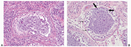


FIGURE 12-14A: Intratubular embryonal carcinoma. Tumor cells line a portion of the seminiferous tubule and fill the lumen. B: IGCNU and intratubular EC. IGCNU is characterized by atypical cells with irregular nuclear contours, prominent nucleoli, and cytoplasmic clearing (thin arrows) whereas the EC cells have larger, more irregular nuclei and nucleoli as well as amphophilic cytoplasm (thick arrows).

ICGNU is commonly encountered adjacent to invasive germ cell tumors, more rarely during the workup of infertility, and in countries where patients with unilateral GCT undergo a diagnostic biopsy of the contralateral gonad. Biopsy of the contralateral side is rarely performed in this country but has been advocated in countries where the incidence of developing a contralateral tumor is high, such as in Scandinavia. In these settings, the cytologic features of ICGNU are identical to what we have described, but the diagnosis can be a bit more challenging because the number of neoplastic cells tends to be more limited. It is important to evaluate well-fixed, well-stained samples and to place close attention to the nuclear detains of the cells. In this setting prudent use of a limited panel of immunohistochemical markers, such as Oct4 and podoplanin, may be very useful. PLAP and C-kit can also be used, with the caveats mentioned above.


Seminoma

Seminomas are the most common GCTs arising in the male gonad, whether they arise in a pure state or mixed with other morphologic types.26,82, 83, 84, 85, 86, 87 “Pure” seminoma account for 30% of testicular GCT and another 15% to 20% contain syncytiotrophoblasts without other germ cell components. Seminoma is extremely rare in children younger than 10 years of age, and uncommon in adolescents.88 Approximately 1% to 2% are bilateral and bilaterality can occur synchronously or asynchronously. Seminomas reach a peak incidence between the fourth and fifth decades of life, which is approximately one decade later than nonseminomatous GCTs. Most patients present with self-identified testicular swelling, approximately 10% have acute groin pain, and fewer than 3% have symptoms secondary to metastatic disease, most commonly lumbar pain resulting from retroperitoneal involvement.26,89 At the time of presentation, clinically about 75% of patients have disease limited to the testis, 20% have retroperitoneal involvement, and 5% have supradiaphragmatic or visceral metastases (Box 12-3).

A subset of seminomas, specifically those containing syncytiotrophoblastic cells, may have mildly elevated
serum levels of β-HCG. Patients with metastases are more likely to have elevated levels of β-HCG; however, elevation of this marker does not appear to be an adverse prognostic marker but rather a marker of bulkier disease or advanced stage.89 AFP levels generally are not elevated in pure seminoma, and any elevations require more thorough sampling of the tumor for a nonseminomatous component. LDH can be elevated in seminoma but this elevation is in no way specific to this disease but generally is a marker of the extent of disease; the larger the tumor burden, the high the serum levels.


Gross Features

Seminomas appear as a fleshy, well-circumscribed, bulging cream or light tan mass that, depending on size, may occupy a variable amount of testicular parenchyma or replace it entirely (Fig. 12-15). Tumor size can be quite variable with some measuring less than a centimeter while rare cases may reach a size of 10 cm or greater. Areas of necrosis may be observed grossly in up to 20% to 25% of cases, with this finding being more common in larger tumors. Some seminomas are associated with a granulomatous reaction, and in these cases the tumor takes on a fibrous and nodular gross appearance. Tumors with trophoblastic elements may be associated with punctate foci of hemorrhage. In general, one section per centimeter of tumor is sufficient sampling. However, tumors with a variegated appearance and those with nodularity must be sampled in a manner that all areas are sampled adequately. It is better to oversample in these cases since the management between pure seminomas and tumors with mixed histologies may be quite different. As previously mentioned, it is important to sample in a manner that allows the examination of the interface between the tumor-parenchyma, tumor-rete testis, tumor-mediastinum, and tumor-adnexa.15 Sections of the spermatic cord should be taken as mentioned earlier in this chapter.


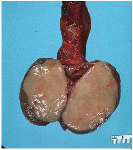
FIGURE 12-15 ▪ Seminoma gross. A well-circumscribed fleshy tan mass occupies most of the testicular parenchyma.


Microscopic Features

Seminoma usually grows in a sheet-like arrangement intersected by thin fibrous septa containing mature lymphocytes (Fig. 12-16). Occasionally, the fibrous septa are thicker, imparting a more nodular appearance to the tumor. Rarely seminomas may exhibit unusual patterns such as cribriform, pseudoglandular, and tubular growth90 (Fig. 12-17A and B). These do not represent separate entities, but rather histologic variants of classic seminoma. Most tumors efface the underlying testicular parenchyma although an interstitial pattern of infiltration may be seen at the periphery in many cases. Rarely the pattern of growth is preferentially interstitial.91 Interestingly, we have encountered a few cases of seminoma in which the component of the tumor with an interstitial pattern of growth took on micropapillary features due to retraction from the surrounding stroma (Fig. 12-18A and B). In two cases, the possibility of a metastatic carcinoma was entertained. The lymphocytes associated with the fibrous septa are mostly T cells, many with γ/δ phenotype.92,93 Some tumors may contain lymphoid aggregates with germinal center formation. Up to 50% of tumors may exhibit a granulomatous reaction that may be so intense as to obscure the underlying neoplasm (Fig. 12-19). Some cases exhibit extensive fibrosis, particularly those associated with a granulomatous reaction or tumors that have undergone partial regression.
Seminomas seen in association with a parenchymal scar should bring to mind the possibility of partial regression of the tumor, which may have been seminoma or possibly another GCT component other than teratoma. This phenomenon may be the explanation in some cases for nonseminomatous metastases in otherwise pure testicular seminomas. Whenever a parenchymal scar suggestive of partial regression is encountered, it is important to include it in the pathology report as a comment since this finding may account for the discrepancy between the pathology of the viable tumor and serum tumor markers.


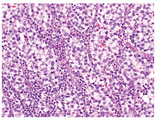
FIGURE 12-16 ▪ Seminoma, classic type. Cells have squaredoff nuclei, clear cytoplasm with little nuclear overlap. Mature lymphocytes are present in the septa.
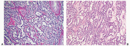


FIGURE 12-17A: Seminoma with a tubular pattern of growth. The cytologic features of the tumor cells are identical to those seen in classic seminoma. B: Seminoma with a tubular pattern of infiltration. At low power it is easy to understand why it might be confused with a SCT. It would be highly unusual for a seminoma to have this pattern of growth throughout the lesion.

Seminoma cells have a moderate amount of clear to pale eosinophilic cytoplasm with prominent cytoplasmic membranes and distinct cell borders (Fig. 12-16). The nuclei are large, round to rhomboid in shape and centrally located. The chromatin is evenly distributed and one or more prominent nucleoli are invariably present. In well-preserved specimens, one is unlikely to encounter nuclear overcrowding and overlap or syncytial growth. Mitotic activity is variable but may be quite brisk, a feature that has no bearing on prognosis. A significant proportion of seminomas contain syncytiotrophoblasts and their presence may be associated with focal areas of hemorrhage (Fig. 12-20). The absence of cytotrophoblast distinguishes these lesions from choriocarcinoma. Seminomas with syncytiotrophoblasts are generally accompanied by serum elevation of human chorionic gonadotropin (HCG), but levels will rarely reach levels above 500 IU/mL.86


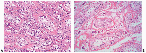
FIGURE 12-18 ▪ Seminoma with an interstitial pattern of infiltration. IGCNU is present, and the walls of the seminiferous tubules are irregularly thickened. A: Seminoma with an interstitial pattern of infiltration. Tumor cells infiltrate among seminiferous tubules. B: Seminoma with an interstitial pattern of infiltration. Notice the retraction artifact, giving the tumor cell a “micropapillary” appearance.


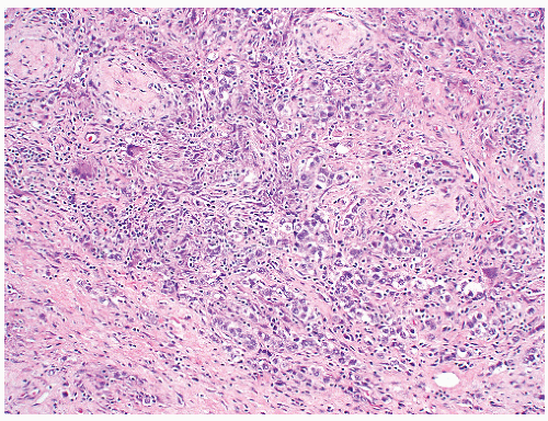
FIGURE 12-19 ▪ Seminoma associated with an intense granulomatous reaction.


Histochemistry and Immunohistochemistry

The expression profile of seminomas is detailed in Table 12-3. Briefly, tumor cells contain glycogen (PAS-positive) and express PLAP, C-kit (CD117), by immunohistochemistry70,71,94, 95, 96, 97, 98, 99, 100 but not cytokeratins, CD30, or inhibin. In our practice, PAS and PLAP are rarely relied upon because of the availability of better discriminating markers. On occasion weak CD30 cytoplasmic immunoreactivity may be encountered in isolated seminoma cells, a finding that should not warrant a change in diagnosis. It is important to remember that CD30 may be expressed in some hematopoietic cells as well so attention to nuclear detail is warranted. A minority of seminoma cells may express focal and weak, dot-like or linear immunoreactivity for cytokeratin AE1/AE3 and CAM 5.2, however, never diffuse and strong staining throughout the cytoplasm (Fig. 12-21). Caution must be taken when interpreting cytokeratin markers since syncytiotrophoblasts are usually strongly immunoreactive, as are its mononuclear variants. If one relies on panels of markers this issue is resolved easily since syncytiotrophoblasts lack immunoreactivity to POU5F1 (OCT4), SALL4, podoplanin, and so on. Like IGCNU, seminoma cells express (OCT4) in a nuclear distribution70,74,101,102 (Fig. 12-22). SALL4 is positive in a nuclear distribution while podoplanin (D2-40) is expressed in a cytoplasmic membranous distribution, once again similar to ICGNU.


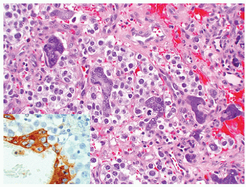
FIGURE 12-20 ▪ Seminoma with syncytiotrophoblasts. Hematoxylin and eosin (H&E) stain reveals multinucleated syncytiotrophoblasts among classic seminoma cells, in the absence of cytotrophoblast. Inset demonstrates immunoreactivity for HCG in syncytiotrophoblastic cells.


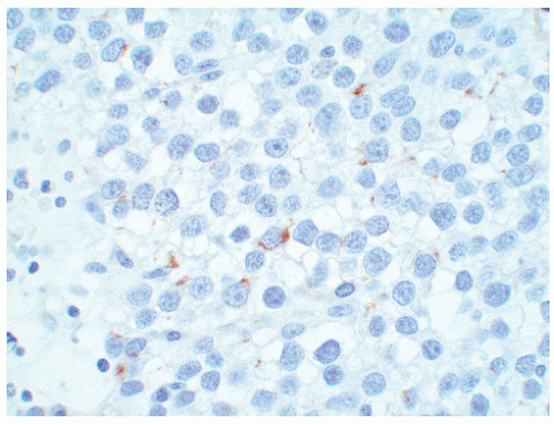
FIGURE 12-21 ▪ Cytokeratin immunoreactivity in classic seminoma. Notice the dot-like cytoplasmic positivity in occasional cells.


Differential Diagnosis

Some seminomas exhibit a significant degree of cytologic atypia103, 104, 105, 106, 107, 108, 109, 110, 111 and this fact led to the now abandoned concept
of “anaplastic seminoma” as a discreet entity with a worse prognosis.31,112 As described, this tumor was characterized by overall morphologic features of a seminoma but containing more pleomorphic cells with nonclear cytoplasm and abundant mitotic figures. Fibrovascular septa and lymphocytes were absent and focal necrosis was commonly seen. This concept did not withstand the test of time since many series later showed that stage for stage there was no difference in clinical outcome between classic and anaplastic seminomas.26,103,113 In addition, it has become quite evident that mitotic activity in seminomas is quite variable and, in fact, may be quite high even in classical cases.105 For the last decade or so, we have used the term “seminoma with atypia” not as a diagnostic entity but rather as a reminder to consider all possible explanations for the atypical morphologic findings (Fig. 12-23).


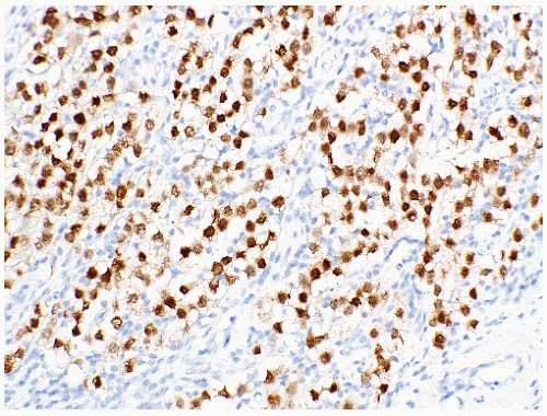
FIGURE 12-22 ▪ OCT4 immunoreactivity in seminoma showing diffuse nuclear staining.


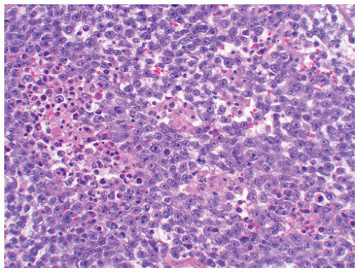
FIGURE 12-23 ▪ Seminoma with atypia. While some tumors may lack a lymphocytic infiltrate and exhibit atypical cytologic features, one must consider other possibilities in the differential diagnosis.

We need to emphasize that the “classic” growth and cytologic features of seminoma are not always present. This fact can bring problems in establishing the correct diagnosis. Poor fixation is a major culprit. In this setting the tumor cells are poorly preserved, the cytoplasm is condensed and eosinophilic, with the nuclei becoming irregular and hyperchromatic. In this setting, isolated necrotic nuclei may be present. In cases of poor tissue fixation, these changes are usually seen in a geographic distribution. The presence of a granulomatous and fibrous reaction mechanically distorts the diffuse growth pattern of the seminoma cells, which is now seen as aggregates or nests of cells with irregular cytoplasmic features. In extreme examples they can mimic neoplastic epithelial and even hematopoietic cells. If the seminoma is associated with an intense lymphocytic infiltrate, the tumor cells may be difficult to identify at low magnification, although the neoplastic component can be readily identified at high magnification, if necessary supplemented by immunohistochemistry. The problem associated with seminomas containing a tubular or interstitial growth pattern has been mentioned and must be kept in mind. Attention to cytologic detail should lead us to the correct diagnosis since in these cases the classic nuclear cytology of seminoma is preserved. A myxoid stromal response is rarely encountered in seminoma, a feature more commonly associated with SS. Once again, nuclear cytology should solve the differential diagnosis, with immunohistochemistry limited to truly difficult cases.

Tumors thought to be seminoma but exhibiting atypical histology should trigger consideration of a differential diagnosis of seminoma, which includes (a) seminoma with “early carcinomatous differentiation,” (b) solid variants of EC or YST, (c) lymphoma, (d) sex cord-gonadal stromal tumor (particularly SCT), (e) SS, (f) monophasic trophoblastic tumor (choriocarcinoma), and (g) metastatic disease, including poorly differentiated carcinoma and melanoma. Some examples are illustrated later in this chapter.

“Early carcinomatous differentiation” refers to areas of transition from seminoma to EC (Fig. 12-24). This concept suggests that seminoma cells are not terminally differentiated but rather, under certain poorly understood circumstances, may differentiate into other GCT types.86,108,114 These tumors are characterized by either focal or diffuse nuclear atypia, nuclear overlapping, and on immunohistologic evaluation may show small clusters with intense membrane-predominant cytokeratin or CD30 (Ber-H2) positivity (Fig. 12-24). We would expect for OCT4 and SALL4 to be retained while C-kit and podoplanin expression would be lost in the EC component. The management of these tumors, whether as seminomas or as mixed GCTs, is controversial and is institution-dependent. Some investigators feel that these tumors should be approached clinically as mixed GCTs since the seminoma has already manifested its propensity to differentiate toward a more aggressive
phenotype. Others would not change management based on this finding alone since, in the absence of other adverse prognostic factors, the patient would likely be placed on surveillance either way. The fact of the matter is that decisions on how to treat these patients are at best empiric because of the rarity of the situation and the absence of clinical studies.


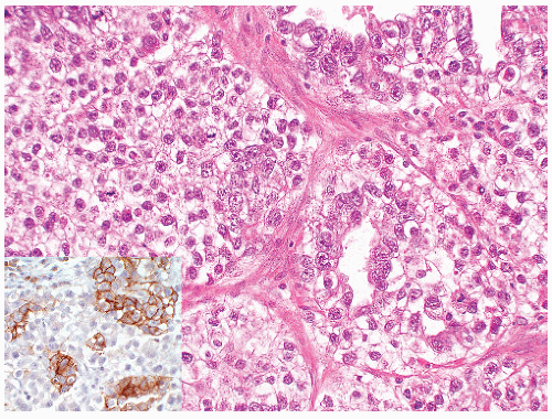
FIGURE 12-24 ▪ Seminoma with early carcinomatous transformation. On the H&E stain you are able to notice subtle changes in cytology of the tumor cells with the EC cells having indistinct cells borders, nuclear overlap, and larger, more irregular nuclei. Inset demonstrates an AE1/AE3 cytokeratin stain in which strong cytoplasmic immunoreactivity is seen in the EC component.

In contrast to a typical seminoma, EC has consistently a higher nuclear grade, irregular chromatin distribution, and syncytial arrangement of the cells (Fig. 12-18). The cytoplasm often is amphophilic to basophilic, the cell borders are poorly defined and glandular, or papillary areas may be present. The solid pattern in YSTs may also mimic seminoma. However, this pattern is usually accompanied by other diagnostic patterns of YST, making the distinction usually easy. YST cells generally have smaller nuclei without prominent nucleoli and may contain hyaline globules. Conversely, edema and microcysts in a seminoma may raise the possibility of a reticular or microcystic pattern of YST. The immunohistochemical profile of each of these tumors is distinct, as seen in Table 12-4. Lymphomas of the testis usually are bilateral tumors, occur in older men, have an interstitial growth pattern, and lack intratubular germ cell neoplasia. The nuclei are generally more convoluted and lack cytoplasmic clearing. Leukocyte common antigen and other lymphoid markers usually are positive, and PLAP is negative.

Jun 10, 2016 | Posted by in UROLOGY | Comments Off on Tumors of the Testis

Full access? Get Clinical Tree

Get Clinical Tree app for offline access