Pathology of the Adrenal Gland


Pathology of the Adrenal Gland


SHAMLAL MANGRAY

RONALD A. DELELLIS


Although considerably less common than diseases of the kidney, urinary bladder, and prostate, disorders of the adrenal glands form a significant proportion of the material submitted for pathologic examination to surgical pathologists. Adrenal diseases may be nonfunctional or may be associated with a complex array of clinical syndromes resulting from abnormalities in the production and secretion of steroid hormones or catecholamines. As with other endocrine diseases, the same clinical syndromes can develop as a consequence of diverse pathophysiologic mechanisms. It is essential, therefore, that the pathologist is familiar with basic pathophysiologic concepts of adrenal disorders and with appropriate ancillary techniques that aid in the differential diagnosis of specific disorders.

The role of the surgical pathologist and cytopathologist has been further complicated by the detection of a variety of mass lesions by ultrasonography, computed tomography (CT), magnetic resonance imaging (MRI), or positron emission tomography (PET) in the workup of patients for reasons other than expected adrenal pathology. The presence of incidental lesions of varying sizes (incidentalomas) has been demonstrated in up to 5% of individuals subjected to abdominal CT studies.1 By far the majority are of cortical origin and a subset of these lesions are subjected to needle core biopsy or fine needle aspiration biopsy, which frequently pose considerable diagnostic challenges.

The purpose of this chapter is to provide an overview of the pathology of the adrenal glands, including the use of immunohistochemical and molecular approaches for the differential diagnosis and prognostic assessment of a wide range of neoplastic and nonneoplastic lesions.


EMBRYOLOGY OF THE ADRENAL GLANDS

The adrenal cortex is first recognizable at 5 to 6 weeks of gestation (9-mm embryo stage) as a proliferation of cells from the peritoneum at the base of the dorsal mesentery close to the cranial aspect of the mesonephros.2 By 8 weeks, the cortical cells separate from the mesothelium and are enveloped by a fibrous capsule. By the second trimester, the cortex includes a broad inner zone composed of large eosinophilic cells referred to as the provisional zone or fetal cortex and an outer zone that is destined to become the adult cortex (Fig. 3-1). Dehydroepiandrosterone sulfate is the major steroid product of the fetal cortex, while cortisol, aldosterone, and sex steroids are the main products of the adult cortex. Relative to total body weight, the weight of the adrenals is maximal at the 4th week of development. At birth, the fetal cortex occupies about 75% of the cortical volume, but shortly thereafter it begins to undergo involution, which is associated with an approximate 50% reduction in glandular weight.3 At birth, the adrenal glands together weigh close to 10 g (Table 3-1). Further involution of the fetal cortex and proliferation of the permanent cortex toward the center of the gland occur simultaneously. As a result the fetal cortex accounts for approximately 20% of the cortical volume by the 12th postgestational week.

The neural crest-derived intra-adrenal (adrenal medulla) and extra-adrenal paraganglia and the sympathetic nervous system are intimately associated during embryonic development. In the 14-mm embryo, the cortical anlage is invaded on its medial aspect by primitive sympathetic cells and nerve fibers that originate from the contiguous prevertebral and paravertebral sympathetic tissue. Some primitive sympathetic cells, however, may penetrate the anlage without associated nerve fibers.4,5 They are first apparent as nodular aggregates in the cortex (Fig. 3-2), where they may form rosettes or pseudorosettes. Mature medullary (chromaffin) cells are identifiable among the primitive sympathetic cells between the 27- and 33-mm stages and gradually increase in number. Nodules of primitive sympathetic cells peak in number and size between 17 and 20 weeks and then decline. Groups of these cells may, however, persist until birth and may also be apparent in early infancy (see “Neuroblastoma”). As the medullary chromaffin cells reach their maximum volume, there is a progressive involution of extra-adrenal chromaffin cells. However, the formation of the adrenal medulla is not completed until the 3rd year of life.


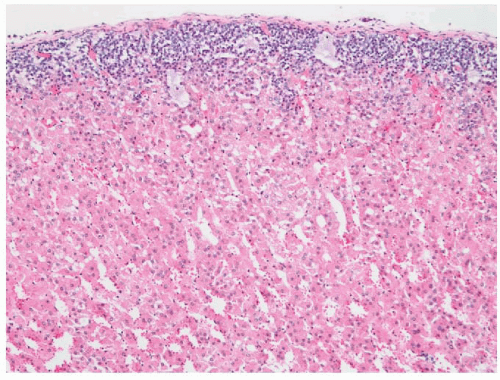
FIGURE 3-1 ▪ Adrenal cortex of a 16-week fetus demonstrating the broad inner zone of eosinophilic cells (provisional zone or fetal cortex) and the thin outer zone that will form the adult cortex.


NORMAL ANATOMY AND PHYSIOLOGY OF THE ADRENAL CORTEX AND MEDULLA

The adrenal glands are present on the superior surfaces of the kidneys. There is considerable variation in reported adrenal weights in different published series (Table 3-1). In adults who have died suddenly, the average adrenal weight is between 4 and 5 g and measures 5 × 3 × 1 cm in the normal adult. However, greater weights occur in hospitalized patients dying after prolonged illnesses, presumably as a result of prolonged stimulation of the glands by adrenocorticotropin during stress.6,7 The right gland has a pyramidal shape, while the left gland has a crescentic shape. Each gland has a tripartite structure consisting of head (medial), body (middle), and tail (lateral) portions.8,9 The central vein emerges from the gland at the junction of the head and body of the gland. Within the gland itself, the muscle bundles of the central vein are eccentric and are oriented toward the medulla.


Table 3-1 ▪ AVERAGE COMBINED WEIGHTS OF ADRENAL GLAND FROM SERIES OF AUTOPSY PATIENTS*


Combined Weight


Age


Mean (grams)


Range (grams)


Fetal (Wk)


30-33


2.3


1.6-3.2


33-38


4.4


3.1-5.8


38-41


6.2


2.0-10.5


Infants and Toddlers (Wk)


0-1


6.3


3.8-8.8


1-3


4.4


3.1-5.8


3-9


2.4


1.8-3.0


9-14


2.0


1.2-2.6


14-32


2.3


1.6-3.2


32-78


2.5


1.6-4.0


Children and Adults (Y)


1.5-4


4.0


0-8.0


4-8


4.1


2.3-5.8


8-14


6.2


3.5-9.0


14-18


8.8


6.0-11.2


18-25


8.0


5.6-9.8


25-35


9.0


6.2-11.4


*Derived from Figure 9-1 in Lack EE. AFIP Atlas of Tumor Pathology (series 4) Tumors of the Adenal Gland and Extraadrenal Paraganglia.


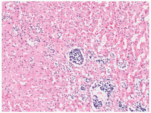
FIGURE 3-2 ▪ Groups of primitive sympathetic cells forming neuroblastic nodules within the fetal cortex in the adrenal gland of a 16-week fetus (same specimen as Fig. 3-1).

In the fresh state, the outer cortex is bright yellow, while the inner cortical zone is brown to tan. The cortex measures approximately 1 mm in thickness in adults and constitutes approximately 90% of the total glandular weight. Cortical extrusions, which are characterized by the presence of nodular groups of cortical cells that extend into the periadrenal fat, are common. They are attached to the adjacent cortex by a small pedicle and are surrounded by a fibrous capsule; however, they may be completely separated from the gland in some instances.

The zones of the cortex include glomerulosa, fasciculata, and reticularis6,10 (Fig. 3-3). The glomerulosa, which is composed of relatively small lipid-poor cells that synthesize mineralocorticoids, accounts for 15% of the cortical volume. The glomerulosa layer is often incomplete so the fasciculata may abut the capsule of the gland directly. The fasciculata, on the other hand, is composed of columns of lipid-rich cells, which synthesize both glucocorticoids and sex steroids and occupy 70% to 80% of the cortical volume. Stimulation of the adrenal by adrenocorticotropic hormone (ACTH) during times of stress leads to depletion of lipid stores from the fasciculata.6 As a result, the cells of this layer become compact and eosinophilic.11 In some patients, the solid cords of the outer cortex are replaced by tubular structures lined by lipid-depleted cells. Proteinaceous material and degenerated cells may also be present within the tubular structures. In some patients dying from infections, chronic renal failure, and overexposure to cold, cells within the zona fasciculata may have intracytoplasmic vacuoles.11 The lipid content becomes replenished (lipid reversion)
during the process of recovery. The remainder of the cortex is composed of the reticularis, which is capable of synthesis of both glucocorticoids and sex steroids. The cells of the reticularis have eosinophilic cytoplasm, scanty lipid vacuoles, and prominent deposits of lipochrome pigment, which are responsible for the brown color of the reticularis. Ultrastructurally, the cells of the glomerulosa contain lamelliform mitochondria while tubulovesicular mitochondria predominate in the fasciculata and reticularis. The intermediate filaments of cortical cells include vimentin and variable amounts of low-molecular-weight cytokeratins.12 Cortical cells are also positive for calretinin, inhibin A, melan-A, and synaptophysin.13, 14, 15, 16


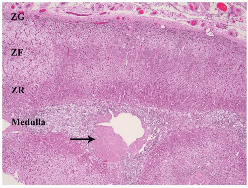
FIGURE 3-3 ▪ Histology of the normal adrenal gland. The three cortical zones (ZG, zona glomerulosa; ZF, zona fasciculata; ZR, zona reticularis), and medulla (MED) are demonstrated. There is slight nodularity of the cortex as occurs in normal adults. Also demonstrated is the eccentric smooth muscle wall of the central vein (arrow).

Focal aggregates of lymphocytes are an incidental finding in the adrenal cortices of normal adults, and increase in frequency with the age of the patient.17 Most of the lymphocytes are of T lineage.18

Cholesterol, which is derived from circulating low-density lipoproteins, is the precursor of all steroid hormones. Following internalization into the cortical cells, the lipoproteins are hydrolyzed with the production of cholesterol esters, which yield cholesterol and free fatty acids.19,20 There are four cytochrome P-450 enzymes that are involved in the biosynthesis of adrenal steroids (P-450scc, P-450c17, P-450c21, P-450c11). The enzyme 3β-hydroxysteroid dehydrogenase does not belong to the P-450 cytochrome family (Fig. 3-4). The synthesis and secretion of glucocorticoids, 18-hydroxysteroids, and androgens are regulated by a complex set of control mechanisms. Hypothalamic corticotropin-releasing hormone
(CRH), a 41-amino-acid polypeptide, reaches the anterior pituitary gland via the hypophyseal portal system, where it stimulates the release of ACTH.20


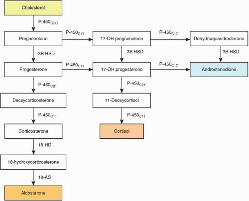
FIGURE 3-4 ▪ Biosynthesis of adrenal steroid hormones. 18-AS, 18-aldehyde synthetase; 3B HSD, 3-β hydroxysteroid dehydrogenase; 18 HD, 18 hydroxylase.


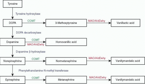
FIGURE 3-5 ▪ Catecholamine biosynthesis and metabolism. COMT, catecholamine-O-methyl transferase; DOPA, 3,4-dihydroxyphenylalanine; MAO/AldDehy, monoamine oxidase and aldehyde dehydrogenase.

In the adrenal cortex, ACTH stimulates cortical cells by activation of intracytoplasmic cyclases that form cyclic adenosine monophosphate and guanosine monophosphate from adenosine triphosphate (ATP) and guanosine triphosphate, respectively. The control of cortisol synthesis is mediated by a complex series of positive and negative feedback loops. Both cortisol and ACTH inhibit the release of CRH, and cortisol also inhibits secretion of ACTH. The secretion of ACTH is normally episodic, with the number and duration of episodes increasing to a peak in the early morning and a nadir in the evening, resulting in the characteristic circadian rhythm of cortisol secretion.20 The secretion of aldosterone is regulated primarily by the renin-angiotensin system.20, 21, 22

The adrenal medulla occupies 8% to 10% of each gland volume in adults and has an average weight of 0.4 g.7 The major portion of the medulla lies within the head of the gland, while the body of the gland contains medullary (chromaffin) cells within its crest and usually within one alar region.8 The average corticomedullary ratio is 5:1 in the head of the gland and 14.7:1 in the body. The tail of the adrenal does not normally contain medullary tissue.

Medullary (chromaffin) cells are arranged in small nests and cords that are separated by a rich capillary network. A few medullary cells, particularly those found in the juxtacortical regions, may have enlarged and hyperchromatic nuclei, which increase in number with age23 in addition to periodic acid-Schiff (PAS) positive hyaline globules.24 At the ultrastructural level, the most characteristic feature of medullary cells is the presence of membrane-bound secretory granules in which catecholamines and other products are stored.25,26 A few ganglion cells are present within the medulla either as single cells or as small-cell clusters. S-100 protein-positive sustentacular cells are present at the peripheries of the medullary cords and nests and are also evident around the ganglion cells. There may also be small collections of lymphocytes and plasma cells within the medulla, but their significance is unknown.

The precursor of the catecholamines is tyrosine, which is converted sequentially to 3, 4-dihydroxyphenylalanine (DOPA), dopamine, norepinephrine, and epinephrine by a series of well-characterized enzymatic steps (Fig. 3-5).27 The major catecholamine product of the medulla is epinephrine, which affects the activities of a wide variety of cells and tissues following its interactions with specific receptors.

The catecholamine content of chromaffin cells has been demonstrated traditionally by a variety of histochemical techniques including the chromaffin reaction.28, 29, 30, 31 In current surgical pathology practice, immunohistochemical stains for chromogranins, chromomembrins, synaptophysin, and regulatory peptides are most commonly utilized for this purpose.32, 33, 34, 35


NONNEOPLASTIC DISORDERS OF THE ADRENAL GLAND


Developmental Disorders

Accessory adrenal tissue (heterotopia) is the most common congenital anomaly of the adrenal gland.36 Most accessory adrenal tissue consists exclusively of cortical tissue, but a
few examples, particularly those in the region of the celiac ganglion, may also contain medullary tissue.37 Based on autopsy studies accessory adrenal tissue is most frequently found in the retroperitoneal space (32%) along the celiac axis, the upper pole of the kidney just beneath the renal capsule (0.1% to 6%), the broad ligament (23%), adnexa of the testes (7.5%), and along the course of the spermatic cord (3.8% to 9.3%).11 Less common sites include the pancreas, spleen, liver, lung placenta, and brain. In surgical pathology series, the most commonly encountered site is in inguinal hernia sacs. One percent of hernia sacs have been reported to contain accessory adrenal tissue.38 Accessory adrenals may undergo hyperplasia in response to increased levels of ACTH and may serve as the site of origin of cortical neoplasms.23,39,40 True heterotopic adrenal glands may be fused with the liver or kidney and are typically surrounded by a common connective tissue capsule.41,42 In such cases, a concern for a metastatic renal cell carcinoma (RCC) or other clear-cell tumors may arise.

Adrenal union (fusion) and adhesion are rare anomalies that are distinguished by the presence (adrenal union) or absence (adhesion) of a connective tissue capsule. Fusion is occasionally associated with midline congenital defects, including spinal dysraphism, indeterminate visceral situs, and the Cornelia de Lange syndrome. Fusion of the adrenal glands can occur in patients with bilateral renal agenesis.43

Aplasia of the adrenal glands has been reported in association with anencephaly, but in most instances, the glands are markedly hypoplastic rather than completely absent.44 In approximately 10% of patients with unilateral renal agenesis, the ipsilateral adrenal gland is also absent. There are four main types of congenital adrenal hypoplasia (OMIM #300200): sporadic with hypopituitarism, autosomal recessive, X-linked cytomegalic type with hypogonadotrophic hypogonadism, and the form associated with glycerol kinase deficiency.44,45 The majority of patients present early in childhood and most typically in infancy; however, occasional individuals with mild forms of these disorders may present as adults. Patients commonly have signs and symptoms of adrenal insufficiency, but other clinical manifestations include hearing loss, coal black pigmentation of the skin, hypogonadism, and precocious puberty.45

In primary hypoplasia, the adult cortex is markedly hypoplastic, but the fetal zone is retained and often has cytomegalic features. This disorder has an X-linked pattern of inheritance and has been associated with mutations or deletions of the DAX-1 gene (Xp21).37,46,47 The miniature adult type of hypoplasia may appear sporadically or as an inherited abnormality with an autosomal recessive pattern of inheritance. Despite their small size, the adrenals have a normal architecture.

The differential diagnosis of primary hypoplasia includes disorders in which adrenal insufficiency precedes other manifestations. For example, if hypoadrenalism occurs prior to neurologic manifestations, patients with adrenoleukodystrophy will present with clinical symptomology simulating that of primary adrenal hypoplasia. Chronic maternal exogenous glucocorticoid administration induces secondary hypoplasia of the adrenal glands in the newborn. In such instances the adrenal glands are small for age, have decreased fetal zone volume, and contain cells with decreased lipid and scattered cytomegalic features.


Adrenal Cytomegaly and Beckwith-Wiedemann Syndrome

Adrenal cytomegaly is characterized by the presence of collections of markedly enlarged (up to 150 µm) eosinophilic cortical cells containing hyperchromatic and pleomorphic nuclei (Fig. 3-6), with occasional nuclear cytoplasmic pseudoinclusions within the fetal cortex. Cytomegaly may be seen in neonates but is more common in premature infants (3% to 7%) and is also relatively common in infants with Rh incompatibility.17 In Beckwith-Wiedemann syndrome, cytomegaly usually affects most of the cells of the fetal cortex and both adrenal glands are typically hyperplastic.11,37,48 Characteristic features of Beckwith-Wiedemann syndrome include macroglossia, prenatal and postnatal overgrowth (gigantism/hemihypertrophy), abdominal wall defects (exomphalos), and pancreatic islet cell hyperplasia leading to hypoglycemia (Table 3-1).

The adrenal glands in Beckwith-Wiedemann syndrome are enlarged and the combined weight of both glands may be as high as 16 g. Because of the cortical hyperplasia the external aspects of the glands appear cerebriform as a result of the presence redundant folds and nodules.11 Microscopic sections demonstrate collections of cells similar to those seen in isolated adrenal cytomegaly, but they are present in greater numbers. The enlarged cells may raise a concern for an infectious etiology, such as cytomegalovirus (CMV) infection, but the cells lack the characteristic eosinophilic nuclear inclusions, perinuclear halos, and granular cytoplasmic inclusions.

Hemorrhagic adrenal cortical macrocysts may also be found.49,50 Patients with Beckwith-Wiedemann syndrome
are predisposed to the development of a variety of malignant tumors including Wilms tumor, adrenal cortical carcinoma, neuroblastoma (NB), hepatoblastoma, and pancreaticoblastoma. However, the hyperplastic cortical cells in this syndrome do not represent a preneoplastic lesion.43,50 Adrenal cortical adenomas and ganglioneuromas have also been reported in affected patients.49 Autosomal dominant inheritance is well established in Beckwith-Wiedemann syndrome, but approximately 85% of cases are actually sporadic. The molecular basis of this syndrome is complex and involves downregulation of imprinted genes within the chromosome 11p15 region (Table 3-2). Other syndromes that predispose to adrenal tumors are discussed in subsequent sections.


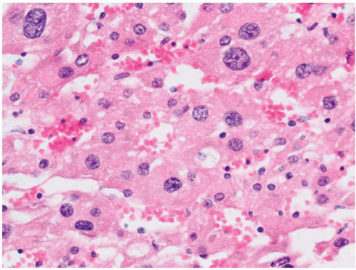
FIGURE 3-6 ▪ Focal cytomegaly of cells of the fetal cortex from a newborn.


Table 3-2 ▪ SYNDROMES ASSOCIATED WITH ADRENALTUMORS


Syndrome


Gene


Chromosome Locus


Adrenal Tumor


Extra-Adrenal Features


Beckwith-Wiedemann (OMIM: 130650)


CDKN1/NSD1, KCNQ1, KCNQ1OT1 (domain 2); IGF2 and H19 (domain 1)


11p15.5


Cortical carcinoma, cortical adenoma, neuroblastoma, ganglioneuroma


Exomphalos, macroglossia, pancreatic islet cell hyperplasia, gigantism/ hemihypertrophy, Wilms tumor, hepatoblastoma, pancreaticoblastoma


Li-Fraumeni syndrome (OMIM:151623)


TP53


17p13.1


Cortical carcinoma


Other neoplasms/cancers


Carney complex (OMIM:160980)


PRKAR1A


17q23-q24


PPNAD


Lentigines and other pigmented skin lesions, myxomas, LCCST of testis, pituitary adenoma.


MEN 1 (Wermer syndrome) (OMIM:131100)


MEN1


11q13


Cortical adenoma


Endocrine lesions of parathyroid, pituitary, pancreas, GI tract lesions, skin lesions


MEN 2A (Sipple syndrome) (OMIM:171400)


RET (exon 10, 11)


10q11.2


Pheochromocytoma


MTC, primary hyperparathyroidism (hyperplasia)


MEN 2B (OMIM:162300)


RET (exon 16)


10q11.2


Pheochromocytoma


MTC, mucosal neuromas, ganglioneuromatosis of intestine, marfanoid habitus, corneal nerve lesions


von Hippel-Lindau disease (OMIM:193300)


VHL


3p26-p35


Pheochromocytoma


Retinal and cranial hemangioblastoma, RCC, cysts of multiple, organs, pancreatic endocrine tumors, ELST of ear


Familial pheochromocytoma-paraganglioma (OMIM:115310)


SDHD, SDHC, SDHB


1p36.1-p35


Pheochromocytoma


Paraganglioma of abdomen, thorax, head, and neck


Neurofibromatosis type 1 (OMIM:162200)


NF1


17q11.2


Pheochromocytoma


Café au lait macules, neurofibromas or plexiform neurofibroma, optic glioma, axillary or inguinal freckling, Lisch nodules, osseous lesions


ELST, endolymphatic sac tumor; GI, gastrointestinal; LCCST, large cell calcifying Sertoli cell tumor; MEN, multiple endocrine neoplasia; MTC, medullary thyroid carcinoma; PPNAD, primary pigmented nodular adrenocortical disease.


Metabolic Disorders


Storage Diseases

Adrenoleukodystrophy (Addison-Schilder disease; OMIM 300100) is a rare, X-linked recessive disorder characterized by progressive demyelination of the central and peripheral nervous system and by adrenal cortical insufficiency.51,52 In the classic form, the disorder is characterized by early behavioral manifestations including inattention, hyperactivity, and emotional lability becoming apparent through school difficulties. Subsequently there are visual symptoms, auditory processing difficulties, and motor incoordination. Once the neurologic manifestations appear, progression of the illness
is tragically rapid and the child is often in a vegetative state within 1 to 2 years. According to Moser et al.53 there are seven phenotypes that include the childhood cerebral form, adrenomyeloneuropathy, adult cerebral, adolescent, adrenal insufficiency without neurologic disease, asymptomatic, and heterozygotes. Clinical manifestations can, therefore, vary widely even within the same family.

The disorder is caused by mutations of the gene on chromosome Xq28 encoding an ATP-binding transporter ALDP—adrenoleukodystrophy protein that is localized in the peroxisomal membrane. The mutations result in the defective oxidation of very long fatty acids54 and the diagnosis can be established by the presence of hexacosanoate and other long-chain fatty acids in cultured skin fibroblasts.55

The adrenal glands in this disorder are grossly atrophic with weights ranging from 1 to 2 g or less, with ballooning and striation of the cells of the inner zona fasciculata and zona reticularis. Groups of ballooned cells form nodules that may undergo degenerative changes with the formation of large cortical vacuoles. The medulla is usually normal. Ultrastructurally, there is proliferation of smooth endoplasmic reticulum and the presence of lamellar inclusions with a trilaminar structure.51 An adult variant with an onset in the second or third decades is known as adrenomyeloneuropathy, a condition that can be associated with unexplained adrenal insufficiency in the absence of neurologic manifestations at first clinical presentation.56 Cortical cells typically appear ballooned and contain linear lamellar inclusions.55,56 A third form of the disease has been reported in women who are carriers of the abnormal gene.

The presence of primary adrenal insufficiency and atrophic adrenal glands may raise the possibility of autoimmune adrenalitis. However, in autoimmune adrenalitis the adrenal glands have a chronic inflammatory infiltrate (see section on “Primary Hypofunction”) and the degenerative changes in the cortical cells described above are lacking. Additionally, correlation with the clinical presentation and autoimmune serology will assist in making the distinction.

Wolman disease (primary familial xanthomatosis) is a rare lipid storage disorder caused by an autosomal recessive deficiency of lysosomal acid lipase.57 The disease is characterized by the accumulation of triglycerides and cholesterol esters in a variety of tissues including the liver, spleen, and adrenal glands. Most of the affected individuals die by the age of 6 months. Typically, the adrenal glands are markedly enlarged even though they often retain their normal configurations. The glands often demonstrate multiple foci of calcification in association with necrosis and fibrosis, and vacuolated zona reticularis cells.57 Other storage diseases (e.g., Niemann-Pick disease) may also result in adrenal enlargement and hypofunction.


Congenital Adrenal Hyperplasia (Congenital Adrenogenital Syndrome)

Congenital adrenal hyperplasia encompasses a spectrum of abnormalities that result from autosomal recessive enzymatic defects in adrenal steroid biosynthesis.58

This group of disorders is caused by deficient activity of one of the following enzymes: 21-hydroxylase (P-450c21), 11β-hydroxylase (P-450c11), 3β-hydroxysteroid dehydrogenase, 17-hydroxylase/17,20-lyase (P-450c17), and 20,22 desmolase (P-450scc). Approximately 95% of cases of congenital adrenal hyperplasia are due to 21-hydroxylase deficiency (OMIM +201910) in which there are four clinically recognized forms: salt wasting (approximately 65%), simple virilizing (approximately 30%), nonclassic (also referred to as attenuated or acquired), and cryptic.59 The worldwide incidence of classic 21-hydroxylase deficiency is 1 in 14,500 births, with a heterozygote frequency of approximately 1 in 60. Affected children have evidence of glucocorticoid deficiency, aldosterone deficiency with salt wasting, and excess adrenal androgen production resulting in virilization. Excess adrenal androgen production is a consequence of the accumulation of 17-hydroxypregnenolone, which is subsequently metabolized to androgenic steroids. Nonclassic (cryptic) 21-hydroxylase deficiency has a frequency of 1 in 100 in certain parts of the United States and is one of the most common autosomal recessive disorders. Affected individuals have mild degrees of cortisol deficiency, normal aldosterone production, and excess production of adrenal androgens. This form of 21-hydroxylase deficiency is most often diagnosed in childhood or early adulthood. A small proportion of individuals with 21-hydroxylase deficiency may have no apparent symptoms.

Masculinization of females in utero usually occurs while males generally appear normal at birth. Postnatally, untreated males as well as females may manifest rapid growth, penile or clitoral enlargement, precocious adrenarche, and early epiphyseal closure with resultant short stature. A mild form of late-onset adrenal hyperplasia due to 21-hydroxylase deficiency can occur in adults and is characterized by hirsutism as the only manifestation in the most attenuated form. The detailed biochemical, genetic, and molecular features of these disorders are discussed elsewhere.58, 59, 60, 61, 62 Prenatal screening of amniotic fluid and neonatal screening by blood spot for elevated upstream hormones is available. Confirmation by testing for mutation of the underling CYP21 gene is then performed.59

Deficiency of 11β-hydroxylase (P-450c11; OMIM #202010) accounts for approximately 5% of all cases of congenital adrenal hyperplasia and is associated with increased production of androgens and deoxycorticosterone.60, 61, 62 As a result, affected patients typically exhibit signs of hyperandrogenism and hypertension. Deficiency of 17α-hydroxylase (OMIM #202110) is responsible for approximately 1% of cases. External genitalia are female in both sexes. Increased levels of deoxycorticosterone are responsible for the hypertension seen in these patients. In 3β-hydroxysteroid dehydrogenase deficiency (OMIM #201810), there is deficiency of all steroid hormones including androstenedione. As a result virilization is less developed, but salt loss is a prominent feature. The adrenal glands are similar to those of the normal fetus.63


Adrenal hyperplasia in the congenital adrenogenital syndromes result from inadequate production of glucocorticoids, leading to stimulation of the cortex by increased pituitary ACTH production. The adrenal glands become markedly enlarged (up to 15 g) with a characteristic cerebriform appearance and a tan-brown color due to lipid depletion of cortical cells.43 This contrasts with ACTH pituitary-dependent adrenocortical hyperplasia in which there is an outer yellow and inner tan-brown appearance as a result of hyperplasia of the fasciculata (see Hyperfunctional States). In paraneoplastic ACTH-dependent adrenocortical hyperplasia, the adrenal glands are usually larger than those seen in congenital adrenal hyperplasia, but they typically appear tan-brown as well because of lipid depletion. In patients with 20,22-desmolase (P-450scc) deficiency, also referred to congenital lipoid hyperplasia, the adrenal glands are pale yellow or white and are characterized on microscopic examination by vacuolated cells, occasionally with formation of cholesterol clefts and an accompanying giant-cell reaction. These disorders are usually treated by replacement of the deficient steroid hormone and surgical correction of ambiguous genitalia or hypospadias.

Rarely, adrenal cortical adenomas and carcinomas may develop, in the setting of congenital adrenal hyperplasia.64,65 Testicular tumors can also arise in affected patients. The testicular lesions are not autonomous neoplasms, since they are dependent on the presence of elevated levels of ACTH for their maintenance.66 They are commonly bilateral and are most typically located in the hilar regions of the testes. The cell of origin is unknown, but the component cells of these lesions contain abundant amounts of eosinophilic cytoplasm, which lack crystalloids of Reinke. In the series reported by Rutgers et al.,66 the diagnosis of congenital adrenal hyperplasia was made only after the appearance of testicular tumors in 18% of the cases.


Hypofunctional States


Primary Hypofunction


Autoimmune Adrenalitis

Autoimmune adrenalitis is responsible for approximately 75% of cases of the inflammatory conditions affecting the adrenal glands in North America and Western Europe, followed by infection and a variety of other conditions (Box 3-1). In idiopathic (autoimmune) Addison disease (chronic adrenocortical insufficiency), the glands are markedly atrophic and the residual cortical tissue is infiltrated by lymphocytes and plasma cells. The presence of a few aggregates of lymphocytes in the adrenal cortex should not be considered evidence of adrenalitis. All layers of the cortex are involved in autoimmune adrenalitis, but the medulla is unaffected. Typically, the capsules of the glands are fibrotic. Both humoral and cell-mediated immune mechanisms have been implicated in the development of autoimmune adrenal hypofunction. Autoantibodies to cortical cells are present in 50% of all patients and in more than 70% of women with newly diagnosed disease. The major targets for autoantibody reactivity are the adrenal cytochrome P-450 enzymes.67 Affected patients can also have antibodies to gonadal, gastric parietal, and thyroid follicular cells.


Adrenocortical hypofunction may be associated with hypofunction of other endocrine glands.68 The type I polyglandular autoimmune syndrome is associated with mucocutaneous candidiasis, alopecia, hypoparathyroidism, adrenal insufficiency, autoimmune thyroiditis, and diabetes mellitus. This form of the disease has also been termed autoimmune polyendocrinopathy-candidiasis-ectodermal dystrophy (APECED). Mutations in the APECED or autoimmune regulatory gene (21q22.3) have been implicated in the development of this syndrome.69,70 The type II polyglandular autoimmune syndrome is characterized by adrenal insufficiency, autoimmune thyroiditis, and insulin-dependent diabetes mellitus. The type I syndrome is inherited as an autosomal recessive trait, while the pattern of inheritance of the type II syndrome is usually dominant.


Infectious Disorders

Infectious disorders including tuberculosis and fungal diseases (histoplasmosis, North and South American blastomycosis, coccidiomycosis, cryptococcosis) can affect both the cortical and medullary regions of the adrenal glands. Although tuberculosis is now a rare cause of adrenal insufficiency in the United States and Western Europe, it is
a common cause in parts of the world where tuberculosis is endemic. In contrast to the shrunken appearance of the adrenal glands in idiopathic Addison disease, the glands in mycobacterial infection are typically enlarged and necrotic. Infection with Mycobacterium avium intracellulare is typically associated with the presence of confluent masses of histiocytes containing acid-fast organisms.

CMV has been identified in the adrenal glands of a large proportion of patients dying of the acquired immunodeficiency syndrome.71 Adrenal cortical necrosis associated with CMV infection can be severe enough to result in acute adrenal insufficiency in some instances. Both herpes simplex and varicella zoster may also involve the adrenal glands and may lead to adrenal cortical insufficiency when they are associated with extensive cortical necrosis.


Amyloidosis

Rarely, amyloid deposition can result in cortical hypofunction.72 Typically, adrenal involvement is associated with extensive systemic amyloid disease of the AA type. The adrenal glands may have a normal shape and size or may be enlarged. In severe cases, the glands are pale tan to yellow. Amyloid deposits typically affect the fasciculata and reticularis zones and are present between the cortical cells and capillary endothelium. The cortical cells ultimately become atrophic as a result of the progressive deposition of intercellular amyloid. In patients with AL disease, the amyloid deposits are usually vascular in distribution.


Adrenal Hemorrhage

Adrenal hemorrhage may develop in a segmental fashion or may involve the entire adrenal (Fig. 3-7).73 This syndrome may be seen in association with sepsis and shock due to meningococcal infection or infection with other bacteria, including Haemophilus influenzae, Streptococcus pneumoniae, and Pseudomonas aeruginosa (Waterhouse-Friderichsen syndrome). Typically, the glands are enlarged and hemorrhagic, with necrosis of both cortical and medullary tissue. Adrenal hemorrhage in the Waterhouse-Friderichsen syndrome is regarded as the consequence rather than the cause of shock. Anticoagulant therapy has also been associated with adrenal hemorrhage. Corticomedullary necrosis of milder degrees can occur in association with hypotension and shock.74 In patients with segmental lesions, examination of the capsular vessels and sinusoids often reveals evidence of thrombus formation. Affected cortical areas show a pattern of ischemic necrosis that ultimately heals by the process of fibrosis.


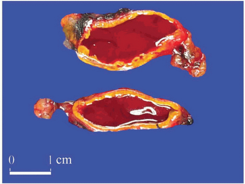
FIGURE 3-7 ▪ Adrenal gland hemorrhage in a patient with cholangiocarcinoma. There was no evidence of metastatic disease.


Miscellaneous Causes of Hypofunction

Adrenal hypofunction may result from bilateral involvement by tumor, most commonly metastatic carcinoma. Typically, clinically apparent hypofunction occurs when more than 95% of the glands are replaced by tumor. A rare case of hypofunction has been reported secondary to involvement by Erdheim-Chester disease, a non-Langerhans histiocytosis that typically involves bone, but in which extraskeletal manifestations are present in up to 50% of cases. In this condition, there is diffuse enlargement of the adrenal gland secondary to infiltration by foamy histiocytes.75

In patients with acquired immune deficiency syndrome (AIDS), involvement of the adrenals by opportunistic infections or neoplasms such as Kaposi sarcoma can lead to significant glucocorticoid insufficiency. However, most AIDS patients also have decreased adrenal reserves characterized by a defect in the production of 17-deoxycorticosteroid by the zona fasciculata. This is associated with morphologic changes of lipid depletion as described earlier in stressrelated changes of the adrenal gland. Also, peripheral resistance to glucocorticoids related to decreased affinity for type II glucocorticoid receptors has been reported in a subset of patients with AIDS.17

Drugs such as 12-methylbenzathracene and hexadimethane can cause direct necrosis of the cortex. Ketoconazole, etomidate, cyanoketone, and trilostane have inhibitory effects on adrenal steroidogenesis while rifampicin and dilantin can lead to increased breakdown of glucocorticoids.17

Patients with increased iron stores can have excess deposition of iron in the adrenal cortex, particularly the zona glomerulosa. High-dose radiation to the abdomen, pelvis, or lumbar regions for treatment of malignancies can lead to fibrosis of the adrenal glands. While the adrenal cortex is relatively resistant to radiation compared to other endocrine organs, fibrosis of the inner cortex, particularly the zone reticularis along with reduction in the zona fasciculata, can occur.17


Secondary Hypofunction

Adrenocortical atrophy may be found in association with lesions primarily affecting the pituitary or hypothalamus, leading to diminished secretion of ACTH.6 The administration of exogenous corticosteroids will produce similar changes as a result of suppression of endogenous ACTH. The adrenal glands in secondary hypofunctional states are considerably smaller than normal, although the overall
configurations of the glands are retained. Typically, the cortex is bright yellow owing to lipid accumulation in the cortical cells, the capsule is fibrotic, and the medulla is unaffected. The zona glomerulosa is usually of normal thickness in these cases.


Hyperfunctional States


Adrenocortical Hyperplasia

Hyperplasia of the adrenal cortex, which represents an increased cortical mass resulting from stimulation of the cortex by ACTH derived from the pituitary gland or from a variety of extrapituitary sources, can be associated with a wide variety of clinical syndromes. Cortical hyperplasia can also selectively involve the zona glomerulosa in patients with idiopathic hyperaldosteronism.


Pituitary/Hypothalamic-based Hyperplasia (Cushing Disease)

Hyperplasia may be the result of stimulation of the adrenal glands by ACTH-producing pituitary adenomas or of hypothalamic stimulation of the pituitary ACTH cells by CRH.76,77 Basophilic pituitary adenomas were originally described in association with hypercortisolism by Harvey Cushing in 1932, and this association has been termed Cushing disease or ACTH-dependent Cushing syndrome. Immunohistochemical studies have shown that ACTHproducing pituitary adenomas are considerably more common than had been recognized on light microscopy alone, and many of them have been classified as microadenomas measuring considerably <1 cm.

Adrenocortical hyperplasia in patients with ACTHdependent Cushing syndrome can be either diffuse or nodular and combinations of diffuse and nodular hyperplasia are common. In diffuse hyperplasia, gland weights may be increased minimally.6 In more advanced cases, the combined average weight is considerably in excess of normal with combined weights of >25 g. The glands have rounded contours rather than the sharp outlines typical of normal glands. The inner portion of the cortex is widened and often appears pale brown or tan. The outer layers of the cortex are typically yellow. On microscopy, the inner brown zone corresponds to lipid-depleted cells of the fasciculata, whereas the cells of the outer cortex are more characteristically vacuolated.78 The glomerulosa in adults with Cushing disease is often difficult to identify, but in children the glomerulosa may also appear slightly hyperplastic.79

In some cases, the cortex may appear nodular with individual nodules measuring <0.5 or 1.0 cm in diameter, depending on varying criteria used by different authors.43 This type of change is classified as “diffuse and micronodular hyperplasia.” If the nodules exceed 1 cm in diameter, the hyperplasia is defined as “diffuse and macronodular type.”

In diffuse and nodular (micro- or macro-)hyperplasia, multiple cortical nodules are present in association with a diffusely hyperplastic cortex (Figs. 3-8 and 3-9). Formation of nodules is often asymmetric. While one adrenal gland may show diffuse and nodular cortical hyperplasia, the contralateral adrenal gland may appear diffusely hyperplastic. Most often, the nodules are composed of admixtures of clear- and compact-type cells. In contrast to the atrophic cortex adjacent to a functioning adenoma, the cortex between or adjacent to the nodules in nodular hyperplasia is diffusely hyperplastic.


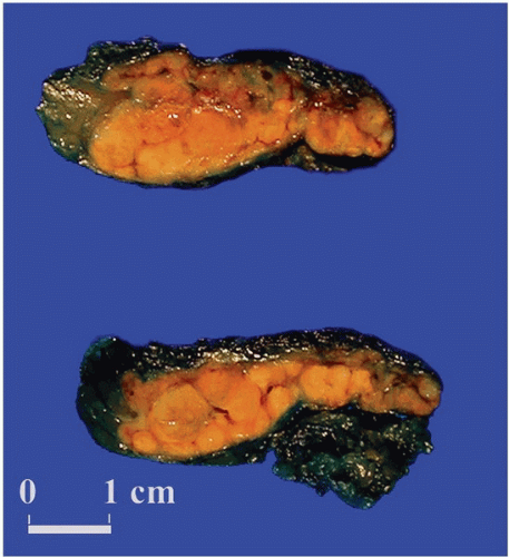
FIGURE 3-8 ▪ Diffuse and nodular adrenocortical hyperplasia. Adrenal gland from a patient with pituitary-dependent Cushing syndrome (Courtesy Dr. A. Mc Nicol, Glasgow, Scotland, UK.).


Adrenocortical Hyperplasia Associated with Paraneoplastic (Ectopic) Production of Adrenocorticotropic Hormone or Corticotropinreleasing Hormone

Hyperplasia can be found in association with a variety of neoplasms producing ACTH, CRH, or both ACTH and CRH.77,80,81 In most series, bronchial carcinoids and smallcell carcinomas are responsible for the paraneoplastic production of these hormones. Other tumors associated with the paraneoplastic ACTH syndrome include pancreatic endocrine neoplasms, medullary thyroid carcinoma, thymic carcinoids, and pheochromocytomas. In patients with the paraneoplastic ACTH syndrome, the adrenals are usually larger (average combined weight of 20 to 30 g) than those seen in association with hyperplasia stemming from pituitary ACTH overproduction. The cortex is diffusely hyperplastic and appears tan-brown throughout its width. On microscopy, there is evidence of diffuse hyperplasia of the fasciculata cells, which are characterized by
a compact or lipid-depleted appearance.82 Foci of nuclear enlargement and hyperchromasia of reticularis cells may be noted, and these features may be particularly striking adjacent to metastatic foci in the glands.23 Both bronchial carcinoids and small-cell bronchogenic carcinomas may also produce CRH.


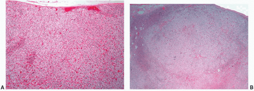
FIGURE 3-9 ▪ Diffuse and nodular adrenocortical hyperplasia. Histologic section of diffuse (A) and nodular hyperplasia (B) from a patient with Cushing syndrome. There is predominantly hyperplasia of the vacuolated fasciculata.


Macronodular Hyperplasia (Massive Macronodular Adrenocortical Disease)

In macronodular hyperplasia with marked adrenal enlargement, the adrenal glands may together weigh up to 180 g, and individual nodules may measure up to 4.0 cm in diameter.37,83,84 Nodules are composed of clear cells, compact cells, or admixtures of these cell types. Macronodular hyperplasia is ACTH-independent, and this entity can involve a single gland in some instances.85 The cortex between the nodules is often atrophic, as might be expected in an ACTH-independent process. This entity, which has also been referred to as massive macronodular adrenocortical disease, has a bimodal age distribution. A small proportion of patients may present during the 1st year of life with association with the McCune-Albright syndrome. Most of the patients present clinically in the fifth decade with a male to female ratio of 1:1. Rare examples of familial massive macronodular adrenocortical disease have also been reported. This disorder has been associated with aberrant (ectopic) expression and regulation of various G-protein-coupled receptors.86 These lesions are treated by bilateral adrenalectomy.17


Primary Pigmented Nodular Adrenocortical Disease—Microadenomatous Hyperplasia of the Adrenal Gland

Primary pigmented nodular adrenocortical disease (PPNAD) is a rare disorder, which is seen in association with ACTHindependent Cushing syndrome and treated by bilateral adrenalectomy. Morphologically, PPNAD is characterized by the presence of multiple pigmented nodules of cortical cells with intervening atrophic cortical tissue.87, 88, 89, 90 The glands may be either smaller than normal or enlarged (range 0.9 to 13.4 g).17 Individual nodules, which can vary in color from gray to black, typically measure from 1 to 3 mm in diameter, although larger nodules measuring up to 3 cm in diameter may also be evident.43 The nodules are composed of large, granular eosinophilic cells that often contain hyperchromatic nuclei with prominent nucleoli (Fig. 3-10). Because of the atypical nuclear features, this entity had been referred to previously as micronodular dysplasia. The black color is due to the presence of lipochrome pigment. The presence of pigmented nodules might raise the possibility of metastatic melanoma, but this will be readily excluded on careful examination and review of the clinical data.

Primary pigmented adrenal cortical disease may occur sporadically or in a familial form that can be associated with
Carney complex (CNC), which includes cardiac myxomas, spotty pigmentation, neurofibromatosis, testicular Leydig or Sertoli cell tumors, mammary myxoid fibroadenomas, and cerebral hemangiomas.89,91 The Carney complex may occur sporadically or may be inherited as an autosomal dominant trait. The gene encoding the protein kinase A type Iα regulatory subunit PRKAR1A has been mapped to 17q22-q24 and loss of heterozygosity (LOH) studies from patients with CNC have revealed mutations in this gene in approximately 50% of affected individuals. No mutations have been found on 2p16. Studies of sporadic and isolated cases of (CNC) have also revealed inactivating mutations of PRKAR1A. The wild-type alleles could be inactivated by somatic mutations consistent with the hypothesis that the gene belongs to the tumor suppressor class.92,93 Some studies have suggested that PPNAD may have an autoimmune etiology.94


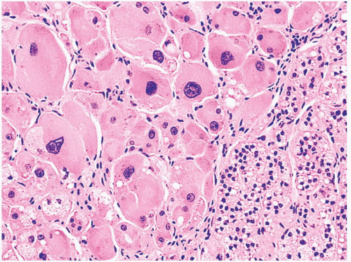
FIGURE 3-10 ▪ Primary pigmented nodular adrenocortical disease (PPNAD). The nodule is composed of large cells with hyperchromatic nuclei. In this photomicrograph pigment is absent.

Howath et al. reported mutations in the gene encoding phosphodiesterase 11A4 (PDE11A4) in cases of PPNAD and other forms of micronodular adrenocortical hyperplasia. LOH and other analyses showed susceptible genes at the 2q31-2q25 locus.95 PDEs regulate cyclic nucleotide levels. The same group subsequently reported that missense mutations of PDE11A were frequently present in patients from the general population with adrenal cortical hyperplasia and adenoma, resulting in speculation that PDE11A genetic defects may be associated with adrenal pathology in a wider clinical spectrum.96


Adrenocortical Hyperplasia Associated with Hyperaldosteronism

Primary hyperaldosteronism is characterized by the excessive secretion of aldosterone from the adrenal glands and is associated with suppression of plasma renin activity with resultant hypokalemia and hypertension. At least six subtypes of primary hyperaldosteronism have been recognized, including aldosterone-producing adenoma, idiopathic hyperaldosteronism, primary adrenal hyperplasia, aldosterone-producing adrenal cortical carcinoma, aldosterone-producing ovarian tumor, and familial hyperaldosteronism (FH).97 FH is subdivided into two groups: FH-I (glucocorticoid-remediable hyperaldosteronism) and FH-II (aldosterone-producing adenoma and idiopathic hyperaldosteronism).


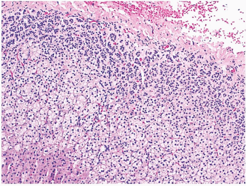
FIGURE 3-11 ▪ Hyperplasia of the zona glomerulosa in a patient with primary hyperaldosteronism.

In approximately 40% of cases of primary hyperaldosteronism, the only apparent adrenal abnormality is hyperplasia of the zona glomerulosa with or without the formation of micronodules.9,43,98,99 Generally, biochemical abnormalities in patients with hyperplasia are less severe than in those with adenomas. On histologic examination, hyperplasia of the glomerulosa is characterized by thickening of this cell layer, with extensions of the glomerulosa extending toward the fasciculata (Fig. 3-11). Micronodules, when present, are usually composed of clear fasciculata-type cells9,23 and are thought to be a consequence of the associated hypertension. In about 10% of cases, it may not be possible to distinguish a micronodule from a true adenoma associated with aldosterone production.


ADRENAL NEOPLASMS

A host of syndromes predispose to adrenocortical or adrenomedullary tumors. In addition to the previously discussed (CNC) and Beckwith-Wiedemann syndrome, other disorders associated with adrenal tumors include Li-Fraumeni syndrome, multiple endocrine neoplasia (MEN) types 1, 2A, and 2B, familial pheochromocytoma-paraganglioma, neurofibromatosis type 1, and von Hippel-Lindau disease (Table 3-2). Unlike Beckwith-Wiedemann syndrome, which predisposes to both cortical and medullary tumors, the other syndromes give rise to either cortical or medullary tumors. While Beckwith-Wiedemann syndrome presents early in infancy and childhood, the manifestations of the other syndromes usually occur later in life; however, features of MEN2B, which include oral, ocular, and gastrointestinal ganglioneuromatosis associated with a Marfanoid habitus, may present at birth.

Adrenal medullary hyperplasia, although viewed as a nonneoplastic lesion, is seen in patients with inherited syndromes that predispose to the development of pheochromocytoma. This change has been reported in association with MEN2A and 2B and von Hippel-Lindau disease, but rare examples have been reported without an apparent associated familial syndrome. This topic is further discussed in “Pheochromocytoma.”


ADRENOCORTICAL NEOPLASMS


Adrenocortical Adenomas

Adrenocortical adenomas represent a heterogeneous group of benign neoplasms that can differentiate toward any of the cortical layers.43,100 Most adenomas are nonfunctional. In order of decreasing frequency, functional adenomas can be associated with the production of mineralocorticoids (Conn syndrome), glucocorticoids (Cushing syndrome), or sex steroids (adrenogenital syndrome). Mixed syndromes can also occur.


Nonfunctional Adrenocortical Adenomas and Cortical Nodules


Epidemiology and Etiopathogenesis

The classification of nonfunctional cortical nodes is controversial. While some studies refer to them as cortical adenomas, others classify them as hyperplastic nodules. In this chapter, the terms “nonfunctional adenoma” and “hyperplastic nodule” will be used interchangeably.

Adrenocortical nodules occur commonly in patients without clinical or biochemical evidence of steroid hormone hypersecretion,43 and they are detected frequently by abdominal imaging techniques. A significant proportion of these lesions occur as “incidentalomas.” Autopsy studies have revealed cortical nodules in approximately 25% of individuals without evidence of biochemical abnormalities. They occur frequently in the adrenals of elderly individuals and in patients with essential hypertension or diabetes mellitus. Most nodules represent foci of compensatory cortical hyperplasia that have developed in response to focal atrophy of the cortex induced by narrowing of adrenal capsular arterioles.101 Nonfunctional cortical nodules may be particularly prominent in patients with aldosterone-secreting adenomas, presumably as a result of the associated hypertension.


Gross Pathology

Although cortical nodules are commonly multicentric and bilateral, dominant nodules measuring up to 2 to 3 cm in diameter or larger may be present. The smaller nodules are often nonencapsulated, whereas the larger single nodules may be surrounded by a fibrous pseudocapsule. The nodules are generally bright yellow with foci of brownish discoloration (Fig. 3-12A). Those arising in the zona reticularis are more homogeneously brown or black.


Microscopic Features

Adenomas have pushing borders with a pseudocapsule derived from compression of the adjacent cortex or expansion of the adrenal capsule.11 They can be composed of small nests,
cords, or alveolar arrangements of vacuolated (clear) cells that most closely resemble those of the normal fasciculata (Fig. 3-12B). Variable numbers of compact-type cells are also present (Fig. 3-12C). Black adenomas are composed exclusively of lipochrome-rich compact cells.6 The nuclear/cytoplasmic ratio is generally low, although a few single cells and small groups of cells may have enlarged and hyperchromatic nuclei. Typically, the nuclei are vesicular with small, distinct nucleoli and little or no mitotic activity. In fine-needle aspiration biopsy specimens, the cells are round to polyhedral with round nuclei and foamy cytoplasm. Numerous naked nuclei in a background of granular to foamy material may be prominent.


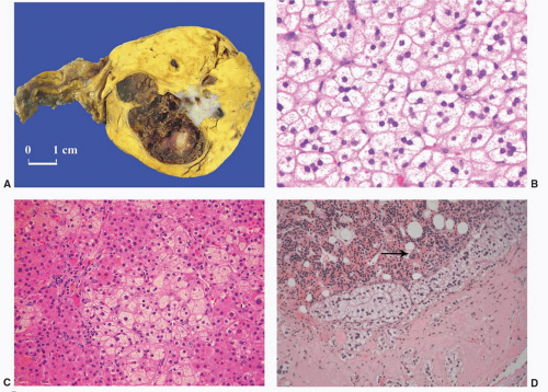
FIGURE 3-12 ▪ Nonfunctional adrenocortical adenoma. The cut surface is bright yellow with central hemorrhage and fibrosis (A) (Courtesy of Dr. A. Matoso, Providence, RI). The tumor is composed predominantly of clear cells that resemble normal cells of the fasciculata (B), but compact cells are also present (C). Myelolipomatous change was present in a section from the central hemorrhagic area (D, arrow megakaryocyte).

Foci of myelolipomatous change, calcification, or ossification may be evident within the nodules, particularly the larger ones (Fig. 3-12D). In contrast to functional adenomas associated with glucocorticoid production, the cortex adjacent to nonfunctional nodules is of normal thickness.


Adenomas Producing Cushing Syndrome


Epidemiology and Etiopathogenesis

Functional adenomas associated with Cushing syndrome occur more commonly in females than in males and are typically unilateral and unicentric. X-chromosome inactivation analyses have shown that some adenomas are clonal while others are polyclonal. Monoclonal adenomas are larger than polyclonal lesions and have a higher prevalence of nuclear pleomorphism.102 This heterogeneity may reflect different pathogenetic mechanisms or different stages of a common multistep process.


Gross Pathology

The tumors are typically unilateral and present as sharply circumscribed masses that usually weigh <50 g and measure 3 to 3.5 cm in average diameter. Larger tumors should be examined with particular care to rule out malignancy.9,23,37 On cross section, adenomas vary from yellow to brown, and occasional examples of heavily pigmented (black) adenomas associated with Cushing syndrome have been reported (Fig. 3-13A). Necrosis is rare, but cystic change is relatively common, particularly in larger tumors.


Microscopic Features

The microscopic features are similar to those of nonfunctional adenomas.11 Adenomas are most often composed of small nests, cords, or alveolar arrangements of vacuolated (clear) cells that resemble those of the normal fasciculata. Generally, adenoma cells are somewhat larger than normal cortical cells. Variable numbers of lipochrome-rich compact-type cells are also evident6 and predominate in cases of black adenomas (Fig. 3-13B). Foci of spindle-cell growth may be present in some cases, and some adenomas may exhibit considerable fibrosis or myxoid change. The nuclear/ cytoplasmic ratio is generally low, although a few single cells and small groups of cells may have enlarged hyperchromatic nuclei. Typically, the nuclei are vesicular with small, distinct nucleoli. Mitotic activity is rare. In fine needle aspiration biopsy specimens, the cells are round to polyhedral with round nuclei and foamy cytoplasm. Numerous naked nuclei in a background of granular to foamy material may
be prominent. Foci of myelolipomatous change or calcification may be seen, particularly in larger adenomas.23 On ultrastructural examination, adenoma cells most closely resemble the cells of the normal fasciculata or reticularis.6


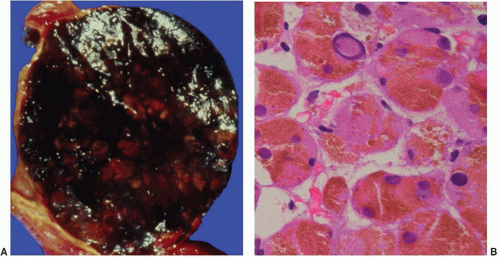
FIGURE 3-13 ▪ Pigmented (black) adenoma associated with Cushing syndrome (A). The tumor is composed predominantly of compact cells containing abundant lipochrome pigment (B).


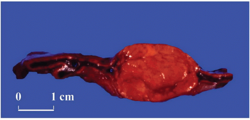
FIGURE 3-14 ▪ Adrenocortical adenoma associated with Conn syndrome. This tumor has a yellow-brown color or orange cut surface rather than a bright yellow appearance that is more characteristic.


Adenomas Producing Conn Syndrome


Epidemiology and Clinical Features

Initial studies suggested that cortical adenomas were responsible for Conn syndrome in up to 90% of cases; however, more recent analyses indicate that adenomas are present in a considerably smaller proportion of cases. As in cases of hyperplasia of the zona glomerulosa, these patients present with symptoms and signs of hypokalemia and hypertension.


Gross Pathology

Most adenomas associated with hyperaldosteronism measure <2 cm in diameter and are round to ovoid in configuration.103 They are usually unilateral, bright yellow in color and are demarcated from the adjacent cortex by a fibrous pseudocapsule. Occasional cases have a yellow brown or orange appearance (Fig. 3-14).


Microscopic Features

The tumor cells, which are usually arranged in small nests and cords, can resemble cells of the glomerulosa, fasciculata, or reticularis or combine the features of both glomerulosa and fasciculata cells (hybrid cells)23,103 (Fig. 3-15A). Some of the tumors may be highly pigmented.104 In patients treated with spironolactone, occasional cells may contain lamellar eosinophilic inclusions (spironolactone bodies) measuring up to 10 µm in diameter. They are often demarcated from the adjacent cytoplasm by a clear halo (Fig. 3-15B). Ultrastructurally, spironolactone bodies resemble myelin figures.

Although most of the tumor cells have relatively small vesicular nuclei and small but distinct nucleoli, some tumors exhibit considerable variation in nuclear size and shape. At the ultrastructural level, the mitochondria manifest tubular or vesicular cristae, although some may have lamelliform cristae typical of the zona glomerulosa. The fasciculata adjacent to aldosterone-secreting adenomas is of normal thickness. Hyperplasia of the zona glomerulosa may be present, however, in association with these tumors.103


Adenomas Producing Adrenogenital Syndromes


Epidemiology and Clinical Features

Benign adrenocortical tumors may be associated with syndromes of virilization or feminization, but the presence of a pure adrenogenital syndrome, particularly feminization, should suggest the possibility of malignancy. Some authors, in fact, consider all feminizing cortical neoplasms as being potentially malignant.


Gross and Microscopic Pathology

Virilizing adenomas are generally larger than those found in the context of pure Cushing syndrome, and a few adenomas associated with adrenogenital syndromes have
weighed up to 500 g.9,23,43 Similar to tumors associated with glucocorticoid overproduction, virilizing adenomas are sharply circumscribed or encapsulated; however, they tend to be red-brown rather than yellow on cross section23 (Fig 3.16A). Smaller tumors have an alveolar pattern of growth, whereas larger tumors tend to have more solid or diffuse growth patterns. Although most tumor cells have a low nuclear/cytoplasmic ratio, single cells and small-cell groups may exhibit considerable nuclear enlargement and hyperchromasia. The cytoplasm is usually eosinophilic and granular (Fig. 3.16B). Rare virilizing tumors contain Reinke crystalloids and have been termed Leydig cell adenomas similar to their testicular counterparts.105,106 On ultrastructural examination, the mitochondria are of the tubulolamellar type. Sex steroid-producing adenomas are not associated with atrophy of the adjacent cortex or the contralateral adrenal gland.


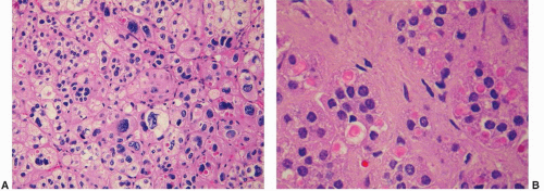
FIGURE 3-15 ▪ Adrenocortical adenoma associated with Conn syndrome. The tumor is composed of an admixture of fasciculata- and glomerulosa-type cells. Cells with enlarged, hyperchromatic nuclei are evident (A). Spironolactone bodies that have a typical lamellar appearance are prominent in this section (B).


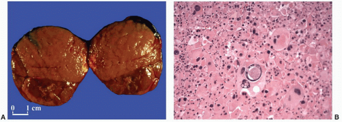
FIGURE 3-16 ▪ Adrenocortical adenoma associated with virilizing syndrome. This tumor from a 2-year-old girl has a brown cut surface (A). The neoplastic cells are eosinophilic and granular with focal nuclear enlargement and hyperchromasia (B). Nuclear pseudoinclusions are also present.


Oncocytic Adrenocortical Tumors


Epidemiology and Clinical Features

Tumors with oncocytic features develop rarely as primary adrenocortical neoplasms. While some behave as benign neoplasms (oncocytomas), others may be malignant, as discussed in the section on adrenocortical carcinomas. Most oncocytomas are nonfunctional,107, 108, 109a but up to a quarter of tumors may be associated with virilization109, 110 or Cushing syndrome.109a,111 A case of a giant oncocytoma arising in retroperitoneal accessory adrenal tissue has been described.112


Gross and Microscopic Features

Typically, adrenal cortical oncocytomas are dark brown, similar to oncocytomas at other sites. Reported cases have ranged in size from 8 g to over 500 g. They have abundant granular eosinophilic cytoplasm (Fig. 3-17), which corresponds to the presence of numerous mitochondria with both lamellar and tubulovesicular cristae and small, electron-dense inclusions.113 However, smaller eosinophilic cells (small oncocytes), may be present and may be quite frequent in some cases.109a The nuclei are enlarged, irregularly shaped, and vesicular with coarse chromatin and prominent nucleoli. Nuclear psuedoinclusions may be seen. Mitotic activity is minimal to absent and there is no evidence of necrosis. The cells have a tendency to be arranged in diffuse sheets, but variable trabecular, alveolar, and microcystic architecture can be seen.

Bisceglia et al.113a suggested that oncocytic adrenocortical tumors be classified as pure when the tumor was composed of 90% oncocytic cells and mixed when the
tumors were composed of 50% to 90% oncocytic cells. Subsequently, Duregon et al.113b classified oncocytic adrenocortical tumors as being pure, mixed, having focal oncocytic features, or being conventional adrenocortical tumors when the oncocytic cells constituted >90%, 50% to 90%, 10% to 49%, and <10% of the tumor cells, respectively (Box 3-2). Because of variations in sampling with fine needle or core biopsies it is appropriate to render a diagnosis of “adrenocortical neoplasm with oncocytic features” in such samples.


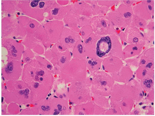
FIGURE 3-17 ▪ Adrenocortical oncocytoma. The cells contain densely granular eosinophilic cytoplasm and pleomorphic nuclei with a pseudoinclusion.

Recognition of adrenocortical oncocytoma and distinction from conventional adrenocortical carcinoma is an important one since these tumors have many of the features that would fulfill criteria for malignancy in conventional adrenocortical neoplasms. Therefore, the criteria for malignancy in oncocytic adrenocortical tumors are different (Box 3-2) and are fully discussed in “Adrenocortical Carcinomas.”

Although the granular cell variants of conventional RCC and chromophobe RCC tend to have eosinophilic cytoplasm, adrenocortical oncocytic neoplasms generally tend to have more voluminous cytoplasm that demonstrate diffuse strong positive granular cytoplasmic staining with the antimitochondrial antibody mES-13.109,109a Immunohistochemical staining with markers for adrenocortical tumors and RCC will assist in the diagnosis. Melan-A-positive staining of neoplastic cells is the most sensitive adrenocortical marker.109,109a As noted in a subsequent section, pheochromocytomas rarely have oncocytic cytoplasm (oncocytic pheochromocytoma), but staining with antibodies to chromogranin will assist in establishing the diagnosis of pheochromocytoma. Interestingly, like oncocytic neoplasms of other organs, 9 of 12 tumors classified by Duregon et al.113b as oncocytic adrenal adenomas were shown to have the 4,977-bp mitochondrial DNA “common deletion” by real-time polymerase chain reaction (PCR) and fluorescent in situ hybridization (FISH). However, this finding was not entirely specific since it was demonstrated in some normal adrenocortical cells and some examples of conventional adrenocortical adenomas.113b


Adrenocortical Carcinomas


Epidemiology and Etiopathogenesis

Adrenocortical carcinomas account for 0.05% to 0.2% of all malignancies and have an incidence of approximately one to two cases per million population per year. They have a bimodal age distribution with a small peak occurring in the first two decades and a larger peak in the fifth decade.114 Adrenocortical carcinomas develop somewhat more commonly in women than in men in most large clinical series, although some studies have demonstrated a slight male predominance. They occur in approximately 1% of patients with the Li-Fraumeni syndrome, with most affected individuals harboring p53 mutations at chromosome locus 17p13. These tumors, in fact, may be the only manifestation of this disorder in childhood.115 The frequency of cortical malignancies is also increased in patients with the Beckwith-Wiedemann syndrome (Table 3-2) and congenital adrenal hyperplasia.

Gene expression profiling studies have demonstrated that the most significantly upregulated genes in carcinomas include ubiquitin-specific protease 4 (USP4) and ubiquitin degradation 1-like (UFD1L). Additional upregulated genes include members of the insulin-like growth factor (IGF) family such as IGF2, IGF2R, IGFBP3, and IGFBP6.116 Giordano et al. also demonstrated increased expression of IGF2 in adrenal cortical carcinomas.117 Downregulated genes in carcinomas include the chemokine (C-X-C motif) ligand 10 (CXCL10), the retinoic acid receptor responder 2, the aldehyde dehydrogenase family member A1 (ALD1f1A1), cytochrome b reductase 1, and glutathione S-transferase A4.117

Similar patterns of gene expression occur in pediatric adrenal cortical tumors with a consistent marked decrease in the expression of all histocompatibility class II genes in carcinomas as compared to adenomas.118 These results parallel the observations by Marx et al.119 that pre- and postnatal adrenals do not express major histocompatibility complex class II antigens in contrast to adult adrenals, which express these antigens.


Clinical Features

Some patients may present with abdominal pain, and up to 30% may have a palpable abdominal mass. The tumors may be associated with Cushing syndrome or evidence of sex steroid overproduction, and mixed syndromes are more common than in patients with cortical adenomas. In exceptional circumstances, mineralocorticoid production may be
present. A significant proportion of cortical carcinomas (up to 75% in some series) may be unassociated with syndromes of hormone overproduction.120 In some instances, patients may show signs of hypoglycemia due to the production of IGFs by the tumor or hypercalcemia due to the production of parathyroid hormone-related peptide.


Gross Pathology

Adrenocortical carcinomas generally weigh more than 100 g in adults, and most often, tumor weight is in excess of 750 g.9,121, 122, 123, 124 Rarely, however, tumors weighing <50 g will metastasize, while a small proportion of tumors weighing more than 1,000 g will not.43 It should be remembered, however, that benign tumors associated with sex steroid overproduction, however, can weigh considerably more than 100 g. Tumor weight is a useful predictor of malignancy in children. Tumors weighing more than 500 g in a series of 23 cases reported by Cagle et al.125 were malignant, whereas only a single tumor weighing <500 g pursued a malignant course.

Many cortical malignancies have a multinodular appearance with individual nodules varying from pink to yellow-tan, depending on their lipid content. Carcinomas associated with feminization or virilization tend to be red-brown, while those associated with Cushing syndrome are more often yellow-tan. Rare cases may have a myxoid appearance. Foci of necrosis, cystic change, hemorrhage, and calcification are common, particularly in large tumors (Fig. 3-18). The larger tumors often invade contiguous structures, including the kidney and liver.


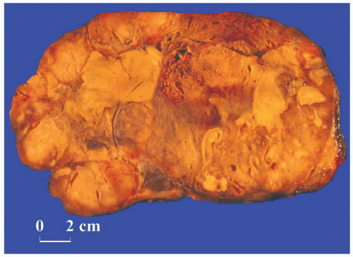
FIGURE 3-18 ▪ Adrenocortical carcinoma. The cut surface of this large tumor shows prominent necrosis.


Microscopic Features

Adrenocortical carcinomas have diverse architectural patterns, including alveolar (Fig. 3-19), trabecular, or solid patterns of growth, and many tumors exhibit admixtures
of these patterns.23,37,43 Many tumors are composed of widened trabeculae separated by endothelium-lined sinusoidal channels (Fig. 3-20A) in contrast to the thin cell cords characteristic of adenomas. Necrosis, particularly in large tumors, may be extensive (Fig. 3-21A). Foci of myxoid change, pseudoglandular patterns, and spindle-cell growth may be prominent in some cases.126 Depending on their lipid content, the cytoplasm may vary from vacuolated to eosinophilic. Some tumors may have eosinophilic globular inclusions resembling those seen in pheochromocytomas. Rare cases may exhibit adenosquamous differentiation.127 There may be considerable variation in the appearance of the nuclei. In some instances, they may appear relatively small and uniform, while in others they may exhibit marked pleomorphism (Fig. 3-20B), coarse chromatin, and multiple enlarged nucleoli. Mitotic activity, including atypical forms (Fig. 3-20C), is often prominent. Nuclear pseudoinclusions may be particularly striking in some cortical carcinomas.


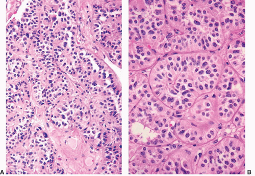
FIGURE 3-19 ▪ Adrenocortical carcinoma. The tumor has an alveolar pattern of growth (A, low power; B, high power).


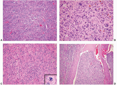
FIGURE 3-20 ▪ Adrenocortical carcinoma. Trabecular growth pattern (A), nuclear pleomorphism (B), prominent mitotic activity (C, inset atypical mitotic figure), and vascular space invasion (D) are demonstrated.

Some adrenocortical carcinomas are composed of oncocytic cells.109,128, 129, 130 While some of these tumors may be associated with Cushing syndrome or feminization,109 others may be nonfunctional. Hoang et al.129 concluded that large tumor size, extracapsular extension, vascular invasion, necrosis, and metastasis are features of malignancy in these tumors, while mitotic rate was less than 1 per 10 high-power fields (HPFs).129 Cytologic atypia and mitotic rate, therefore, were not reliable criteria for the prediction of biologic behavior of these neoplasms according to this study.129 On the other hand, Bisceglia et al.109 have also reviewed the criteria for the distinction of benign and malignant adrenal oncocytic tumors.109 According to these authors, major criteria for malignancy included high mitotic rate, atypical mitoses, and venous invasion while minor criteria included large tumor size, necrosis, capsular invasion, and sinusoidal invasion (Box 3-2, section on Oncocytic Adrenocortical Tumors). The presence of one major criterion was sufficient for the diagnosis of malignancy while one to four minor criteria were sufficient for a diagnosis of tumors of uncertain malignant potential. The absence of all criteria indicated benignancy.

Rarely, adrenocortical carcinomas may contain sarcomatous foci (carcinosarcoma). In the case reported by
Fischler et al.,131 the sarcomatous component had features of rhabdomyosarcoma and stained positively for musclespecific actin and desmin. Although this tumor was associated with virilization, the case reported by Decorato et al.132 was nonfunctional. Recently, Thway et al.132a reported an example of oncocytic adrenocortical carcinosarcoma in a 45-year-old man with pleomorphic rhabdomyosarcoma that metastasized to the mesentery, hilar lymph nodes, lungs, and brain over the 11-month terminal course from diagnosis.


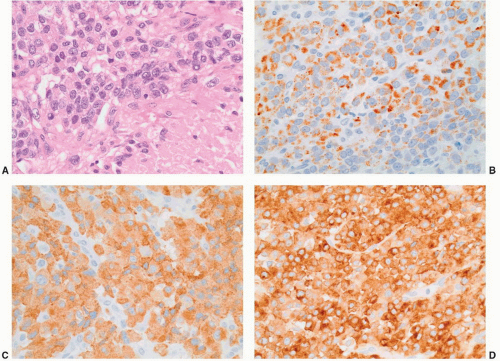
FIGURE 3-21 ▪ Adrenocortical carcinoma. There was extensive necrosis of this tumor and viable tumor cells have high nuclear to cytoplasmic ratio (A, hematoxylin-eosin section; necrosis in right lower aspect). Neoplastic cells stain positively with antibodies to melan-A (B), inhibin (C), and synaptophysin (D).

In fine needle aspiration biopsy samples, cortical carcinomas generally contain single cells and poorly cohesive cell clusters in a necrotic background. Although there may be considerable nuclear atypia and mitotic activity, some cortical carcinomas appear deceptively bland.126 According to Ren et al.133 common cytologic features include hypercellularity, necrosis, nuclear pleomorphism, mitotic figures, and prominent nucleoli. Twenty percent of their cases exhibited all five features while necrosis and/or mitoses were found in every case.


Differential Diagnosis and Ancillary Studies

Adrenocortical carcinomas must be distinguished from cortical adenomas and a variety of secondary tumors involving the adrenal gland, including RCC, hepatocellular carcinoma (HCC), metastatic carcinoma, and liposarcoma. Immunohistochemistry may be of particular value in discriminating these tumors,12,134, 135, 136, 137, 138, 139 (Table 3-3; Box 3-3). The most commonly utilized immunohistochemical antibodies for adrenal cortical tumors are those that react with melan-A, calretinin, and inhibin A (Fig. 3-21) and synaptophysin. The monoclonal antibody A103, which reacts with melan-A, an antigen recognized by cytotoxic T cells and expressed in melanocytes, has also been used for the identification of adrenocortical and other steroid hormone-producing tumors.14 With the exception of melanoma, the only tumors that are reactive with A103 are adrenocortical adenomas and carcinomas, testicular Leydig cell tumors, and ovarian Sertoli-Leydig cell tumors. Antibodies to inhibin A also provide an additional useful approach for the identification of steroid-producing
cells. Renshaw and Granter15 have demonstrated that inhibin A and A103 are both useful for the identification of adrenal cortical neoplasms and that A103 is marginally more specific and inhibin A slightly more sensitive. Calretinin is also expressed in adrenal cortical neoplasms and is a useful adjunct in cases where stains for inhibin A are negative. Jorda et al.13 demonstrated that almost 75% of cortical neoplasms were positive for inhibin A; however, when calretinin was added, the numbers of tumors staining positively for the two markers increased to 94% (31/33 cases).


Table 3-3 ▪ DIFFERENTIAL DIAGNOSIS OF ADRENOCORTICAL CARCINOMA


Only gold members can continue reading. Log In or Register to continue

Stay updated, free articles. Join our Telegram channel

Jun 10, 2016 | Posted by in UROLOGY | Comments Off on Pathology of the Adrenal Gland

Full access? Get Clinical Tree

Get Clinical Tree app for offline access


Limited Panel of Antibodies


Tumor


CK


VIM


Mel-A


INH


CAL


SYN


CHR


S-100


EMA


pCEA


Other Antibodies


Cortical carcinoma


+/-


+


+


+


+


+/-


D2-40+, SF-1+


Pheochromocytoma


+/-


+


+


Hepatocellular carcinoma


+


+/-


+/-


+


AFP+, Hep+, CD10+ (pCEA pattern)


Renal cell carcinoma


+


+