Fig. 26.1
Everting sutures in the distal anal canal widen the anal canal and shorten the distance between surgeon and tumor
The rectum, especially its lowest part, is wider than most other parts of the colon and also has a thicker wall. This allows for tumors to grow to a larger size without becoming symptomatic, but it also allows for the rectum to be stretched. That helps with visualization as well as closure at the end of the procedure. The rectum starts bending posteriorly a few centimeters above the dentate line. This anatomy may, in some situations, limit visualization and the ability to reach higher lesions. This can be partly overcome by placing the patient in lithotomy position, which is the preferred position for patients with posterior lesions.
In the anterior projection the lower rectum borders the prostate in men and the posterior vaginal wall in women. In addition, the anterior peritoneal reflection can be quite low, especially in thin older women. Entry into the peritoneal cavity should be avoided to limit the systemic inflammatory response. A prone-jackknife position (Fig. 26.2) would be more appropriate for anterior or laterally based lesions.
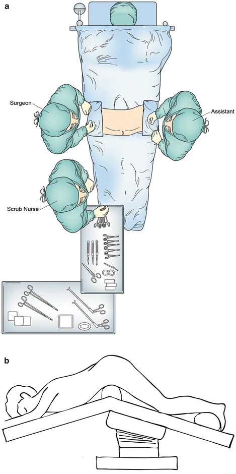
Fig. 26.2
Patient positioning and operating room setup. The patient is placed in prone-jackknife position
Nerve Blocks and Retraction
Perianal and pudendal nerve blocks should be performed with a local anesthetic (e.g., combination of short-acting lidocaine and long-acting bupivicaine with epinephrine). This decreases bleeding during the procedure, helps to relax the sphincter complex, and provides good pain control after surgery. Since good visualization is the key to a successful transanal resection, a headlight or a lighted retractor is extremely important. For lower tumors, a variety of retractors may be used, e.g., Pratt-bivalve, Fansler, Sawyer, or Hill-Furguson retractor anal speculum. For deeper lesions, everting sutures, along with a LoneStar retractor, can be used to provide better exposure and to pull the anorectal junction distally.
Examination and Marking
After a thorough exam to make sure that the mass is accessible by the transanal approach, the margins of resection should be marked. At least a 1-cm margin from the border of the tumor should be taken in all directions. Electrocautery can be used to mark the margin. Occasionally, traction sutures can be placed both distal and proximal to the tumor to pull a high tumor down within reach. These sutures will help control the proximal resection margin, which may retract after removal of the specimen. However, the presence of multiple sutures in a narrow operative field can be bothersome; an Allis clamp can also be used to pull down the rectal wall and grasp the specimen. An alternative approach for lesions in the mid-rectum involves making an elliptical incision distal to the lesion and then continuing the dissection behind the rectal wall as the proximal margin is approached.
Resection of the Specimen
Electrocautery can be used to perform a full-thickness resection along the previously marked margin (Fig. 26.3). Special care should be taken not to grasp the tumor or damage its margin. The key to successful resection is slow, meticulous incision of the bowel wall and mesorectal tissue with proper control of the submucosal and mesorectal blood vessels. Upon completion, the perirectal fat should be visualized to confirm a complete full-thickness excision (Fig. 26.4).
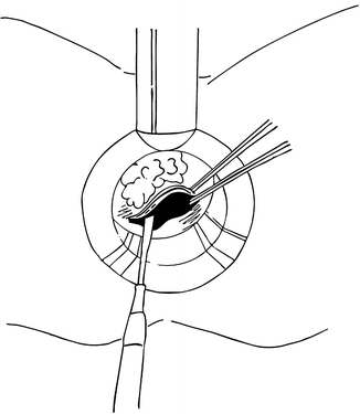
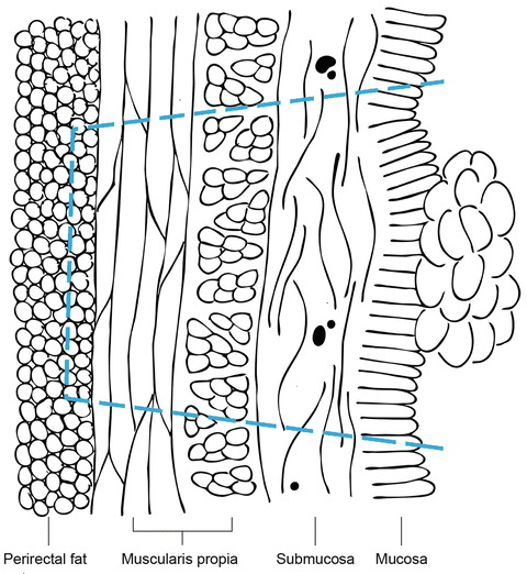
Fig. 26.3
Operative diagram showing the resection of the specimen with electrocautery beginning at the distal margin
Fig. 26.4
Diagram indicating the extent of a full-thickness resection of a rectal lesion
Once the specimen is removed, it should be carefully oriented on a pin board clearly marking its orientation (proximal, distal, left, and right). The use of pins also helps to keep the margins from shrinking during processing of the specimen. Since careful orientation of the specimen is critically important, pathologists should be called if possible to explain the specimen and its orientation. In our institution, all transanal excisions are processed with a “rectal cancer protocol,” which includes inking the resection in quadrants (four colors) with deep and peripheral mucosal margins inked the same colors and then fixing the specimen prior to submitting it for histologic examination. Frozen section exam of the margins may also be considered.
Closure of the Defect
Once the specimen is removed, the area is irrigated and hemostasis is ensured. The defect should be closed if possible. Although most transanal lesions will be below the peritoneal reflection and closure is not mandatory, the defect should be closed to decrease the rates of postoperative bleeding and expedite recovery. The defect should be closed transversely to prevent stricturing and obstruction. Absorbable sutures such as 2-0 and 3-0 vicryl should be used for this full-thickness closure. The wound can be closed in either a continuous running or interrupted fashion. Other absorbable sutures may be used.
< div class='tao-gold-member'>
Only gold members can continue reading. Log In or Register to continue
Related posts:


Stay updated, free articles. Join our Telegram channel

Full access? Get Clinical Tree


