Fig. 2.1
Operative positioning for laparoscopic mobilization of the stomach
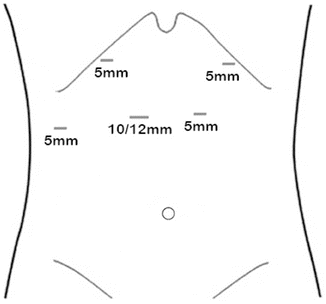
Fig. 2.2
Trocar placement for laparoscopic mobilization of the stomach
Gastric Mobilization
The dissection is started in the hiatus. The right crus is exposed using an energy device and blunt dissection. The LigaSure (Covidien) and Harmonic Scalpel (Ethicon) are both adequate for these steps. We typically remove the peritoneal lining around the crus, but do not routinely excise muscle fibers unless the tumor is adherent. The dissection from the crus is followed anteriorly and over to the left crus. If there is a hiatal hernia, it is helpful to reduce the sac and completely separate the sac from the crura. For patients who do not have a hiatal hernia or only a small hernia, we divide some of the right crural fibers to enlarge the hiatus so that it will easily accommodate the gastric conduit.
Next, the right gastroepiploic artery is visually identified. The stomach is separated from the omentum and mesocolon by retracting the omentum caudal to the point of transection and dividing it away from the gastroepiploic artery. We avoid trauma to the gastric conduit by minimizing retraction of the stomach or avoiding retraction altogether. We usually harvest an omental flap by leaving a pedicled portion of the omentum attached to the conduit perfused by two to three branches of the gastroepiploic arcade; any more than that would be too bulky. This tongue of omentum is dissected directly off the colon. The dissection continues parallel to the left gastroepiploic artery until the short gastric vessels are identified. The short gastric arteries are all serially transected using a vessel sealing device and the stomach is completely mobilized off the spleen and left crus. Additional attachments to the mesocolon are then freed medially. To prevent paraesophageal hernia and to allow maximum mobility of the stomach, the mesocolon should be completely separated from the stomach. Extreme caution must be taken in the vicinity of the takeoff of the right gastroepiploic artery from the gastroduodenal artery to avoid accidental injury of either artery. The lesser sac is then dissected, freeing the stomach from the pancreas. While the assistant retracts the gastric conduit up, the left gastric artery pedicle is dissected from the celiac axis. Nodal tissue is carefully dissected and swept towards the stomach (Fig. 2.3). Once this is complete, the left gastric pedicle is transected using an endovascular stapler cartridge.
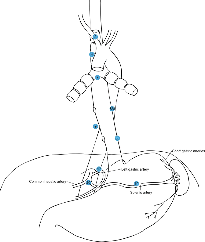
Fig. 2.3
Key lymph node stations during Ivor Lewis esophagogastrectomy
Before the gastric conduit is created, we check to ensure that the stomach is circumferentially freed, and that the pylorus easily reaches the hiatus. A Kocher maneuver is not necessary for an Ivor Lewis esophagectomy. The posterior gastroesophageal (GE) junction is then dissected and the mediastinal esophagus is circumferentially dissected as cephalad as possible. It is easy to enter one or both pleural cavities during the mediastinal dissection, so it is best to wait until the latter part of the laparoscopic procedure to perform this dissection. A pleural defect can be a nuisance during laparoscopy and impair the surgeon’s ability to insufflate the abdomen adequately.
Creating the Gastric Conduit
The NGT is then pulled back into the pharynx. A point on the lesser curvature of the stomach between the right and left gastric arteries is identified just proximal to the incisura. Collateral vessels overlying this point are divided. Medium/thick tissue staple cartridges are then used to create the conduit, firing multiple stapler loads up until a point on the gastric fundus. We do not oversew the staple line. Ideally the gastric conduit is no smaller than 4 cm in width. The stomach is then sewn back to the specimen with a single mattress stitch. Alternatively, the last 2 cm of the stomach can be left undivided while creating the gastric conduit and then later divided within the chest. We leave a quarter inch penrose drain around the GE junction, secured by a suture to facilitate retrieval and dissection within the chest.
< div class='tao-gold-member'>
Only gold members can continue reading. Log In or Register to continue
Related posts:


Stay updated, free articles. Join our Telegram channel

Full access? Get Clinical Tree


