Fig. 15.1
Operative positioning for open right colectomy. The surgeon stands on the left side of the patient with the assistant on the right side
Incision
A midline incision starting 6–8 cm above the umbilicus and continuing 2–3 cm below the umbilicus is most commonly utilized for open right colectomy (Fig. 15.2a). Alternatively, a transverse incision starting in the midline 2–3 cm above the umbilicus and continuing for 6–8 cm laterally on the right side of the abdomen is another option (Fig. 15.2b). With the transverse incision, the right rectus muscle is divided. In the non-obese patient, the transverse incision is similar in size to a laparoscopic extraction incision and results in minimal postoperative discomfort.
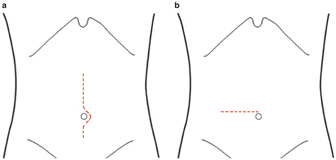
Fig. 15.2
Abdominal wall incisions for open right colectomy. (a) Mid-line peri-umbilical incision. (b) Right transverse incision
Exploration
Upon entrance to the abdomen, the peritoneal cavity is explored and the liver is palpated. Biopsy of suspicious lesions should be performed. Particular attention is also directed to the omentum and in women the ovaries are also inspected. Preoperatively, potential oophorectomy should be discussed and a plan developed regarding identification of any ovarian pathology.
Colon Mobilization
With the assistance of a self-retaining retractor and the patient’s right side up, the lateral white line of Toldt is taken down with cautery starting at the cecum and proceeding superiorly along the ascending colon to the hepatic flexure. Care must be taken to avoid dissecting too deep into the retroperitoneum to avoid injury to the ureter, kidney, and duodenum. If the dissection proceeds in the correct plane, mandatory identification of the ureter is not necessary but in some instances the cecum and terminal ileum can have significant retroperitoneal attachments requiring identification of the ureter in order to avoid injury. The best way to identify the right ureter is to find it as it crosses the external iliac artery at the pelvic inlet and then follow it proximally from this point.
At the level of the hepatic flexure, it is important to identify the duodenum. The surgeon should be aware that the proximal transverse colon attachments are thicker and more vascular than the lateral attachments to the ascending colon. After the omental attachments to the proximal transverse colon are divided, the entire right colon is now fully mobilized and attached only by its mesentery. The omentum is divided from its free edge to the mid-transverse colon so that it is also resected en bloc with the right side of the colon.
Large cecal or ascending colon cancers can invade the abdominal sidewall requiring an en bloc resection of the tumor and abdominal wall. After en bloc resection, the abdominal wall resection area should be outlined with surgical clips to allow for better localization of the tumor bed for postoperative radiation. Rarely, a bulky tumor at the hepatic flexure or proximal transverse colon may extend into the duodenum and pancreas, requiring a Whipple procedure to complete the en bloc resection. This unusual circumstance should be identifiable on preoperative computed tomography (CT) scans and the operation should be planned accordingly.
< div class='tao-gold-member'>
Only gold members can continue reading. Log In or Register to continue
Related posts:


Stay updated, free articles. Join our Telegram channel

Full access? Get Clinical Tree


