Nonneoplastic Diseases of the Kidney


Nonneoplastic Diseases of the Kidney


DAVID J. GRIGNON

MARK A. WEISS


The surgical pathologist is called on to examine kidneys removed for a wide range of nonneoplastic conditions. This chapter is not meant as a comprehensive review of nonneoplastic kidney diseases purposely excludes those conditions that are generally considered within the area of “medical kidney diseases” that pathologists are exposed to primarily through renal biopsy. For the purposes of this chapter all transplantation-related nephropathology including resection of allografts is excluded. For those diseases that are associated with renal neoplasia, the detailed discussion of the tumors is covered in Chapter 2.


EMBRYOLOGY

A detailed discussion of the embryologic development of the kidney is beyond the scope and purpose of this chapter as are the many advances in our understanding of the molecular aspects of embryogenesis. The reader is referred to more comprehensive sources if required.1, 2, 3, 4 The following discussion will focus on those aspects of embryology relevant to understanding the pathologic conditions covered in this chapter.

The kidney develops from the mesoderm and forms through three successive stages: the pronephros, mesonephros, and metanephros. Each of these overlaps with the preceding step as development progresses.


Pronephros

The pronephros forms from the most caudal end of the nephrogenic cord and is short-lived, appearing at the end of the 3rd week of gestation and disappearing by day 25. The pronephric ducts are the only part that persist and are incorporated into the mesonephros.


Mesonephros

The mesonephros develops from the midportion of the nephrogenic cord beginning on day 24 and results in the formation of the urogenital ridge. A series of glomeruli, each with a tubule and a capillary, form with a connection to the developing aorta. These tubules join an excretory duct, the mesonephric (Wolffian) duct. The mesonephros is a temporary functional excretory organ. These structures successively involute and by the end of the first trimester have disappeared.


Metanephros

The permanent kidney develops from the metanephros that first appears in the 5th week and becomes functional in the 9th week (Fig. 1-1). The metanephric mesenchyme, formed from the caudal end of the nephrogenic tube, gives rise to the kidney parenchyma with the ureteric bud being the origin of the collecting ducts, calyces, renal pelvis, and ureter.


Collecting System Formation

The ureteric bud develops as an outgrowth from the mesonephric duct. Signaling pathways critical to ureteric bud induction and development include GDNF and GFRα1. The WT1 gene is involved in inducing GDNF expression. Defects in restrictive signaling pathways such as Spry-1 allow for multiple ureteric buds to form and may be related to duplicate ureters.4 As the growing ureteric bud comes in contact with the metanephric mesenchyme it undergoes a series of dichotomous branchings. The Wnt-11 and GREM1 genes are among the most important in this process. The tip of the bud is known as the ampulla as is the tip of each subsequent branch as it develops. The first three to six of these give rise to the renal pelvis and major calyces (Fig. 1-2). BMP-4 is a key factor present in the metanephric mesenchyme controlling this process. Abnormalities in BMP-4 have been related to a wide range of anomalies including renal hypoplasia/dysplasia, hydroureter, megaureter, ectopic ureter, ureteral duplication, and ureteropelvic obstruction.4 These eventually coalesce to form the structure as we see it
at birth. The continued branching and growth result in the formation of the minor calyces and ever-increasing numbers of collecting ducts (Fig. 1-3).


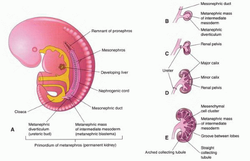
FIGURE 1-1 ▪ Embryologic development of the kidney. A: Sketch of a lateral view of a 5-week embryo showing the primordium of the metanephros. B-E: Sketches showing successive stages in the development of the metanephric diverticulum or ureteric bud (5th to 8th weeks). Note the development of the ureter, renal pelvis, calices, and collecting ducts. (From Moore KL, Presaud TVN. The Developing Human: Clinically Oriented Embryology. 7th ed. Philadelphia, PA: Saunders; 2003, with permission.)


Nephron Formation

The nephrons develop from condensations in the metanephric mesenchyme with a condensation related to each tip of the branching ureteric bud ampullae (Fig. 1-4). The condensation forms an S-shaped structure (the nephrogenic vesicle) with a space that becomes continuous with the lumen of the growing collecting tubule. The nephrogenic vesicle elongates with one end forming the proximal tubule and the loop of Henle. The initial period of nephron formation ends after the 14th week of gestation. The nephrons from this initial period are localized to the corticomedullary junction. In the second period of nephrogenesis, additional glomeruli develop as arcades from the no longer branching ampullae of the ureteric bud. In the third period of nephrogenesis, between weeks 22 and 36, the ampullae grow peripherally to the outermost part of the cortex giving rise to another four to seven nephrons. The last nephrons formed are located in the subcapsular region. It is estimated that there are between 0.7 and 1.5 million glomeruli per kidney.


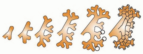
FIGURE 1-2 ▪ Development of the renal pelvis. Diagram showing branching of the ureteral bud. (Reprinted from Mills SE. Histology for Pathologists. 3rd ed. Philadelphia, PA: Lippincott Williams & Wilkins; 2008. Modified from Potter EL. Normal and Abnormal Development of the Kidney. Chicago, IL: Year Book; 1972, with permission.)

After 36 weeks, the ampullae regress and disappear with no additional nephrons being formed. During this last part of fetal development, the tubules lengthen and become more tortuous.


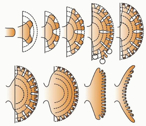
FIGURE 1-3 ▪ Development of renal calyces and papillae. Diagram showing coalescence of the third to fifth generation of branches of the primordial calyx with inward prolapse of the renal papilla. (Reprinted from Mills SE. Histology for Pathologists. 3rd ed. Philadelphia, PA: Lippincott Williams & Wilkins; 2008. Modified from Potter EL. Normal and Abnormal Development of the Kidney. Chicago, IL: Year Book; 1972, with permission.)


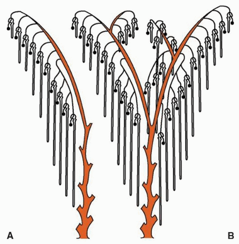
FIGURE 1-4 ▪ Arrangement of nephrons at birth as revealed by microdissection. A: Usual pattern. B: Possible variations. (Reprinted from Mills SE. Histology for Pathologists. 3rd ed. Philadelphia, PA: Lippincott Williams & Wilkins; 2008. Modified from Potter EL. Normal and Abnormal Development of the Kidney. Chicago, IL: Year Book; 1972, with permission.)


ANATOMY AND HISTOLOGY

In this section, the anatomy and histology of the kidney relevant to the types of specimens encountered by the surgical pathologist will be reviewed. Detailed descriptions of the anatomy and histology of the kidney are available in more comprehensive references.5,6


Gross Anatomy

The kidneys are paired organs located in the retroperitoneum. The superior poles are usually located at the level of T12/L1 with the lower poles at L3, although the upper and lower extremes can range from T11 to L5. The right kidney is usually 1 to 2 cm lower than the left. The kidneys are related to the diaphragm at the superior posterior aspect, the psoas muscle posteriorly and the quadratus lumborum and aponeurosis of the transversus abdominus muscle laterally. Anteriorly the right kidney is juxtaposed to the liver, right colonic flexure, descending part of the duodenum, and the small intestine. On the left, the anterior surface relates to the stomach, spleen, pancreas, left colonic flexure, descending colon, and jejunum. Both superior poles are intimately related to the adrenal glands. They are surrounded by fat within Gerota fascia (Fig. 1-5). Gerota fascia is a thin fibromembranous structure that is in continuity with the transversalis fascia; it surrounds the perirenal adipose tissue superiorly, laterally, and medially but does not cover the hilar or inferior boundaries.

In newborns, the kidneys weigh between 13 and 44 g; they increase in size with age reaching 115 to 166 g in adult females and 125 to 170 g in adult males.7 The increase in weight is related to increased tubular and interstitial tissues; the number of glomeruli remains constant. With advancing age, the kidneys begin to decrease in weight due primarily to cortical atrophy. The average adult kidney measures 11 to 12 cm in length, 5 to 7 cm in width, and 2.5 to 3 cm in thickness. The kidneys are surrounded by a well-developed fibrous capsule that can be readily stripped from the cortical surface; the capsule covers the outer convexities of the kidney but is not well-developed in the hilar region where the vascular and collecting system structures enter the renal parenchyma (Fig. 1-6). In fact, there is no capsule covering the renal cortical tissue of the columns of Bertin where the columns come in contact with the renal sinus adipose tissue. This has been shown to represent an important site of tumor extension beyond the renal parenchyma (Fig. 1-7).8,9

In some adults, the kidney retains the prominent fetal lobulations that are evident at birth (Fig. 1-8). In most kidneys, however, these largely disappear with the exception of two grooves extending from the hilum dividing the kidney into three poorly defined regions: a middle zone and the upper and lower poles. The cut surface reveals a continuous layer of pale tan cortex, measuring about 1 cm in thickness and making up the entire outer aspect of the renal

parenchyma (Fig. 1-9). Cortical tissue extending from the outer aspect to the renal sinus (columns of Bertin) separates individual medullary pyramids and their draining calyces. The medulla is a darker red brown than the cortex and has a striated appearance. It can be divided into an outer or peripheral zone and an inner zone or papillae. A single unit of a pyramid with its associated cortex is the equivalent of a unipapillary kidney. In humans, the fusion of these individual units results in a multipapillary type of kidney.


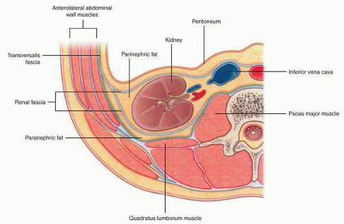
FIGURE 1-5 ▪ Kidney anatomy. Cross section of kidney at the level of renal hilum showing relationship to fat and renal (Gerota) fascia. (From Drake RL, Vogl AW, Mitchell AWM. Gray’s Anatomy for Students. 2nd ed. Philadelphia, PA: Churchill Livingstone; 2010:357, with permission.)


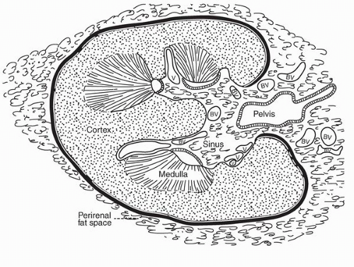
FIGURE 1-6 ▪ Kidney anatomy. Line drawing of cross section of kidney at the level of the renal hilum illustrating the extent of the renal capsule (thick line) and in particular its absence in the renal sinus area. (From Murphy WM, Beckwith JB, Farrow GM. Tumors of the Kidney, Bladder and Related Urinary Structures, AFIP Fascicle. 3rd series. Washington, DC: American Registry of Pathology; 1994, with permission.)


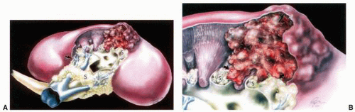
FIGURE 1-7 ▪ Renal sinus and tumor invasion (A,B). Drawing illustrating the propensity of tumors to infiltrate into the renal sinus soft tissue at a location lacking a renal capsule (arrow, B). (From Bonsib SM, Gibson D, Mhoon M, et al. Renal sinus involvement in renal cell carcinomas. Am J Surg Pathol 2000;24:452, with permission.)


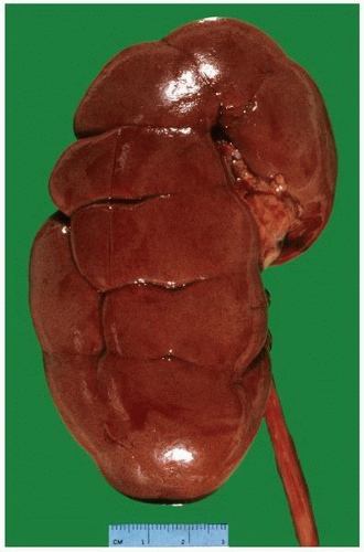
FIGURE 1-8 ▪ Kidney with prominent fetal lobulations. Kidney from a 37-year-old woman with prominent grooves highlighting the normal fetal lobulations that usually disappear with maturity.


Vascular Structure

The arterial supply to each kidney originates from the aorta with a main renal artery. In most kidneys, the renal artery divides into anterior and posterior divisions that pass anterior and posterior to the renal pelvis. Within the hilum, these further divide into variable numbers of segmental branches (most often the anterior division divides into four segmental branches while the posterior division continues as a single segmental branch). The main renal artery also gives rise to the suprarenal artery supplying the adrenal gland and a ureteric artery supplying the ureter. Deviation from this pattern is, however, common.

Within the renal sinus, each segmental artery gives rise to multiple lobar arteries (the vessels that typically enter the parenchyma) that then give rise to the interlobar then the arcuate arteries, followed by the interlobular arteries and finally the arterioles.10 These are all end arteries and so occlusion leads to downstream infarction. The efferent arterioles form a complex capillary network that supplies the cortical tubules and the juxtamedullary glomeruli. The medulla derives its blood supply mainly from the efferent arterioles of the juxtamedullary glomeruli.

The venous return largely parallels the vascular supply with interlobular, arcuate, and interlobar veins paired with their corresponding artery. In contrast to the arterial system, the venous system includes a complex network of anastomoses. The interlobar veins join to form the main renal vein that passes anterior to the renal pelvis.


Lymphatic System

The major lymphatic drainage system follows the vascular system.11 The lymphatics begin at the level of the interlobular arteries. No lymphatic channels are present in the area of the glomeruli and associated tubules. The main lymphatics drain into the hilar and periaortic lymph nodes. A second
lymphatic system present within the renal capsule drains the outermost portion of the cortex and eventually communicates with the major lymphatic system in the hilar region.


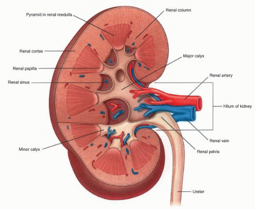
FIGURE 1-9 ▪ Kidney anatomy. Line drawing of the cut surface of the kidney. (From Drake RL, Vogl AW, Mitchell AWM. Gray’s Anatomy for Students. 2nd ed. Philadelphia, PA: Churchill Livingstone; 2010:358, with permission.)


Nerve Supply

The nerve supply of the kidney is sympathetic in type and is derived from the celiac plexus and then branches to the renal plexus via the splanchnic nerves. The nerve fibers originating in the renal plexus follow the arterial supply to the cortical region and innervate the juxtaglomerular apparatus and the renal vasculature. Nerve endings also interact with tubules, particularly the thick part of the ascending loop of Henle. Sensory fibers in the kidney follow the sympathetic nerves to the T10-T11 region.


Microscopic Anatomy

The renal cortex is covered by the renal capsule which is composed of dense fibrous connective tissue (Fig. 1-10). In the cortex of the kidney, the components are arranged in two distinct patterns defined by the cortical labyrinth and the medullary rays. The major components of the cortical labyrinth are the glomeruli and the proximal tubules, but this area also includes distal convoluted tubules, connecting tubules, and the most proximal portions of the collecting ducts (Fig. 1-11). The medullary rays contain collecting ducts, and the proximal and distal straight tubules. The outer medulla contains the straight portion of the proximal tubules, the thin descending limb of the loop of Henle, and the thin and thick ascending limb of the loop of Henle as well as portions of the collecting ducts. The inner medulla (papilla) includes the thin descending and ascending limbs of the loop of Henle and the collecting ducts (Fig. 1-12).


Glomerulus

The normal glomerulus is about 200 µm in diameter. It consists of the glomerular tuft that is suspended in Bowman space, a fluid-filled space that empties into the proximal convoluted tubule (Fig. 1-13). The glomerular tuft consists of a complex capillary network supported by the glomerular
mesangium and fed by the afferent arteriole and drained by an efferent arteriole. The glomerular tuft contains three types of cells: endothelial, mesangial, and epithelial (podocytes) cells. The glomerular visceral epithelial cells (podocytes) transition into the parietal epithelial cells that line Bowman space. These squamous-like cells form a complete layer on the inner surface of Bowman capsule.


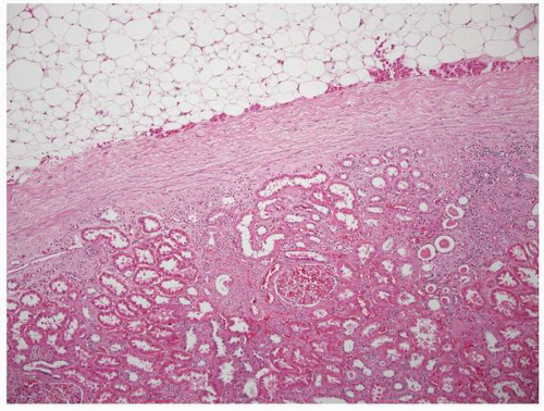
FIGURE 1-10 ▪ Renal capsule. External surface of the kidney illustrating a well-defined fibrous capsule covering the surface of the renal cortex and separating the parenchyma from the perinephric fat.

The juxtaglomerular apparatus is located at the vascular pole of the glomerulus. This structure is composed of specialized epithelial cells, vascular smooth muscle cells, the macula densa of the distal tubule, and specialized cells of the extraglomerular mesangium (lacis cells). This complex structure is responsible for regulating glomerular hemodynamics.


Proximal Tubule

The proximal tubule originates at the urinary end of the glomerulus and includes both a proximal convoluted and a distal straight portion. The lining cells are cuboidal to columnar with densely eosinophilic cytoplasm. Ultrastructural examination shows that the cells contain abundant mitochondria and have a prominent brush border.


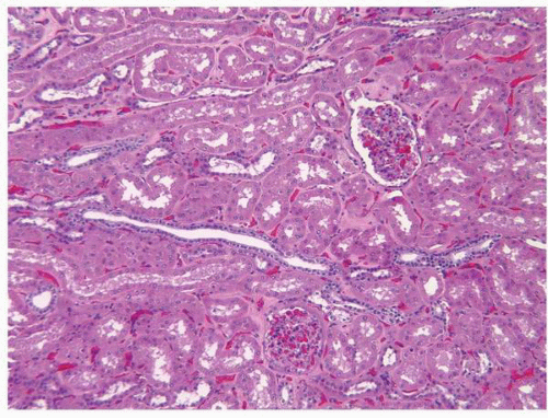
FIGURE 1-11 ▪ Renal cortex. Normal renal cortex containing glomeruli and tubules of the proximal and distal nephron.


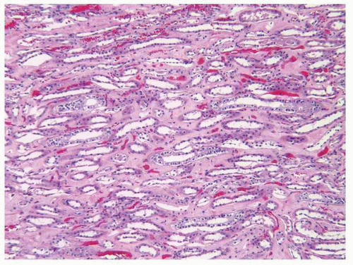
FIGURE 1-12 ▪ Renal medulla. Normal renal medulla with distal tubules and collecting ducts.


Loop of Henle

The loop of Henle consists of a descending thin limb, an ascending thin limb, and an ascending thick limb. At the beginning of the descending thin limb, the proximal tubular cells change to a flattened inconspicuous squamous-like epithelium. In general, the cells throughout the thin limbs of the loop of Henle have nuclei that bulge into the tubular lumens and a surface that contains few to no microvilli. The thick ascending limb of the loop of Henle is considered to be part of the distal tubule.


Distal Tubule

The distal tubule includes three distinct components; the thick ascending limb, the distal convoluted tubule, and the macula densa. In the medullary portion of the thick ascending limb, the lining consists of low cuboidal cells with eosinophilic cytoplasm and apically located nuclei. These cells have no brush border. The cells of the cortical segment are shorter but otherwise similar. In the distal convoluted tubule, the cells are also cuboidal with pale eosinophilic cytoplasm and closely packed nuclei. They have short microvilli along the surface. At the junction with the connecting tubule the cells are intermingled with the connecting tubule and intercalated cells.


Collecting Duct

The collecting ducts begin in the cortex and make up much of the medullary rays as they descend and then pass into the medulla and finally terminate in the papilla. The collecting ducts are lined by two types of cells: the principal cells
and the intercalated cells. The intercalated cells are largely restricted to the cortical and outer medullary segments of the collecting ducts. By light microscopy, both the principal cells and intercalated cells are cuboidal with pale cytoplasm and centrally located nuclei. The principal cells tend to get taller in the more distal portion of the collecting duct with the cells being columnar in the distal collecting ducts (ducts of Bellini).


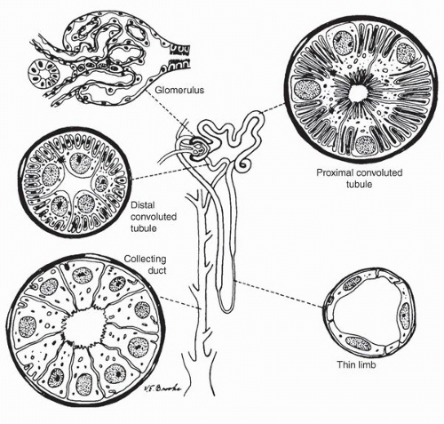
FIGURE 1-13 ▪ Normal histology of the nephron. Line drawing illustrating the various components of the nephron from the glomerulus to the collecting ducts. (From Bennington JL, Kradjian R. Renal Carcinoma. Philadelphia, PA: WB Saunders; 1967, with permission.)


Interstitium

The interstitium of the kidney includes a mixture of cells and stromal matrix. The predominant cell type within the interstitium is the fibroblast. The fibroblasts have specialized functions in different areas of the kidney. The medullary interstitial cell is a specialized lipid-laden fibroblast. These cells are involved in control of blood flow and produce prostanoids that have an antihypertensive effect.12 Other cells normally present are dendritic cells, macrophages, and lymphocytes. The matrix includes many substances including types I, II, and VI collagen, fibronectin, and sulfated and nonsulfated glycosaminoglycans.


Renal Sinus

The junction between the renal cortex and the renal sinus fat is not well-defined by a fibrous capsule (Fig. 1-14). Often there is a loose hypocellular connective tissue between the renal parenchymal tissue and the adipose tissue within the sinus. The sinus contains an abundance of thin-walled vascular and lymphatic channels embedded within the fat.


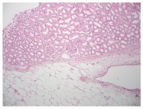
FIGURE 1-14 ▪ Renal sinus. In the area of the renal sinus there is no capsule separating the renal parenchyma from the sinus fat. Note tubular epithelium immediately adjacent to fat cells.


CONGENITAL ANOMALIES

Congenital anomalies involving the kidney and urinary tract have become known under the general label “congenital abnormalities of the kidney and urinary tract” or CAKUT.13 The use of this broad umbrella recognizes that in many cases multiple anomalies are present and further that single gene mutations can give rise to diverse abnormalities.14 The frequency is 3 to 6 per 1,000 births, and they are a significant cause of morbidity and mortality in the 1st year.15 Congenital anomalies are responsible for an estimated 50% of cases of end-stage renal failure in childhood and continue to be responsible for a small percentage in the 20- to 30-year-old age group.16,17


Renal Agenesis and Hypoplasia


Renal Agenesis


Clinical Features

Renal agenesis refers to the complete absence of one or rarely both kidneys.1,18,19 This can occur sporadically or in the setting of a large number of syndromes (Table 1-1).20 The majority of patients with renal agenesis have other abnormalities involving the genitourinary tract.20 In males, the most common organs involved are the epididymis, vas deferens, and seminal vesicle, with seminal vesicle cysts being among the more frequent. The association of unilateral renal agenesis with seminal vesicle cysts is known as Zinner syndrome.21 In females, the fallopian tube, uterus, and vagina can all have associated abnormalities. Patients with unilateral agenesis may be asymptomatic and the abnormality found incidentally even in adulthood.22 In other cases, it is symptoms related to an associated anomaly that bring the patient to medical attention. It is recommended that close evaluation of patients be undertaken when absence of a kidney is discovered incidentally.23

Bilateral renal agenesis (Potter syndrome) occurs in 0.1 of 1,000 births and is incompatible with life.1,14 The typical manifestation is severe oligohydramnios. Oligohydramnios, irrespective of cause, results in a characteristic phenotype that includes Potter facies; positional deformities of the hips, knees, and feet; and hypoplastic lungs (Fig. 1-15A and B). Infants with Potter syndrome are either stillborn or die shortly after birth due to respiratory failure.


TABLE 1-1 ▪ SYNDROMES ASSOCIATED WITH RENAL AGENESIS


Trisomy 13


Trisomy 18


Müllerian aplasia syndrome


Fraser syndrome


William syndrome


Cloacal exstrophy


Sirenomelia


Hereditary renal dysplasia


VATER syndrome


Many others


Pathology

The apparent absence of a kidney does not always indicate renal agenesis and ultimately the diagnosis of true renal agenesis requires confirmation of the absence of the kidney at autopsy or surgery. In unilateral agenesis, the remaining kidney may be significantly enlarged and hyperplastic.


Renal Hypoplasia


Clinical Features

Renal hypoplasia is defined by the presence of kidneys that are significantly smaller than normal. The kidneys are normally differentiated and should be distinguished from abnormal small kidneys. The condition can result from deficient metanephric induction, metanephric blastema, or postnatal renal growth.24,25 Bernstein and Gilbert-Barness26 have recognized three distinct categories of renal hypoplasia: oligonephric hypoplasia, simple hypoplasia, and unirenicular hypoplasia. Simple hypoplasia is rare and usually bilateral.26,27 Clinical diagnosis is based on the identification of a significant reduction in kidney size (more than 2 standard deviations), no evidence of scarring by dimercaptosuccinic acid scan, and compensatory hyperplasia of the contralateral kidney.14 Confirmation does require pathologic evaluation to exclude dysplasia.

Oligonephric hypoplasia (oligomeganephronia) is characterized by severe polyuria and polydipsia developing before the age of 2 years. This is associated with dehydration, anorexia, failure to thrive, and growth retardation. The condition progresses to renal failure before the age of 20 years and patients do well with renal transplantation. Mutations in the PAX2 gene have been described in some cases of oligonephric hypoplasia.28

Segmental hypoplasia (Ask-Upmark kidney) is now considered to be an acquired condition and is discussed later in this chapter.


Pathology


Gross Features.

Both kidneys are affected except in cases associated with contralateral agenesis or dysplasia. They are smaller than normal with a mean weight of 20 g (Fig. 1-16).24,26 The lobes and the number of pyramids are usually reduced in number and the pelvis and calyces are normally formed.


Microscopic Features.

In simple hypoplasia, there may be reduced numbers of glomeruli, but the kidneys are otherwise morphologically normal. In oligonephric hypoplasia, there are reduced numbers of markedly enlarged glomeruli (Fig. 1-17). Tubules are also enlarged and dilated with the formation of small cysts. The finding of any dysplastic changes excludes the diagnosis.


Abnormalities in Form and Position


Rotation Abnormality


Clinical Features

During development, the kidneys move from the pelvis into the abdomen and as part of that transition the kidneys rotate from an anterior-facing renal pelvis to the normal medial
orientation. This rotation can be disrupted with the renal pelvis remaining oriented to the anterior or less frequently over rotation results in a more posterior orientation. These abnormalities may have no clinical consequences and might be identified incidentally.22 In others, the abnormal position of the renal pelvis and ureter may result in obstruction and the associated complications including hydronephrosis, obstructive nephropathy, and lithiasis.


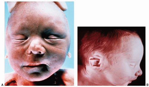
FIGURE 1-15 ▪ Potter syndrome. Infant born with bilateral renal agenesis. Characteristic features of the face include (A) the widely spaced eyes, flattened nose, prominent inner canthic folds, and the receding chin; (B) the lateral view highlights the flattened nose, receding chin, and the low set ears with little cartilage.


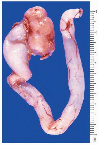
FIGURE 1-16 ▪ Renal hypoplasia. The kidney is small but largely retains a normal reniform shape.


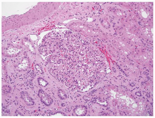
FIGURE 1-17 ▪ Oligomeganephronia. Enlarged glomeruli in the setting of renal hypoplasia.


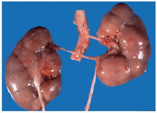
FIGURE 1-18 ▪ Malrotation. The right kidney is abnormally rotated, resulting in a flattened, disc shape. Note also the aberrant renal arteries.


Pathology

Malrotated kidneys are often abnormal in shape. Most characteristic is a flattened or discoid shape (Fig. 1-18). Microscopically the kidney is normal. Secondary changes related to obstruction and its consequences may be seen.


Horseshoe Kidney


Clinical Features

Renal fusion refers to the joining of the two kidneys to form a single structure. The most common expression of renal fusion is the horseshoe kidney due to fusion of the lower poles of the two developing kidneys. This anomaly, which is more common in males, occurs in 1:350 to 1:2,000 births.29,30 These are essentially all ectopic in location; the majority located anterior to the aorta and vena cava with others located posterior to the great vessels. The pelves face anterior. In many cases, there are additional abnormalities. Many of these remain asymptomatic unless complicated by nephrolithiasis and/or obstruction with its associated complications.31 Renal cell carcinoma develops in horseshoe kidneys in a frequency similar to normally located, nonfused kidneys.32,33 There is an apparent unusually high frequency of renal carcinoid tumors in horseshoe kidneys.34,35 Squamous cell carcinoma has also been described in association with nephrolithiasis.36


Pathology

The kidneys are typically fused at the lower pole and may or may not be associated with other abnormalities (Figs. 1-19 and 1-20). The amount of renal parenchyma in the isthmus is variable. The arterial supply is often anomalous. Abnormalities of the ureteral location are also common. The renal parenchyma is normal. Secondary changes resulting from obstruction and its complications may be present.


Malposition (Renal Ectopia)


Clinical Features

Ectopic location of the kidney is a relatively common congenital anomaly with an estimated prevalence of 1:1,000. The kidney can be located in the thorax but is more often located inferiorly in the pelvis. When the kidney is on the same side as the ureter and blood supply, the ectopy is termed simple; when located on the opposite side it is referred to as crossed. In crossed ectopy, fusion is also often present. Ectopy is frequently associated with other congenital
anomalies, including anomalies of the cardiovascular and central nervous systems. The malposition is typically associated with aberrant vascular and ureteral locations resulting in obstructive phenomena. Many ectopic kidneys are dysplastic. If undetected in childhood, these may present in adulthood with pain, hematuria, recurrent infections, lithiasis, and obstruction. Treatment depends on the specific type of ectopy and its clinical associations.


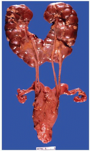
FIGURE 1-19 ▪ Horseshoe kidney. In this example, there is a fairly large isthmus at the lower poles, bilateral bifid ureters, and prominent fetal lobulations.


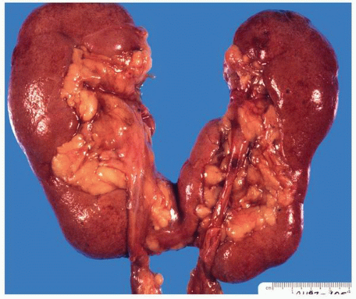
FIGURE 1-20 ▪ Horseshoe kidney. In this case the isthmus joins the lower pole of the right with the lower midportion on the left.


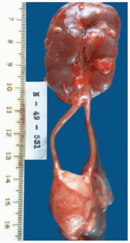
FIGURE 1-21 ▪ Crossed fused ectopia. There is a fused single kidney with two ureters.


Pathology

Ectopic kidneys are often abnormally shaped with a disclike appearance frequent in pelvic kidneys (Figs. 1-21 and 1-22). The gross appearance reflects other associated abnormalities. A wide range of microscopic features can range from essentially normal to dysplastic.


CYSTIC DISEASES

Cystic diseases of the kidney have long been considered among the most difficult to understand categories of kidney pathology that the surgical (or autopsy) pathologist is called upon to resolve. The past decade has seen remarkable growth in the understanding of the genetics and pathogenesis of this diverse group of conditions.37 These have been classified in a variety of ways over the years. Table 1-2 outlines the diseases discussed in this section organized by whether they are known to be inherited, acquired, or uncertain.


FIGURE 1-22 ▪ Ectopia. The right kidney is located in the pelvis at the bifurcation of the aorta. There is also malrotation with the kidney having a disc-like shape.


Cysts in Hereditary Syndromes


Autosomal Recessive (Infantile) Polycystic Disease


Genetics

Autosomal recessive polycystic kidney disease is present in 1 per 6,000 to 1 per 14,000 live births.38,39 It is inherited as an autosomal recessive defect, and the genetic defect has been
localized to the PKHD1 (polycystic kidney and hepatic disease) gene on chromosome 6.40 This gene codes for the fibrocystin/polyductin protein.41 The protein is localized to the primary cilium within renal epithelial cells of the collecting ducts. It is critical to ciliary function and within the cilium is primarily found in the basal body.42


TABLE 1-2 ▪ CYSTIC DISEASES OF THE KIDNEY


Inherited disorders


Autosomal recessive (infantile) polycystic kidney disease


Autosomal dominant (adult) polycystic kidney disease


Nephronophthisis


Medullary cystic kidney disease


von Hippel-Lindau disease


Tuberous sclerosis


Primarily nonhereditary disorders


Multicystic renal dysplasia


Glomerulocystic disease


Medullary sponge kidney


Simple cyst


Acquired cystic kidney disease


Segmental cystic disease


Cyst-like lesions


Pyelocalyceal diverticulum (cyst)


Perinephric pseudocyst


Clinical Features

The clinical expression of the disorder is considered to represent a continuous spectrum; however, for practical purposes two general categories of expression predominate.43,44 In the more common pattern, the presentation is with marked abdominal distension caused by gross renal enlargement at birth (Fig. 1-23). This may lead to intrapartum dystocia manifest as Potter syndrome.1 These infants usually die shortly after birth secondary to pulmonary insufficiency; therapy is generally supportive only.45 In individuals presenting later in childhood, the picture is dominated by both the renal disease and the associated hepatic fibrosis. Renal involvement is variable with about one-half retaining renal function and the remainder progressing to renal failure.38 Hypertension may be severe and lead to heart failure. Hepatic fibrosis manifests predominantly as portal hypertension with bleeding esophageal varices being a common and frequently fatal complication46; liver function usually remains normal. Other associations include cholangitis and hypersplenism. Treatment in these patients may include renal transplantation and surgical shunting procedures for the portal hypertension.47 Recent clinical experience has demonstrated significant improvement in survival for these patients. Perinatal mortality is in the 30% to 50% range; for patients who survive past 30 days, a 5-year survival rate of up to 87% has been reported.48


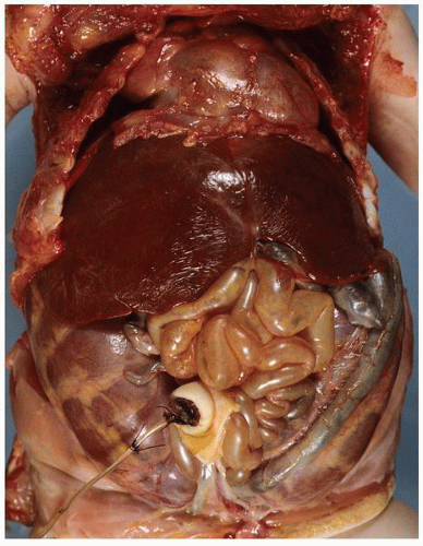
FIGURE 1-23 ▪ Autosomal recessive polycystic kidney disease. In situ photograph of a neonate at autopsy. The markedly enlarged kidneys can be seen filling the abdomen and pelvis. Note the marked displacement of the liver upward resulting in severe compromise of the chest cavity. The lungs were severely hypoplastic.


Pathology


Gross Features.

The kidneys are bilaterally grossly enlarged weighing up to 10 times normal in patients with severe disease (Fig. 1-24). A normal reniform shape is maintained. The cortical surface is smooth with numerous small cysts (1 to 2 mm). The cut surface reveals radially arranged cylindrical cysts replacing the cortex and medulla with loss of definition of the corticomedullary junction (Fig. 1-25). In those individuals surviving into childhood, the findings are much more variable. The cysts tend to be fewer, more haphazardly distributed, and larger; the kidneys may be normal or enlarged.46


Microscopic Features.

The pathogenesis of cyst formation is believed to be related to abnormal ciliary function.41 The cysts in the cortex are elongate and fusiform with a cuboidal epithelial lining (Fig. 1-26). In the medulla, the shape of the cysts is more variable. The cysts originate from collecting ducts with the proximal tubules and glomeruli being spared. In patients diagnosed at an older age, the cysts tend to be much less uniform in appearance. Dysplastic elements such as immature tubules and cartilage are not found.


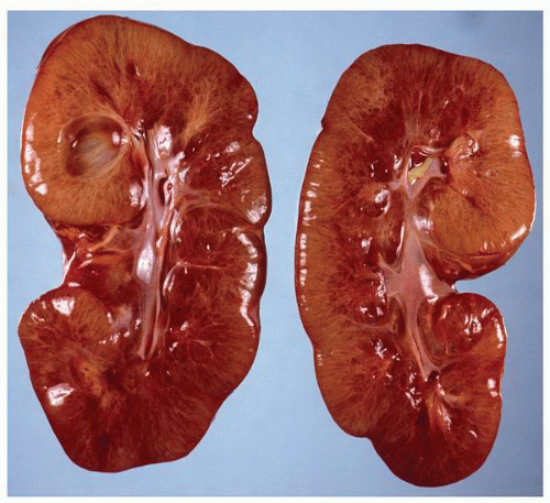
FIGURE 1-24 ▪ Autosomal recessive polycystic kidney disease. Bilateral kidneys at autopsy showing markedly enlarged kidneys with normal reniform appearance. The long tubular cysts can be appreciated in this photograph to result in a radiating pattern outward toward the cortical surface (see Fig. 1-25).


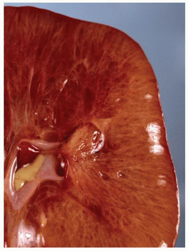
FIGURE 1-25 ▪ Autosomal recessive polycystic kidney disease. Close-up view of the cut surface of the kidney illustrating the elongated fusiform cysts involving the cortex and medulla.


Autosomal Dominant (Adult) Polycystic Kidney Disease


Genetics

Adult or autosomal dominant polycystic disease is among the most common inherited disorders with about 1 in 600 to 1 in 1,000 persons being affected.49,50 The defective gene in 85% to 90% of cases has been localized to the short arm of chromosome 16 (PKD1, 16p13.3).51 The remaining cases are related to the PKD2 gene at 4q13-23 with evidence that a third gene PKD3 may also be involved.51 The PKD1 and PKD2 genes encode for the polycystin 1 and polycystin 2 proteins, respectively. These proteins localize to the primary cilia of renal epithelial cells.52 The TSC1 gene is located near the PKD1 gene, and in some patients both genes are affected resulting in kidneys with overlapping features of tuberous sclerosis and autosomal dominant polycystic kidney disease.53


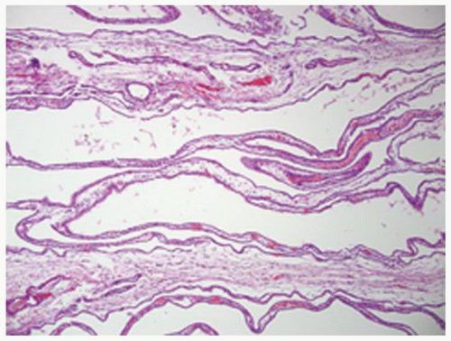
FIGURE 1-26 ▪ Autosomal recessive polycystic kidney disease. The elongated, saccular cysts are lined by a flattened to cuboidal epithelium.


Clinical Features

There is a very high rate of penetrance, and most affected individuals will show some signs of the disease.54 Autosomal dominant polycystic kidney disease is the most common inherited cause of end-stage renal disease in the United States.55 The majority of cases present in the third or fourth decades with development in infancy or childhood infrequent. Ultrasound abnormalities are detectable in 85% of patients by age 25.56 In children, the kidneys are of normal size.57 Most patients present with abdominal pain and palpably enlarged kidneys; gross or microscopic hematuria and hypertension may also be present. In symptomatic patients, renal failure ensues on average 10 years after presentation. Associated abnormalities, particularly hepatic cysts with fibrosis and cerebral aneurysms, may also have significant clinical impact (Table 1-3).58, 59, 60, 61, 62

Treatment is symptomatic with dialysis or transplantation required in about one-half of affected patients.55,56,63 The most common renal complications (excluding endstage renal disease) are pain, hematuria, and infection. Hypertension develops in almost all patients and is a major part of the management required. In general, the native kidneys are not resected as even limited function is considered valuable.64 The most common indications for native nephrectomy are recurrent infection, pain, persistent hematuria, and recurrent nephrolithiasis. Renal calculi complicate the condition in up to 35% of patients. For those with large calculi that fail medical management, open surgery or percutaneous nephrolithotomy may be necessary.65,66 Understanding of the genetics of this disorder has identified a number of
molecular pathways that are potential targets for therapy67,68 (Fig. 1-27). In transplanted patients, the major causes of death are cardiovascular disease, infection, malignancies, and cerebrovascular causes.55


Table 1-3 ▪ EXTRARENAL MANIFESTATIONS OF AUTOSOMAL DOMINANT POLYCYSTIC KIDNEY DISEASE (APPROXIMATE FREQUENCY)


Hepatic cysts (>90%)


Seminal tract cysts (40%)


Bronchiectasis (35%)


Pericardial effusion (35%)


Mitral valve prolapse (25%)


Abdominal hernia (10%)


Intracranial aneurysms (8%)


Arachnoid membrane cysts (8%)


Spinal meningeal cysts (<2%)


Pancreatic cysts (10%)


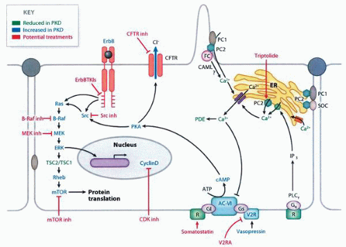
FIGURE 1-27 ▪ Autosomal dominant polycystic kidney disease. Cellular changes with polycystic kidney disease. Components and pathways that are down-regulated and up-regulated are indicated. Potential treatments that target these defective pathways are shown in red. (From Harris PC, Torres VE. Polycystic kidney disease. Annu Rev Med 2009;60:325, with permission.)

The association with malignant tumors is controversial.69 It has been estimated that the number of cases of renal cell carcinoma in patients with autosomal dominant polycystic kidney disease is no more than could be expected by chance.68 Other observations such as a higher incidence of multifocal and bilateral tumors in these patients indicate an increased risk of tumor development.70 The frequent development of epithelial proliferations including papillary adenomas in these cysts also would support the latter point of view.71


Pathology


Gross Features.

Involved kidneys are diffusely enlarged, weighing on average 5 to 10 times the normal72 (Fig. 1-28). There is diffuse involvement of the kidney parenchyma by unilocular cysts with the normal reniform shape of the kidney being generally maintained (Fig. 1-29). The cysts range from less than a millimeter to several centimeters. Most contain translucent clear to straw-colored fluid, but hemorrhagic fluid and blood clots may be present. The amount of intervening stroma depends on the degree of advancement of the process.


Microscopic Features.

The diagnosis of autosomal dominant polycystic kidney disease requires the demonstration of normal renal elements in the septa between the cysts (Fig. 1-30). These elements may be distorted by secondary changes related to compression or pyelonephritis. The cysts affect any part of the nephron from the glomerulus to the distal collecting ducts.54 The cysts are lined by flattened to cuboidal epithelial cells. In over 90% of kidneys, areas of epithelial hyperplasia can be identified within the cysts ranging from increased numbers of cell layers to papillary proliferations (Fig. 1-31A and B).71 In some cases, the papillary proliferations can become more prominent and complex (Fig. 1-32).


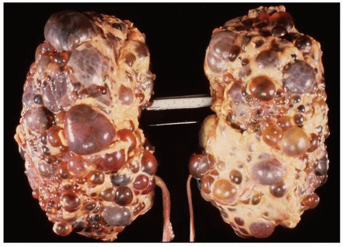
FIGURE 1-28 ▪ Autosomal dominant polycystic kidney disease. Bilateral nephrectomy specimen with markedly enlarged kidneys, normal reniform shape, and the external surface distorted by innumerable cysts of variable size.


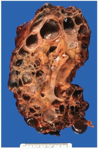
FIGURE 1-29 ▪ Autosomal dominant polycystic kidney disease. Cut surface of a kidney with innumerable cysts replacing the entire kidney. In this 57-year-old patient there is no grossly visible normal parenchyma.


Medullary Nephronophthisis


Genetics

Nephronophthisis refers to multiple disease complexes having essentially identical pathologic findings and mutations involving the NPHP family of genes (NPHP1 to NPHP9).73, 74, 75, 76, 77 These are also referred to as the juvenile, infantile, and adolescent forms. They are autosomal recessive inherited disorders.74,78 The pure renal juvenile form is related to a gene NPHP1 on chromosome 2q1379 with an infantile form related to NPHP2 and an adolescent form to
NPHP3. The NPHP3 gene has been implicated in a small subset of patients with the infantile form.76 The NPHP genes code for a series of proteins known as nephrocystins that are expressed in primary cilia or centrosomes of renal epithelial cells (Fig. 1-33).75,80 It has been suggested that this group of diseases is best classed as “ciliopathies.”81 Cysts presumably develop due to altered signaling pathways resulting in cell polarity and tissue maintenance abnormalities.80


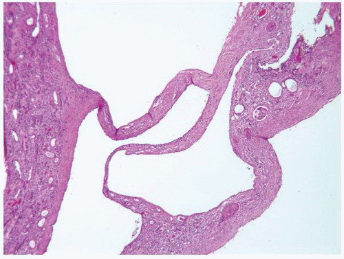
FIGURE 1-30 ▪ Autosomal dominant polycystic kidney disease. Several cysts with varying amounts of stroma containing residual renal parenchyma. In the wider septa residual normal epithelial elements are apparent.
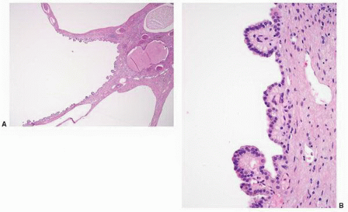


FIGURE 1-31A,B: Autosomal dominant polycystic kidney disease. The photomicrograph shows several cysts with varying amounts of stroma and residual parenchyma between them. In one of the cysts, there are multiple small papillae protruding into the lumen. These are a relatively common finding and are covered by cuboidal epithelium without cytologic atypia.


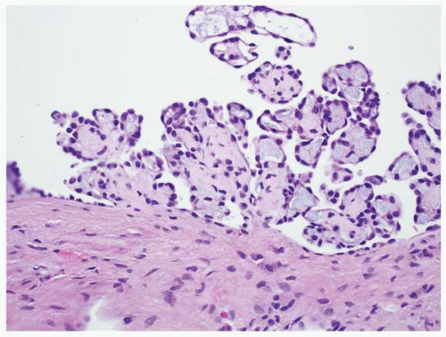
FIGURE 1-32 ▪ Autosomal dominant polycystic kidney disease. Infrequently the papillae become more complex but retain the very bland cytologic features. The nature of these proliferations is unknown, but there is no evidence to suggest they have any clinical significance.


Clinical Features

The incidence varies geographically with approximately 1 in 50,000 to 1 in 100,000 births affected in North America.75 The presentation is dependent on the type. Both sexes are affected equally. The juvenile form is most common and is estimated to be responsible for 5% to 10% of cases of end-stage renal failure in children.75 Most patients present between 4 and 6 years of age with polyuria and polydipsia related to a decrease in urinary concentrating ability and loss of sodium conservation. The diagnosis is confirmed by radiologic studies with renal parenchymal hyperechogenicity and loss of corticomedullary differentiation. Development of small medullary cysts occurs later. End-stage renal disease develops in the early teens but can occur later. In 10% to 20% of patients, there are extrarenal manifestations with ocular, neurologic, liver, and musculoskeletal being most frequent (Table 1-4).74,75 The adolescent form is associated with the development of end-stage renal disease at an older age (mean 19 years vs. 13 years for the juvenile form). The infantile form progresses to end-stage renal disease much more rapidly, usually by 2 years of age. Clinically severe hypertension is common.

Treatment is supportive with most patients developing end-stage renal disease requiring dialysis or transplantation within 5 to 10 years of diagnosis.82 The disease does not recur after transplantation.83


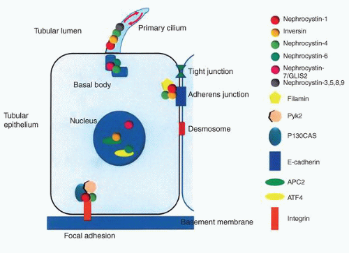
FIGURE 1-33 ▪ Nephronophthisis. Schematic representation of a tubular epithelial cell and the subcellular localization of the nephrocystin proteins. Most of the nephrocystin proteins interact with one another forming a nephrocystin complex. (From Salomon R, Saunier S, Niaudet P. Nephronophthisis. Pediatr Nephrol 2009;24:2338, with permission.)


Pathology


Gross Features.

The process is bilateral with affected kidneys being normal or slightly smaller than normal with thinning of both the cortex and the medulla.84 Cysts are multiple, <2 cm in size, and are concentrated at the corticomedullary junction but may involve the medulla and pyramids (Fig. 1-34).85 In the infantile form the kidneys can be enlarged mimicking autosomal recessive polycystic kidney disease. In some cases no cysts are recognizable grossly.


TABLE 1-4 ▪ EXTRARENAL MANIFESTATIONS OF NEPHRONOPHTHISIS


Ocular


Retinitis pigmentosa


Coloboma


Isolated oculomotor apraxia


Nystagmus


Ptosis


Neurologic


Mental retardation


Cerebellar ataxia


Hypopituitarism


Liver


Fibrosis


Biliary duct proliferation


Skeletal


Short ribs


Phalangeal cone-shaped epiphyses


Postaxial polydactyly


Skeletal dysplasia


Other


Situs inversus


Cardiac malformations


Ectodermal dysplasia


Microscopic Features.

The cysts derive from the loop of Henle, distal convoluted tubules, and collecting ducts and are lined by a flattened to squamous type of epithelium.86,87 The remainder of the parenchyma shows diffuse interstitial
fibrosis, tubular basement membrane thickening, atrophic dilated tubules, periglomerular fibrosis, and numerous sclerotic glomeruli resulting in features similar to that found in chronic pyelonephritis. The dilated tubules contain Tamm-Horsfall protein. Ultrastructurally the tubular basement membranes show patchy basement membrane thickening with duplication alternating with areas of splitting and thinning. The basement membrane changes are seen in the juvenile and adolescent but not the infantile forms.75


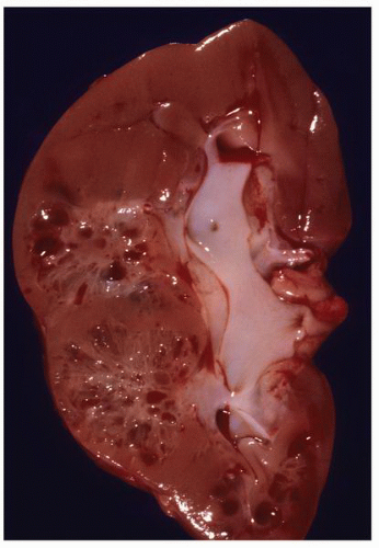
FIGURE 1-34 ▪ Nephronophthisis. Kidney from a patient with juvenile nephronophthisis showing multiple small cysts within the renal medullary pyramids.


Medullary Cystic Disease


Genetics

This is inherited as an autosomal dominant disease that includes two and possibly a third gene. Medullary cystic disease was first linked to a gene on chromosome 1q21-q23 that is now known as the medullary cystic kidney disease 1 gene (MCKD1); however, the specific gene involved has not yet been identified.88 A second gene, MCKD2, has been localized to chromosome 16p12.89 This is also the site of mutation in familial juvenile hyperuricemic nephropathy and some cases of glomerulocystic disease. The MCKD2 gene codes for the protein uromodulin (Tamm-Horsfall protein). The specific type of mutation of the uromodulin gene has a modest effect on kidney survival.90 A third possible gene at 1q41 has been proposed.91


Clinical Features

Medullary cystic disease and nephronophthisis have similar clinical features. Patients present with polydipsia and polyuria, have cysts at the corticomedullary junction and in the medulla, and ultimately can develop end-stage renal disease. The mean age for the development of end-stage renal failure (type 1, 62 years; type 2, 32 years) is older than for nephronophthisis.74 The clinical features are quite variable. There is a known association with hyperuricemia and gout.92,93 The cysts are often clinically undetectable and are bilateral in a minority of patients.


Pathology


Gross Features.

There is considerable overlap in the pathologic features of medullary cystic disease and nephronophthisis. The kidneys are normal to slightly small with cysts localized to the corticomedullary junction and the medulla (Fig. 1-35). The cysts tend to be smaller than in nephronophthisis.


Microscopic Features.

The findings are similar to nephronophthisis described above including the characteristic basement membrane changes. In type 2 medullary cystic kidney disease, immunohistochemistry for thrombomodulin demonstrates abnormal dense deposits in the renal tubular epithelial cells.94 Because of the association with hyperuricemia, changes of gouty nephropathy might also be present.


von Hippel-Lindau Disease


Genetics

von Hippel-Lindau disease is inherited as an autosomal dominant genetic disorder due to a defect in the von Hippel-Lindau gene (VHL) located at 3p25-p26.95,96 It occurs in one of every 30,000 to 50,000 live births.97 Different types of mutations have been identified with specific patterns of expression.98 Truncating mutations are associated with all the typical features of the disease except the development of pheochromocytoma (type 1). In contrast, missense mutations result in a high risk for the development of pheochromocytoma (type 2).99


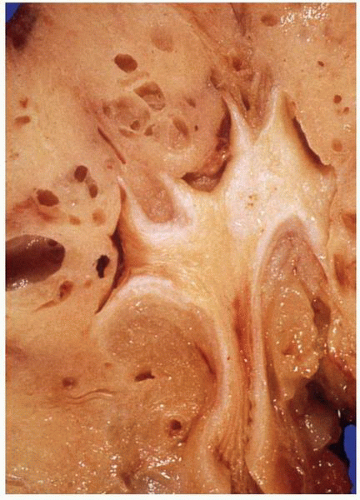
FIGURE 1-35 ▪ Medullary cystic disease. Kidney showing multiple cysts within the renal medullary areas.


Clinical Features

Renal cysts are detected in up to 75% of affected individuals.97,100 In addition to renal tumors, patients also develop tumors of the adrenal gland (pheochromocytoma), pancreas (islet cell tumors, serous cystic tumors), central nervous system (hemangioblastoma), petrous bone (endolymphatic sac tumors),101 and retina (angioma). Males and females can have papillary cystadenoma of the epididymis and the broad ligament, respectively (Table 1-5). Renal cell carcinoma develops in about 50% of patients with many having multiple bilateral tumors.97,102,103


Pathology


Gross Features.

The kidneys are generally normal in size unless involved by large tumors. Several lesions occur in the kidney including benign cysts, cysts with intracystic tumor formation, adenomas, and renal cell carcinoma (Fig. 1-36).100,104 The cysts can be unilateral or bilateral and are variable in size but are generally small (<5 cm). Larger
cysts can be multilocular. Tumors are typically multifocal with a variegated appearance (Fig. 1-37). In a study of 33 kidneys from 23 patients with von Hippel-Lindau disease, 190 solid lesions were identified.105


Table 1-5 ▪ EXTRARENAL MANIFESTATIONS OF VON HIPPEL-LINDAU DISEASE


Cerebellar hemangioblastoma


Retinal hemangioblastoma


Spinal hemangioma


Ependymoma


Endolymphatic tumor of the inner ear


Pancreatic cysts


Pancreatic serous cystadenoma


Pancreatic islet cell tumors


Hepatic cysts


Hepatic adenoma


Splenic cysts


Splenic angioma


Epididymal papillary cystadenoma


Broad ligament papillary cystadenoma


Café au lait spots (skin)


Syringobulbia


Syringomyelia


Microscopic Features.

The cysts are lined by an attenuated epithelium or cuboidal to columnar cells with clear to eosinophilic cytoplasm (Figs. 1-38 and 1-39). These may show stratification, and even solid nodules of clear cell renal cell carcinoma can be found inside the cysts.106 In 138 cystic lesions examined microscopically by Paraf et al.,105 103 (75%) were simple cysts, 20 (14%) were atypical with a multilayered clear cell epithelium, and 15 (11%) were cystic clear cell renal cell carcinomas. Hemorrhage into the cysts is common, and hemosiderin-laden macrophages can be present in the cyst lumen or surrounding fibrous tissue. The clear cells lining the cysts have an immunohistochemical expression profile similar to that of clear cell renal cell carcinoma. In a study of grossly normal renal parenchyma, Walther et al.107 found numerous microscopic abnormalities ranging from cysts to tumors. Based on these findings, the authors estimated that the average kidney in von Hippel-Lindau disease contains approximately 1,000 small cysts and 600 small tumors.107 The parenchyma can contain microscopic foci of
clear cell proliferation, and individual abnormal clear cells can be present within tubules (Fig. 1-40). These also have an immunohistochemical profile similar to that of clear cell renal cell carcinoma.


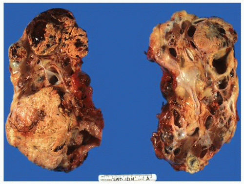
FIGURE 1-36 ▪ von Hippel-Lindau disease. Bilateral nephrectomy from a young woman showing multiple variably sized cysts and multiple clear cell renal cell carcinomas.


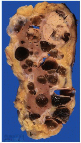
FIGURE 1-37 ▪ von Hippel-Lindau disease. Cut surface of a nephrectomy specimen illustrating multiple cysts with the development of tumors evident within several of the cysts.


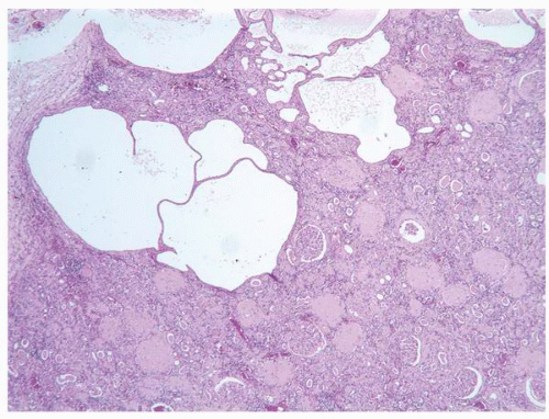
FIGURE 1-38 ▪ von Hippel-Lindau disease. Multiple variably sized cysts lined by flattened epithelium. There are changes secondary to chronic pyelonephritis in the background.


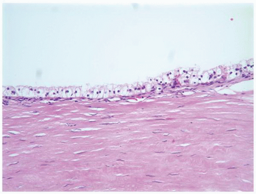
FIGURE 1-39 ▪ von Hippel-Lindau disease. Example of a cyst lined by clear cells with morphologic features typical of the cells of clear cell renal cell carcinoma.


Tuberous Sclerosis


Genetics

The tuberous sclerosis complex is an autosomal dominant inherited disorder with a high (95%) degree of penetrance.108 The prevalence is estimated at 1:11,000 births.109 The disease is due to germline mutations of the TSC1 (9q34) or TSC2 (16p13.3) genes.110,111 The disease manifestations tend to be less severe in the TSC1-related cases.112 The protein products of the TSC1 and TSC2 genes are hamartin and tuberin, respectively. The adult polycystic kidney disease gene PKD1 is located in the immediate vicinity of the TSC2 gene and in some patients both genes are affected resulting in features of both conditions being found in the same kidney (TSC2/ADPKD1 contiguous gene syndrome).53 The TSC1 and TSC2 proteins have a critical role in cell proliferation and death through mTOR-dependent and mTOR-independent signaling pathways.113


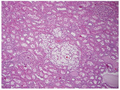
FIGURE 1-40 ▪ von Hippel-Lindau disease. Tiny focus of clear cell renal cell carcinoma. Note the edge of a small cyst in the right lower corner that is lined by clear cells similar to those illustrated in Figure 1-39.


Table 1-6 ▪ MANIFESTATIONS OF TUBEROUS SCLEROSIS COMPLEX


Major features


Renal angiomyolipoma


Lymphangioleiomyomatosis


Facial angiofibroma


Ungula and periungual fibroma


Hypomelanotic macules


Shagreen patches


Retinal hamartoma and astrocytoma


Cortical tubers


Subendymal nodules and giant cell astrocytoma


Cardiac rhabdomyoma


Minor features


Renal cysts


Hamartomatous rectal polyp


Bone cysts


Cerebral white matter migration lines


Gingival fibroma


Enamel dental pits


Retinal achromic patches


Confetti skin lesions


Clinical Features

Tuberous sclerosis is a complex disease characterized by multiple manifestations involving multiple organs.114,115 Currently the diagnosis of tuberous sclerosis is defined by a combination of major and minor features (Table 1-6).116 The diagnosis requires two or more distinct lesions rather than multiple single lesions in one organ. Although the clinical picture is often dominated by neurologic complications such as mental retardation and seizures, the renal lesions are significant. The combination of cysts and angiomyolipomas may reduce the functional renal mass and result in renal failure. In the TSC2/ADPKD1 contiguous gene syndrome, severe cystic kidney disease may be present at birth.117 Second, angiomyolipomas can become large and can be complicated by retroperitoneal hemorrhage. Finally these patients have a significantly increased risk for the development of renal cell carcinoma at a young age including in children.118 Targeted therapies hold promise in the treatment of the associated neurodevelopmental disorders, angiomyolipoma, and other manifestations of this disease.119, 120, 121


Pathology


Gross Features.

Indications for nephrectomy in patients with tuberous sclerosis are usually an enlarging angiomyolipoma or the presence of a solid tumor suspicious for renal cell carcinoma. The combination of characteristic cysts and angiomyolipomas is typical of this inherited disorder
(Fig. 1-41).122,123 In one study, 80% of children with tuberous sclerosis developed renal lesions by age 10 with angiomyolipomas (75%) being more common than cysts (17%).124 Approximately 50% of patients develop renal cysts. The cysts are generally small and can be located in the cortex and medulla. There may only be a few cysts or they can be sufficiently numerous to produce a sponge-like appearance. The tumors range from a few millimeters to several centimeters and have a variable appearance depending on the proportion of the components; hemorrhage is common.


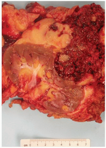
FIGURE 1-41 ▪ Tuberous sclerosis. Nephrectomy specimen with multiple angiomyolipomas (small yellow nodules). The large tumor is an epithelioid angiomyolipoma with rupture and a retroperitoneal hemorrhage.


Microscopic Features.

The cysts are characteristically lined by tall cells with granular eosinophilic cytoplasm and large nuclei resembling proximal tubular epithelial cells (Figs. 1-42 and 1-43). Pseudostratification and papillary tufting can be present (Fig. 1-44).125 Cysts involving the glomeruli are also a characteristic feature. The cysts can contain eosinophilic secretions. In the absence of grossly visible tumors, there are frequently microscopic angiomyolipomas (Figs. 1-45 and 1-46). The pathology of the tumors in tuberous sclerosis is covered in detail in Chapter 2.


Multicystic Renal Dysplasia

Although the pathogenesis continues to be the subject of debate, it is generally accepted that most cases of renal dysplasia are related to urinary tract obstruction or urinary reflux during kidney development.24,26 Multicystic dysplastic kidneys are also a feature of many genetic disorders (Table 1-7).14,52


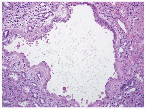
FIGURE 1-42 ▪ Tuberous sclerosis. Cyst lined by cells with moderate to abundant amphophilic cytoplasm.


Clinical Features

Renal dysplasia is defined by the presence of abnormal renal organization with abnormal differentiation of metanephric elements. An associated urinary tract anomaly is identifiable in up to 90% of affected individuals; among the most frequent are ureteral atresia and urethral valves. In dysplasia related to reflux, the changes can be focal and the kidney can retain some level of function; otherwise, dysplastic kidneys are characteristically nonfunctional.

Multicystic dysplastic kidneys present as a flank mass in newborns and the diagnosis can be confirmed by ultrasound.57,126 In up to 40% of affected patients, there are contralateral urinary tract abnormalities.127 Nephrectomy has
been the standard therapy, but conservative treatment with careful monitoring for possible tumor development is an alternative.128 Nephroblastoma and renal cell carcinoma arising in dysplastic kidneys are described.129,130 Intrauterine diagnosis with subsequent treatment prior to birth has been tried with varied success.


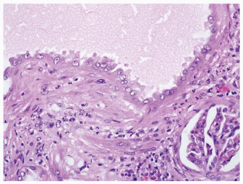
FIGURE 1-43 ▪ Tuberous sclerosis. High-power photomicrograph of the lining epithelium in a cyst.


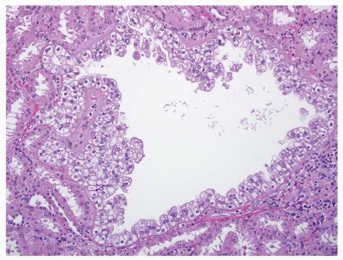
FIGURE 1-44 ▪ Tuberous sclerosis. In this cyst the lining epithelium is pseudostratified and shows cytoplasmic clearing.


Pathology


Gross Features.

Most dysplastic kidneys are small, abnormally shaped, and have multiple cysts (Figs. 1-47 and 1-48).131 In other cases, the dysplastic kidney consists of only a small nodule of rudimentary metanephric tissue. The cysts in dysplastic kidneys communicate,132 and their size and location may correlate with the level of the obstruction.133 Dysplasia can be segmental or focal.134,135


Microscopic Features.

The histologic hallmarks of dysplastic kidneys are lobar disorganization, primitive ducts, and metaplastic cartilage136 (Fig. 1-49). There is only primitive development of the renal medulla. Collecting ducts are surrounded by a thick fibromuscular collar and may become cystic (Fig. 1-50). Small nests of hyaline cartilage are found in the cortex but not in all cases (Fig. 1-51). Nodules of renal blastema may be present (Fig. 1-52).129,131 The presence of keratinizing squamous epithelium has been described (Fig. 1-53).137,138


FIGURE 1-45 ▪ Tuberous sclerosis. A small subcapsular angiomyolipoma that is composed entirely of smooth muscle.


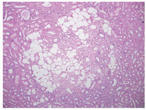
FIGURE 1-46 ▪ Tuberous sclerosis. A small intraparenchymal angiomyolipoma with fat and smooth muscle.


Glomerulocystic Disease

Glomerular cysts are defined by dilation of Bowman space to two or three times the normal size. Glomerulocystic kidney

disease as an entity is reserved for inherited types of the disease. The major form is inherited as an autosomal dominant condition. The subtypes include (i) autosomal dominant glomerulocystic kidney disease related to uromodulin gene (UMOD) mutations, (ii) familial hypoplastic glomerulocystic kidney disease due to mutations in the TCF2 gene (hepatocyte nuclear factor 1β), and (iii) other genetic causes.139 Glomerular cysts occur in a wide range of other conditions including autosomal dominant polycystic kidney disease, autosomal recessive kidney disease, numerous syndromes, and urinary obstruction with or without dysplasia (Table 1-8).139


Table 1-7 ▪ MALFORMATION SYNDROMES ASSOCIATED WITH RENAL DYSPLASIA


Syndrome


Gene(s)


Alagille syndrome


JAG1, NOTCH2


Bardet-Biedl syndrome


BBS1-BBS11


Branchiootorenal syndrome


EYA1, SIX1, SIX2


Di George syndrome


Del. 22q11


Hypothyroidism, sensorial deafness, renal anomalies


GATA3


Fraser syndrome


FRAS1, FREM2


Kallmann syndrome


KALL1, FGFR1


Renal cysts and diabetes syndrome


TCTF2


Simpson-Golabi-Behmel syndrome


GPC3


Smith-Lemli-Opitz syndrome


DHCR7


Townes-Brocks syndrome


SALL1


Cornelia de Lange syndrome


NIPBL


Zellweger syndrome


PEX family


Pallister-Hall syndrome


GLI3


Beckwith-Wiedemann syndrome


p57(KIP2)


Meckel-Gruber syndrome


Unknown


Adapted from Sanna-Cherchi S, Caridi G, Weng PL, et al. Genetic approaches to human renal agenesis/hypoplasia and dysplasia. Pediatr Nephrol 2007;22:1675-1684.


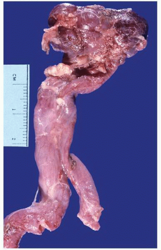
FIGURE 1-47 ▪ Multicystic dysplasia. The kidney consists of a small irregularly, somewhat reniform shaped piece of soft tissue. The cysts are not grossly visible on the surface. There is an associated bifid hydroureter.


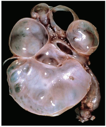
FIGURE 1-48 ▪ Multicystic dysplasia. In this example, the residual kidney is almost entirely cystic with little solid tissue.


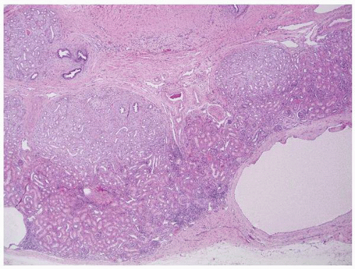
FIGURE 1-49 ▪ Multicystic dysplasia. This is an example of segmental cystic dysplasia with preserved renal parenchyma, cysts, and immature stroma with entrapped tubules.


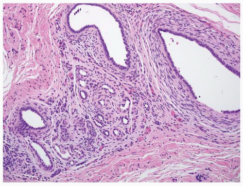
FIGURE 1-50 ▪ Multicystic dysplasia. Small tubules are surrounded by a cuff of immature stroma.


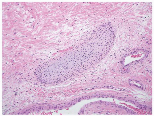
FIGURE 1-51 ▪ Multicystic dysplasia. There is a nest of cartilage within loose fibrous connective tissue.


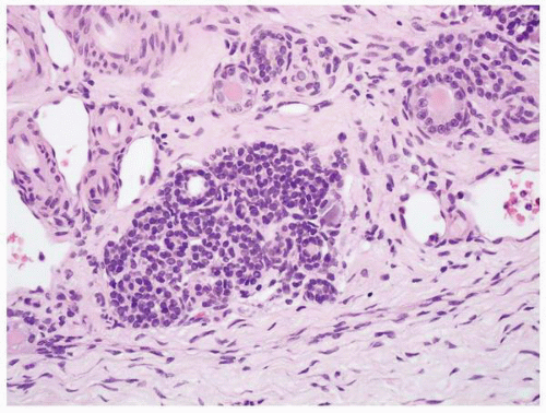
FIGURE 1-52 ▪ Multicystic dysplasia. In this example, there is a nephrogenic rest with adjacent tubules and loose fibrous connective tissue.


Clinical Features

The clinical significance is dependent on the underlying etiology. Diagnosis is made by radiology examination. In the fetus and neonate, differentiation from other cystic diseases may not be possible.140 In the inherited types, the disease may present early or late. In early onset, renal insufficiency develops in infants. Progression to end-stage renal disease occurs over a highly variable period of time. In more aggressive cases, renal failure can occur within 3 years of diagnosis.139 In adults, the disease is usually asymptomatic and an infrequent cause of end-stage renal disease.


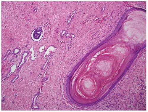
FIGURE 1-53 ▪ Multicystic dysplasia. In this unusual case, the lining of a cyst shows keratinizing squamous metaplasia.


Table 1-8 ▪ CONDITIONS WITH GLOMERULAR CYSTS


Glomerular cystic kidney diseases (inherited)


Autosomal dominant glomerulocystic kidney disease


Familial hypoplastic glomerulocystic kidney disease


Other inherited variants


Other inherited conditions and syndromes


Autosomal dominant polycystic kidney disease


Autosomal recessive polycystic kidney disease


Tuberous sclerosis


von Hippel-Lindau disease


Familial juvenile nephronophthisis


Congenital nephrotic syndrome of the Finnish type


Many others


Glomerular cysts in dysplastic kidneys


Renal dysplasia associated with congenital obstruction


Zellweger syndrome


Meckel syndrome


Many others


Miscellaneous other conditions


Hemolytic uremic syndrome


Systemic lupus erythematous


Sjögren syndrome


And many others


Adapted from Lennerz JK, Spence DC, Iskandar SS, et al. Glomerulocystic kidney: one hundred-year perspective. Arch Pathol Lab Med 2010;134:583-605.


Pathology


Gross Features.

In cases associated with autosomal dominant or autosomal recessive kidney disease, the gross features are those of the associated condition. In the inherited forms, the kidneys may be enlarged with variable numbers of small cysts (1 to 3 mm) grossly visible.


Microscopic Features.

By definition the glomerular space is two to three times the normal size. The cysts are variable in size. In even large cysts, small residual glomerular tufts may be identifiable (Fig. 1-54). In cases related to other disorders, the histopathology of that condition is present (Table 1-8).


Medullary Sponge Kidney

The etiology of medullary sponge kidney is unknown.74 The GDNF gene has been implicated as having a role.141 There is no evidence that it is an inherited condition although the hypothesis that papillary duct ectasia is a congenital anomaly is favored.52 It has been described in association with Marfan syndrome, Ehlers-Danlos syndrome, and Caroli disease.


Clinical Features

Medullary sponge kidney is a sporadic condition found in 1:5,000 live births.142 Males and females are affected equally,
and it is seen in association with hemihypertrophy in about 10% of cases.143 The condition is usually asymptomatic and is discovered incidentally in children or in patients in their third or fourth decades presenting with renal lithiasis144,145; the latter occurs in about 50% of patients.146 Diagnosis is usually made by computed tomographic urography.147 These patients rarely progress to chronic renal failure but complications such as recurring urolithiasis, pyelonephritis, and septicemia may cause significant clinical problems.148 Treatment is generally directed at the complications.


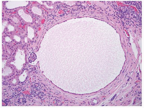
FIGURE 1-54 ▪ Glomerulocystic disease. An example of a glomerular cyst in a case of adult polycystic kidney disease.


Pathology


Gross Features.

The kidneys are normal or slightly enlarged in most cases. The cysts are small (<5 mm) and localized to the medullary pyramids and papillary tips (Fig. 1-55).145 Small calculi are often present within the cysts.149


Microscopic Features.

The cysts derive from the collecting ducts; in addition to cysts, ectatic ducts in the papillae are also present. The cysts are lined by urothelial, columnar, or squamous epithelium.143,149 Interstitial fibrosis and inflammation are present, and features resembling dysplasia including cartilage can be found.143


Simple Cysts

The cysts are believed to develop from diverticula that are thought to arise in the distal convoluted or collecting tubules.150 These increase in frequency with advancing age, correlating with the increasing occurrence of cysts.151


Clinical Features

Simple renal cysts have an increasing prevalence with advancing age and are found in up to 50% of kidneys at autopsy.151,152 Simple cysts can be single or multiple and are often unilateral. These are usually asymptomatic and their major significance lies in differentiating them from renal neoplasms.84 In most instances, this is accomplished by radiologic evaluation but some cases are aspirated and others require surgical exploration. Symptomatic cysts have been managed by multiple approaches including percutaneous aspiration, sclerotherapy, cyst decortication, cystectomy, and cystoretroperitoneal shunt.153, 154, 155


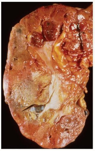
FIGURE 1-55 ▪ Medullary sponge kidney. The kidney shows several small cysts that include several at the corticomedullary junction.


Pathology


Gross Features.

The cysts may be solitary or multiple and typically are unilocular although a multilocular appearance can be seen (Fig. 1-56A and B). Most are under 5 cm, but much larger cysts have been described. The fluid is under tension and is straw-colored. Cysts can be hemorrhagic or may become infected (Fig. 1-57). The inner lining is most often smooth and glistening; a shaggy, irregular surface and/or a thick wall should heighten suspicions of a malignant tumor.


Microscopic Features.

The cysts are lined by a flattened single-layered epithelium that can be difficult to demonstrate in larger lesions (Fig. 1-58). There may be associated atrophy of adjacent renal parenchyma with fibrosis, but in general the kidney is not diseased other than age-related changes. The lack of renal disease is helpful in distinguishing this from acquired cystic kidney disease. Cases complicated by hemorrhage or infection can have a thickened wall with hemosiderin-laden macrophages and a mixed inflammatory infiltrate (Fig. 1-59).


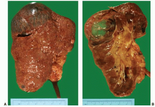
FIGURE 1-56 ▪ Cortical cyst. A large, intact benign cortical cyst can be seen bulging from the external surface of the kidney (A). The opened cyst shows a thin translucent lining (B).


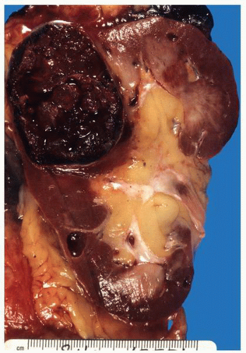
FIGURE 1-57 ▪ Cortical cyst. This is an example of a benign cortical cyst complicated by intracystic hemorrhage.


Acquired Cystic Disease

Acquired cystic disease is a well-described complication of hemodialysis and peritoneal dialysis but can also occur in patients with prolonged azotemia without dialysis.156, 157, 158, 159 It can develop on a background of any chronic renal disease and is seen in up to 50% of all patients on hemodialysis.160 The incidence increases with increasing time on dialysis.161


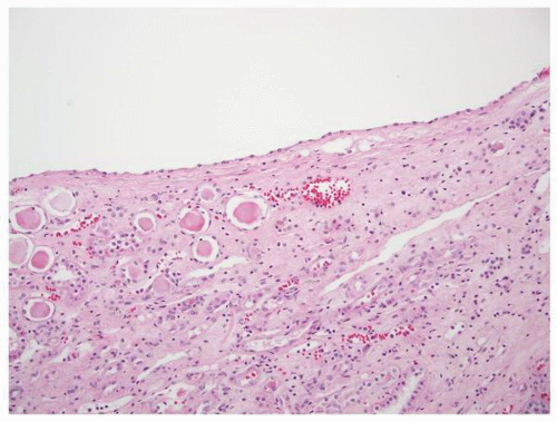
FIGURE 1-58 ▪ Cortical cyst. The lining of an uncomplicated cyst has a flattened epithelium and a thin wall of connective tissue.


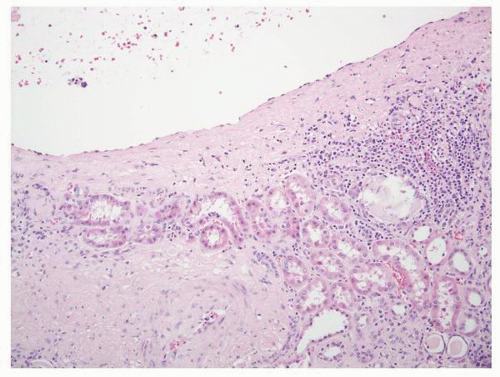
FIGURE 1-59 ▪ Cortical cyst. The wall of this cortical cyst is thickened with dense fibrous tissue, and there is an associated chronic inflammatory infiltrate.

The pathogenesis remains unknown with many theories having been presented over the years.157


Clinical Features

The diagnosis requires the demonstration of a minimum of five renal cysts in each kidney of patients with end-stage renal disease.159 Most patients have no symptoms directly related to the development of acquired cystic disease. If symptoms do develop, they usually relate to bleeding resulting in gross or microscopic hematuria and rarely significant retroperitoneal, subcapsular, or intrarenal hemorrhage.162 Of most importance is the association with the development of renal cell carcinoma.156 Chronic dialysis and acquired cystic disease are associated with an estimated 10- and 50-fold increased risk of renal cell carcinoma, respectively.156,163, 164, 165 Acquired cystic disease does not require specific treatment. Most authors advocate periodic imaging studies with computed tomography to evaluate for the development of carcinoma. In the past, nephrectomy has been recommended if a tumor larger than 3 cm develops84; more recently, early detection and treatment has been stressed.166,167 Interestingly, transplantation reduces the risk of tumor development even with the native kidneys left in situ.157 Renal cell carcinoma can also develop in acquired cystic disease occurring in allografts.168 Surgical intervention may also be indicated in some cases complicated by hemorrhage.


Pathology


Gross Features.

In most cases, the kidneys are smaller than normal although there is a wide variation in size (Figs. 1-60 and 1-61). In rare cases, the kidney can be enlarged.169 The number of cysts is highly variable, and a minimum of five is recommended as a diagnostic criterion to separate this from simple cysts.159 The cysts can occur anywhere but are predominantly cortical. Size ranges from a few millimeters to several centimeters; however, most are small (2 mm or less).169 The cysts contain translucent fluid, but hemorrhage may be seen. In 15% of kidneys, single or multiple grossly visible tumor nodules are present and in 5% these are more than several centimeters in size (Fig. 1-61). Such lesions should be well-sampled for histologic evaluation, particularly any hemorrhagic or yellow tumors that are more likely to represent clear cell carcinoma.


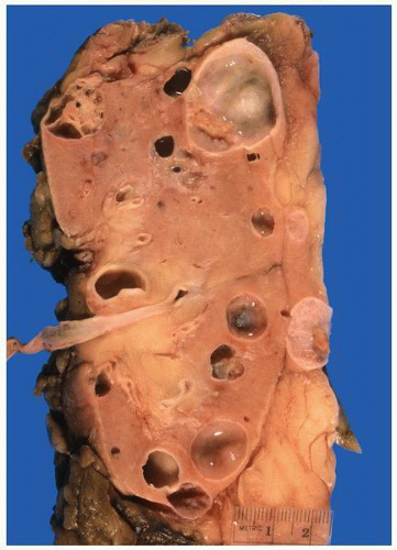
FIGURE 1-60 ▪ Acquired cystic disease. The cut surface of this relatively normal sized kidney shows multiple cysts. The cysts are varied in size and in one a multilocular architecture is present (upper left). Histologically this was a clear cell papillary renal cell carcinoma.


Microscopic Features.

The cysts can arise from any part of the tubular structure including proximal and distal tubules and collecting ducts (Fig. 1-62).170 The kidney parenchyma shows changes of end-stage renal disease with interstitial fibrosis, sclerotic glomeruli, and atrophic tubules. In patients on dialysis, calcium oxalate crystal deposition is present in most cases (Fig. 1-63). The cysts are lined by a singlelayered cuboidal to columnar epithelium that can show a range of hyperplastic changes up to and including formation of adenomas and carcinomas169,171,172 (Figs. 1-64 and 1-65). Atypical epithelial proliferations within cysts have been shown to have cytogenetic abnormalities including gains in chromosomes 7, 12, 17, 20, and Y, supporting the hypothesis that these represent the precursor of at least some of the tumors that develop in these kidneys.173 Papillary adenomas are a frequent finding in the renal parenchyma (Fig. 1-66). Tumors may develop within the cysts or in the adjacent parenchyma.172 The pathology of neoplasms developing in this setting is described in detail in Chapter 2.


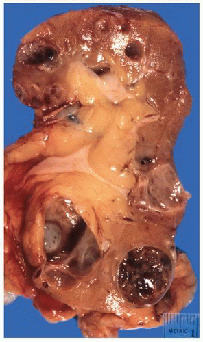
FIGURE 1-61 ▪ Acquired cystic disease. In this example the kidney is small and contains multiple cysts. There is a hemorrhagic tumor arising within a cyst in the lower pole. Histologically the tumor was an acquired cystic disease-associated renal cell carcinoma.


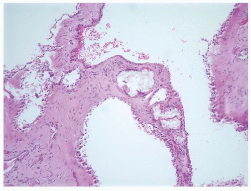
FIGURE 1-62 ▪ Acquired cystic disease. Several cysts in this image are lined by a single- to multilayered cuboidal epithelium. There is fibrotic stroma between the cysts that contains several calcium oxalate crystal deposits.


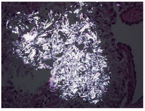
FIGURE 1-63 ▪ Acquired cystic disease. Calcium oxalate crystals as seen with polarized light.


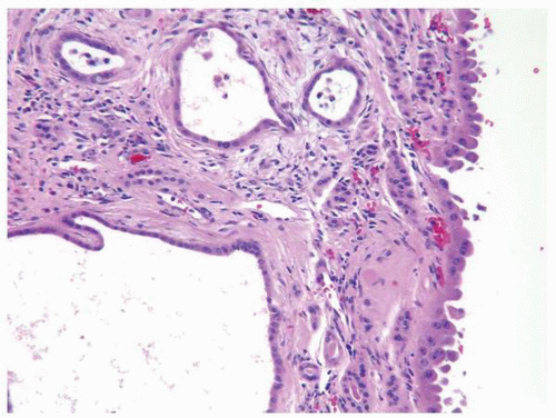
FIGURE 1-64 ▪ Acquired cystic disease. One of the cysts in this photomicrograph is lined by cells with moderate amphophilic cytoplasm and large nuclei that contain prominent nucleoli.


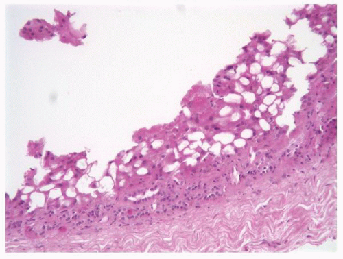
FIGURE 1-65 ▪ Acquired cystic disease. An example of a cyst lined by a pseudostratified epithelium. The cells have abundant eosinophilic cytoplasm and cytoplasmic vacuoles producing a sieve-like appearance.


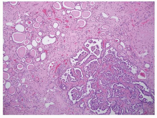
FIGURE 1-66 ▪ Acquired cystic disease. Papillary adenoma developing in a kidney with acquired cystic disease.

Jun 10, 2016 | Posted by in UROLOGY | Comments Off on Nonneoplastic Diseases of the Kidney

Full access? Get Clinical Tree

Get Clinical Tree app for offline access