Mineral and Bone Disorder in Chronic Kidney Disease


Mineral and Bone Disorder in Chronic Kidney Disease


Elvira O. Gosmanova

Darryl L. Quarles


Historically, renal osteodystrophy (ROD) was a term used to describe the metabolic bone disease caused by abnormalities in mineral homeostasis resulting from kidney failure.1 In recent years, the recognition of a complex endocrine regulation of mineral and bone metabolism, the prominent extra skeletal manifestations of chronic kidney disease (CKD), and the association between abnormalities in mineral metabolism and increased morbidity and mortality in patients with kidney failure led to formulation of a new term—chronic kidney disease-mineral and bone disorder (CKD-MBD) (Table 77.1)1—which describes a broader clinical syndrome, including metabolic/endocrine abnormalities, parathyroid gland dysfunction, bone disease, and unique CKD-associated cardiovascular risk factors as well as other adverse clinical outcomes, such as fractures and vascular and soft tissue calcifications (Fig. 77.1). In this chapter we review separate components of CKD-MBD, clinical manifestations, and general principles of CKD-MBD treatment.

Four organ systems, including the gut (absorption of calcium [Ca] and inorganic phosphate [Pi]), kidneys (reabsorption and excretion of Ca, Pi, and calcitriol synthesis), bones (interchange of Ca and Pi with extracellular pool, and FGF23 secretion), and parathyroid gland (parathyroid hormone [PTH] secretion) are involved in regulating mineral homeostasis2 and each play a role in the pathogenesis of CKD-MBD (Fig. 77.2).


BIOCHEMICAL ABNORMALITIES IN CKD-MBD


Calcium

There are three Ca pools in our body. The majority of Ca (99%) is found in bone. The remaining Ca is either intracellular (mostly protein bound), and extracellular (45% protein bound and 55% free calcium). PTH is a key regulator of serum calcium levels. The extracellular Ca concentration is tightly regulated by changes in PTH through sensing of calcium by the calcium-sensing receptor (CaSR)3 and through actions of PTH on bone and kidney. In turn, Ca controls PTH secretion, synthesis, and degradation as well as parathyroid cell hypertrophy and hyperplasia.4,5 PTH directly regulates the excretion of Ca by the kidney and also influences the exchange between bone and extracellular pools. PTH stimulates gut calcium absorption indirectly through the stimulation of 1,25(OH)2D3 production by the kidney, which in turn activates calcium absorption through vitamin D receptor (VDR)-dependent mechanisms. Decrements in 1,25(OH)2D3 levels occur prior to elevations of PTH during the progressive loss of glomerular filtration rate (GFR). The increments in PTH maintain serum calcium concentrations in the normal range until late in the course of CKD (Fig. 77.3A,B).6


Phosphorus

Pi is required for cellular function and skeletal mineralization and excess phosphate is associated with soft tissue and vascular calcifications. Serum Pi level is less tightly regulated than Ca, but is maintained in normal range through a complex interplay between intestinal absorption, exchange with intracellular and bone storage pools, and renal tubular reabsorption. Pi is abundant in the diet, and intestinal absorption of Pi is stimulated by 1,25(OH)2D. The kidney is a major regulator of Pi homeostasis, where increases or decreases in its Pi reabsorptive capacity under the control of various hormones determine serum Pi levels. The crucial regulated step in Pi homeostasis is the transport of Pi across the renal proximal tubule via the type II sodium-dependent phosphate (Na/Pi) cotransporter 2a (NPT2a) and 2c (NPT2c). PTH and FGF23 are the two principal hormones that regulate NPT2 translocation to the proximal tubular brush border membrane. PTH inhibits renal phosphate reabsorption due to reductions in membrane expression of NPT2. FGF23, a bone-derived hormone originally identified as the causative factor in inherited and acquired hypophosphatemic disorders also inhibits proximal tubular phosphate transport through mechanisms that remain to be defined. Increments in FGF23 appear to precede elevations of PTH in CKD, but both work in concert to prevent elevations in serum Pi by increasing renal phosphate excretion.6,7,8,9,10,11,12,13,14 Like Ca, serum Pi levels remain in the normal range until late in the course of
CKD, typically when glomerular filtration rate (GFR) is <30 to 40 mL/min/m2.6,8,11,15


TABLE 77.1 Classification of Chronic Kidney Disease: Mineral and Bone Disorder


CKD-MBD is either one or combination of the following:


▪ Abnormalities of calcium, phosphorus, PTH, or vitamin D metabolism, as measured by laboratory values


▪ Abnormalities in bone turnover, mineralization, volume, linear growth, or strength, as measured mainly by bone histology


▪ Vascular or other soft tissue calcifications


CKD-MBD, chronic kidney disease-mineral and bone disorder; PTH, parathyroid hormone.


Adopted from KDIGO clinical practice guideline for the diagnosis, evaluation, prevention, and treatment of Chronic Kidney Disease-Mineral and Bone Disorder (CKD-MBD). Kidney Int Suppl. 2009;(113):S1-130.


Vitamin D

Decrements in both 25(OH)D and 1,25(OH)2D occur early in the course of CKD-MBD. Low levels of vitamin D are associated with increased mortality in CKD and treatment with vitamin D analogues are believed to have a survival benefit.16 The mechanism for decreased circulating 25(OH)D levels in CKD are not well understood, but may result from poor nutritional status caused by chronic illness. Patients with CKD, however, may also be refractory to nutritional vitamin D supplementation, suggesting other mechanisms for decreased 25(OH)D levels.

1,25(OH)2D3, the active form of vitamin D, is synthesized from 25 (OH)D by 1α-hydroxylase- cytochrome P450, family 27, subfamily B, polypeptide 1(CYP27B1) located in the kidney proximal tubule. CYP27B1 is stimulated by PTH and inhibited by FGF23. Both 25(OH)D and 1,25(OH)2D are catabolized by 25-hydroxyvitamin D324-hydroxylase (CYP24), which is also present in the proximal tubule. CYP24 is stimulated by FGF23 and inhibited by PTH. Decrements in 1,25(OH)2D occur early in CKD. Diminished 1,25(OH)2D levels are seen with early GFR decline to less than 60 to 70 mL/min/m2 and are inversely related to elevation in FGF23.8 The reductions in 1,25(OH)2D in CKD were thought to be the result of reduced production of this hormone caused by the diseased kidney, but more recently it has been recognized that the suppression of 1,25(OH)2D production is a regulated process due to the effects of FGF23 on CYP27B1-mediated production and/or CYP24-mediated degradation of 1,25(OH)2D.17,18,19 Reduced GFR can additionally contribute to 1,25(OH)2D deficiency via decrease in renal uptake of 25-hydroxyvitamin D by proximal tubular cells for its activation to 1,25(OH)2D through decrease in amount of filtered 25-hydroxyvitamin D bound to vitamin D-binding protein available for uptake. Moreover, CKD leads to a decrease in kidney megalin content that is essential for the internalization of 25-hydroxyvitamin D into proximal tubular cells.20

The principal functions of 1,25(OH)2D are to promote active intestinal absorption of Ca and Pi, suppress PTH gene transcription in the parathyroid gland, stimulate bone formation and resorption in bone, as well as regulate the innate immune response in other tissues. Alterations in 1,25(OH)2D directly suppresses PTH gene expression through a genomic action21 via VDR on parathyroid cells. 1,25(OH)2D also indirectly regulates parathyroid gland function through elevations in serum calcium and stimulation of CaSR. Mouse genetic studies suggest that CaSR is dominant to VDR in regulation of parathyroid gland function, because calcium exerts PTH and VDR regulation in absence of any vitamin D source,22 whereas ablation of VDR in the parathyroid gland results in hyperparathyroidism that can be rescued by raising serum calcium levels.23,24 Several additional factors lead to impaired action of 1,25(OH)2D on parathyroid gland in uremia, such as diminished activation of VDR with reduced 1,25(OH)2D levels in CKD, and decrease in parathyroid VDR content, especially when nodular parathyroid hyperplasia is present.


FGF23

Elevations in circulating FGF23 levels are one of the earliest abnormalities in CKD-MBD and are strongly associated with increased all-cause mortality.25,26 Elevations of FGF23

inversely correlate with GFR.8,27,28,29 Patients with end-stage renal disease (ESRD) have markedly elevated levels of FGF23 that parallels with degree of hyperphosphatemia30 and secondary hyperparathyroidism (SHPT).31


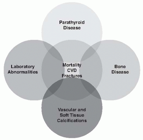
FIGURE 77.1 Spectrum of pathology in chronic kidney disease-mineral and bone disorder. (Modified from KDIGO clinical practice guideline for the diagnosis, evaluation, prevention, and treatment of Chronic Kidney Disease-Mineral and Bone Disorder (CKD-MBD). Kidney Int Suppl. 2009;(113):S1-130.)


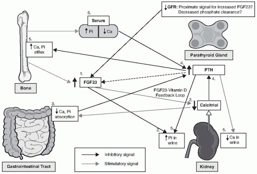
FIGURE 77.2 The schematic representation of chronic kidney disease-mineral and bone disorder pathogenesis. Ca, calcium; Pi, phosphorus. 1, Increase in FGF23; 2, suppression of calcitriol and increased urinary phosphate; 3, decreased gastrointestinal calcium and phosphorus absorption; 4, increased parathyroid hormone; 5, increased bone resorption/calcium and phosphorus bone efflux; increased phosphorus and decreased calcium in urine; 6, maintenance of serum phosphorus and calcium in normal range until stage V chronic kidney disease.


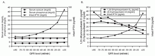
FIGURE 77.3 Relationship between glomerular filtration rate (GFR) and parathyroid hormone (PTH), calcium, phosphorus, and calcitriol levels in patients with chronic kidney disease. A: Median values of serum calcium, phosphorus, and intact PTH by GFR levels. B: Median values of 1,25-dihydroxyvitamin D,25-hydroxyvitamin D, and intact PTH by GFR levels. (Adopted from Levin A, Bakris GL, Molitch M, et al. Prevalence of abnormal serum vitamin D, PTH, calcium, and phosphorus in patients with chronic kidney disease: results of the study to evaluate early kidney disease. Kidney Int. 2007;71(1):31-38.)

FGF23 is a 32-kDa protein with an N-terminal region containing the FGF-homology domain and a novel 71-amino acid C terminus32,33 that interacts with FGF receptor (FGFR) in the presence of the members of Klotho family of proteins.34,35 In vitro studies indicate that Klotho is an essential cofactor for FGF23 to activate FGFR.34,36,37 Circulating FGF23 is mainly produced and secreted by osteoblasts and osteocytes in bone.38 The target organs for FGF23 are defined by the coexpression of the membrane form of Klotho and FGFR.37,39 Klotho is expressed in high levels in parathyroid gland, kidney, and several other organs; however, the kidney is the principal physiologically defined target for FGF23, where it inhibits phosphate reabsorption and 1,25(OH)2D synthesis. The parathyroid gland is also a target for FGF23 action, but it is not clear if FGF23 stimulates or inhibits PTH secretion. Elevated levels of FGF23 in human disease and mouse models are associated with hyperparathyroidism (HPT),40,41 likely due to the effect of FGF23 to suppress 1,25(OH)2D leading to the secondary development of HPT. In contrast, in vitro studies demonstrated that FGF23 activates extracellular regulated kinases 1/2 – Egr-1 pathway leading to inhibition of PTH mRNA expression and PTH secretion from parathyroid cells.42,43 Additionally, FGF23 suppresses parathyroid cell proliferation and increases CaSR and VDR expression in normal parathyroid gland.44 In individuals with normal kidney function, FGF23 exerts negative feedback on the parathyroid gland; however, the fact that in CKD patients PTH remains high despite elevated FGF23 suggests the presence of resistance to FGF23 action. This possibility was reinforced by a finding of a reduced Klotho and FGFR expression in surgically removed parathyroid glands from uremic patients.42

The mechanism of increased FGF23 in CKD is poorly understood. The increase in serum FGF23 is not explained by reduced FGF23 clearance; and the proximate stimulus in early CKD that leads to increments in FGF23 are not clear. Nevertheless, FGF23 production is likely increased to counteract Pi retention due to reduced nephron mass by promoting urinary Pi excretion.45 Elevations in FGF23 precede increments in PTH in CKD46 and animal studies show that blockade of FGF23 by neutralizing antibodies lead to normalization of 1,25(OH)2D and PTH levels in models of CKD.47 On the other hand, there is also strong evidence supporting the ability of PTH to stimulate FGF23 expression in bone in patients with CKD. In this regard, parathyroidectomy reduces FGF23 in humans with ESRD and animal models of kidney failure.48,49 Recent studies also demonstrate the ability of PTH to directly stimulate FGF23 expression in osteoblast cultures and overexpression of a constitutively active PTH stimulates FGF23 expression in bone of transgenic mice. Regardless, the discovery and elucidation of FGF23 functions as phosphaturic50,51 and 1,25(OH)2D counter-regulatory hormone8,10,51 provided new insight for the understanding of SHPT. Primary decrease in Pi excretion due to loss of functioning renal mass when GFR falls below ˜70 mL/min/m2 somehow leads to increase in FGF23 secretion from bone, which in turn inhibits renal Pi reabsorption and suppresses production of 1,25(OH)2D.52 Reduction in 1,25(OH)2D, leads to increase in PTH production.12 Both PTH and elevated FGF23 work in concert to increase Pi excretion and to maintain normal serum Pi. Further loss of renal function and elevations of PTH further stimulate FGF23 in an abnormal positive feed forward loop.


Parathyroid Hormone

As noted previously, elevations in PTH occur early in the course of CKD, just after increments in FGF23 and decrements in 1,25(OH)2D and before demonstrable alterations in serum calcium and phosphate levels.

PTH actions are mediated through PTH receptor (PTH1R) in the kidney—which inhibits renal Pi reabsorption, increases renal tubular calcium excretion, and increases 1,25(OH)2D production—and in osteoblasts in bone, which stimulates bone formation and osteoclastic bone resorption.53 Chronic elevation of PTH in SHPT leads to increased bone remodeling which plays a crucial role in mineral homeostasis by providing access to the stores in bones’ Ca and Pi. PTH orchestrates a coordinated process of increased bone resorption by osteoclasts followed by new bone formation by osteoblasts. PTH stimulates osteoclast formation indirectly by binding to its receptor (PTH1R) on osteoblastic cells. This in turn triggers production of receptor activator of NFkB ligand (RANKL) and suppresses the RANKL decoy receptor osteoprotegerin (OPG), thereby stimulating maturation of osteocytes by RANKL.54 PTH also increases osteoblast number and activity, possibly through release of growth factors from bone matrix during its resorption, although the mechanism is not entirely understood.55 However, the net result of these changes by continuous PTH stimulation in SHPT is the loss of cortical bone and increased bone fragility. Additionally, PTH was implicated in reduced red cell production by causing marked bone marrow fibrosis.56 Interestingly, PTH can exert anabolic or catabolic action on the bone depending on whether it acts on the bone in continuous or pulsatile fashion. Intermittent administration of PTH inhibits osteoblast apoptosis and increases osteoblast number, whereas chronic administration of PTH increases mostly osteoclast number.57 The PTH1R is also found in nonclassical PTH target tissues such as breast, skin, heart, blood vessels, liver, and other tissues.

PTH is secreted as linear protein consisting of 84 amino acids also called intact PTH (iPTH). Interaction of the 1-34 amino acid N-terminal portion of PTH is required for activation of PTH1R. In addition to full length PTH, other PTH fragment are produced from 1-84 PTH in parathyroid gland and the liver, such as bioactive N-terminal fragment (1-34), as well as various C-terminal fragments that are found in blood.
There is increase in half-life of circulating PTH and especially C-terminal fragments observed in serum of patients with uremia, possibly due to reduced clearance as the kidney is one of the principal sites for the degradation of PTH and its fragments. Patients with advanced CKD also exhibit abnormal ratio in serum between circulating 1-84 PTH and its fragments as compared with healthy controls.58 Conventional two-site immunoassays for intact (1-84) PTH can register long N-truncated C terminal PTH fragments that lack full N terminal region (1-34) necessary for PTH1R activation. These long N-truncated C terminal fragments accumulate disproportionally to 1-84 PTH in kidney failure and may constitute up to 50% or more to total PTH immunoreactivity, as compared to 15% to 20% in normal subjects. Some of these fragments have been identified as 7-84 PTH and studies in animal models demonstrated that 7-84 PTH can antagonize effects of 1-84 PTH on increased bone turnover and serum Ca levels.59 It has been documented that patients with CKD have impaired serum Ca response to PTH and higher PTH levels are required to maintain eucalcemia. Several possible explanations of bone PTH resistance include presence of inhibitors, such as 7-84 PTH and elevated osteoprotegerin, as well as downregulation of PTH1R mRNA in animal models and patients with CKD.60,61

As noted previously, CaSR is the major regulator of PTH secretion and production as well as parathyroid gland hyperplasia. VDR plays an important modulating role on PTH gene transcription. Hyperphosphatemia may stimulate PTH secretion independently from low Ca or 1,25(OH)2D62,63 through poorly defined posttranslational mechanisms.21,64,65


Clinical Significance of Abnormal Biochemistries in Chronic Kidney Disease

The growing body of evidence links disordered values of all CKD-MBD laboratory markers and all-cause and cardiovascular mortality in patients with CKD. In the international study of ESRD patients, lowest mortality was observed for Ca at 8.6 to 10.0 mg per dL, corrected to albumin Ca of 7.6 to 9.5 mg per dL, phosphorus at 3.6 to 5.0 mg/dL, and PTH between 101 and 300 pg per mL, with the highest mortality for Ca or corrected to albumin Ca levels greater than 10.0 mg per dL, Pi levels greater than 7.0 mg per dL, and PTH levels greater than 600 pg per mL.66 However, recent meta-analysis challenged the association between levels of Ca and PTH and all-cause or cardiovascular mortality, whereas still strongly supporting the association between rising levels of Pi and these outcomes.67 There is also an evidence of possible nonlinear U-shape or J-shape association between levels of Ca, Pi, PTH, and mortality with both very low and high levels predicting poor outcomes.68,69 FGF23 has also been strongly linked in several large observational studies to all-cause mortality in CKD patients both with earlier stages not requiring renal replacement therapy (RRT) as well as hemodialysis.25,26,70 Additionally, higher FGF23 levels are associated with the faster progression of CKD to need of RRT.70,71,72 The strong adverse association between disordered markers of CKD-MBD and mortality and risk of ESRD progression necessitates the need of clinical control studies aiming to improve these outcomes in CKD patients.


BONE ABNORMALITIES

Bone is central to the pathogenesis of CKD-MBD because it is: (1) a reservoir for calcium and phosphate; (2) a target for PTH, which activates PTH receptors located in osteoblasts to increase osteoblast-mediated bone resorption and to stimulate osteoclast mediated bone resorption through the release of Rank ligand; (3) a target for 1,25(OH)2D, which binds to VDR:RXR complexes to activate gene transcription in both osteoblasts and osteoclasts; and (4) the principal source of the phosphaturic and VDR hormone FGF23, which is made by osteoblasts and osteocytes.

Renal osteodystrophy (ROD) is a general term to describe the variety of skeletal histologic abnormalities that result from the changes in hormones and calcium/phosphate homeostasis in CKD.73 The classification of ROD is based on quantitative bone histomorphometric analysis of bone biopsy that measures bone turnover (i.e., bone formation rates and resorption), mineralization of extracellular matrix, and trabecular bone volume and cortical porosity) (Tables 77.2 and 77.3). Based on the degree of bone remodeling and mineralization abnormalities, bone biopsy diagnoses typically include osteitis fibrosa cystica (characterized by excessive PTH-mediated increases in bone formation and resorption accompanied by peritrabecular fibrosis, woven osteoid, and increased cortical porosity), osteomalacia (characterized by excess unmineralized osteoid and prolonged mineralization lag time), and adynamic bone (characterized by severely diminished bone formation and resorption). Milder forms of these abnormalities can occur and combinations of abnormal bone turnover and mineralization can occur (referred to as mixed uremic osteodystrophy). Additionally, cortical osteopenia due to excess PTH and osteoporosis due to loss of trabecular bone volume can be found in CKD and lead to increased fracture risks. Other systemic abnormalities leading to skeletal abnormalities such as β2-microglobulin amyloidosis and acidosis induced demineralization can also occur in patients with CKD.

The majority of epidemiologic data on ROD were obtained from cross-sectional analysis of bone biopsies in predialysis patients or patients on RRT; therefore, accurate data on patients with earlier stages of CKD are uncertain. The reported prevalence of ROD in CKD stage 4 and 5 ranges from 62% to 100%5; however, given the importance of bone remodeling as a target for PTH and 1,25(OH)2D in the maintenance of calcium metabolism, virtually all patients in the late stages of CKD would be expected to have high turnover ROD, either osteitis fibrosa (OF) or mixed uremic osteodystrophy (MUO).74
Although data are incomplete, the epidemiology of ROD appears to have changed in the last three decades, with a decline of OF and a higher prevalence of low bone remodeling states of uncertain clinical significance and etiology. Types of ROD also vary depending whether or not the patient already started RRT and on modality of RRT, with low turnover bone remodeling being the most common lesion in predialysis patients (27%—48%) and patients on peritoneal dialysis (48%-62%), whereas OF (32%-37%) and low turnover bone remodeling (32%—36%) occur with similar frequency in hemodialysis patients. Mixed disease represents about 10% to 13% of cases of ROD, and low turnover osteomalacia is present in 3% to 8% of patients. We lack diagnostic tools to accurately assess bone remodeling and mineralization, other than bone biopsy, making it difficult to determine the type and magnitude of bone abnormalities in an individual patient with CKD.


TABLE 77.2 Classification of Bone Disease in Chronic Kidney Disease Patients


Renal Osteodystrophy


High-turnover bone disease (represented by increased bone formation rate, increased osteoblastic/ osteoclastic activity and number, reduced osteoid volume, and high peritrabecular fibrosis surface area)


▪ Osteitis fibrosa (associated with severe hyperparathyroidism)


▪ Mild disease (associated with mild to moderate hyperparathyroidism)


Low-turnover bone disease (low bone formation rate is characterized as being equal to or below the lower value observed in normal individuals)


▪ Osteomalacia (defined as markedly increased osteoid volume and thickness with decreased fibrosis and defective bone mineralization)


▪ Adynamic bone disease (characterized by paucity of bone cells with severely reduced osteoid seams and absence of fibrosis)


Mixed uremic osteodystrophy (includes findings of increased osteoid volume and fibrosis surfaces and may present with different degrees of bone formation rate that vary from high to normal and low)


Osteopenia and Osteoporosis


Other Causes of Bone Pathology in CKD


▪ Acidosis


▪ β2-microglobulin amyloidosis


CKD, chronic kidney disease.


Adopted from Sprague SM. The role of the bone biopsy in the diagnosis of renal osteodystrophy. Semin Dial. 2000;13(3):152-155.


TABLE 77.3 TMV Classification System for Renal Osteodystrophy


Turnover


Mineralization


Volume


Low


Normal


Low


Normal


Abnormal


Normal


High


High


TMV, bone turnover, mineralization, and volume.


Adopted from Moe S, Drueke T, Cunningham J, et al. Definition, evaluation, and classification of renal osteodystrophy: a position statement from Kidney Disease: Improving Global Outcomes (KDIGO). Kidney Int. 2006;69(11):1945-1953.


When GFR declines below 60 mL/min/m2,75 excess PTH and decrements in 1,25(OH)2D are the major factors leading to abnormalities of high bone remodeling and abnormal mineralization in CKD that characterize OF and MUO. The pathogenesis of PTH and 1,25(OH)2D alterations in CKD were discussed earlier in this chapter.

Low turnover bone disease is at the opposite end of the bone remodeling spectrum and is characterized by a diminished bone formation rate, a paucity of bone cells, an absence of fibrosis, and an abnormal bone mineralization. Adynamic bone disease (ABD) and osteomalacia are variants of low turnover bone disease in CKD. A reduction in the osteoid accumulation and number of bone remodeling sites are predominant features of ABD, which represent a primary defect in osteoblast-mediated bone formation or osteoclast-mediated bone resorption, whereas increased relative osteoid defines the presence of osteomalacia, which is a primary defect in the mineralization of extracellular matrix.

The cause of low turnover bone disease in CKD is poorly understood and it is likely to be a multifactorial condition. First reports of low turnover bone disease were osteomalacic lesions associated with aluminum toxicity; however, it was quickly recognized that low turnover bone disease can occur without aluminum accumulation in the bone. Presently, the emphasis on pathogenesis of ABD in CKD is placed on oversuppression of circulating PTH levels and concomitant skeletal resistance to PTH actions due to downregulation of PTH1R. Exposure to high Ca through the use of Ca-containing phosphate binders and dialysate with high Ca is a risk factor for ABD. Metabolic acidosis and uremia-induced oxidative stress are additional CKD-related risk factors that can induce low turnover bone disease via suppression of active vitamin D and collagen synthesis and reduction of osteoblast life span, respectively.76 An advanced age, presence of diabetes,
hypogonadism, and a treatment with corticosteroids are also important clinical conditions associated with low turnover bone disease.77 There is growing evidence linking ABD to the malnutrition-inflammation complex syndrome. Higher rates of ABD are reported in peritoneal dialysis patients with low albumin levels.78 Additionally, several proinflammatory cytokines such as interleukin-1β and interleukin-6 were shown to inhibit PTH secretion in vitro.79,80 Therefore, the development of ABD is influenced by patient characteristics, as well as treatment options for CKD-MBD (Fig. 77.4).


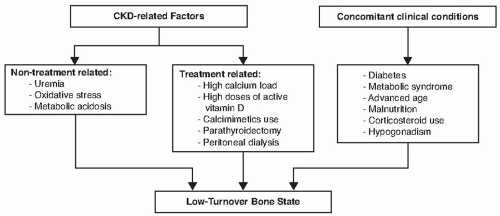
FIGURE 77.4 Low-turnover bone state risk factors.

PTH is the most widely used surrogate marker of bone turnover (Table 77.4). Although relatively low to normal iPTH levels (<50-100 pg per mL) in ESRD patients are associated with biopsy proven ABD, higher iPTH levels (>300 pg per mL) can be also be seen in patients with biopsy-proven ABD,81 especially in African Americans.82,83 There are several proposed explanations of this variability of iPTH levels and ABD. First, iPTH assays and their ability to discriminate between whole PTH and its fragments differ across the studies. Some PTH fragments, such as 7-84PTH, can be actually inhibitory on bone formation and these fragments tend to accumulate in ESRD; therefore, higher PTH may not be equivalent of presence of high biointact PTH. Additionally, treatment modalities
may influence bone formation rate independently from PTH levels. Lastly, PTH is not a bone-derived marker and therefore may never be a fully accurate indicator of bone turnover. At present, it is unknown what levels of PTH are associated with ABD in patients with less severe CKD not yet on renal replacement therapy. Bone-specific alkaline phosphatase (BSAP) may be an additional useful marker of ABD. Low levels of BSAP predict ABD and BSAP correlates with bone turnover in ESRD patients treated with hemodialysis.82


TABLE 77.4 Factors Regulating Parathyroid Hormone Secretion


Factor


Mechanism


Decreased PTH secretion


Calcium


Direct activation of CaSR leading to posttranslational decrease in PTH secretion


Calcitriol


Direct inhibition of preproPTH gene transcription via VDR


Indirect inhibition via increase in CaSR in parathyroid gland


FGF23


Direct inhibition of PTH mRNA expression


Increased PTH secretion


High phosphorus


Posttranslational increase in PTH via stabilization of PTH mRNA


Low calcium


Indirectly via increase in unbound to calcium calreticulin that inhibits calcitriol action on PTH secretion


Direct decrease in activation of CaSR


FGF23


Indirect increase in PTH secretion through decrease in calcitriol synthesis


PTH, parathyroid hormone; CaSR, calcium-sensing receptor; VDR, vitamin D receptor.


Fracture Risks in Chronic Kidney Disease

Patients with ESRD have fourfold increased risk of fractures; and the highest risk (10- to 100-fold increase) of fractures is observed in ESRD patients below age 65 as compared with age-matched individuals from the general population.84 The risk of fractures is also augmented in early CKD.85,86 Vertebral and hip fractures are shown to independently increase all-cause mortality in CKD patients.87,88 The fracture risk in patients with low turnover bone disease remains controversial as no biopsy-proven studies are available investigating the association between adynamic bone disease and fractures in CKD patients. Because ABD is linked to PTH oversuppression, several studies demonstrated the association between relatively low to normal PTH levels and the risk of vertebral and hip fractures.89,90 However, in a case-control study, dialysis patients who underwent parathyroidectomy were found to have 32% lower risk for hip fractures, and 31% lower risk for any fractures as compared with matched controls.91

May 29, 2016 | Posted by in NEPHROLOGY | Comments Off on Mineral and Bone Disorder in Chronic Kidney Disease

Full access? Get Clinical Tree

Get Clinical Tree app for offline access