Membranous Nephropathy


Membranous Nephropathy


Laurence Beck Jr.

David Salant


Membranous nephropathy (MN) is one of the leading causes of primary nephrotic syndrome in adults. It is recognized by its characteristic subepithelial immune deposits as visualized by immunofluorescence and electron microscopy, in addition to the thickened glomerular basement membrane (GBM) that gives the disease its name. Primary MN is a glomerulus-specific autoimmune disease and accounts for about 75% to 80% of cases of MN. Recent work has found that most patients with primary MN have circulating autoantibodies to the phospholipase A2 receptor (PLA2R); the remainder can be considered idiopathic MN. Secondary MN accounts for the remaining 20% to 25% of cases. It may be a feature of systemic autoimmune disease, chronic infections, malignancy, or therapeutic drugs, and is rarely if ever associated with anti-PLA2R antibodies. The course of primary MN is variable, and may be marked by spontaneous remissions and relapses. Although a proportion of those patients who fail to remit may have persistent proteinuria with maintained renal function, another 30% to 40% will progress to end-stage renal disease (ESRD). MN recurs in the kidney allograft in up to 40% of those cases that are transplanted. When treatment is deemed necessary, often for those with high levels of nephrotic-range proteinuria or worsening renal disease, immunosuppressive agents such as cyclophosphamide and cyclosporine have been shown to be effective. Several other agents have shown promise in small studies and may also turn out to be useful agents for the treatment of MN.


EPIDEMIOLOGY

Primary (or idiopathic) MN has been and remains the leading cause of adult nephrotic syndrome in many Caucasianpredominant populations, and is second only to focal and segmental glomerulosclerosis (FSGS) in others.1,2,3 The estimated annual incidence of MN is 1 in 100,000.4,5 Despite its relatively high incidence in Caucasian populations, it can be found worldwide in all racial groups. It is most common in the fourth through sixth decades, but can also occur in children or adolescents as well as the very elderly.6,7 The primary form of MN is more common in males, with a male to female ratio of approximately 2:1. Secondary MN, related to autoimmune diseases, infections, malignancy, or drugs may also occur at any age, and is the form most often seen in children (especially hepatitis B-associated8). Malignancy-associated MN is more often a disease of older patients.


PRIMARY MEMBRANOUS NEPHROPATHY

As mentioned previously, primary MN is the most common form of this disease, representing 75% to 80% of all cases. It is a glomerulus-specific autoimmune disorder, characterized by the presence of subepithelial immune deposits containing IgG4 and associated in the majority of cases with circulating IgG4 autoantibodies to PLA2R, a glycoprotein exposed on the podocyte surface (see Pathogenesis). The remainder of cases thought to be primary in nature may reflect a distinct disease with autoantibodies to another podocyte or glomerular protein, patients that remain proteinuric after the disappearance of anti-PLA2R antibodies, or in fact cases of undiagnosed secondary disease. The primary form may recur after renal transplantation in up to 40% of cases.9


SECONDARY MEMBRANOUS NEPHROPATHY

Secondary cases of MN are suspected when the characteristic pathologic findings of MN are found in conjunction with another systemic condition or are associated with the use of a therapeutic agent or toxin. Due to a lack of available serologic markers (e.g., anti-PLA2R autoantibodies) that could help to rule out primary disease, secondary cases were previously assumed to be present when MN was found in conjunction with one of the well- or lesser-known associations (Table 51.1).10,11 There are many single case reports of MN occurring in conjunction with rare autoimmune diseases, infections, cancers, or therapeutic agents. It should be kept in mind that many of these may instead represent a coincidental finding of primary MN with these disorders, rather than a
disorder that is truly responsible for secondarily causing MN. Evidence for secondary MN comes in situations in which treatment of the underlying process (infection, autoimmune disease, malignancy) or removal of an offending drug is temporally associated with resolution of the nephrotic syndrome, but this still does not guarantee causation because primary MN undergoes spontaneous remission in one third of cases.


TABLE 51.1 Causes of Membranous Nephropathy


Primary


Anti-PLA2R-associated


Idiopathic


Secondary


Autoimmune diseases


Systemic lupus erythematosus (class V lupus nephritis)


Other: rheumatoid arthritis, autoimmune thyroid disease, IgG4-related systemic disease


Infection


Hepatitis B


Other chronic infections: hepatitis C, HIV, syphilis, schistosomiasis


Alloimmunization


Fetomaternal alloimmunization


Graft-versus-host disease following hematopoietic stem cell transplantation


De novo membranous nephropathy in the renal allograft


Drugs or toxins


Nonsteroidal anti-inflammatory drugs and Cox-2 inhibitors


Mercury-containing compounds


Other: gold salts, D-penicillamine, bucillamine


Malignancy


Solid tumors (colon, stomach, lung, prostate)


Others: non-Hodgkin lymphoma, chronic lymphocytic leukemia, melanoma


Despite these caveats, MN has been repeatedly found to be secondary to lupus and hepatitis B; in addition to being the most common secondary forms, the strength of the association is also the clearest. Malignancy-associated MN is another important secondary cause to be discussed later, but historically the association has been more controversial. In many cases, the pathologic lesion in secondary MN is similar to that of primary MN. However, there are often subtleties in terms of the location of the deposits, type of immunoglobulin deposited, or other additional features that are more supportive of a secondary cause.


Autoimmune Conditions

Various rheumatologic disorders have been described in association with MN (Table 51.1), of which systemic lupus erythematosus (SLE) is the most common. Ten to 20% of patients with lupus nephritis have an International Society of Nephrology/Renal Pathology Society Class V (membranous) lesion with predominantly subepithelial deposits (see Chapter 53). Clinically, the presentation is that of the nephrotic syndrome and is indistinguishable from idiopathic MN. The majority of these patients are young females, and the onset of the nephrotic syndrome may predate the development of other signs and symptoms of SLE. In a substantial proportion of these patients, the antinuclear antibody (ANA) titer may be low or undetectable, and the complement levels are usually normal. Therefore, there should be a high degree of suspicion for SLE in any young female with the nephrotic syndrome who is found to have MN by renal biopsy. With more established disease, ANA and anti-double-stranded DNA antibodies may be present, and complement levels may be slightly depressed. Several pathologic features on biopsy such as the presence of mesangial and/or subendothelial deposits, as well as the precise IgG subclass present in the deposits, may distinguish lupus-associated secondary MN from primary MN (see section on Pathology). Otherwise, the course of lupus-associated MN resembles that of the idiopathic form, with a good long-term prognosis for renal survival as compared to other forms of lupus nephritis.12,13

Rheumatoid arthritis is another autoimmune condition that has been historically linked to MN. However, this has usually, but not always, been in the setting of concurrent treatment of the arthritis with agents such as gold salts, D-penicillamine, or bucillamine (which are no longer commonly used), or nonsteroidal anti-inflammatory agents.14 In these cases, proteinuria develops soon after exposure to the drug and resolves slowly over a period of months after the offending agent is withdrawn. The pathologic lesion is often identical to that of primary MN.

There are other autoimmune and systemic disorders that have been suggested to be rare secondary causes of MN, including autoimmune thyroid disease (Graves disease and Hashimoto thyroiditis),10,15,16 IgG4-related systemic disease,17,18 and sarcoidosis.19,20 Whether or not these are truly causative etiologies or rather coincidental findings (that are likely to be reported in the literature due to this rare association of distinctive diseases) is not known at this point. As a case in point, a recent report described a patient with MN in which the diagnosis of sarcoidosis and the onset of proteinuria were temporally associated; however, the patient tested positive for anti-PLA2R autoantibodies,21 which suggests that the MN was in fact primary.


Infectious Diseases

Numerous infectious diseases have been associated with the development of MN (Table 51.1). In all cases, these represent chronic infections with longstanding and persistent antigenemia. The argument for an etiologic role of the
infectious disease is strengthened when the nephrotic syndrome resolves with treatment of the infection, or when antigens produced by the microorganism are consistently found within the immune deposits.

The role of chronic infection with hepatitis B virus (HBV) is particularly strong and was first noted by Combes and colleagues in 1971.22 HBV infection may account for 30% to 40% of cases of MN in Asia and is particularly prevalent in children in endemic areas, many of whom are asymptomatic carriers with no history of active hepatitis.8,23,24,25 It is particularly noteworthy that the incidence of HBV-associated MN declined following the implementation of an active immunization program.26 The serum transaminases tend to be normal or only mildly elevated, and the serology is positive for surface antigen, anti-core antibody, and usually e antigen. It appears that it is the e antigen and cationic anti-e antibody that are primarily deposited in the glomeruli.8,25 HBV infection, along with membranous lupus nephritis, is the only other form of MN that may be associated with hypocomplementemia.24 Although there may be spontaneous resolution of proteinuria in children, successful treatment of the underlying viral infection in adults with antiviral agents such as entecavir or lamivudine is typically necessary to achieve remission of the nephrotic syndrome.

MN has also been associated with many other chronic infectious diseases, although there is less evidence of causality, and MN is often not the predominant histologic lesion. For example, there are a number of case reports of MN in patients with chronic hepatitis C virus (HCV) infection,27,28 but this agent is much more frequently associated with mixed cryoglobulinemia and the development of a membranoproliferative glomerulonephritis (MPGN) lesion. A membranous pattern has also been reported in patients infected with human immunodeficiency virus (HIV),29,30,31 hepatosplenic schistosomiasis,32 and congenital or acquired syphilis33,34,35; however, other forms of immune complex glomerulonephritis are more usual in these diseases. In several cases, microbial antigens such as those from treponemes in syphilis were found within the immune deposits.36,37 As in lupus nephritis, the exact nature of the immune complex may determine whether it ultimately forms in a subepithelial versus a mesangial or subendothelial location.


Malignancy

The association of MN with cancer has long been a point of contention, in part due to the implications of screening for malignancy in a patient who has no other potential secondary causes for their MN. Proponents argue that screening a patient for malignancy may reveal an early occult tumor, whereas opponents argue that, because primary MN and malignancy are both diseases that occur with increased frequency in older individuals, the finding of both diseases in the same person is coincidental. The first report of a possible link between carcinoma and MN came in 196638 and this association has been reviewed virtually every decade since.39,40,41,42,43,44 Although some may argue that detection bias can explain the association (i.e., patients who are found to have MN on biopsy are more likely to be screened for malignancy than their age-matched counterparts), a recent study that restricted the definition of malignancy-associated MN only to those in which the tumor was clinically evident before or at the time of the diagnosis of MN still found a higher than expected incidence of cancer compared to age- and gender-adjusted national cancer rates.43 Thus, solid tumors, such as those of the gastrointestinal tract (colon and stomach), lung, and prostate, do appear to be detected in patients with MN at a greater frequency than would be expected for an age-matched national cohort. MN may also rarely occur secondary to non-Hodgkin lymphoma or chronic lymphocytic leukemia.45 The association of MN and malignancy is strengthened by the temporal association, in several reports, of remission of the nephrotic syndrome following removal or treatment of the tumor. Some investigators have found evidence of tumor antigens such as CEA within the subepithelial immune deposits, and have been able to elute glomerular antibodies with reactivity to the tumor.46,47

Given evidence that seems to support both sides of the issue, it is likely that malignancy may be etiologically connected to MN in certain cases, but may only be coincidentally present with primary MN in other cases. This is reflected by a recent report that assayed 10 cases of malignancy-associated MN for the presence of anti-PLA2R antibodies.48 In 3 of 10 cases, there was evidence of circulating anti-PLA2R antibodies and the predominant glomerular IgG subclass on examination of the biopsy material was IgG4, suggesting a coincidental occurrence of primary MN with a tumor. In the remaining seven cases, however, the patients were anti-PLA2R negative, and the immune deposits were not positive for IgG4, suggesting a truly secondary cause of MN.49 Future studies such as this may clarify the relationship between the two disease processes, and a positive test for autoantibodies may obviate the need for an extensive malignancy workup. For the time being, however, it is worth making sure that an elderly patient who is found to have MN on biopsy has had age- and gender-appropriate cancer screening, such as colonoscopy, prostate examination (and prostate-specific antigen testing), mammography, and chest imaging in patients with a history of past or current smoking.


Drugs and Toxins

Drug-associated MN can occur at any age and typically develops within 6 to 12 months of exposure to the offending agent, but the onset may be delayed for 3 to 4 years.50 Historically, gold salts, D-penicillamine, and bucillamine used in the treatment of rheumatoid arthritis have been strongly linked to MN, although these agents are no longer in widespread use. The most common therapeutic agents currently implicated are the nonsteroidal anti-inflammatory drugs (NSAIDs), with mercury-containing compounds reflecting the most frequently encountered toxic exposure. The latter can be found as ingredients in certain skin-lightening agents, which have been linked to the development of MN in several reports.51,52 Discontinuation of the drug leads to resolution of the proteinuria in virtually all cases.50,53 However, studies with penicillamine,
gold, and bucillamine indicate that protein excretion may continue to rise for several months after the cessation of therapy.50 The mean time to resolution of the proteinuria is 9 to 12 months, although 2 to 3 years is required in some cases.

Although NSAID-induced nephrotic syndrome is more commonly associated with minimal change disease, it is evident that MN can also occur.54,55 The association of NSAIDs with MN was illustrated in a study of 125 patients with a biopsy diagnosis of MN55; 23% reported regularly using NSAIDs and 13 of them were likely to have had NSAID-associated MN, as they demonstrated resolution of proteinuria within 1 to 36 weeks of discontinuing NSAIDs and had no recurrence of proteinuria at follow-up (5 months to 13 years). Many of the patients who developed MN had been treated with diclofenac, but probably any NSAID can be involved,55 including cyclooxygenase-2 (Cox-2) inhibitors.56


Alloimmunity

MN may develop in situations when the immune system encounters non-self-antigens,57 such as in renal transplantation or after allogeneic hematopoietic stem cell transplantation (HSCT). Although patients with a previous history of primary MN may develop recurrent disease in their allograft, more common is de novo MN, which may represent an alloimmune reaction to minor histocompatibility antigens on the allograft podocytes. The MN that occurs post-HSCT is likely to be a humoral manifestation of graft-versus-host disease, and is the most common cause of the nephrotic syndrome after HSCT.58,59 It is of note that, like primary MN, these cases predominate in males, as opposed to the other causes of nephrotic syndrome after HSCT such as minimal change disease. A rare neonatal form of reversible MN due to fetomaternal alloimmunization has been described in babies born to mothers deficient in neutral endopeptidase (NEP), a protein expressed on podocytes (see Pathogenesis).60,61


Miscellaneous Conditions

Another form of pediatric MN was recently described in which circulating antibodies were found to be reactive with a cationic form of bovine serum albumin (BSA).62 BSA, likely derived from cow’s milk and absorbed as an undigested or partially digested protein, was detected in the glomerular immune deposits along with IgG. Moreover, specific anti-BSA antibodies could be eluted from the biopsy specimen in one case. MN has also been reported with diabetes, with or without associated diabetic nephropathy.63 Although this may reflect a coincidental occurrence of MN with another common disease, there was evidence of porcine insulin within the immune deposits by immunostaining, and an improvement in proteinuria after switching from porcine to human insulin in a small case series.64 There are also several reports of MN co-occurring with ANCA-positive crescentic glomerulonephritis. However, a recent report looking at the frequency of the two conditions in all renal biopsies performed at a single referral center concluded that the association was likely due to coincidence.65


PATHOLOGY

The name membranous nephropathy derives from the histopathologic appearance of the glomeruli of advanced cases of the disease in which expansion of the GBM is clearly visible on light microscopy and there is a paucity of inflammatory cells. Earlier in the course of the disease, however, the glomeruli may appear normal by light microscopy and further studies with immunofluorescence and electron microscopy are necessary for diagnosis. Conceptually, it is useful to think of the disease process as beginning with the formation or deposition of immune complexes beneath the podocyte, which then leads to podocyte injury and the deposition of new extracellular matrix between and around the immune deposits, culminating in a morphologically thickened GBM. Whereas several disparate conditions may underlie the development of MN and give rise to the formation of subepithelial immune deposits, as noted previously, the final pattern of injury is strikingly similar with some subtle differences discussed later. Although the histologic hallmarks of this disease—including GBM “spikes” visualized with the use of silver stains, and the fine granular deposition of IgG in a capillary loop pattern on immunofluorescence—were first described by Jones,66 and Mellors and Ortega,67 respectively, over 60 years ago, a definitive pathologic diagnosis depends on identifying the immune deposits with electron microscopy.


Light Microscopy

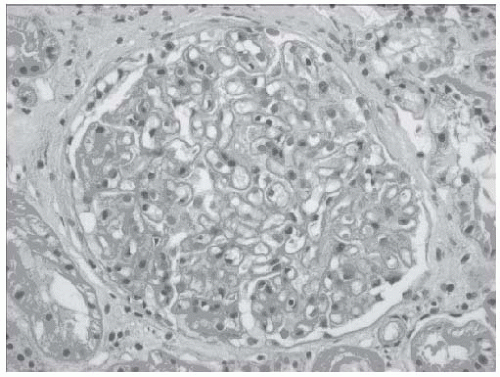
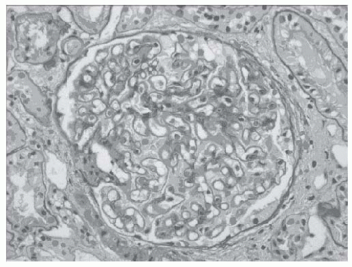
Light microscopy, with either hematoxylin and eosin (H&E) or periodic acid-Schiff (PAS) staining, reveals diffuse and generally uniform thickening of the GBM (Figs. 51.1 and 51.2). The heterogeneous character of the thickened GBM is best
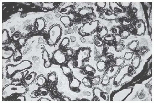
seen by silver methenamine (Jones’ stain), which binds to basement membrane components but is not taken up by the immune deposits (Fig. 51.3). This staining, in appropriately advanced disease, reveals “spikes” of GBM present between deposits when the GBM is sectioned in cross-section, or “craters” or “pock-marks” caused by the nonsilver stained immune deposits when a tangential section of the GBM is encountered. These findings are pathognomonic for MN. The formation of immune deposits and the basement membrane response proceeds in stages according to the duration of disease (and repair). Ehrenreich and Churg (Table 51.2) classified this progression into four morphologic stages, which are more appropriate for describing the pathologic findings than correlating with clinical findings or prognosis.


FIGURE 51.1 Hematoxylin and eosin stain (×250) of a glomerulus from a patient with idiopathic membranous nephropathy. There is diffuse thickening of the basement membrane without associated hypercellularity of the glomerular tuft. Inflammatory infiltrates are not seen and the capillary loops are widely patent. (Courtesy of Dr. Helen Cathro.)


FIGURE 51.2 Periodic acid-Schiff stain of a glomerulus from a patient with idiopathic membranous nephropathy (×250). The basement membrane surrounding the capillary loops is diffusely thickened. (Courtesy of Dr. Helen Cathro.)


FIGURE 51.3 Jones silver stain (×250) of a glomerulus from a patient with idiopathic membranous nephropathy demonstrating “spikes” corresponding to newly synthesized basement membrane surrounding immune complexes. (Courtesy of Dr. Edward Klatt.)


TABLE 51.2 Pathologic Staging of Membranous Nephropathy


Stage


Electron Microscopy


I


Subepithelial electron-dense deposits


II


Subepithelial electron-dense deposits with intervening basement membrane (“spikes”)


III


Incorporation of subepithelial electron-dense deposits into the basement membrane


IV


Reabsorption of deposits with loss of electron-dense deposits and development of lucent area in the basement membrane


Remodeling of basement membrane and loss of electron-dense deposits


From Ehreneich T, Churg J. Pathology of membranous nephropathy. Pathol Annu. 1968;3:145.


Other compartments of the glomerulus usually appear normal. There is no evidence of mesangial cell proliferation or expansion except in the setting of SLE and other secondary forms. Importantly, there is typically no evidence of inflammatory cell infiltration (which argues against the continued use of the term “membranous glomerulonephritis”). Experimental studies suggested that this is on account of the subepithelial location of the immune deposits, which are separated from the capillary lumen and thereby unable to recruit inflammatory effector cells, as more readily occurs when immune deposits form in a mesangial or subendothelial location.68

With longer duration of disease and/or sustained heavy nephrotic proteinuria, tubulointerstitial damage can occur which is associated with decreased glomerular filtration rate (GFR) and a worsened renal prognosis. Similarly, lesions of secondary FSGS may also develop, also portending a worse prognosis and persistent proteinuria that is likely to be unresponsive to immunosuppression.


Immunofluorescence Microscopy

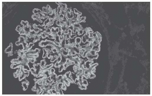
The finding of granular deposits of IgG in a capillary loop pattern on immunofluorescence is the sine qua non of both primary and secondary MN (Fig. 51.4). With the exception of class V lupus nephritis which may present with a “full house” pattern on immunofluorescence,69 the deposits are predominantly IgG, with minimal staining for IgA and IgM, and tend to spare the mesangium. The complement component C3 is often seen, with the exception of very early disease. Although not typically performed, the characterization of IgG subclasses often helps to differentiate
primary from secondary disease, as the predominant IgG subclass in primary MN is IgG4. Secondary causes, in most cases, have a predominance of non-IgG4 subclasses, most notably in lupus-associated69,70,71 and malignancy-associated MN.49 The presence of C1q, an early component of the classical complement pathway, may also help distinguish between primary and secondary cases. Strong C1q staining is not typically found in primary MN (less than 20% of cases)69,72 but is more common in lupus-associated MN.


FIGURE 51.4 Immunofluorescence staining (anti-IgG) of a glomerulus from a patient with idiopathic membranous nephropathy (×250). Diffuse granular staining along the basement membrane is evident and corresponds to the deposition of immune complexes. Mesangial areas are free of immune deposits.

May 29, 2016 | Posted by in NEPHROLOGY | Comments Off on Membranous Nephropathy

Full access? Get Clinical Tree

Get Clinical Tree app for offline access