Mechanisms of Diuretic Action


Mechanisms of Diuretic Action


David H. Ellison

Ewout J. Hoorn

Robert W. Schrier


The term diuretic derives from the Greek diouretikos, which means, “to promote urine.”* Even though many substances promote urine flow, the term diuretic is usually taken to indicate a substance that can reduce the extracellular fluid volume by increasing urinary solute and water excretion. In 1553, Paracelsus recorded the first truly effective medical treatment for dropsy (edema), namely inorganic mercury (calomel). Inorganic mercury remained the mainstay of diuretic treatment until the first part of the 20th century. In 1919, the ability of organic mercurial antisyphilitics to affect diuresis was discovered by Vogl, then a medical student. This observation led to the development of effective organic mercurial diuretics that continued to be used through the 1960s. In 1937, the antimicrobial sulfanilamide was found to cause metabolic acidosis. This drug was soon thereafter shown to inhibit the enzyme carbonic anhydrase, which had been discovered in 1932. Pitts then showed that sulfanilamide inhibited sodium (Na) bicarbonate reabsorption in dogs and Schwartz showed that sulfanilamide could induce diuresis when administered to patients with congestive heart failure. Soon, more potent sulfonamide-based carbonic anhydrase inhibitors were developed, but these drugs suffered from side effects and limited potency. Nevertheless, a group at Sharp & Dohme Inc. was stimulated by these developments to explore the possibility that modification of sulfonamide-based drugs could lead to small molecules that enhanced Na excretion with chloride rather than bicarbonate, thus better enhancing depletion of the extracellular fluid, which is composed primarily of sodium chloride (NaCl) and water. The result of this program was the synthesis of chlorothiazide and its marketing in 1957. This drug ushered in the modern era of diuretic therapy and revolutionized the clinical treatment of edema.

The search for more potent classes of diuretics led to the development of ethacrynic acid and furosemide in the United States and Germany, respectively. The safety and efficacy of these drugs led them to replace the organic mercurials as drugs of choice for severe and resistant edema. Spironolactone, marketed in 1961, was developed after the properties and structure of aldosterone had been discovered and steroidal analogues of aldosterone were found to have aldosterone-blocking activity. Triamterene was initially synthesized as a folic acid antagonist, but was found to have diuretic and potassium (K)-sparing activity. The identification of the arginine vasopressin (AVP) receptor subtypes led to the more recent development of vasopressin antagonists, which have recently entered clinical practice. The identification and cloning of natriuretic peptides led to the development of drugs with similar effects.

The availability of safe, effective, and relatively inexpensive diuretic drugs has made it possible to treat edematous disorders and hypertension effectively. Incidentally, driven by clinical drug development, specific ligands that interact with discrete Na and Cl transport proteins in the kidney were developed, permitting these transport proteins to be identified. Subsequently, these ligands were used to clone the Na and Cl transport proteins that mediate the bulk of renal Na and Cl reabsorption. The diuretic-sensitive transport proteins that have been cloned include the sodium hydrogen exchanger (NHE3; gene symbol SLC9A3), the bumetanide-sensitive Na-K-2Cl cotransporter (NKCC2; gene symbol SLC12A1), the thiazide-sensitive Na-Cl cotransporter (NCC; gene symbol SLC12A3), and the epithelial Na channel (ENaC; gene symbols SCNN1A, SCNN1B, SCNN1G). The information derived from molecular cloning has also permitted identification of inherited human diseases that are caused by mutations in these diuretic-sensitive transport proteins. The phenotypes of several of these disorders resemble the manifestations of chronic diuretic administration. Thus, the development of clinically useful diuretics permitted identification and later cloning of specific ion transport pathways. The molecular cloning is now helping to define mechanisms of diuretic action and diuretic side effects. The use of animals in which diuretic-sensitive transport pathways have been “knocked out” permits a clearer understanding of which diuretic effects result directly or secondarily from actions of the drugs on specific ion transport pathways and which effects result from actions on other pathways or other organ systems.


NORMAL RENAL NaCl HANDLING

The normal human kidneys filter approximately 23 moles of NaCl in 150 liters of fluid each day. According to data from NHANES III, typical dietary sodium consumption in the United States is 4.3 g daily for men and 2.9 g daily for women.1 The sex difference reflects differences in caloric intake, not differential food choices. As 17 mEq of Na is 1 g of table salt and 43 mEq of Na is in 1 g of sodium, men typically consume 100 mEq of Na and women typically consume 67 mEq on a daily basis. To maintain balance, renal NaCl excretion must equal this, ignoring the modest losses in feces and sweat. Under normal circumstances, approximately 99.2% of the filtered NaCl is reabsorbed by kidney tubules generating a normal fractional sodium excretion of <1% (100 mEq in urine per 23,000 mEq filtered = 0.4%). Sodium, chloride, and water reabsorption by the nephron is driven by the metabolic energy provided by ATP. The ouabain-sensitive Na/K ATPase is expressed at the basolateral cell membrane of all Na transporting epithelial cells along the nephron. This pump maintains large ion gradients across the plasma membrane, with the intracellular Na concentration maintained low and the intracellular K concentration maintained high. Because the pump is electrogenic and associates with a K channel in the same membrane, renal epithelial cells have a voltage across the plasma membrane oriented with the inside negative relative to the outside.

The combination of the low intracellular Na concentration and the plasma membrane voltage generates a large electrochemical gradient favoring Na entry from lumen or interstitium. Specific diuretic-sensitive Na transport pathways are expressed at the apical (luminal) surface of cells along the nephron, permitting vectorial transport of Na from lumen to blood. Along the proximal tubule, where approximately 50% to 60% of filtered Na is reabsorbed, an isoform of the Na/H exchanger is expressed at the apical membrane. Along the thick ascending limb, where approximately 20% to 25% of filtered Na is reabsorbed, an isoform of the Na-K-2Cl cotransporter is expressed at the apical membrane. Along the distal convoluted tubule, where approximately 5% of filtered Na is reabsorbed, the thiazide-sensitive Na-Cl cotransporter is expressed. Along the connecting tubule (CNT) and cortical collecting duct (CCD), where approximately 3% of filtered Na is reabsorbed, the amiloride-sensitive epithelial Na channel is expressed. These apical Na transport pathways are the primary targets for diuretic drug action.


TABLE 66.1 Effects of Diuretics on Electrolyte Excretion


Na


Cl


K


Pi


Ca


Mg


Osmotic diuretics6,205,388,389,390


image(10%-25%)


image(15%-30%)


image(6%)


image(5%-10%)


image(10%-20%)


image(>20%)


Carbonic anhydrase inhibitors57,98,205


image(6%)


image(4%)


image(60%)


image(>20%)


imageor ⇔ (<5%)


image(<5%)


Loop diuretics98,177,205,344,391,392


image(30%)


image(40%)


image(60%-100%)


image(>20%)


image(>20%)


image(>20%)


DCT diuretics177,205,391,393


image(6%-11%)


image(10%)


image(200%)


image(>20%)


image


image(5-10%)


Collecting duct diuretics177,205,344


image(1%-5%)


image(6%)


image(8%)


image


Figures indicate approximate maximal fractional excretions of ions following acute diuretic administration in maximally effective doses. image indicates that the drug increases excretion; image indicates that the drug decreases excretion; ⇔ indicates that the drug has little or no direct effect on excretion. During chronic treatment, effects often wane (Na excretion), may increase (K excretion during distal convoluted tubule diuretic treatment), or may reverse as with uric acid (not shown).


Na, sodium; Cl, chloride; K, potassium; Pi, phosphate; Ca, calcium; Mg, magnesium.


This chapter discusses the molecular and physiologic bases for diuretic action in the kidney. Although some aspects of clinical diuretic usage are discussed, physiologic principles and mechanisms of action are emphasized. Several recent texts provide detailed discussions of diuretic treatment of clinical conditions.2 Extensive discussions of diuretic pharmacokinetics are also available.3,4

A rational classification of diuretic drugs (Table 66.1) is based on the primary nephron site of action. Such a scheme emphasizes that different chemical classes of drugs can affect the same ion transport mechanism and exhibit many of the same clinical effects and side effects. Furthermore, although
most diuretic drugs affect transport processes along several nephron segments, most owe their clinical effects primarily to their ability to inhibit Na transport by one particular nephron segment. An exception is the osmotic diuretics. Although these drugs were initially believed to inhibit solute and water flux primarily along the proximal tubule, subsequent studies have revealed effects in multiple segments. Other diuretics, however, are classified according to their primary site of action.


OSMOTIC DIURETICS

Osmotic diuretics are substances that are freely filtered at the glomerulus, but are poorly reabsorbed (Fig. 66.1). The pharmacologic activity of drugs in this group depends entirely on the osmotic pressure exerted by the drug molecules in solution. It does not depend on interaction with specific transport proteins or enzymes. Mannitol is the prototypical osmotic diuretic.5 Because the relationship between the magnitude of diuretic effect and concentration of osmotic diuretic in solution is linear, all osmotic diuretics are small molecules. Other agents considered in this class include urea, sorbitol, and glycerol.


Urinary Electrolyte Excretion

Although osmotic agents do not act directly on transport pathways, ion transport is affected. Following mannitol infusion, sodium, potassium, calcium, magnesium, bicarbonate, and chloride excretion rates increase (see Table 66.1). Rates of sodium and water fractional reabsorption are reduced by 27% and 12%, respectively, following the infusion of mannitol.6 Reabsorption of magnesium and calcium is also reduced in the proximal tubule and loop of Henle. In contrast, phosphate reabsorption is only inhibited slightly by mannitol in the presence of parathyroid hormone.7


Mechanism of Action

The functional consequences that result from intravenous infusion of mannitol include an increase in cortical and medullary blood flow; a variable effect on glomerular filtration rate; an increase in sodium, water, calcium, magnesium, phosphorus, and bicarbonate excretion; and a decrease in medullary concentration gradient. The most pronounced effect observed with mannitol is a brisk diuresis and natriuresis. The mechanisms by which mannitol produces a diuresis include: (1) an increase in osmotic pressure in the lumens of the proximal tubule and loop of Henle, thereby retarding the passive reabsorption of water and (2) an increase in renal blood flow and washout of medullary tonicity.


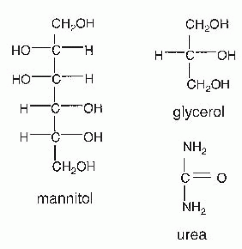
FIGURE 66.1 Structures of osmotic diuretics.

Mannitol is freely filtered at the glomerulus and its presence in tubule fluid minimizes passive water reabsorption. Normally, within the proximal tubule, sodium reabsorption creates an osmotic gradient for water reabsorption. When an osmotic diuretic is administered, however, the osmotic force of the nonreabsorbable solute in the lumen opposes the osmotic force produced by sodium reabsorption. Isosmolality of tubule fluid is preserved because molecules of mannitol replace sodium ions reabsorbed. However, sodium reabsorption eventually stops because the luminal sodium concentration is reduced to a point where a limiting gradient is reached and net transport of sodium and water ceases. The validity of this mechanistic explanation has been confirmed by stationary micropuncture studies. Quantitatively, mannitol has a greater effect on inhibiting Na and water reabsorption in the loop of Henle than in the proximal tubule. Free-flow micropuncture studies following mannitol infusion in dogs demonstrated a modest decrease in fractional reabsorption of sodium and water by the proximal tubule, but a much larger effect by the loop of Henle.7 Within the loop of Henle, the site of action of mannitol appears to be restricted to the thin descending limb, decreasing water reabsorption.8 In the thick ascending limb, Na reabsorption continues, in proportion to its delivery to this segment. The sum of net transport in the thin and thick limbs determines the net effect of mannitol in the loop of Henle. Further downstream in the collecting duct, mannitol reduces sodium and water reabsorption.9


Renal Hemodynamics

During the administration of mannitol, its molecules diffuse from the bloodstream into the interstitial space. In the interstitial space, the increased osmotic pressure draws water from the cells to increase extracellular fluid volume. This effect increases total renal plasma flow.9 Cortical and medullary blood flow rates both increase following mannitol infusion.9 Single nephron glomerular filtration rate (GFR), on the other hand, increases in cortex but decreases in medulla.8 The mechanisms by which mannitol reduces the GFR of deep nephrons are not known, but it has been postulated that mannitol reduces efferent arteriolar pressure. Micropuncture studies examining the determinants of GFR in superficial nephrons have demonstrated that the increase in single nephron GFR results from an increase in single nephron plasma flow and a decrease in oncotic pressure.10 The net effect of mannitol on total kidney GFR has been variable, but most studies indicate that the overall effect is to increase GFR.10


The combination of enhanced renal plasma flow and reduced medullary GFR washes out the medullary osmotic gradient by reducing papillary sodium and urea content. Experimental studies indicate that the osmotic effect of mannitol to increase water movement from intracellular to extracellular space leads to a decrease in hematocrit and in blood viscosity. This fact contributes to a decrease in renal vascular resistance and increase in renal blood flow. In addition, secretion of vasodilatory substances is stimulated by mannitol infusion. Both prostacyclin (PGI2)11 and atrial natriuretic peptide12 could mediate the effect of mannitol on renal blood flow. The vasodilatory effect of mannitol is reduced when the recipient is pretreated with indomethacin or meclofenamate, suggesting that PGI2 is involved in the vasodilatory effect. Alterations in renal hemodynamics contribute to the diuresis observed following administration of mannitol. An increase in medullary blood flow rate reduces medullary tonicity primarily by decreasing papillary sodium and urea content13 and increasing urine flow rate.14


Pharmacokinetics

Mannitol is not readily absorbed from the intestine5; therefore, it is routinely administered intravenously. Following infusion, mannitol distributes in extracellular fluid with a volume of distribution of approximately 16 liters15; its excretion is almost entirely by glomerular filtration.16 Of the filtered load, less than 10% is reabsorbed by the renal tubule, and a similar quantity is metabolized, probably in the liver. With normal glomerular filtration rate, plasma half-life is approximately 2.2 hours.


Clinical Use

Mannitol is used prophylactically to prevent acute kidney injury.17 In the past, it was administered to patients with established acute kidney injury, but it has proven ineffective in this situation. Mannitol improves renal hemodynamics in a variety of situations of impending or incipient acute kidney injury. Mannitol (along with hydration and sodium bicarbonate) has been recommended by some,18,19 but not all20 investigators for the early treatment in myoglobinuric acute kidney injury and to prevent posttransplant acute kidney injury.21 Mannitol is frequently used perioperatively to treat patients undergoing cardiopulmonary bypass surgery. The beneficial effects may relate to its osmotic activity thereby reducing intravenous fluid requirement22 and its ability to act as a free radical antioxidant.23 Although some studies have shown a beneficial effect when used prophylactically to treat patients at risk for contrast nephropathy,24 most prospective controlled studies have not found mannitol beneficial in preventing acute kidney injury and it is not currently recommended.25,26

Mannitol is used for short-term reduction of intraocular pressure.27 By increasing the osmotic pressure, mannitol reduces the volume of aqueous humor and the intraocular pressure by extracting water. Mannitol also decreases cerebral edema and the increase in intracranial pressure associated with trauma, tumors, and neurosurgical procedures,22,23,28 although hypertonic saline appears more effective for this purpose.29

Mannitol and other osmotic agents have been used to treat dialysis disequilibrium.30,31 This syndrome is characterized by acute symptoms during or immediately following hemodialysis, and is especially common when dialysis is first initiated. Most significant symptoms are attributable to disorders of the central nervous system such as headache, nausea, blurred vision, confusion, seizure, coma, and death. Rapid removal of small solutes such as urea during dialysis of patients who are markedly azotemic is associated with the development of an osmotic gradient for water movement into brain cells producing cerebral edema and neurologic dysfunction. Dialysis disequilibrium syndrome can be minimized by slow solute removal, using low blood flow and short treatment times; raising plasma osmolality with saline or mannitol can also be employed.


PROXIMAL TUBULE DIURETICS (CARBONIC ANHYDRASE INHIBITORS)

Through the development of carbonic anhydrase inhibitors, important compounds were discovered that have utility as therapeutic agents and as research tools. Carbonic anhydrase
inhibitors have a limited therapeutic role as diuretic agents, however, because they are only weakly natriuretic when employed chronically. They are used primarily to reduce intraocular pressure in glaucoma, to enhance bicarbonate excretion in metabolic alkalosis or chronic hypercapnia, and to prevent mountain sickness. Structures of carbonic anhydrase inhibitors are shown in Figure 66.2.


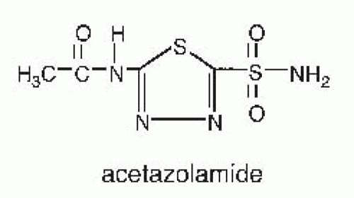
FIGURE 66.2 Structure of a carbonic anhydrase inhibitor.


Urinary Electrolyte Excretion

Through their effects on carbonic anhydrase in the proximal tubule, carbonic anhydrase inhibitors increase bicarbonate excretion by 25% to 30% (see Table 66.1). The increase in sodium and chloride excretion is smaller than might be expected, because these ions are reabsorbed by more distal segments of the nephron.37 However, a residual small but variable amount of sodium is excreted along with bicarbonate (Table 66.1). Calcium and phosphate reabsorption are also inhibited along the proximal tubule by carbonic anhydrase inhibitors. Because distal calcium reabsorption is stimulated by increased distal delivery, fractional calcium excretion does not increase. In contrast, phosphate appears to escape distal reabsorption resulting in an increase in fractional excretion of phosphate by ˜3%. Although proximal tubule magnesium transport is inhibited by carbonic anhydrase inhibitors, fractional excretion of magnesium is either unchanged or increased as a result of variable distal reabsorption.38

Carbonic anhydrase inhibitors increase potassium excretion. It is likely that several indirect effects contribute to the observed kaliuresis. Carbonic anhydrase inhibition could block proximal tubule potassium reabsorption and increase delivery to the distal tubule, but this has not been established clearly. Whereas carbonic anhydrase inhibitors decrease proximal tubule sodium, bicarbonate, and water absorption during both free flow micropuncture and microperfusion, the effects of carbonic anhydrase inhibitors on proximal tubule potassium transport have been less consistent. In free flow micropuncture studies, carbonic anhydrase inhibition did not affect proximal tubule potassium reabsorption,39 whereas it did reduce net reabsorption by microperfused proximal tubules.40 The effect of carbonic anhydrase inhibitors on the proximal tubule ion transport does, however, facilitate an increase in tubular fluid flow rate and sodium and bicarbonate but not chloride delivery to the distal nephron. This effect is thought to increase the concentration of bicarbonate in the distal tubule lumen, which increases lumen negative voltage41 and increases flow rate,42 factors known to increase potassium secretion by the distal tubule. Carbonic anhydrase inhibitors can also produce a luminal composition that is low in chloride and high in bicarbonate. This luminal fluid composition has been demonstrated to stimulate potassium secretion by the distal nephron independent of a change in lumen negative voltage.43


Mechanism of Action

In the kidney, carbonic anhydrase inhibitors act primarily on proximal tubule cells to inhibit bicarbonate absorption.44 Carbonic anhydrase, a metalloenzyme containing one zinc atom per molecule, is important in sodium bicarbonate reabsorption and hydrogen ion secretion by renal epithelial cells. The biochemical, morphologic, and functional properties of carbonic anhydrase have been reviewed.45 Carbonic anhydrase isoforms (CA) can be categorized into four groups: (1) cytosolic, I, II, III, VII; (2) mitochondrial, V; (3) membrane associated IV, IX, XII, XIV; and (4) secreted, VI.45 Carbonic anhydrases regulate cellular H ion secretion through catalyzing the formation of HCO3 from OH and CO2 and by binding to transporters and directly regulating activity. There are three major renal carbonic anhydrases. Type II carbonic anhydrase (CAII) is distributed widely comprising more than 95% of the overall activity in kidney and is sensitive to inhibition by sulfonamides. CAII is expressed in the cytoplasm and facilitates the secretion of H ions by catalyzing the formation of HCO3 from OH and CO2 (see equation 66.3). In addition, CAII binds to the C-terminal region of NHE1 and likely regulates the transport efficiency of Na/H exchange. CAIV is bound to renal cortical membranes, comprising up to 5% of the remaining overall activity in rodent kidney, and is also sensitive to sulfonamides. Carbonic anhydrase activity at basolateral and luminal plasma membranes of proximal tubule cells and luminal membrane of intercalated cells catalyzes the dehydration of intraluminal carbonic acid generated from secreted protons. The carbonic anhydrase activity at the basolateral and luminal plasma membranes of proximal tubule cells is thought to be due in part to CAIV.46 CAIV has been shown to also bind to the extracellular loop of NaHCO3 transporter 1 (NBC1) regulating its transport activity.47 Evidence for the physiologic importance for carbonic anhydrase is apparent as a deficiency of CAII leads to a renal acidification defect resulting in renal tubular acidosis. Furthermore, metabolic acidosis leads to an adaptive increase in both CAII and IV carbonic anhydrase mRNA expression in kidney48 suggesting the importance of both carbonic anhydrase isoforms in this disorder. CAXII is also expressed in proximal tubules and collecting ducts and may contribute to the carbonic anhydrase activity in these segments.49,50,51

Normally the proximal tubule reabsorbs 80% of the filtered load of sodium bicarbonate and 60% of the filtered load of sodium chloride. Early studies by Pitts and micropuncture studies by DuBose and others indicated that hydrogen ion secretion is responsible for bicarbonate absorption and renal acidification. The cellular mechanism by which proximal
tubules reabsorb bicarbonate is depicted in Figure 66.3. The effect of carbonic anhydrase to accelerate bicarbonate is a result of the reactions that occur in both luminal fluid and in the cell. The mechanism of carbonic anhydrase action in luminal fluid,52 is shown here, where E represents the carbonic anhydrase enzyme:


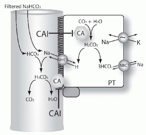
FIGURE 66.3 Mechanisms of diuretic action in the proximal tubule. The figure shows functional model of proximal tubule (PT) cells. Many transport proteins are omitted from the model, for clarity. Carbonic anhydrase (CA) catalyzes inside the cell the formation of HCO3 from H2O and CO2. This is the result of the two-step process (please see equations in the text for additional details). Bicarbonate leaves the cell via the Na, HCO3, cotransporter.186,187 A second pool of carbonic anhydrase is located in the brush border (CA). This participates in disposing of carbonic acid, formed from filtered bicarbonate and secreted H+. Both pools of carbonic anhydrase are inhibited by acetazolamide and other carbonic anhydrase inhibitors (see text for details).


Luminal Fluid


Note that the addition of reactions 1 and 2 leads to the classic reaction 3. In this scheme, the enzyme is viewed as a superhydroxylator.

Luminal carbonic anhydrase prevents H from accumulating in tubule fluid, which would eventually stop all Na/H exchange. Once formed, carbon dioxide diffuses rapidly from the lumen into the cell across the apical membrane.

The mechanism by which intracellular carbonic anhydrase participates in net H+ secretion is functionally the reverse of the reactions shown previously.


Intracellular Fluid


In this case, the enzyme splits water, thereby providing an hydroxyl ion to form bicarbonate. The bicarbonate ions then exit the basolateral membrane via Na(HCO3)3 cotransport.53 Thus, in the early proximal tubule, the net effect of the process described results in the isosmotic reabsorption of NaHCO3. The lumen chloride concentration increases because water continues to be reabsorbed, thereby producing a lumen positive potential. These axial changes provide an electrochemical gradient for transport of chloride, via paracellular and transcellular pathways. The latter pathway for chloride likely involves an exchange of Cl with anions, including oxalate and formate, operating in parallel with a Na/H proton exchanger. The dual operation of these parallel exchangers results in net NaCl absorption.54

Carbonic anhydrase inhibitors act primarily on proximal tubule cells, where approximately 60% of the filtered load of sodium chloride is reabsorbed. Despite the magnitude of sodium chloride reabsorption in the proximal tubule segment, the natriuretic potency of carbonic anhydrase inhibitors is relatively weak. Several factors explain this observation. First, proximal sodium reabsorption is mediated by carbonic anhydrase-independent as well as carbonic anhydrase-dependent pathways. Second, the increased sodium delivery to distal nephron segments is largely reabsorbed by these distal nephron segments. Third, carbonic anhydrase inhibitors generate a hyperchloremic metabolic acidosis further reducing the effects of subsequent doses of carbonic anhydrase inhibitor. Metabolic acidosis also produces resistance to carbonic anhydrase action. Following the induction of metabolic acidosis, the Ki for bicarbonate absorption by membrane impermeant carbonic anhydrase inhibitors was increased by a factor of 100 to 500, suggesting that metabolic acidosis is associated with changes in the physical properties of the carbonic anhydrase protein.55 For these reasons, carbonic anhydrase inhibitors alone are rarely used as diuretic agents.

Following carbonic anhydrase inhibitor administration, proximal tubule bicarbonate reabsorption declines between 35% and 85%. Additional sites of action of carbonic anhydrase inhibitors include proximal straight tubule or loop of Henle, distal tubule, and the collecting and papillary collecting ducts. Yet, despite the effect of carbonic anhydrase inhibitors on proximal tubules as well as other nephron segments, compensatory reabsorption of bicarbonate at other downstream
tubular sites limits net fractional excretion of bicarbonate to ˜25% to 30%, even during acute administration.56,57

The relative contributions of membrane-bound and intracellular components of cellular carbonic anhydrase have been examined. Both species contribute to bicarbonate absorption. The role of membrane-bound carbonic anhydrase was addressed in studies that employed carbonic anhydrase inhibitors with different abilities to penetrate proximal tubule cell membranes. Benzolamide is charged at normal pH and does not penetrate cell membranes well, whereas acetazolamide enters the cell relatively easily.58 Proximal tubular perfusion of benzolamide inhibits bicarbonate reabsorption by 90%59 indicating that luminal carbonic anhydrase inhibition contributes importantly to bicarbonate absorption. Inhibition of luminal carbonic anhydrase causes lumen pH to decrease because of the continued secretion of hydrogen ions into the tubule lumen.59 The conclusion that tubular fluid is in direct contact with membrane carbonic anhydrase was substantiated by the use of dextran-bound carbonic anhydrase inhibitors.60,61 In proximal tubules perfused in vivo, Lucci et al. determined that dextran-bound inhibitors, which inhibit only luminal carbonic anhydrase, decreased proximal tubule bicarbonate absorption by approximately 80% and reduced lumen pH.61

Although these studies establish the importance of luminal carbonic anhydrase, they also support an important role for intracellular and basolateral carbonic anhydrase. Both acetazolamide and benzolamide inhibit proximal tubule bicarbonate reabsorption to a similar degree yet they produce opposite effects on tubule fluid pH, suggesting that intracellular carbonic anhydrase contributes to proximal tubule luminal acidification. Furthermore, inherited deficiency of the predominant renal carbonic anhydrase, CAII, causes proximal renal tubular acidosis.62

The expression of carbonic anhydrase in the basolateral membrane of proximal tubule cells suggests that this membrane-bound enzyme also has an important role in basolateral bicarbonate transport. Although it is well known that carbonic anhydrase inhibitors inhibit intracellular generation of substrate for the transporter,63,64 the direct interaction between CAIV and NBC1, the sodium/bicarbonate cotransporter,47 suggests the possibility that carbonic anhydrase inhibitors may also directly regulate anion transport activity. Functional studies using an impermeant carbonic anhydrase inhibitor, p-fluorobenzyl-aminobenzamide, that is 1% as permeable as acetazolamide, demonstrated the importance of basolateral membrane-bound carbonic anhydrase. p-Fluorobenzyl-aminobenzamide reduced fluid and bicarbonate absorption when applied to the basolateral membrane of rabbit proximal tubules perfused in vitro.50

In the collecting duct, carbonic anhydrase facilitates acid secretion that is mediated by a vacuolar H adenosine triphosphatase (H-ATPase)65 and a P-type gastric H-K-ATPase.66,67,68 Luminal administration of acetazolamide produced an acid disequilibrium pH in the outer medullary collecting duct suggesting the contribution of luminal carbonic anhydrase.69

Using a membrane-impermeant carbonic anhydrase inhibitor (F-3500; aminobenzamide coupled to a nontoxic polymer polyoxyethylene), bicarbonate absorption was reduced confirming the presence of carbonic anhydrase in the luminal membrane of the outer medullary collecting duct.55 The Ki for inhibition of bicarbonate absorption was 5 µM, consistent with the inhibition of CAIV.


Renal Hemodynamics

Inhibition of carbonic anhydrase decreases GFR acutely. Systemic acetazolamide infusion decreased GFR by 30%. Single nephron glomerular filtration rate (SNGFR) was 23% lower during acetazolamide infusion partly because increased solute delivery to the macula densa activates the tubuloglomerular feedback (TGF) mechanism, which reduces GFR. Similar results were observed following infusion of benzolamide.70 Nevertheless, the effects of carbonic anhydrase inhibitors to reduce GFR are not simply the result of TGF activation. Sarala8- angiotensin I, an angiotensin II antagonist, prevented the decrease in SGNFR suggesting the involvement of local angiotensin II in response to benzolamide.70 Further, infusion of benzolamide into targeted adenosine-1 receptor knockout mice (i.e., mice that lack a TGF response) reduced GFR by 21%.71 Taken together, these results suggest complex mechanisms by which carbonic anhydrase inhibitors reduce GFR.


Pharmacokinetics

Acetazolamide is well absorbed from the gastrointestinal (GI) tract. More than 90% of the drug is plasma protein bound. The highest concentrations are found in tissues that contain large amounts of carbonic anhydrase (e.g., renal cortex, red blood cells). Renal effects are noticeable within 30 minutes and are usually maximal at 2 hours. Acetazolamide is not metabolized but is excreted rapidly by glomerular filtration and proximal tubular secretion. The half-life is approximately 5 hours and renal excretion is essentially complete in 24 hours.16 In comparison, methazolamide is absorbed more slowly from the GI tract, and its duration of action is long, with a half-life of approximately 14 hours.


Clinical Use

As noted, these drugs are almost never used as first-line diuretics because of the availability of much more potent drugs. Daily use produces systemic acidemia from an increase in urinary excretion of bicarbonate. Nevertheless, acetazolamide can be administered for short-term therapy, usually in combination with other diuretics, to patients who are resistant or who do not respond adequately to other agents.76 The rationale for using a combination of diuretic agents is based on summation of their effect at different sites along the nephron.

The major indication for the use of acetazolamide as a diuretic agent is in the treatment of patients with metabolic alkalosis accompanying edema77,78 or the treatment of chronic respiratory acidosis in chronic obstructive lung disease.79,80 In patients with cirrhosis, congestive heart failure, or nephrotic syndrome, aggressive diuresis with loop diuretics promotes intravascular volume depletion and secondary hyperaldosteronism, conditions that promote metabolic alkalosis. Administration of sodium chloride to correct the metabolic alkalosis simply exacerbates the edema. Acetazolamide can improve metabolic alkalosis by decreasing proximal tubule bicarbonate reabsorption thereby increasing the fractional excretion of bicarbonate. An increase in urinary pH (>7.0) indicates enhanced bicarbonaturia. However, it should be noted that potassium depletion should be corrected prior to acetazolamide use because acetazolamide will also increase potassium excretion. The time course of the acetazolamide effect is rapid. In critically ill patients on ventilators, following the correction of fluid and electrolyte disturbances, intravenous acetazolamide produced an initial effect within 2 hours and a maximum effect in 15 hours.81

Acetazolamide is used effectively to treat chronic open-angle glaucoma. The high bicarbonate concentration in aqueous humor is carbonic anhydrase dependent and oral carbonic anhydrase inhibition can be used to reduce aqueous humor formation. Topical formulations of carbonic anhydrase inhibitors were 82, and these drugs are now available to treat glaucoma.

Acute mountain sickness usually occurs in climbers within the 12 to 72 hours of ascending to high altitudes. Symptoms include headache, nausea, dizziness, and breathlessness. Carbonic anhydrase inhibitors improve symptoms and arterial oxygenation.83

The administration of acetazolamide has been used in the treatment of familial hypokalemic periodic paralysis,84,85 a disorder characterized by intermittent episodes of muscle weakness and flaccid paralysis. Its efficacy may be related to a decrease in influx of potassium as a result of a decrease in plasma insulin and glucose86 or to metabolic acidosis. Carbonic anhydrase inhibitors can also be used as an adjunct treatment of epilepsy,87 pseudotumor cerebri,88 and central sleep apnea.89

By increasing urinary pH, acetazolamide has been used effectively in certain clinical conditions. Acetazolamide is used to treat cystine and uric acid stones by increasing their solubility in urine, although urinary alkalinization is no longer recommended for prevention of tumor lysis syndrome.90 Acetazolamide in combination with sodium bicarbonate infusion has been used to treat salicylate toxicity, but acetazolamide is now considered to be contraindicated in this situation. Other indications for CAIs are experimental but emerging and include possible application of CAIs in conditions as diverse as obesity, cancer, and infection.91


LOOP DIURETICS

The loop diuretics inhibit sodium and chloride transport along the loop of Henle and macula densa. Although these drugs also impair ion transport by proximal and distal tubules under some conditions, these effects probably contribute little to their action clinically. The loop diuretics available in the United States include furosemide, bumetanide, torsemide, and ethacrynic acid (Fig. 66.4).

Loop diuretics are organic anions. Studies that utilized radiolabeled bumetanide suggest that loop diuretics bind to one of the chloride (anion) sites on the transporter.92 According to this model, the loop diuretic would bind because of its negative charge (and its shape) and then inhibit the transport of ions because it is not transported. Studies utilizing chimeric cloned proteins, comprising portions of different members of the cation chloride cotransporters, however, have indicated that diuretic binding and ion affinities are properties of the central hydrophobic domain of the proteins.93 Isenring and colleagues94,95 found that transmembrane domains 2 to 6 and 10 to 12 play roles in defining loop diuretic affinity, whereas chloride affinity is regulated by transmembrane domains 4 and 7.95 This suggests that loop diuretics do not simply bind to one of the chloride sites on the transporter. The results of the chimeric studies, however, have been complex and it would appear that interactions between various transmembrane domains might reconcile the apparent differences in results. Recent models, based on crystallization of related proteins, may provide more definitive information about these results.96


Urinary Electrolyte and Water Excretion

Loop diuretics increase the excretion of water, Na, K, Cl, phosphate, magnesium, and calcium (see Table 66.1). The doseresponse relationship between loop diuretic and urinary Na and Cl excretion is sigmoidal (Fig. 66.5). The steep dose response relation has led many to refer to loop diuretics as “threshold” drugs.3 Loop diuretics have the highest natriuretic and chloruretic potency of any class of diuretics; they are sometimes called “high ceiling” diuretics for this reason. Loop diuretics can increase Na and Cl excretion up to 25% of the filtered load. If administered during water loading, solute-free water clearance (CH2O) decreases and osmolar clearance increases, although the urine always remains dilute. This effect
contrasts with that of osmotic diuretics which increase osmolar clearance and CH2O.97 During hydropenia, loop diuretics impair the reabsorption of solute-free water (image). During maximal loop diuretic action, the urinary Na concentration is usually between 75 to 100 mM.98 Because urinary K concentrations during furosemide-induced natriuresis remain low, this means that the clearance of electrolyte free water (CH2Oe) is increased when loop diuretics are administered during conditions of water diuresis or hydropenia.98 This effect of loop diuretics has been exploited to treat hyponatremia, when combined with normal or hypertonic saline.99,100


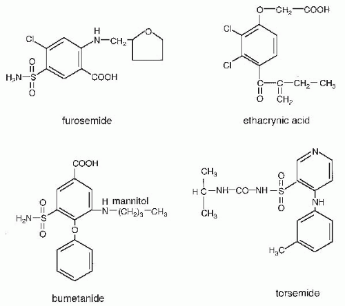
FIGURE 66.4 Structures of loop diuretics.


Mechanisms of Action


Sodium and Chloride Transport

The predominant effect of loop diuretic drugs is to inhibit the electroneutral Na-K-2Cl cotransporter at the apical surface of thick ascending limb cells. The loop of Henle, defined as the region between the last surface proximal segment and the first surface distal segment, reabsorbs from 20% to 50% of the filtered Na and Cl load101; approximately 10% to 20% is reabsorbed by thick ascending limb cells. The model in Figure 66.6 shows key components of Na, K, and Cl transport pathways in a thick ascending limb cell. As in other nephron segments, the Na/K ATPase at the basolateral cell membrane maintains the intracellular Na concentration low (approximately 10-fold lower than interstitial) and the K concentration high (approximately 20-fold higher than interstitial). Potassium channel(s)102 in the basolateral cell membrane permit K to diffuse out of the cell, rendering the cell membrane voltage oriented with the intracellular surface negative, relative to extracellular fluid. A chloride channel in the basolateral cell membrane permits Cl to exit the cell.102 Together with the apical K channel, described below, this chloride channel generates a transepithelial voltage, oriented in the lumen-positive direction.

The transporter inhibited by loop diuretics is a member of the cation chloride cotransporter family.93,103 This protein—referred to as the bumetanide-sensitive cotransporter, first isoform (BSC-1), or as the Na-K-2Cl cotransporter, second isoform (NKCC2)—is encoded by the gene SLC12A1. It apparently comprises 12 membrane-spanning domains, exists as a dimer,96 and is expressed at the apical membrane of the thick ascending limb104 and macula densa (MD) cells.105,106 A K channel (ROMK) is also present in the same membrane, permitting potassium to recycle from the cell to the lumen.107 Greger et al. showed that the asymmetrical orientation of channels (apical versus basolateral) and the action of the Na/K ATPase and Na-K-2Cl cotransporter combine to create a transepithelial voltage that is oriented with the lumen positive, with respect to the interstitium.108 This lumen-positive potential drives absorption of Na+, Ca2+, and Mg2+ via the paracellular pathway. The paracellular component of Na reabsorption comprises 50% of the total transepithelial Na transport by thick ascending limb cells.109 It should be noted, however, that both the transcellular and the paracellular components of Na transport are inhibited by loop diuretics, the former directly and the latter indirectly. The thick ascending limb is virtually impermeable to water. The combination of solute absorption and water impermeability determines the role of the thick ascending limb as the primary diluting segment of the kidney.

Although direct inhibition of ion transport is the most important natriuretic action of loop diuretics, other actions may contribute. Thick ascending limb cells have been shown to produce prostaglandin E2 following stimulation with furosemide,110
perhaps via inhibition of prostaglandin dehydrogenase.111,112 Blockade of cyclooxygenase reduces the effects of furosemide to inhibit loop segment chloride transport in rats,113,114 and this effect appears to be important clinically because nonsteroidal anti-inflammatory drugs (NSAIDs) are common causes of diuretic resistance (see below). Increases in renal prostaglandins may also contribute to the hemodynamic effects of loop diuretics, described later.


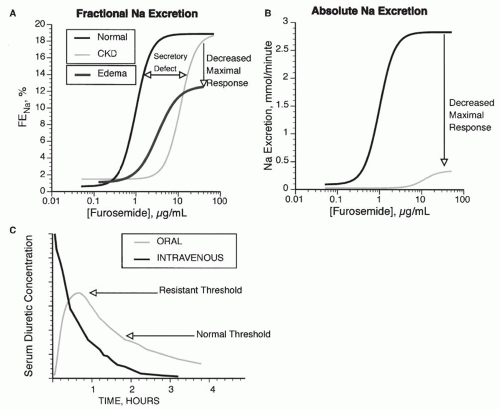
FIGURE 66.5 Dose response curve for loop diuretics. A: The fractional Na excretion (FENa) as a function of loop diuretic concentration. Compared with normal patients, patients with chronic renal failure (CKD) show a rightward shift in the curve, owing to impaired diuretic secretion. The maximal response is preserved when expressed as FENa, but when expressed as absolute Na excretion (B), maximal natriuresis is reduced in patients with CKD. Patients with edema demonstrate a rightward and downward shift, even when expressed as FENa (A). C: Compares the response to intravenous and oral doses of loop diuretics. In a normal individual (Normal), an oral dose may be as effective as an intravenous dose because the time above the natriuretic threshold (indicated by the normal line) is approximately equal. If the natriuretic threshold increases (as indicated by the dashed line, from an edematous patient), then the oral dose may not provide a high enough serum level to elicit natriuresis.


Calcium and Magnesium Transport

Loop diuretics increase the excretion of the divalent cations calcium (Ca) and magnesium (Mg). Although a component of magnesium and calcium absorption by thick ascending limbs may be active (especially when circulating parathyroid hormone levels are high115), a large component of their absorption is passive and paracellular, driven by the transepithelial voltage. As described above, active NaCl transport by thick ascending limb cells leads to a transepithelial voltage, oriented in the lumen positive direction. The paracellular pathway in the thick ascending limb expresses claudin-16 (paracellin-1) and claudin-19, which interact to form a tight junction that mediates both magnesium and calcium movement.116,117 Mutations in these genes lead to the clinical syndrome familial hypomagnesemia and hypercalciuria (FHHNC), an autosomal recessive tubular disorder that is frequently associated with renal failure.118 The positive voltage in the lumen, relative to
the interstitium, drives calcium and magnesium absorption through the paracellular pathway. Loop diuretics, by blocking the activity of the Na-K-2Cl cotransporter at the apical membrane of thick ascending limb cells, reduce the transepithelial voltage toward or to 0 mV. This stops passive paracellular calcium and magnesium absorption.


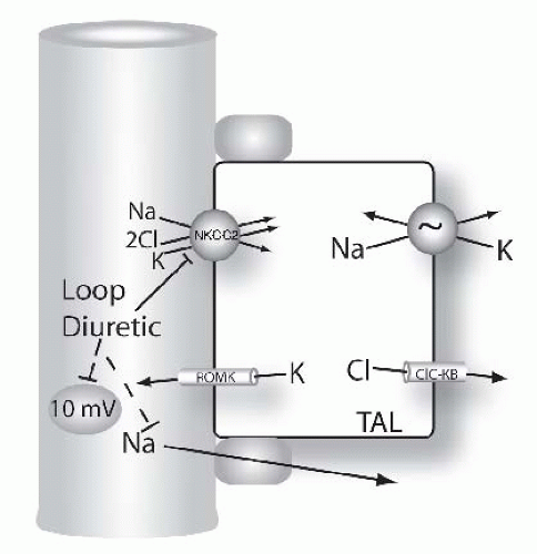
FIGURE 66.6 Mechanisms of diuretic action along the loop of Henle. Figure shows model of thick ascending limb (TAL) cells. Na and Cl are reabsorbed across the apical membrane via the loop diuretic-sensitive Na-K-2Cl cotransporter, NKCC2. Loop diuretics bind to and block this pathway directly. Note that the transepithelial voltage along the TAL is oriented with the lumen positive relative to blood (circled value, given in millivolts, mV). This transepithelial voltage drives a component of Na (and calcium and magnesium, see Fig. 66.9) reabsorption via the paracellular pathway. This component of Na absorption is also reduced by loop diuretics because they reduce the transepithelial voltage.


Renin Secretion

In addition to enhancing Na and Cl excretion, effects that result directly from inhibiting Na and Cl transport, loop diuretics also stimulate renin secretion. Although a component of this effect is frequently related to contraction of the extracellular fluid volume (see later), loop diuretics also stimulate renin secretion by inhibiting Na-K-2Cl cotransport directly. Macula densa cells, which control renin secretion, sense the NaCl concentration in the lumen of the thick ascending limb.119 High luminal NaCl concentrations in the region of the macula densa lead to two distinct but related effects. First, they activate the tubuloglomerular feedback (TGF) response, which suppresses GFR. Second, they inhibit renin secretion. The relation between these two effects is complex and has been reviewed,120 but both effects appear to result largely from NaCl movement across the apical membrane.121 Most of the ion transport pathways of macula densa cells are expressed by thick ascending limb cells. This includes the loop diuretic-sensitive Na-K-2Cl cotransporter (NKCC2) at the apical surface.105,106 Under normal conditions, an increase in luminal NaCl concentration in the thick ascending limb raises the NaCl concentration inside macula densa cells.121 Because the activity of the basolateral Na/K ATPase is lower in macula densa cells than in surrounding thick ascending limb cells,120 the cell NaCl concentration is much more dependent on luminal NaCl concentration in macula densa than in thick ascending limb cells.122 When luminal and macula densa cell NaCl concentrations decline, production rates of nitric oxide and prostaglandin E2 are stimulated. Although the mechanisms by which Na and Cl transport regulate nitric oxide and prostaglandin production rates are not known, both mediators appear to participate importantly in effecting renin secretion. Interestingly, loop diuretics also may stimulate renin secretion by inhibiting NKCC1, the secretory form of the three ion cotransport mechanism. Genetic deletion of NKCCC1 leads to an increase in plasma renin activity and a failure of renin exocytosis in response to furosemide,119 suggesting a role for alternative pathways.

The constitutive (neuronal) isoform of nitric oxide synthase (nNOS) is expressed at high levels by macula densa cells, but not by other cells in the kidney.123 Nitric oxide produced by macula densa cells has a paracrine effect to increase cellular cAMP in adjacent juxtaglomerular cells. Cyclic AMP through protein kinase A helps to stimulate renin secretion. In juxtaglomerular cells, nitric oxide may act by increasing cellular cGMP which inhibits phosphodiesterase 3,124 leading to phosphodiesterase 3 inhibition and cAMP accumulation. Several laboratories reported that furosemide-induced stimulation of renin secretion is dependent on an intact nitric oxide system because nonspecific nitric oxide inhibition interferes with this phenomenon.125,126,127 More recent studies, however, utilized knockout models to examine the role of nitric oxide in diuretic-induced renin secretion. Using this approach, it appears that neither neuronal nor endothelial nitric oxide synthases are required for loop diuretic-induced renin secretion. Instead, nitric oxide appears to play a permissive, rather than necessary, role in facilitating diuretic-induced renin secretion.128

Prostaglandin production also participates in regulating renin secretion. Cyclooxygenase, COX-2, is expressed by macula densa cells and by interstitial cells in the kidney.129,130,131,132 This isoform is often found only after induction by inflammatory cytokines. Blockade of prostaglandin synthesis either by nonspecific cyclooxygenase inhibitors133 or by specific COX-2 blockers134,135 reduces both the natriuresis induced by loop diuretics and dramatically inhibits the renin secretory response. These results have been corroborated in humans.136


Renal Hemodynamics

GFR and renal blood flow (RBF) tend to be preserved during loop diuretic administration,137 although GFR and RPF can decline if extracellular fluid volume contraction is severe. Loop diuretics reduce renal vascular resistance and increase
RBF under experimental conditions.138,139 This effect is believed related to the diuretic-induced production of vasodilatory prostaglandins (discussed previously).

Another factor that may contribute to the tendency of loop diuretics to maintain GFR and RBF despite volume contraction is their effect on the TGF system. The sensing mechanism that activates the TGF system involves NaCl transport across the apical membrane of macula densa cells by the loop diuretic sensitive Na-K-2Cl cotransporter.140 Under normal conditions, when the luminal concentration of NaCl reaching the macula densa rises, GFR decreases via TGF. To a large degree, the TGF-mediated decrease in GFR is believed to be due to afferent arteriole constriction. In response to changes in NaCl transport across the apical membrane of macula densa cells, ATP is released across the basolateral membranes through a NaCl sensitive ATP-permeable large-conductance (380 pS) anion channel.141 ATP appears to be degraded to adenosine which activates A1 adenosine receptor (P1 purinergic receptor class) expressed on afferent arteriole,142,143 as reviewed.140 Loop diuretic drugs block TGF by blocking the sensing step of TGF.144 In the absence of effects on the macula densa, loop diuretics would be expected to suppress GFR and RPF by increasing distal NaCl delivery and activating the TGF system. Instead, blockade of the TGF permits GFR and RPF to be maintained.


Systemic Hemodynamics

Acute intravenous administration of loop diuretics increases venous capacitance.145 Some studies suggest that this effect results from stimulation of prostaglandin synthesis by the kidney.146,147 Other studies suggest that loop diuretics have effects in peripheral vascular beds as well.148 Pickkers and coworkers examined the local effects of furosemide in the human forearm. Furosemide had no effect on arterial vessels, but did cause dilation of veins, an effect that was dependent on local prostaglandin production.149 More recently, loop diuretic-induced vasodilation was shown to depend on increased nitric oxide production.150 Although venodilation and improvements in cardiac hemodynamics frequently result from intravenous therapy with loop diuretics, the hemodynamic response to intravenous loop diuretics may be more complex.151 Johnston et al. reported that low dose furosemide increased venous capacitance, but that higher doses did not.152 It was suggested that furosemide-induced renin secretion leads to angiotensin II-induced vasoconstriction. This vasoconstrictor might overwhelm the prostaglandin-mediated vasodilatory effects in some patients. In two series, 1 to 1.5 mg per kg furosemide boluses, administered to patients with chronic heart failure, resulted in transient deteriorations in hemodynamics (during the first hour), with declines in stroke volume index, increases in left ventricular filling pressure,153 and exacerbation of heart failure symptoms. These changes may be related to activation of both the sympathetic nervous system and the renin/angiotensin system by the diuretic drug. Evidence for a role of the renin/angiotensin system in the furosemide-induced deterioration in systemic hemodynamics includes the temporal association between its activation and hemodynamic deterioration,153 and the ability of angiotensin-converting enzyme (ACE) inhibitors to prevent much of the pressor effect.154 The effects of renal denervation on sympathetic responses to furosemide were studied. These results confirm that the effects are mediated by both direct renal nerve traffic and indirectly, by activation of the renin/angiotensin axis.155,156 Many other studies have shown that acute loop diuretic administration frequently produces a transient decline in cardiac output; whether diuretic administration increases or decreases left atrial pressure acutely may depend primarily on the state of underlying sympathetic nervous system and renin/angiotensin axis activation.


Pharmacokinetics

The three loop diuretics that are used most commonly— furosemide, bumetanide, and torsemide—are absorbed quickly after oral administration, reaching peak concentrations within 30 minutes to 2 hours. Furosemide absorption is slower than its elimination in normal subjects; thus, the time to reach peak serum level is slower for furosemide than for bumetanide and torsemide. This phenomenon is called “absorption-limited kinetics,” as the rate of absorption is often slower than the rate of elimination.3 The bioavailability of loop diuretics varies from 50% to 90% (Table 66.2); furosemide bioavailability is approximately 50%4; when furosemide dosing is switched from intravenous to oral, the dose may need to be increased to compensate for its poor bioavailability.3 The half-lives of the loop diuretics available in the United States vary, but all are relatively short (ranging from approximately 1 hour for bumetanide to 3 to 4 hours for torsemide). The half-lives of muzolimine, xipamide, and ozolinone, none of which are available in the United States, are longer (6 to 15 hours).

Loop diuretics are organic anions that circulate tightly bound to albumin (>95%), thus their volume of distribution is small except during extreme hypoproteinemia.157 Approximately 50% of an administered dose of furosemide is excreted unchanged into the urine. The remainder appears to be eliminated by glucuronidation, probably by the kidney. Torsemide and bumetanide are eliminated both by hepatic processes and through renal excretion. The differences in metabolic fate mean that the half-life of furosemide is altered by renal failure, whereas this is not true for torsemide and bumetanide. Similar to CAIs and thiazides, loop diuretics gain access to the tubular fluid almost exclusively by proximal secretion. The peritubular uptake is mediated by the organic anion transporters OAT1 and OAT3, whereas the apically located multidrug resistance-associated protein 4 (Mrp-4) mediates secretion into the tubular fluid. Mice lacking OAT1, OAT3, or Mrp-4 are remarkably resistant to both loop and thiazide diuretics, illustrating the functional importance of these proteins.158,159,160 In humans, polymorphisms in the gene encoding the organic anion transporter OATP1B1 resulted in a slower elimination of torsemide.161
Interestingly, the response to loop diuretics is also associated with polymorphisms in the genes encoding the more distal sodium transporters NCC and ENaC.162


TABLE 66.2 Pharmacokinetics of Loop Diuretics


Elimination Half-Life (hours)


Oral Bioavailability (%)


Healthy


Renal Disease


Liver Disease


Heart Failure


Furosemide


10-100


1.5-2


2.8


2.5


2.7


Bumetanide


80-100


1


1.6


2.3


1.3


Torsemide


80-100


3-4


4-5


8


6


Adapted from the data in Brater DC. Diuretic therapy. N Engl J Med. 1998;339:387-395.


May 29, 2016 | Posted by in NEPHROLOGY | Comments Off on Mechanisms of Diuretic Action

Full access? Get Clinical Tree

Get Clinical Tree app for offline access