Immunobiology and Immunopharmacology of Renal Allograft Rejection


Immunobiology and Immunopharmacology of Renal Allograft Rejection


Choli Hartono

Terry B. Strom

Manikkam Suthanthiran


Renal transplantation is the treatment of choice for patients with irreversible renal failure and has moved from a high risk, experimental procedure to a safe, clinical procedure in the relatively short time of five decades.1 The substantive gains in patient and graft survival owe much to an improved understanding of the antiallograft repertoire, better preservation of donor kidneys, judicious usage of immunosuppressive drugs and monoclonal/polyclonal antibodies, and the clinical application of infection prophylaxis protocols.


IMMUNOBIOLOGY OF RENAL TRANSPLANTATION


The Antiallograft Response

Allograft rejection is contingent on the coordinated activation of alloreactive T cells and antigen-presenting cells (APCs). Through the intermediacy of cytokines and cell-to-cell interactions, a heterogeneous contingent of lymphocytes, including CD4+ helper T cells, CD8+ cytotoxic T cells, antibody-forming B cells, and other proinflammatory leukocytes are recruited into the antiallograft response (Fig. 81.1 and Table 81.1).


T Cell Activation and the Immunologic Synapse: Signal One

The immunologic synapse consists of a multiplicity of T cell-surface protein forms and clusters, thereby creating a platform for antigen recognition and generation of crucial T cell activation-related signals.2 The synapse begins to form when the initial adhesions between certain T cell (e.g., CD2, LFA-1) and APC surface proteins (e.g., CD58, ICAM-1) are formed (Table 81.2). These physical contacts between T cells and APCs provide an opportunity for the antigen reactive T cells to recognize cognate antigen. Antigen-driven T cell activation, a highly coordinated, preprogrammed process, begins when T cells recognize intracellularly processed fragments of foreign proteins (approximately 8 to 16 amino acids) embedded within the groove of the major histocompatibility complex (MHC) proteins expressed on the surface of APCs.3,4,5 Some of the recipient’s T cells directly recognize the allograft (i.e., donor antigen [s] presented on the surface of donor APCs) and this process is termed direct recognition whereas other T cells recognize the donor antigen after it is processed and presented by self-APCs6 (Fig. 81.1) and this process is designated indirect recognition.

The T cell antigen receptor (TCR)-CD3 complex is composed of clonally distinct TCR α and β peptide chains that recognize the antigenic peptide in the context of MHC proteins and clonally invariant CD3 chains that propagate intracellular signals originating from antigenic recognition (Fig. 81.2).2,7,8 The TCR variable, diversity, junction, and constant region genes (i.e., genes for regions of the clonespecific antigen receptors) are spliced together in a cassettelike fashion during T cell maturation.7 A small population of T cells expresses TCR γ and σ chains instead of the TCR α and β chains.

CD4 and CD8 proteins, expressed on reciprocal T cell subsets, bind to nonpolymorphic domains of human leukocyte antigen (HLA) class II (DR, DP, DQ) and class I (A, B, C) molecules, respectively (Fig. 81.1 and Table 81.2).2,7 A threshold of TCR to MHC-peptide engagements is necessary to stabilize the immunologic synapse stimulating a redistribution of cell-surface proteins and coclustering of the TCR/CD3 complex with the T cell-surface proteins.8,9,10 Additional T cell surface proteins such as CD5 proteins join the synapse.9,10 The TCR cluster already includes integrins (e.g., LFA-1) and nonintegrins (e.g., CD2)2,8,9 that have created T cell-APC adhesions. Hence, antigen recognition stimulates a redistribution of cell-surface proteins and coclustering of the TCR/CD3 complex with the T cell-surface proteins2,7,8,9 and signaling molecules. This multimeric complex functions as a unit in initiating T cell activation.

Following activation by antigen, the TCR-CD3 complex and coclustered CD4 and CD8 proteins are physically associated with intracellular protein-tyrosine kinases (PTKs)
of two different families, the src (including p59fyn and p56lck) and ZAP 70 families.2 The CD45 protein, a tyrosine phosphatase, contributes to the activation process by dephosphorylating an autoinhibitory site on the p56lck PTK. Intracellular domains of several TCR/CD3 proteins contain activation motifs that are crucial for antigen-stimulated signaling. Certain tyrosine residues within these motifs serve as targets for the catalytic activity of src family PTKs. Subsequently, these phosphorylated tyrosines serve as docking stations for the SH2 domains (recognition structures for select phosphotyrosinecontaining motifs) of the ZAP-70 PTK. Following antigenic engagement of the TCR/CD3 complex, select serine residues of the TCR and CD3 chains are also phosphorylated.2,5


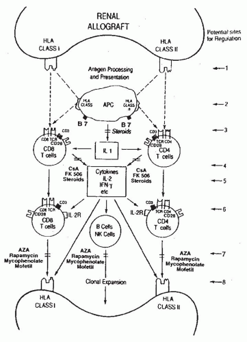
FIGURE 81.1 The antiallograft response. Schematic representation of human leukocyte antigens (HLA), the primary stimuli for the initiation of the antiallograft response, cell-surface proteins participating in antigenic recognition and signal transduction, contribution of the cytokines and multiple cell types to the immune response,and the potential sites for the regulation of the antiallograft response.Site /:Minimizing histocompatibility between the recipients and the donor (e.g., HLA matching).Site 2: Prevention of monokine production by antigen-presenting cells (e.g., corticosteroids). Site 3: Blockade of antigen recognition (e.g.,OKT3 mAbs).S/fe4: Inhibition of T cell cytokine production (e.g., cyclosporin A [CsA]).Sife 5: Inhibition of cytokine activity (e.g.,anti-interleukin-2 [IL-2] antibody). S/fe 6: Inhibition of cell cycle progression (e.g.,anti-IL-2 receptor antibody). Site 7: Inhibition of clonal expansion (e.g.,azathioprine [AZA]). Site 8: Prevention of allograft damage by masking target antigen molecules (e.g., antibodies directed at adhesion molecules). HLA class I: HLA-A, B, and C antigens; HLA class II: HLA-DR, DP,and DQ antigens. IFN-γ, interferon-y; NK cells, natural killer cells.

The waves of tyrosine phosphorylation triggered by antigen recognition encompass other intracellular proteins and are a cardinal event in initiating T cell activation. Tyrosine phosphorylation of the phospholipase Oγ1 activates this coenzyme and triggers a cascade of events that leads to full expression of T cell programs: hydrolysis of phosphatidylinositol 4,5-biphosphate (PIP2) and generation of two intracellular messengers, inositol 1,4,5-triphosphate (IP3) and diacylglycerol (Fig. 81.2).11 IP3, in turn, mobilizes ionized calcium from intracellular stores, while diacylglycerol, in the presence of increased cytosolic free Ca2+, binds to and translocates protein kinase C (PKC)—a phospholipid/Ca2+-sensitive protein serine/threonine kinase—to the membrane in its enzymatically active form.5,11 Sustained activation of PKC is dependent on diacylglycerol generation from hydrolysis of additional lipids, such as phosphatidylcholine.

The increase in intracellular free Ca2+ and sustained PKC activation promote the expression of several nuclear regulatory proteins (e.g., nuclear factor of activated T cells
[NF-AT], nuclear factor kappa B [FN-κB] , activator protein 1 [AP-1]) and the transcriptional activation and expression of genes central to T cell growth (e.g., interleukin-2 [IL-2] and receptors for IL-2 and IL-15).2,5,12


TABLE 81.1 Cellular Elements Contributing to the Antiallograft Response


Cell Type


Functional Attributes


T cells


The CD4+ T cells and the CD8+ T cells participate in the antiallograft response. CD4+ T cells recognize antigens presented by HLA class II proteins; CD8+ T cells recognize antigens presented by HLA class I proteins. The CD3/TCR complex is responsible for recognition of antigen and generates and transduces the antigenic signal.


CD4+ T cells


CD4+ T cells function mostly as helper T cells and secrete cytokines such as IL-2, a T cell growth/death factor, and IFN-γ, a proinflammatory polypeptide that can upregulate the expression of HLA proteins as well as augment cytotoxic activity of T cells and NK cells. Recently three main types of CD4+ T cells have been recognized: CD4+ TH1, CD4+ TH2, and CD4 TH17. IL-2 and IFN-γ are produced by CD4+ TH1 type cells, IL-4 and IL-5 are secreted by CD4+ TH2 type cells, and IL-17 family of cytokines CD4 + CD17 cells. Each cell type can regulate the secretion of the other and the regulated secretion is important in the expression of host immunity.


CD8+ T cells


CD8+ T cells function mainly as cytotoxic T cells. A subset of CD8+ T cells expresses suppressor cell function. CD8+ T cells can secrete cytokines such as IL-2 and IFN-γ and can express molecules, such as perforin, granzymes that function as effectors of cytotoxicity.


APCs


Monocytes/macrophages and dendritic cells function as potent APCs. Donor’s APCs can process and


present donor antigens to recipient’s T cells (direct recognition) or recipient’s APCs can process and present donor antigens to recipient’s T cells (indirect recognition). The relative contribution of direct recognition and indirect recognition to the antiallograft response has not been resolved. Direct recognition and indirect recognition might also have differential susceptibility to inhibition by immunosuppressive drugs.


B cells


B cells require T cell help for the differentiation and production of antibodies directed at donor antigens. The alloantibodies can damage the graft by binding and activating complement components (complement-dependent cytotoxicity) and/or binding the Fc receptor of cells capable of mediating cytotoxicity (antibody-dependent, cell-mediated cytotoxicity).


NK cells


The precise role of NK cells in the antiallograft response is not known. Increased NK cell activity has been correlated with rejection. NK cell function might also be important in immune surveillance mechanisms pertinent to the prevention of infection and malignancy.


APCs, antigen presenting cells; IFN, interferon; IL, interleukin; HLA, human leukocyte antigen; NK, natural killer; TCR, T cell antigen receptor.


Reproduced from Suthanthiran M, Morris RE, Strom TB. Transplantation immunobiology.


In: Walsh PC, Retik AB, Vaughn ED Jr, et al., eds. Campbell’s Urology, 7th ed. Philadelphia, PA: Saunders; 1997:491, with permission.


Calcineurin, a Ca2+– and calmodulin-dependent serine/threonine phosphatase, is crucial to Ca2+-dependent, TCR-initiated signal transduction.13,14 Inhibition by cyclosporine and tacrolimus (FK-506) of the phosphatase activity of calcineurin is considered central to their immunosuppressive activity15


Costimulatory Signals: Signal Two

Signaling of T cells via the TCR/CD3 complex (signal one) is necessary, but insufficient, to induce T cell proliferation; full activation of T cells is dependent on both the antigenic signals and the costimulatory signals (signal two) engendered by the contactual interactions between cell-surface proteins expressed on antigen-specific T cells and APCs (Fig. 81.3 and Table 81.2).16,17 The interaction of the CD2 protein on the T cell surface with the CD58 (leukocyte function-associated antigen 3 [LFA-3]) protein on the surface of APCs, and that of the CDlla/CD18 (LFA-1) proteins with the CD54 (intercellular adhesion molecule 1 [ICAM-1]) proteins,18 and/or the interaction of the CD5 with the CD72 proteins10 aids in imparting such a costimulatory signal.

Recognition of the B7-1 (CD80) and B7-2 (CD86) proteins expressed upon CD4+ T cells generates a very
powerful T cell costimulus.19 A subset of monocytes and dendritic cells constitutively express CD80 and CD86 at low levels and cytokines (e.g., granulocyte-macrophage colony-stimulating factor [GMCSF] or interferon-γ [IFN-γ]) stimulate heightened expression of CD80 and CD86 on monocytes, B cells, and dendritic cells.19 Many T cells express B7-binding proteins (i.e., CD28 proteins that are constitutively expressed on the surface of CD4 + T cells and CTLA-4 [CD152]), a protein whose ectodomain is closely related to that of CD28, and is expressed upon activated CD44- and CD84- T cells. CD28 binding of B7 molecules stimulates a Ca2+-independent activation pathway that leads to stable transcription of the IL-2, IL-2 receptors, and other activation genes resulting in vigorous T cell proliferation.19 For some time, the terms CD28 and the costimulatory receptor were considered synonymous by some, but the demonstration that robust T cell activation occurs in CD28-deficient mice indicated that other receptor ligand systems contribute to signal two.20 In particular, the interaction between CD40 expressed upon APCs and CD40 ligand (CD154) expressed by antigen-activated CD44- T cells has received great attention as a potent second signal.21


TABLE 81.2 Cell-Surface Proteins Important for T Cell Activationa


T Cell Surface


APC Surface


Functional Response


Potential Consequence of Blockade


LFA-1 (CDlla, CD18)


ICAM (CD54)


Adhesion


Immunosuppression


ICAM1 (CD54)


LFA-1 (CDlla, CD18)


Adhesion


Immunosuppression


CD8, TCR, CD3


MHCI


Antigen recognition


Immunosuppression


CD4, TCR, CD3


MHCII


Antigen recognition


Immunosuppression


CD2


LFA3 (CD58)


Costimulation


Immunosuppression


CD40L(CD154)


CD40


Costimulation


Immunosuppression


CD5


CD72


Adhesion


Immunosuppression


CD28


B7-1 (CD80)


Costimulation


Anergy


CD28


B7-2 (CD86)


Costimuation


Anergy


CTLA4 (CD152)


B7-1 (CD80)


Inhibition


Immunostimulation


CTLA4 (CD152)


B7-2 (CD86)


Inhibition


Immunostimuation


aRceptor/counterreceptor pairs that mediate interactions between T cells and APCs are shown in this table. Inhibition of each protein-to-protein interaction, except the CTLA4-B7.1/B7.2 interaction, results in an abortive in vitro immune response. Initial contact between T cells and APCs requires an antigen-independent adhesive interaction. Next, the T cell antigen-receptor complex engages processed antigen presented within the antigen-presenting groove of MHC molecules. Finally, costimulatory signals are required for full T cell activation. An especially important signal is generated by B7-mediated activation of CD28 on T cells. Activation of CD28 by B7.2 may provide a more potent signal than activation by B7.1. CTLA4, present on activated but not resting T cells, imparts a negative signal. Monoclonal antibodies directed at the T cell CD2 protein, used as component of a preconditioning regimen, has been associated with tolerance to histoincompatble human renal allografts.23


APC, antigen-presenting cell; ICAM, intercellular adhesion molecule; LFA, leukocyte function-associated; MHC, major histocompatibility complex. Reproduced from Suthanthiran M, Morris RE, Strom TB. Transplantation immunobiology In: Walsh PC, Retik AB, Vaughn ED Jr, et al., eds. Campbell’s Urology, 7th ed. Philadelphia: WB Saunders, 1997:491, with permission.


The delivery of the antigenic first signal and the costimulatory second signal leads to stable transcription of the IL-2, several T cell growth-factor receptors, and other pivotal T cell activation genes (Table 81.2). The Ca2+-independent costimulatory CD28 pathway is relatively more resistant to inhibition by cyclosporine or FK-506 as compared to the calcium-dependent pathway of T cell activation. Whereas the interactions between B7 proteins and its counter receptor CD28 result in positive costimulation, the interactions between B7 proteins by CTLA-4, a protein primarily expressed on activated T cells, result in the generation of a negative signal to T cells. This coinhibitory signal is a prerequisite for peripheral T cell tolerance.22

The formulation that full T cell activation is dependent on the costimulatory signal, as well as the antigenic signal, is most significant, as T cell molecules responsible for costimulation and their cognate receptors on the surface
of APCs then represent target molecules for the regulation of the antiallograft response. Indeed, transplantation tolerance has been induced in experimental models by targeting a variety of cell-surface molecules that contribute to the generation of costimulatory signals, and tolerance to histoincompatible human kidney allografts has been accomplished with a conditioning regimen that includes monoclonal antibodies directed at the CD2 protein.23


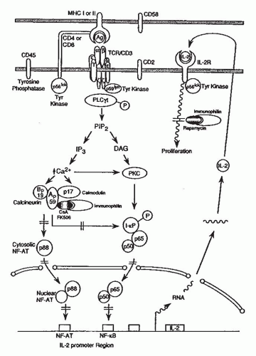
FIGURE 81.2 Signal transduction in T cells and mechanisms of action of cyclosporin A (CsA), FK-506, or rapamycin.Signaling molecules and transmembrane signaling events participating in the transduction of antigenic signals from the plasma membrane of the T cells to the nucleus are schematically shown. The sites of action of the drug (CsA/FK-506/rapamycin)-immunophilin complex are also shown.kg, antigen; Ap59 and Bp19, subunits of calcineurin;DAG, diacylglycerol; lB, inhibitory factor kappa B;IL.-2, interleukin-2; immunophilin, cyclophilin or FK-binding protein; IP3, inositol 1,4,5-triphosphate; MHC, major histocompatibility complex; NF-AT, nuclear factor of activated T cells; NF-κB, nuclear factor kappa B; P, phosphotyrosine; PIP2, phosphatidylinositol 4,5-biphosphate; PKC, protein kinase C;PLCγ1, phospholipase C gamma-1; Tyr kinase, tyrosine kinase. (Adapted from Schreier MH, Baumann G, Zenke G, et al. Inhibition of T-cell signaling pathways by immunophilin drug complexes: Are side effects inherent to immunosuppressive properties? Transplant Proc 1993;25:502.)


lnterleukin-2/lnterleukin-l 5 Stimulated TCell Proliferation

Autocrine type of T cell proliferation occurs as a consequence of the T cell activation-dependent production of IL-2 and the expression of multimeric high affinity IL-2 receptors on T cells (Fig. 81.2) formed by the noncovalent association of three IL-2-binding peptides (α β, γ).12,24,25,26 IL-15 is a paracrine-type T cell-growth factor family member with very similar overall structural and identical T cell stimulatory qualities to IL-2.12 The IL-2 and IL-15 receptor complexes share β and γ chains that are expressed in low abundance upon resting T cells; expression of these genes is amplified in activated T cells. The α-chain receptor components of the IL-2 and IL-15 receptor complexes are distinct and expressed upon activated, but not resting, T cells. The intracytoplasmic domains of the IL-2 receptor β and γ chains are required for intracellular signal transduction. The ligand-activated, but not resting, IL-2/IL-15 receptors are associated with intracellular PTKs.12,27,29 Raf-1, a protein serine/threonine kinase associates with the intracellular domain of the shared β chain,30 and this association and the kinase activity are prerequisites to IL-2/IL-15-triggered cell proliferation. Translocation of IL-2 receptor-bound Raf-1 serine/threonine kinase into the cytosol requires IL-2/IL-15-stimulated
PTK activity. The ligand-activated common γ chain recruits a member of the Janus kinase family, Jak 3, to the receptor complex that leads to activation of a member of the STAT family Activation of this particular Jak-STAT pathway is essential for the proliferation of antigen-activated T cells. The subsequent events leading to IL-2/IL-15-dependent proliferation are not fully resolved; however, IL-2/IL-15—stimulated expression of several DNA binding proteins including Bcl-2, c-jun, c-fos, and c-myc contributes to cell cycle progression.31,32 It is interesting and probably significant that IL-2, but not IL-15, triggers apoptosis of many antigen-activation T cells. In this way, IL-15-triggered events may be more detrimental to the antiallograft response than those initiated by IL-2. As IL-15 is not produced by T cells, IL-15 expression is not regulated by cyclosporine or tacrolimus.


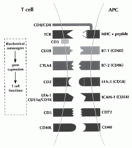
FIGURE 81.3 T cell/antigen-presenting cell contact sites. In this schema of T cell activation, the antigenic signal is initiated by the physical interaction between the clonally variant T cell antigen receptor (TCR) α-β-heterodime, and the antigenic peptide displayed by MHC on antigen-presenting cells (APCs).The antigenic signal is transduced into the cell by the CD3 proteins.The CD4 and the CD8 antigens function as associative recognition structures, and restrict TCR recognition to class II and class I antigens of MHC, respectively. Additional T cell- surface receptors generate the obligatory costimulatory signals by interacting with their counterreceptors expressed on the surface of the APCs.The simultaneous delivery to the T cells of the antigenic signal and the costimulatory signal results in the optimum generation of second messengers (such as calcium), expression of transcription factors (such as nuclear factor of activated T cells), and T cell growth-promoting genes (such as IL-2).The CD28 antigen as well as the CTLA4 antigen can interact with both the B7-1 and B7-2 antigens.The CD28 antigen generates a stimulatory signal, and CTLA4, unlike CD28, generates a negative signal. CD, cluster designation; ICAM-1, intercellular adhesion molecule-1; LFA-1, leukocyte function-associated antigen-1;MHC, major histocompatibility complex. (From Suthanthiran M.Transplantation tolerance: fooling mother nature. Proc Natl Acad Sci U S A. 1996;93:12072.)


Humoral Rejection

Antibody-mediated rejection (AMR) is a form of humoral rejection wherein antibodies directed at the donor HLA antigens (DSAs) serve as the main effector for the immune response directed at the allograft. Antibodies directed at non-HLA antigens such as endothelial cell associated antigens and MHC class I-related chain A antigens (MICA) have also been implicated in the pathogenesis of AMR. Whereas most acute T cell mediated rejections (TMRs) are responsive to steroid therapy, AMR is typically steroid-resistant and requires additional treatment such as plasmapheresis, anti-B cell, and intravenous immunoglobulin (IVIG) therapy. The incidence of AMR has been estimated at less than 10% but appears to be on the rise due to multiple reasons including acute TMR being effectively prevented by current immunosuppressive regimens, better definition of AMR, and transplantation of individuals with humoral presenitization and repeat transplants. Patients with AMR invariably harbor anti-HLA DSA although, in certain cases, histopathologic evidence of AMR may be apparent without any anti-HLA DSA. Acute AMR may occur within 1 week after engraftment even in the setting of antithymocyte globulin induction therapy. The diagnosis of AMR requires the presence of C4d complement staining in the peritubular capillaries in addition to peritubular capillary inflammation with polymorphonuclear and mononuclear leukocytes or the presence of fibrinoid changes/transumural arterial inflammation or acute tubular necrosis (ATN)-like tissue injury.33 In the current Banff classification schema, those who present with histolgic features consistent with AMR but without concurrent intragraft C4d deposition or circulating DSA are classified as supicious for AMR—it is possible that the offending antibodies may be of the noncomplement fixing IgG subtypes and/or non-HLA antibodies (because most screening assays for DSA utilize HLA as target antigens).

A novel form of humoral rejection has also been documented. Antibodies directed against two epitopes of the angiotensin II type I (ATO receptor have been associated with refractory vascular allograft rejection in a series of 16 patients and these patients did not have anti-HLA antibodies at the time of incident humoral rejection.34


Immunobiology and Molecular Diagnosis of Rejection

The net consequence of cytokine production and acquisition of cell-surface receptors for these transcellular molecules is the emergence of antigen-specific and graft-destructive T cells
and antibody producing B cells/plasma cells (Fig. 81.1). Cytokines facilitate not only the T cell effector arm and TCR but also the B cell/plasma cell arm by promoting the production of cytopathic antibodies. Moreover, cytokines such as IFN-y and tumor necrosis factor-α (TNF-α) can amplify the ongoing immune response by upregulating the expression of HLA molecules as well as costimulatory molecules (e.g., B7) on graft parenchymal cells and APCs (Fig. 81.1). We and others have demonstrated the presence of antigen-specific cytotoxic T lymphocytes (CTL) and anti-HLA antibodies during or preceding a clinical rejection episode.35,36 We have detected messenger RNA (mRNA) encoding the CTL-selective serine protease (granzyme B), perforin, Fasligand attack molecules, and immunoregulatory cytokines, such as IL-10 and IL-15, in human renal allografts undergoing acute rejection.37 Indeed, these gene expression events may anticipate clinically apparent rejection. More recent efforts to develop a noninvasive method for the molecular diagnosis of rejection have proved rewarding. Using either peripheral blood38 or urinary leukocytes39 rejection-related, gene expression events evident in renal biopsy specimens are robustly detected in peripheral blood or urinary sediment specimens. Initial results from large-scale multicenter trials (e.g., Clinical Trials in Organ Transplantation, CTOT-04) support the hypothesis that noninvasive diagnosis of acute TMR is feasible by measurement of genes encoding cytotoxic attack molecules in urine, and the urinary cell mRNA profiles may anticipate the future development of acute TMR.40 We speculate as well that a noninvasive, molecular diagnostic approach to rejection would be of value toward the detection of insidious, clinically silent rejection episodes that, although rarely detected through standard measures, are steroid-sensitive but usually lead to chronic rejection.41


TABLE 81.3 Mechanisms of Action of Small Molecule Immunosuppressantsa


Immunosuppressant


Subcellular Site(s) of Action


Azathioprine


Inhibits purine synthesis


Corticosteroids


Blocks cytokine gene expression


CsA/tacrolimus


Blocks Ca2+-dependent T cell activation pathway via binding to calcineurin


Mycophenolate mofetil


Inhibits inosine monophosphate dehydrogenase and prevents de novo guanosine and deoxyguanosine synthesis in lymphocytes


Sirolimus/everolimus


Blocks IL-2 and other growth factor signal transduction; blocks CD28-mediated costimulatory signals


Leflunomide/FK778


Inhibits dihydroorotate dehydrogenase—a key enzyme for de novo pyramidine biosynthesis


FTY720


Phosphorylated FTY720 binds sphingolipid 1-phosphate receptor and prevents SIP signaling of cells; sequestration of lymphocytes within the lymph nodes and prevention of cell egress into the peripheral circulation


CsA, cyclosporin A; IL, interleukin.


Immunopharmacology of Allograft Rejection


Glucocorticosteroid

Glucocorticosteroids inhibit T cell proliferation, T cell-dependent immunity, and cytokine gene transcription (including IL-1, IL-2, IL-6, IFN-γ, and TNF-α gene).42,43,44 Although no single cytokine can reverse the inhibitory effects of corticosteroids on mitogen-stimulated T cell proliferation, a combination of cytokines is effective.45 The glucocorticoid and glucocorticoid-receptor bimolecular complex block IL-2 gene transcription via impairment of the cooperative effect of several DNA-binding proteins.46 Corticosteroids also inhibit formation of free NF-κB, a DNA-binding protein required for cytokine and other T cell-activation gene expression events (Fig. 81.1 and Table 81.3).47


Azathioprine

Azathioprine (AZA), a thioguanine derivative of 6-mercap-topurine,48 is a purine analog, acts as a nonspecific inhibitor of purine biosynthesis, and is an effective antiproliferative agent (Fig. 81.1 and Table 81.3).48,49 In a randomized conversion trial from mycophenolate mofetil (MMF) to AZA in 48 stable kidney transplant recipients at 6 months following engraftment, it was observed that acute rejection rates
were comparable (4.5% vs. 3.8%) after a 6-month observation period in the MMF (n = 22) or AZA (n = 26) arm. The trial participants received cyclosporine and prednisone as maintenance immunosuppressive therapy and antithymocyte globulin induction was used in 27% of the recipients maintained on MMF and 46% in the AZA conversion group. It is worth noting that high-risk patients including retransplant recipients, highly sensitized, and those with a history of steroid-resistant rejection were all excluded from the trial.50


The Calcineurin Inhibitors:Cyclosporine and Tacrolimus (FK-506)

Cyclosporine, a small cyclic fungal peptide, and FK-506, a macrolide antibiotic, block the Ca2+-dependent antigen triggered T cell activation (signal one) (Fig. 81.2).51 The immunosuppressive effects of cyclosporine and FK-506 are dependent on the formation of a heterodimeric complex that consists of the drug cyclosporine or FK-506 and its respective cytoplasmic receptor “immunophilin” proteins, cyclophilin and FK-binding protein (FKBP), respectively. The heterodimeric cyclosporine-cyclophilin complex and the FK-506-FKBP complex target and bind calcineurin and inhibit its phosphatase activity (Table 81.3). The inhibition of the enzymatic activity of calcineurin is considered central to the immunosuppressive effects of cyclosporine and FK-506.

One of the well-documented consequences of calcium/calmodulin dependent activation of calcineurin is dephosphorylation of cytoplasmic NF-AT in T cells, import of NF-AT into the nucleus, binding of NF-AT with its nuclear partmer, and transcription of the IL-2 gene. The cyclosporine-FK-506 mediated inhibition of phosphatase activity of calcineurin results in the lack of dephosphorylation of cytoplasmic NF-AT and retention of the phosphorylated NF-AT in the cytoplasm. In addition to inhibiting the expression of NF-AT, cyclosporine also inhibits other DNA-binding proteins, such as NF-κB and AP-1.52

The phosphorylation status of transcription factors can also affect their DNA binding ability and interaction with the rest of the transcriptional machinery. For example, the DNA binding activities of c-jun increase upon dephosphorylation.

Blockade of cytokine gene activation does not totally account for the antiproliferative effect of cyclosporine and FK-506. It is significant that cyclosporine as well as FK-506, in striking contrast to their inhibitory activity on the induced expression of IL-2, enhance the expression of transforming growth factor-β (TGF-β).53,54 Because TGF-(3 is a potent inhibitor of T cell proliferation and generation of antigen-specific CTL,55 heightened expression of TGF-β must contribute to the antiproliferative/immunosuppressive activity of cyclosporine/tacrolimus. This TGF-jS inducing effect of cyclosporine/tacrolimus also suggests a mechanism for some of the complications (e.g., renal fibrosis and tumor metastasis) of therapy with calcineurin inhibitors, because TGF-β is a fibrogenic and proangiogenic cytokine.


Mycophenolate Mofetil and Enteric-Coated Mycophenolate Sodium

MMF is a semisynthetic derivative of mycophenolic acid (MPA). MMF inhibits allograft rejection in rodents, diminishes proliferation of T and B cells, decreases generation of cytotoxic T cells, and suppresses antibody formation.56,57,58 MMF inhibits inosine monophosphate dehydrogenase (IMP-DH), an enzyme in the de novo pathway of purine synthesis. Lymphocytes are dependent on this biosynthetic pathway to satisfy their guanosine requirements (Table 81.3).58 Early clinical trials have utilized MMF to replace azathioprine in the cyclosporine- and steroid-based immunosuppressive regimen. These controlled, prospective trials have shown a diminished incidence of early acute rejection episodes.58,59,60 Although follow-up studies over a 3-year period have indicated an advantage for MMF over azathioprine,60 a recent randomized trial comparing MMF with azathioprine in recipients of a first kidney transplant from a deceased donor found similar levels of acute rejection in the first 6 months of transplantation.61

Enteric-coated mycophenolate sodium (EC-MPS) was developed to improve the gastrointestinal tolerability of MPA. An international phase III, randomized, double-blinded, parallel group trial demonstrated the therapeutic equivalence of MMF and EC-MPS.62 The two parallel groups received equivalent concomitant antibody induction, corticosteroids, and calcineurin inhibitor (CNI) therapy. At 12 months, the incidence of acute rejection, graft loss, and death was comparable for both treatment groups. Interestingly, in the phase III pivotal trial gastrointestinal complications were not significantly different between MMF and EC-MPS. Within 12 months of enrollment, dose changes were required for gastrointestinal adverse events in 19.5% versus 15% of subjects (P

Only gold members can continue reading. Log In or Register to continue

Stay updated, free articles. Join our Telegram channel

May 29, 2016 | Posted by in NEPHROLOGY | Comments Off on Immunobiology and Immunopharmacology of Renal Allograft Rejection

Full access? Get Clinical Tree

Get Clinical Tree app for offline access