Liver Disease and the Kidney


Liver Disease and the Kidney


Pere Ginès

Andrés Cárdenas

Elsa Solà

Robert W. Schrier


The presence of abnormalities of kidney function in patients with liver diseases has been recognized for several decades.1 More than a century ago, Frerichs in Europe and Flint in the United States reported the association between liver diseases and kidney dysfunction.2,3 These reports described the development of oliguria in patients with chronic liver disease in the setting of normal kidney histology and proposed the first pathophysiologic interpretation of kidney dysfunction in liver disease by linking the abnormalities in kidney function to disturbances in the systemic circulation. Since then, the relationship between the liver and kidney function has been the object of a considerable amount of research and substantial progress has been made in the last two decades with regard to the pathophysiology and management of renal dysfunction in liver diseases. Several books have been published specifically devoted to this topic.4,5,6,7,8,9,10,11,12,13

Most derangements of renal function in liver diseases occur in patients with cirrhosis and are pathophysiologically related to the presence of an expanded extracellular fluid volume which leads to the development of ascites and/or edema. This chapter deals with the pathophysiology, clinical features, and treatment of ascites and renal functional abnormalities in cirrhosis. The abnormalities in kidney function due to other liver diseases are not discussed.


RENAL ABNORMALITIES IN CIRRHOSIS

Most abnormalities of kidney function in cirrhosis are of functional origin (i.e., they occur in the absence of significant alterations in kidney histology).14,15,16,17,18 These abnormalities are usually referred to as functional renal abnormalities, as opposed to nonfunctional renal abnormalities, which may also develop in patients with cirrhosis (i.e., glomerulonephritis).

The most common functional renal abnormalities in cirrhotic patients are an impaired ability to excrete sodium, an impaired ability to excrete solute-free water, and a reduction of the glomerular filtration rate (GFR) secondary to vasoconstriction of the renal circulation. Sodium retention is a key factor in the expansion of the extracellular fluid volume and development of ascites and edema, whereas solute-free water retention is responsible for dilutional hyponatremia. Renal vasoconstriction, when severe, leads to hepatorenal syndrome (HRS). Chronologically, sodium retention is the earliest alteration of kidney function observed in patients with cirrhosis, whereas dilutional hyponatremia and HRS are late findings. In most patients, abnormalities of kidney function usually worsen with time as the liver disease progresses. However, in some patients, a spontaneous improvement or even normalization of sodium and solute-free water excretion may occur during the course of their disease.19,20,21 This improvement in renal function occurs particularly in patients with alcoholic cirrhosis after abstinence from alcohol. Spontaneous improvement of renal function after the development of type-1 HRS (see later) is extremely unusual.22,23


Sodium Retention and Ascites

Sodium retention is the most frequent abnormality of kidney function in patients with cirrhosis and ascites. The existence of sodium retention in cirrhosis was first documented more than 60 years ago when methods to measure electrolyte concentration in organic fluids became available.24,25,26 Since then, it has been well established that sodium retention plays a key role in the pathophysiology of ascites and edema formation in cirrhosis. The amount of sodium retained within the body is dependent on the balance between the sodium ingested in the diet and the sodium excreted in the urine. As long as the amount of sodium excreted is lower than that ingested, patients accumulate ascites and/or edema. The important role of sodium retention in the pathogenesis of ascites formation is supported by the fact that ascites can disappear just by reducing the dietary sodium content in some patients or by increasing the urinary sodium excretion with the administration of diuretics in others.26,27 Conversely, a high-sodium diet or diuretic withdrawal leads to the reaccumulation of ascites.25,26 The achievement of a negative sodium balance (i.e., excretion higher than intake) is the essence of pharmacologic therapy of ascites. Although no studies assessing the chronologic relationship between sodium retention and the formation of ascites have been performed in patients with
cirrhosis, studies in experimental animals have provided conclusive evidence indicating that sodium retention precedes ascites formation, further emphasizing the important role of this abnormality of renal function in the pathogenesis of ascites in cirrhosis.28,29,30,31,32 This observation suggests that sodium retention is the cause and not the consequence of ascites formation in cirrhosis.

The severity of sodium retention in cirrhosis with ascites varies considerably from patient to patient. Some patients have relatively high urinary sodium excretion, whereas urine sodium concentrations are very low or even undetectable in others (Fig. 68.1). The proportion of patients with marked sodium retention depends on the population of cirrhotic patients considered. Most patients who require hospitalization because of severe ascites have marked sodium retention (less than 10 mEq per day). Sodium retention is particularly intense in patients with ascites refractory to diuretic treatment.33,34 By contrast, in a population of cirrhotic patients with mild or moderate ascites, the proportion of patients with marked sodium retention is low and most patients excrete more than 10 mEq per day spontaneously (without diuretic therapy). The response to diuretic treatment is usually better in patients with moderate sodium retention than in those with marked sodium retention.27,35,36


Nephron Sites of Sodium Retention

In healthy subjects approximately 95% of filtered sodium is reabsorbed in the renal tubules. Approximately 60% to 70% is absorbed in the proximal tubules, another 30% to 40% gets absorbed in the thick ascending limb, and 5% to 10% of sodium is reabsorbed in the collecting ducts.37

In many instances, sodium retention in cirrhosis is due to increased tubular reabsorption of sodium because it occurs in the presence of normal or only moderately reduced GFR.27,38 The exact contribution of the different segments of the nephron to this increased sodium reabsorption is not completely known. Micropuncture studies in rats with cirrhosis and ascites have demonstrated an enhanced reabsorption of sodium in the proximal tubule.28,39 On the other hand, it has been shown that the development of a positive sodium balance and the formation of ascites in cirrhotic rats can be prevented by aldosterone antagonists, which suggests that the collecting ducts are important sites of the increased sodium reabsorption in experimental cirrhosis.31,40,41 Studies assessing the protein abundance of renal tubular sodium transporters in rats with CCL4- induced cirrhosis showed an increased expression of the sodium chloride cotransporters of the distal tubule (NCC/TSC) and the epithelial sodium channel of the collecting duct (ENaC), both of which are regulated by aldosterone, consistent with a major role of hyperaldosteronism in sodium retention in this animal model.41 An increased abundance of the Na+-K+-2Cl cotransporter of the thick ascending limb (NKCC/BSC1) and a decreased abundance of the proximal sodium transporters (sodium hydrogen exchanger type 3-NH-3, and sodium phosphate cotransporter isoform 2-NaPi-2) was also found, consistent with increased sodium reabsorption in the ascending limb of the loop of Henle and reduced reabsorption in the proximal tubule.41 Other factors such as the influence of calcium on the bumetanide-sensitive Na+K+2Cl cotransporter (BSC-1) located in the luminal membrane of epithelial cells lining the thick ascending limb of the loop of Henle may play a role in sodium retention.41


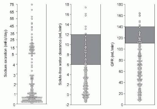
FIGURE 68.1 Individual values of sodium excretion, solute-free water clearance, and glomerular filtration rate in a large series of patients with cirrhosis and ascites without diuretic therapy and under a low-sodium diet. Lines indicate normal ranges. For urine sodium normal range is 80 to 100 mEq per day.

Investigations in patients with cirrhosis have also provided discrepant findings as to the most important nephron site of sodium retention. Results from earlier studies using sodium, water, or phosphate clearances to estimate the tubular handling of sodium suggest that the distal nephron is the main site of sodium retention.42,43,44,45 Results of studies using lithium clearance, which estimates sodium reabsorption
in the proximal tubule, suggest that cirrhotic patients with ascites show a marked increase in proximal sodium reabsorption.46,47 Nevertheless, distal sodium reabsorption is also increased, especially in patients with more avid sodium retention.47 Clinical studies using spironolactone to antagonize the mineralocorticoid receptor indicate that this agent induces natriuresis in a large proportion of cirrhotic patients with ascites without renal failure, which supports a major role for increased sodium reabsorption in distal sites of the nephron in these patients.36,48,49,50,51 Taken together, these results suggest that in patients with cirrhosis without renal failure, an enhanced reabsorption of sodium in both proximal and distal tubules contributes to sodium retention. Potential mediators of this increased sodium reabsorption include changes in the hydrostatic and colloidosmotic pressures in the peritubular capillaries and increased activity of the sympathetic nervous system and the renin-angiotensin-aldosterone system (RAAS). Sodium retention is usually more marked in patients with renal failure than in those without renal failure due to both a reduction in filtered sodium load and a more marked activation of sodium-retaining mechanisms.


Clinical Consequences

Because sodium is retained together with water isoosmotically in the kidneys, sodium retention is associated with fluid retention, leading to expansion of extracellular fluid volume and increased amount of fluid in the interstitial tissue. In some patients with advanced cirrhosis, the total extracellular fluid volume may increase up to 40 L or even more (compared to the average 14 L in a 70-kg healthy adult), which represents an approximate cumulative gain of 3,400 mEq of sodium (26 L of excess extracellular fluid volume times 130 mEq per L). In most patients with advanced cirrhosis, sodium retention is manifested by the development of ascites. The most common clinical symptom of ascites is discomfort due to abdominal swelling. In cases with marked accumulation of fluid, physical activity and respiratory function may be impaired. Other clinical consequences related to the presence of ascites are the appearance of abdominal wall hernias and hydrocele and spontaneous infection of ascitic fluid (also known as spontaneous bacterial peritonitis).52 These complications, especially infection, contribute markedly to the increased morbidity and mortality associated with the presence of ascites.

Accumulation of fluid in the subcutaneous tissue, as edema, is also common in patients with cirrhosis and sodium retention and in most cases occurs concomitantly with the existence of ascites. Edema is most commonly observed in the lower extremities, but generalized edema may occur as well. Mild or moderate pedal edema may decrease or even disappear during bed rest and reappear during the daytime, reflecting an increased natriuresis in the supine position as compared with the upright position.53,54 Both hypoalbuminemia and increased venous pressure in the inferior vena cava due either to constriction of the vena cava within the liver or increased intra-abdominal pressure caused by ascites may contribute to the high incidence of edema in cirrhotic patients with ascites. Leg edema may occur in patients with cirrhosis treated with either surgical portacaval shunts or transjugular intrahepatic portosystemic shunts (TIPS), presumably because of the increased pressure in the inferior vena cava secondary to these procedures.

Other clinical manifestations of sodium retention in cirrhosis include pleural and/or pericardial effusions. Hepatic hydrothorax is defined as a pleural effusion in patients with cirrhosis without associated cardiac and/or pulmonary disease. This complication occurs in approximately 10% of patients with cirrhosis.55,56 In most cases the effusion is mild or moderate, more frequent on the right side, and associated with the presence of ascites. Left-sided effusions are uncommon. Occasionally, large right pleural effusions may exist in the absence of clinically evident ascites and constitute the main manifestation of the disease.56,57 These pleural effusions are very difficult to manage, usually recur after therapy, and are due to the existence of anatomic defects in the diaphragm which cause a communication between the peritoneal and pleural cavities. The gradient between the positive intra-abdominal pressure and the negative intrathoracic pressure explains the passage of the fluid formed in the peritoneal cavity to the pleural cavity. Although less commonly than ascitic fluid, pleural fluid may also become infected spontaneously, a condition known as spontaneous bacterial empyema.58 Finally, between one and two thirds of cirrhotic patients with ascites also have mild or moderate pericardial effusions as demonstrated by echocardiography.59 These disappear after the elimination of ascites and are not associated with clinical symptoms.


Assessment of Sodium Excretion in Clinical Practice

The assessment of the urinary excretion of sodium is very useful in the clinical management of patients with cirrhosis and ascites because it allows the precise quantification of sodium retention. Urine must be collected under conditions of fixed and controlled sodium intake (usually a low-sodium diet of approximately 90 mEq per day during the previous 5 to 7 days), as sodium intake may influence sodium excretion. Although the measurement of sodium concentration in a spot of urine may provide a rough estimate of sodium excretion, the assessment of sodium excretion in a 24-hour period is preferable because it is more representative of sodium excretion throughout the day and takes into account the urine output.

In clinical practice, sodium excretion should be measured without diuretic therapy when patients with ascites are first seen or when there are signs suggestive of disease progression (e.g., marked increase in ascites or edema despite compliance with the sodium-restricted diet and diuretic therapy). Baseline sodium excretion is one of the best predictors of the response to diuretic treatment and is very helpful to establish the therapeutic schedule in cirrhotic patients with ascites. Patients with marked sodium retention
(i.e., urine sodium <10 mEq per day) in whom a positive sodium balance is anticipated despite a restriction in sodium intake should be started on moderately high doses of aldosterone antagonists (e.g., spironolactone 100 to 200 mg per day) alone or in association with loop diuretics (e.g., furosemide 40 mg per day). Conversely, patients with moderate sodium retention (i.e., urine sodium > 10 mEq per day) would likely respond to low doses of aldosterone antagonists (i.e., spironolactone 25 to 100 mg per day). The use of higher doses of spironolactone in these latter patients may induce overdiuresis and cause dehydration, hypovolemic hyponatremia, and prerenal renal failure. Besides its importance in helping establish the dose of diuretics, the intensity of sodium retention also provides prognostic information in patients with ascites. Patients with baseline urine sodium lower than 10 mEq per day have a median survival time of only 1.5 years compared with 4.5 years in patients with urine sodium higher than 10 mEq per day (Fig. 68.2).60,61,62 Finally, the measurement of sodium excretion in patients under diuretic therapy is very useful to monitor the response to treatment.


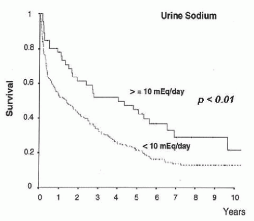
FIGURE 68.2 Long-term survival according to sodium excretion in a series of 204 patients with cirrhosis admitted to the hospital for the treatment of ascites.


Water Retention and Dilutional Hyponatremia

Since the pioneer studies by Papper and Saxon and Shear and colleagues,63,64 it is well known that a derangement in the renal capacity to regulate water balance occurs in advanced cirrhosis. Cirrhotic patients without ascites usually have normal or only slightly impaired renal water handling as compared with healthy subjects. Therefore, in these patients total body water, plasma osmolality, and serum sodium concentration are normal and hyponatremia does not develop, even in conditions of excessive water intake. By contrast, an impairment in the renal capacity to excrete solute-free water is common in patients with ascites and usually it occurs late after the development of sodium retention.63,64,65,66,67 In patients with ascites there is a direct correlation between urinary sodium excretion and water excretion as estimated by urine flow after a water load.64,67 However, no correlation exists between these two parameters when only patients with marked sodium retention are considered. Therefore, sodium retention is necessary but not sufficient for the development of solute-free water retention in cirrhotic patients.

As with sodium retention, the impairment of solute-free water excretion is not uniform in all patients with ascites; rather, it varies markedly from patient to patient (Fig. 68.1). In some patients, water retention is moderate and can only be detected by measuring solute-free water excretion after a water load. These patients are able to eliminate water normally and maintain a normal serum sodium concentration as long as their fluid intake is kept within normal limits, but they may develop hyponatremia when fluid intake is increased. In other patients, the severity of the disorder is such that they retain most of their regular water intake causing hyponatremia and hypoosmolality. Therefore, hyponatremia in cirrhosis with ascites is almost always dilutional in origin since it occurs in the setting of an increased total body water. Hyponatremia is paradoxical in that it is associated with sodium retention and a marked increase in total body exchangeable sodium. The occurrence of spontaneous dilutional hyponatremia requires a profound impairment in solute-free water excretion, since it usually develops with a solute-free water clearance after a water load below 1 mL per minute.65

Hyponatremia in cirrhosis is currently defined as a reduction in serum sodium below 130 mEq per L.68 The prevalence of hyponatremia using this cutoff is 22%. If the cutoff level of 135 mEq per L is used, the prevalence increases up to 49%.69 The presence of dilutional hyponatremia in a cirrhotic patient is associated with a poor survival (Fig. 68.3).62,65,70,71,72,73,74,75,76,77,78,79 The development of dilutional hyponatremia after a precipitating event such as hemorrhage or infection is associated with a better prognosis when compared to the spontaneous appearance of this complication.80 This is possibly related to a higher incidence of renal dysfunction and a more advanced stage of decompensated cirrhosis associated with spontaneous dilutional hyponatremia.

Several factors may aggravate the impairment of solute-free water excretion in cirrhotic patients and precipitate the appearance of hyponatremia. These include treatment with diuretics or nonsteroidal anti-inflammatory drugs (NSAIDs), large-volume paracentesis without plasma volume expansion,67,81,82,83 bacterial infections, and treatment with terlipressin for variceal bleeding.84 Hyponatremia may also develop after the administration of hypotonic fluids in patients with ascites.


Mechanisms of Impaired Renal Water Handling

The pathogenesis of water retention in cirrhosis and dilutional hyponatremia is complex and probably involves several factors, including a reduced delivery of filtrate to the
ascending limb of the loop of Henle, reduced renal synthesis of prostaglandins, and nonosmotic hypersecretion of arginine vasopressin (AVP).68,85,86,87 Although definitive data about the relative importance of these factors in the pathogenesis of hyponatremia in patients with cirrhosis is lacking, it is likely that AVP hypersecretion plays a major role. This contention is supported by studies in animals and patients with cirrhosis showing that the administration of vaptans, drugs that antagonize the tubular effects of AVP (V2 receptor antagonists), improve solute-free water excretion and increase serum sodium concentration.88,89,90,91,92,93,94,95 However, it is important to note there is a significant number of patients in whom hyponatremia does not improve despite the administration of vaptans, thus suggesting that factors other than AVP play also a role in the pathogenesis of solute-free water retention in cirrhosis. In patients with renal failure it is likely that besides AVP, a reduced distal delivery of filtrate due to decreased filtered load and increased proximal sodium and water reabsorption plays a role in solute-free water retention.


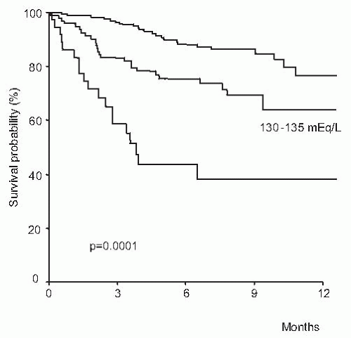
FIGURE 68.3 One-year survival before transplantation in a series of 308 patients with cirrhosis according to different values of serum sodium. (Reproduced with permission from Londoño MC, Cárdenas A, Guevara M, et al. MELD score and serum sodium in the prediction of survival of patients with cirrhosis awaiting liver transplantation. Gut. 2007;56:1283-1290.)


Clinical Consequences

The consequence of an impairment in solute-free water excretion is the development of dilutional hyponatremia. As indicated previously, dilutional hyponatremia in cirrhotic patients is defined as serum sodium < 130 mEq per L in the presence of an expanded extracellular fluid volume, with ascites and/or edema.68 It is associated with sodium retention and increased total body sodium and should be distinguished from hypovolemic hyponatremia that, although less common, may develop in cirrhotic patients with ascites and edema who are maintained on high doses of diuretics and sodium restriction after resolution of ascites and edema. There is limited information on the clinical consequences specifically caused by hyponatremia in cirrhosis because hyponatremia almost always occurs in the setting of advanced liver failure, which causes a wide array of clinical manifestations. Therefore, the precise identification of the clinical consequences of hyponatremia versus those of other causes has so far not been possible. This has been further hindered by the lack of an effective treatment of hyponatremia.

Hyponatremia and neurologic function. In patients without liver disease, hyponatremia is primarily associated with a broad variety of neurologic manifestations related to the existence of brain edema, such as headache, disorientation, confusion, focal neurologic deficits, seizures, and, in some cases, death due to cerebral herniation.96 Severity of neurologic symptoms in patients with hyponatremia without liver disease correlates roughly with the levels of osmolality and sodium in the extracellular fluid. However, rather than the absolute reduction in serum sodium levels, the most important factor in determining the severity of neurologic symptoms is the rate of fall in serum sodium levels, patients with acute hyponatremia having a much higher incidence of neurologic symptoms than those with chronic hyponatremia.

Studies specifically assessing neurologic symptoms in cirrhosis with hyponatremia are lacking. However, the clinical experience indicates that significant neurologic manifestations such as headache, focal motor deficits, seizures, and cerebral herniation are very uncommon. It is likely that the relatively low incidence of neurologic manifestations in patients with cirrhosis and dilutional hyponatremia is related to the fact that in most of these patients hyponatremia is chronic rather than acute, and this gives sufficient time for the brain to adjust to hypo-osmolality of the extracellular fluid. The effects of hyponatremia on brain function have to be discussed in light of the recent hypothesis that proposes a role for a low-grade cerebral edema in the pathogenesis of hepatic encephalopathy.97 According to this hypothesis, ammonia and other neurotoxins act synergistically to induce a low-grade cerebral edema as a result of swelling of astrocytes, which is mainly due to increased intracellular content of glutamine, secondary to ammonia metabolism. The cerebral edema would not be sufficient to cause an increase in intracranial pressure, but astrocyte swelling would result in a number of alterations of neurologic function, which would facilitate the development of hepatic encephalopathy. Evidence for such a low-grade cerebral edema derives from experimental and human studies using magnetic resonance.98,99,100 In this context of low-grade cerebral edema, hyponatremia may represent a second osmotic hit to astrocytes, causing further depletion of osmotic counteractive systems (i.e., organic osmolytes). In this situation, cells
would probably not tolerate a further challenge to cell volume, and encephalopathy would develop due to any other osmotic stimulus, including situations associated with an increased ammonia load to the brain (gastrointestinal hemorrhage, infection) or further impairment in serum sodium concentration (Fig. 68.4). Several lines of evidence support the existence of a relationship between hepatic encephalopathy and low serum sodium concentration. First, serum sodium levels and serum ammonia levels are major factors determining electroencephalographic abnormalities in cirrhosis.101 Second, in patients treated with transjugular intrahepatic portosystemic shunts, hyponatremia is a major risk factor for hepatic encephalopathy.102 Third, in patients treated with diuretics (a clinical situation associated with a high incidence of hepatic encephalopathy), hyponatremia is a risk factor for hepatic encephalopathy (P. Ginès, unpublished data). Finally, serum sodium has been shown to be an independent predictive factor of hepatic encephalopathy in several series of patients with advanced cirrhosis.103,104,105


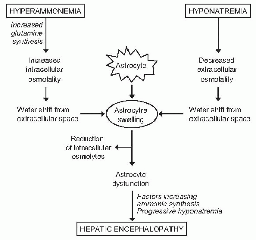
FIGURE 68.4 Proposed interaction between hyperammonemia and hyponatremia on brain astrocytes and possible pathogenic relationship with hepatic encephalopathy. (Reproduced with permission from Ginès P, Guevara M. Hyponatremia in cirrhosis: pathogenesis, clinical significance, and management. Hepatology. 2008;48:1002-1010.)

Hyponatremia and Complications of Cirrhosis. Besides hepatic encephalopathy, hyponatremia has also been reported to be associated with other complications of cirrhosis, yet information is limited. Specifically, hyponatremia is a frequent finding in patients with cirrhosis and bacterial infections.106 In the majority of patients, hyponatremia occurs in close association with renal failure and correlates with a poor prognosis. Patients with ascites and hyponatremia constitute a unique population with a very high risk of developing HRS.23 On the other hand, low serum sodium levels are a very common finding in patients with HRS. Information on the impact of hyponatremia on health-related quality of life in patients both with and without liver disease is very limited. In patients with cirrhosis, hyponatremia impairs quality of life because patients require a restriction of daily fluid intake to prevent further reductions in serum sodium concentration, and this is usually poorly tolerated. Moreover, in a recent study in a large population of patients with cirrhosis, hyponatremia was an independent predictive factor of the impaired health-related quality of life.107

Hyponatremia and Liver Transplantation. Patients with cirrhosis and hyponatremia are at increased risk of neurologic complications after transplantation, central pontine myelinolysis being the most severe, related to a rapid change in serum sodium in the early postoperative period.108,109 The existence of hyponatremia before transplantation is associated not only with an increased risk of neurologic complications after transplantation, but also with an increased risk of renal failure and infectious complications, greater use of blood products, longer duration of hospital stay, and, more importantly, increased short-term mortality after transplantation.110,111


Renal Vasoconstriction and Hepatorenal Syndrome

Investigations performed by Sherlock, Schroeder, and Epstein during the late 1960s and early 1970s provided conclusive evidence indicating that the renal failure of functional origin—the so-called hepatorenal syndrome (HRS)—was due to a marked vasoconstriction of the renal circulation.112,113,114 Further studies showed that, besides the striking renal vasoconstriction present in patients with HRS, mild to moderate degrees of vasoconstriction in the renal circulation are very common in patients with cirrhosis and ascites.115,116,117,118 It has also been recognized that this vasoconstriction leading to HRS may
be triggered by some precipitating factors, particularly bacterial infections.119,120,121 When renal perfusion is estimated by sensitive clearance techniques, such as para-aminohippurate or inulin clearances, in a population of hospitalized patients with ascites, normal values are found in only one fifth of cases. In another 15% to 20%, renal hypoperfusion is very intense and meets the criteria of HRS. In the remaining patients, mild or moderate reductions in renal perfusion exist (Fig. 68.1). These latter patients show slightly increased serum creatinine and/or blood urea nitrogen (BUN) levels in baseline conditions (in the absence of diuretic therapy). This moderate renal vasoconstriction is clinically relevant for several reasons: first, it is often associated with marked sodium and water retention and the presence of refractory ascites122; second, it predisposes to the development of HRS23,120,123; and third, it is associated with an impaired survival.62,73


Definition of Hepatorenal Syndrome

The most recent definition of HRS proposed by the International Ascites Club, which is the most widely accepted, is as follows: “Hepatorenal syndrome is a potentially reversible syndrome that occurs in patients with cirrhosis, ascites and liver failure, as well as in patients with acute liver failure or alcoholic hepatitis. It is characterized by impaired renal function, marked alterations in cardiovascular function and over-activity of the sympathetic nervous system and renin-angiotensin systems. Severe renal vasoconstriction leads to a decrease of glomerular filtration rate. It appears spontaneously, but can also follow a precipitating event.” This description was first proposed in 1999 and was adapted in 2007.122,124 Although in the former definition, the existence of an ongoing bacterial infection precluded the diagnosis of HRS, with the current definition HRS can be diagnosed in the presence of an infection except in cases with septic shock.124


Pathogenic Mechanisms

The pathophysiologic hallmark of HRS is a vasoconstriction of the renal circulation.114,122,125,126 Studies of renal perfusion with renal arteriography,133Xe washout technique, para-aminohippuric acid excretion, and duplex Doppler ultrasonography have demonstrated the existence of marked vasoconstriction in the kidneys of patients with HRS, with a characteristic reduction in renal cortical perfusion.113,126,127,128,129,130,131,132 The functional nature of HRS has been conclusively demonstrated by the lack of significant morphologic abnormalities in the kidney histology,15,16,17,18,133 the normalization or improvement of renal function after liver transplantation,134,135,136,137,138 and the reversibility of the syndrome by pharmacologic treatment with vasoconstrictors and albumin.139

The mechanism of this vasoconstriction is likely multifactorial involving changes in systemic hemodynamics, increased pressure in the portal venous system, activation of vasoconstrictor factors, and suppression of vasodilator factors acting on the renal circulation (discussed later). Contrary to the previous belief of marked vasodilation in extrarenal beds, other vascular beds besides the renal circulation are also vasoconstricted in patients with HRS, including the extremities and the cerebral circulation.140,141,142,143 This indicates the existence of a generalized arterial vasoconstriction in nonsplanchnic vascular beds of patients with HRS and confirms that the only vascular bed responsible for arterial vasodilation and reduced total peripheral vascular resistance in cirrhosis with HRS is the splanchnic circulation.


Clinical and Laboratory Findings

HRS is a common complication of patients with cirrhosis. In patients with ascites, the probability of developing HRS during the course of the disease was reported as 18% at 1 year and 40% after 5 years of follow-up (Fig. 68.5).23 The occurrence of HRS has been investigated in two recent studies. In one study of 129 patients, 22% of patients developed HRS during a follow-up period of 3.5 years.144 In another study including 562 consecutive patients admitted to the hospital with renal failure, the frequency of HRS was 49% (associated with infection in 38% of cases and non-associated in 11%).145 The clinical manifestations of patients with HRS include a combination of signs and symptoms related to renal, circulatory, and liver failure. Nonetheless, there are no specific clinical findings in HRS.

Renal failure in HRS may have a rapid or insidious onset and is associated almost constantly with intense urinary sodium retention (urine sodium < 10 mEq per L), and spontaneous dilutional hyponatremia (serum sodium < 130 mEq per L).122,125,145 HRS may occur in two different clinical patterns, according to the intensity and form of onset of renal failure (Table 68.1).122,124,146 Type 1 HRS is the classic type of HRS and represents the end of the spectrum of changes in renal perfusion in cirrhosis. The dominant clinical features of type 1 HRS are those of acutely severe renal failure
with rapid increase in serum levels of urea and creatinine and low urine volume in some cases, but not all of them. Type 1 HRS is characterized by a rapid and progressive impairment of renal function as defined by a doubling of the initial serum creatinine to a level higher than 2.5 mg per dL in less than 2 weeks. Despite an important reduction of GFR in these patients, serum creatinine levels are commonly lower than values observed in patients with acute renal failure of similar intensity with respect to the reduction in GFR, but without liver disease.125,132,147,148 This is probably due to the lower endogenous production of creatinine secondary to reduced muscle mass in patients with cirrhosis compared with patients without liver disease. Type 1 HRS is associated with a very low survival expectancy, the median survival time being only 2 weeks (Fig. 68.6).146 Type 2 HRS is characterized by a more subtle course with serum creatinine levels around 1.5 to 2.0 mg per dL. Patients are usually in a better clinical condition than those with type 1 HRS and their survival expectancy is longer, approximately 6 months (Fig. 68.6).146 The dominant clinical feature of these patients is diuretic-resistant ascites due to the combination of intense sodium retention, reduced GFR, and marked activation of antinatriuretic systems.122,124,146 Severe spontaneous hyperkalemia is an uncommon feature of HRS. However, marked hyperkalemia may occur if patients are treated with aldosterone antagonists, especially patients with type 1 HRS. Severe metabolic acidosis and pulmonary edema, which are frequent complications of acute renal failure of patients without liver disease, are uncommon findings in patients with HRS. Because HRS is a form of functional renal failure, the characteristics of urine are those of prerenal azotemia, with low urine sodium concentration, and increased urine osmolality and urine-to-plasma osmolality ratio.122,149 Urine volume is not extremely reduced—in a recent series the average urine volume in 60 patients with HRS was 733 mL per day145 and in some cases urine sodium concentration is not extremely reduced.149,150 Table 68.2 shows the current diagnostic criteria of HRS.124


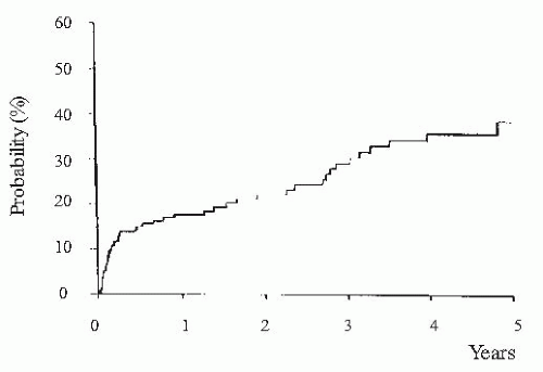
FIGURE 68.5 Probability of developing hepatorenal syndrome in a series of 234 nonazotemic cirrhotic patients with ascites. (Reproduced with permission from Ginès A, Escorsell A, Ginès P, et al. Incidence, predictive factors, and prognosis of the hepatorenal syndrome in cirrhosis with ascites. Gastroenterology. 1993;105:229.)


TABLE 68.1 Clinical Types of Hepatorenal Syndrome


Type 1


Rapid and progressive impairment of renal function as defined by a doubling of the initial serum creatinine to a level higher than 2.5 mg/dL or a 50% reduction of the initial 24-hour creatinine clearance to a level lower than 20 mL/min in less than 2 weeks.


Type 2


Impairment in renal function (serum creatinine >1.5 mg/dL) that does not meet the criteria of type 1.


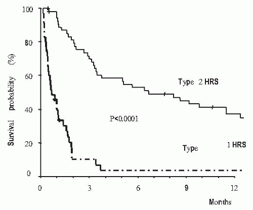
FIGURE 68.6 Survival of patients with cirrhosis according to the type of hepatorenal syndrome. (Reproduced with permission from Alessandria C, Ozdogan O, Guevara M, et al. MELD score and clinical type predict prognosis in hepatorenal syndrome: Relevance to liver transplantation. Hepatology. 2005;41:1282-1289.)

Circulatory failure in patients with HRS is characterized by arterial hypotension (most patients have a mean arterial pressure in the range of 70 mm Hg), and low total systemic vascular resistance, despite marked activation of the vasoconstrictor systems and the existence of severe vasoconstriction in several vascular beds, as already discussed.122,142,143
In addition, several studies have shown that cardiac output is low in patients with HRS, either in absolute values or relative to the reduction in total systemic vascular resistence.151,152,153 This reduction in cardiac output may contribute to the reduction in the effective arterial blood volume and subsequent renal vasoconstriction.152,153,154 In a longitudinal study in patients with cirrhosis it was shown that a reduction in cardiac output was associated with the occurrence of HRS.155 Similarly, in a small series of patients with cirrhosis, those with a low cardiac output had a greater risk of HRS development.156


TABLE 68.2 Diagnostic Criteria of Hepatorenal Syndrome


1.


Cirrhosis with ascites.


2.


Serum creatinine >133 mmol/L (1.5 mg/dL).


3.


No improvement of serum creatinine (decrease to a level of ≤133 mmol/L) after at least 2 days with diuretic withdrawal and volume expansion with albumin. The recommended dose of albumin is 1 g/kg of body weight per day up to a maximum of 100 g/day.


4.


Absence of shock.


5.


No current or recent treatment with nephrotoxic drugs.


6.


Absence of parenchymal kidney disease as indicated by proteinuria >500 mg/day, microhematuria (>50 red blood cells per high power field) and/or abnormal renal ultrasonography.


Adapted from Salerno F, Gerbes A, Ginès P, et al. Diagnosis, prevention and treatment of hepatorenal syndrome in cirrhosis. Gut. 2007;56:1310-1318.


Finally, the third type of clinical manifestations of HRS is related to the existence of liver failure. The majority of patients have features of advanced liver disease with hyperbilirubinemia, elevated prothrombin time, thrombocytopenia, hepatic encephalopathy, hypoalbuminemia, poor nutritional status, and a large amount of ascites. In general, patients with type 1 HRS have more severe liver failure compared with patients with type 2 HRS.146


Precipitating Factors

In some patients, HRS develops without any identifiable precipitating factor, whereas in others it occurs in close chronologic relationship with bacterial infections, particularly spontaneous bacterial peritonitis.119,122,124,157 Approximately one third of patients with spontaneous bacterial peritonitis develop renal failure during or immediately after infection, and in the absence of shock, which is currently defined as HRS119,124 and occurs in the setting of a further decrease in effective arterial blood volume of patients with ascites, as indicated by a marked activation of vasoconstrictor systems, and increased serum and ascitic fluid levels of cytokines.120,157 In approximately one third of patients with spontaneous bacterial peritonitis, HRS is reversible after resolution of infection. However, in the remaining patients HRS is not reversible after the resolution of the infection. Patients who develop type 1 HRS after spontaneous bacterial peritonitis have a dismal outcome, with an almost 100% hospital mortality if not treated appropriately (see below).119,120 Similarly, large-volume paracentesis (> 5 L) without albumin expansion may precipitate type 1 HRS in up to 15% of cases.83 This is one of the main reasons that supports the administration of intravenous albumin when large-volume paracenteses are performed.158,159 Gastrointestinal bleeding has been classically considered as a precipitating factor of HRS.149 However, the development of renal failure after this complication is not very common in patients with cirrhosis (approximately 10%) and occurs mainly in patients with hypovolemic shock, in most cases associated with ischemic hepatitis, which suggests that renal failure in this setting is probably related to the development of acute tubular necrosis (ATN) and not to HRS.160 Diuretic treatment has also been classically described as a precipitating factor of HRS, but there is no clear evidence to support such a relationship. There are several predictive factors in patients with cirrhosis and ascites associated with a greater risk of developing

HRS.23 For the most part these are related to circulatory and renal function and include severe urinary sodium retention, spontaneous dilutional hyponatremia, and low mean arterial blood pressure (< 80 mm Hg). Interestingly, neither the degree of liver failure, as assessed by classic parameters of liver function (serum bilirubin, albumin, and prothrombin time) or the Child-Pugh classification, correlate with the risk of developing HRS.23


FACTORS INVOLVED IN FUNCTIONAL RENAL ABNORMALITIES IN CIRRHOSIS


Circulatory Abnormalities


Hepatic and Splanchnic Circulation

The existence of cirrhosis causes marked structural abnormalities in the liver that result in severe disturbance of intrahepatic circulation causing increased resistance to portal flow and subsequent hypertension in the portal venous system.172 Progressive collagen deposition and formation of nodules alter the normal vascular architecture of the liver. Moreover, selective deposition of collagen in the space of Disse, the space between sinusoidal cells and hepatocytes, may constrict the sinusoids, resulting in further mechanical obstruction to flow.173,174 In addition to this passive resistance to portal flow there is an active component of intrahepatic resistance, which is due to the contraction of hepatic stellate cells (myofibroblastlike cells) present in sinusoids and terminal hepatic venules175,176,177 and low levels of intrahepatic vasodilators. The contraction of these cells is affected by endogenous vasoconstrictors and can be modulated by vasodilators and drugs that antagonize the vasoconstrictor factors.178,179,180 Moreover, there is a strong body of evidence indicating that despite the overproduction of the vasodilator nitric oxide (NO) in the splanchnic and systemic circulation in cirrhosis, there is a reduced production of NO in the intrahepatic circulation of cirrhotic livers that contributes to the increased intrahepatic resistance characteristic of portal hypertension.181,182,183 There is also evidence that besides the role of fibrosis and vasoactive factors, increased hepatic neoangiogenesis and inflammation can play a role in the pathogenesis of increased intrahepatic resistance in experimental cirrhosis.184

Portal hypertension induces profound changes in the splanchnic circulation.185,186,187,188 Classically, portal hypertension was considered to cause only changes in the venous side of the splanchnic circulation. However, studies in experimental animals indicate that portal hypertension also causes marked changes in the arterial side of the splanchnic vascular bed. In the venous side, the main changes consist of increased pressure and formation of portocollateral circulation, which causes the shunting of blood from the portal venous system to the systemic circulation. In the arterial side, there is marked arterial vasodilation which increases portal venous inflow.185,186,187,188,189 This high portal venous inflow plays an important role in the increased pressure in the portal circulation and may explain, at least in part, why portal pressure remains increased despite the development of collateral circulation. This arteriolar vasodilation is also responsible for marked changes in splanchnic microcirculation that may predispose to increased filtration of fluid. It has been shown that chronic portal hypertension causes a much greater increase in intestinal capillary pressure and lymph flow than does an acute increase in portal pressure of the same magnitude.190,191 This is probably due to a loss of the normal autoregulatory mechanism of the splanchnic microcirculation. The acute elevation of venous pressure in the intestine elicits a strong myogenic response, which leads to a reduction in blood flow. This phenomenon is thought to be a homeostatic response to protect the intestine against edema formation. This protective mechanism is not operative in chronic portal hypertension and arteriolar resistance is reduced and not increased.191,192 The resultant increases in capillary pressure and filtration may be important factors in the formation of ascites in cirrhosis. The mechanism(s) by which portal hypertension induces splanchnic arteriolar vasodilation is not completely understood although a number of vasoactive mediators have been proposed (and will be discussed subsequently).185

Several lines of evidence indicate that portal hypertension is a major factor in the pathogenesis of ascites. First, patients with early cirrhosis without portal hypertension do not develop ascites or edema. Moreover, a certain level of portal hypertension is required for ascites formation. Ascites rarely develops in patients with portal pressure below 10 mm Hg, as assessed by the difference between wedged and free hepatic venous pressure (normal portal pressure: 5 mm Hg).193,194,195,196 Second, cirrhotic patients treated with surgical portosystemic shunts for the management of bleeding gastroesophageal varices have lower risk of developing ascites than do patients treated with procedures that obliterate gastroesophageal varices but do not affect portal pressure (i.e., sclerotherapy, esophageal band ligation).197 Finally, reduction of portal pressure with side-to-side or end-to-side portacaval anastomosis or TIPS (placement of a stent between a hepatic vein and the intrahepatic portion of the portal vein using a transjugular approach) is associated with an improvement of ascites, renal function, and suppression of antinatriuretic systems198,199 in cirrhotic patients with fluid retention. The mechanism(s) by which portal hypertension contributes to renal functional abnormalities and ascites and edema formation is not completely understood, yet three pathogenic mechanisms have been
proposed: (1) alterations in the splanchnic and systemic circulation which result in activation of vasoconstrictor and antinatriuretic systems and subsequent renal sodium and water retention; (2) hepatorenal reflex due to increased hepatic pressure which would cause sodium and water retention; and (3) putative antinatriuretic substances escaping from the splanchnic area through portosystemic collaterals that would have a sodium-retaining effect in the kidney. There is a large body of evidence supporting the first of these three pathogenic mechanisms.


Systemic Circulation

The development of portal hypertension is associated with marked hemodynamic changes not only in the hepatic and splanchnic circulation but also in the systemic circulation. These changes, which have been well characterized in human and experimental cirrhosis, consist of reduced systemic vascular resistance and arterial pressure, increased cardiac index, increased plasma volume, and activation of systemic vasoconstrictor and antinatriuretic factors. These changes in systemic hemodynamics appear before the formation of ascites and are more marked as the disease progresses.40,188,200,201,202,203,204,205 The hemodynamic profile of patients with cirrhosis in different stages of the disease is summarized in Table 68.3. The factor that appears to trigger all these hemodynamic changes of cirrhosis is an arterial vasodilation located mainly in the splanchnic circulation.185,186,187,188,189,205,206,207 The existence of a splanchnic arterial vasodilation causes an abnormal distribution of blood volume, which results in a reduction of central blood volume (heart, lungs, and aorta) that is sensed by arterial and cardiopulmonary receptors. This central underfilling triggers a neurohormonal response by activating the SNS, RAAS, and arginine vasopressin (AVP). This explains why systemic vasoconstrictor factors are activated despite an increased plasma volume that in normal conditions would suppress the activation of these systems. Investigations in patients with cirrhosis have assessed central blood volume by measuring the mean circulation time of an indicator or by magnetic resonance imaging.205,208,209,210,211 These studies have confirmed that central blood volume is reduced in patients with cirrhosis, particularly in those with ascites and correlates directly with systemic vascular resistance and inversely with portal pressure, indicating that the greater the vasodilation and the pressure in the portal system, the lower the central blood volume. The crucial role played by the reduced central blood volume in the activation of vasoconstrictor systems has been further corroborated by studies showing that improvement of central blood volume by the combination of expansion of plasma volume or head-out water immersion and administration of pressor agents, suppresses the activation of vasoconstrictor systems.212,213,214,215 Whether or not arterial vasodilation occurs also in nonsplanchnic territories is still controversial but most data indicate that the splanchnic circulation accounts for most, if not all, of the reduced arterial resistance in patients with cirrhosis.185,205,216


TABLE 68.3 Hemodynamic Profile of Patients with Cirrhosis in Different Stages of Disease


Preascitic Cirrhosis


Cirrhosis with Ascites


Hepatorenal Syndrome


Cardiac output


Normal or increased


Increased


Normal or reduced


Arterial pressure


Normal


Normal or reduced


Reduced


Systemic vascular resistance


Normal or reduced


Reduced


Markedly reduced


Plasma volume


Normal or increased


Increased


Increased


Portal pressure


Normal or increased


Increased


Increased


Vasoconstrictor systems activity


Normal


Increaseda


Markedly increased


Renal vascular resistance


Normal


Normal or increased


Markedly increased


Brachial or femoral vascular resistance


Normal or reduced


Normal or increased


Increased


Cerebral vascular resistance


Normal


Increased


Increased


a May be normal in 20%-30% of patients.


Despite extensive investigation, the mechanism(s) responsible for arterial vasodilation in cirrhosis is not completely understood. Several explanations have been proposed, including opening of arteriovenous fistulas, reduced sensitivity to vasoconstrictors, and increased circulating levels of vasodilator substances.185,187,207,216,217,218,219,220 This latter mechanism has
been the most extensively studied. Increased production of NO, carbon monoxide, glucagon, endocannabinoids, prostaglandins, vasoactive intestinal peptide, adenosine, bile salts, platelet activating factor, substance P, calcitonin gene-related peptide, natriuretic peptides, and adrenomedullin have been proposed as possible factors of the development of splanchnic arterial vasodilation.185,191,205,216,221,222,223,224,225,226,227,228,229,230,231 At present, most available data, obtained mainly from experimental cirrhosis, indicate that NO is the main mediator of arterial vasodilation in cirrhosis (Table 68.4).232 NO synthesis from cirrhotic arterial vessels is markedly increased compared to that of normal vascular tissue. This increased NO synthesis appears to be generalized, except for the intrahepatic circulation, but predominates in the splanchnic territory. Among the different isoforms of NO synthase, the constitutive form appears to be the one responsible for the increased NO synthesis. The normalization of NO synthesis in experimental cirrhosis by the administration of inhibitors of NO synthesis is associated with a marked improvement of splanchnic and systemic hemodynamics, suppression of the increased activity of the RAAS and AVP concentration, increased sodium and water excretion, and reduction or disappearance of ascites.233 So far, only few studies have investigated the effect of acute NO synthesis inhibition in patients with cirrhosis on systemic hemodynamia and/or renal function, with discrepant findings.234,235

Stay updated, free articles. Join our Telegram channel

May 29, 2016 | Posted by in NEPHROLOGY | Comments Off on Liver Disease and the Kidney

Full access? Get Clinical Tree

Get Clinical Tree app for offline access