Hemolytic Uremic Syndrome, Thrombotic Thrombocytopenic Purpura, and Acute Cortical Necrosis


Hemolytic Uremic Syndrome, Thrombotic Thrombocytopenic Purpura, and Acute Cortical Necrosis


Marina Noris

Giuseppe Remuzzi

Piero Ruggenenti


HEMOLYTIC UREMIC SYNDROME AND THOMBOTIC THROMBOCYTOPENIC PURPURA

The term thrombotic microangiopathy (TMA) defines a lesion of arteriolar and capillary vessel wall thickening with intraluminal platelet thrombosis and a partial or complete obstruction of the vessel lumina. Laboratory features of thrombocytopenia and microangiopathic hemolytic anemia are almost invariably present in patients with TMA lesions and reflect the consumption and disruption of platelets and erythrocytes in the microvasculature. Depending on whether renal or brain lesions prevail, two pathologically indistinguishable but somehow clinically different entities have been described: hemolytic uremic syndrome (HUS) and the thrombotic thrombocytopenic purpura (TTP). Because HUS can involve extrarenal manifestations and TTP can sometimes involve severe renal disease, the two can be difficult to distinguish on clinical grounds only.1 Newly identified pathophysiologic mechanisms, however, have allowed for the differentiation of the two syndromes on a molecular basis (Table 55.1). Independent of the initial cause, all different forms of HUS and TTP share a similar trend of circulating blood to form thrombi in the microvasculature because of primary endothelial damage, as in Shiga toxin-associated HUS; uncontrolled complement activation, as in atypical HUS; or abnormal cleavage of von Willebrand factor, as inTTP.


Clinical Features


Hemolytic Uremic Syndrome

The term HUS was introduced in 1955 by Gasser and coworkers2 in their description of an acute fatal syndrome in children characterized by hemolytic anemia, thrombocytopenia, and severe renal failure. HUS occurs most frequently in children under the age of 5 years (incidence, 5 to 6 children/100,000/year compared to an overall incidence of 0.5 to 1/100,000/year. Most cases (over 90% of those in children) are associated with infection by Shigalike toxin (Stx), producing Escherichia coli (Stx-HUS).3 In 90% of cases, Stx-HUS is preceded by diarrhea, which is often bloody. Usually patients are afebrile. Streptococcus pneumoniae causes a distinctive form of HUS, accounting for 40% of cases not associated with Stx-producing bacteria.4

Approximately 10% of HUS cases are classified as atypical, caused neither by Stx-producing bacteria nor by Streptococcus .5 Atypical HUS occurs at any age, can be familial or sporadic, and has a poor outcome; 50% progress to end-stage renal disease (ESRD) and 25% may die in the acute phase.4,6 Neurologic symptoms and fever can occur in 30% of patients. Pulmonary, cardiac, and gastrointestinal manifestations can also occur.4,6


Thrombotic Thrombocytopenic Purpura

TTP was first described in 1925 by Moschcowitz7 in a 16-year-old female patient with a fulminant febrile attack, hemolytic anemia, bleeding, renal failure, and neurologic involvement. Pathologic changes were characterized by widespread hyaline thrombosis of small vessels. It is a rare disease, with an incidence of approximately 2 to 4 cases per 1 million persons per year.8,9 It is more common in women (female to male ratio, 3:2 to 5:2) and in whites (white to black ratio, 3:1). Although the peak incidence is in the third and fourth decades of life, TTP can affect any age group.9,10,11 Thrombotic thrombocytopenic purpura classically presents with the pentad of thrombocytopenia, microangiopathic hemolytic anemia, fever, and neurologic and renal dysfunction.7 Thrombocytopenia is essential for the diagnosis; most patients present with values below 60,000 per microliter.9,11 Purpura is minor and can be absent. Retinal hemorrhages can be present; however, bleeding is rare. Neurologic symptoms can be seen in over 90% of patients during the entire course of the disease. Central nervous system involvement mainly represents thrombo-occlusive disease of the grey matter, but can also include headache, cranial nerve palsies, confusion, stupor, and coma. These features are transient but recurrent. Up to half of patients who present with neurologic involvement may be left with sequelae. Renal insufficiency may occur. One group has reported 25% of patients with creatinine clearance of less than 40 mL per minute. Low-grade
fever is present in one-quarter of patients at the diagnosis, but can often be seen as a consequence of plasma exchange. Less common manifestations include acute abdomen, pancreatitis, and sudden death.11


TABLE 55.1 Classification of Hemolytic Uremic Syndrome (HUS) and Thrombotic Thrombocytopenic Purpura (TTP) According to Clinical Presentation and Underlying Etiology


Clinical Presentation


Etiology


Hemolytic Uremic Syndrome


– Stx-associated


Infections by Stx-producing bacteria


– Neuraminidase associated


Infections by Streptococcus pneumoniae


– Atypical


Familial


Mutations: CFH,40%-45%; CFI, 5%-10%; C3, 8%-10%; MCP, 7%-15%; THBD, 9%; CFB, 1%-2%


Sporadic


Idiopathic


Mutations: CFH, 15%-20%; CFI, 3%-6%; C3, 4%-6%; MCP, 6%-10%; THBD, 2%; CFB, 2 cases


Anti-CFH antibodies: 6%-10%


– Pregnancy-associated


Mutations: CFH, 20%; CFI, 15%


– HELLP syndrome


Mutations: CFH, 10%; CFI, 20%; MCP, 10%


– Drugs


Mutations: rare CFH mutations, the large majority unknown


– Transplantation (de novo aHUS)


Mutations: CFH, 15%; CFI, 16%


– HIV


Unknowna


– Malignancy


Unknowna


Thrombotic Thrombocytopenic Purpura


-Congenital


Homozygous or compound heterozygous mutations in ADAMTS13 gene


– Idiopathic


Anti-ADAMTS13 autoantibodies


– Secondary


– Ticlopidine clopidogrel


Anti-ADAMTS13 autoantibodies (ticlopidine, 80%-90%, clopidogrel, 30%)


– HSC transplantation


Unknown, rarely low ADAMTS13 levels


– Malignancies


Unknown, rarely low ADAMTS13 levels


– HIV


HIV virus, rarely low ADAMTS13 levels


SLE, APL, and other autoimmune disease


Depends on the specific primary diseases


a a No published data on frequency of complement gene mutations or anti-CFH autoantibodies.


Stx, Shiga toxin; CFH, complement factor H; CFI, complement factor I; HELLP, hemolytic anemia, elevated liver enzymes, and low platelet count; MCP, membrane cofactor protein; THBD, thrombomodulin; aHUS, atypical hemolytic uremic syndrome; HSC, hematopoietic stem cell transplantation; SLE, systemic lupus erythematosus; APL, antiphospholipid syndrome.


Laboratory Findings

Laboratory features of thrombocytopenia and microangiopathic hemolytic anemia are almost invariably present in patients with TMA lesions and reflect consumption and disruption of platelets and erythrocytes in the microvasculature.5,11,12 Hemoglobin levels are low (less than 10 g per deciliter in more than 90% of patients). Reticulocyte counts are uniformly elevated. The peripheral smear reveals an increased schistocyte number (Fig. 55.1), with polychromasia, and often, nucleated red blood cells. The latter may represent not only a compensatory response, but also may represent damage to the bone marrow-blood barrier resulting from intramedullary vascular occlusion. Detection of fragmented
erythrocytes is crucial to confirm the microangiopathic nature of the hemolytic anemia, provided that heart valvular disease and other anatomic artery abnormalities that may cause erythrocyte fragmentation are excluded. Other indicators of intravascular hemolysis include elevated lactate dehydrogenase (LDH), increased indirect bilirubin, and low haptoglobin levels.5,11,12 The Coombs test is negative. Moderate leukocytosis may accompany the hemolytic anemia. Thrombocytopenia is uniformly present in HUS and TTP. It may be severe, but is usually less so in patients with predominant renal involvement.13 The presence of giant platelets in the peripheral smear or reduced platelet survival time (or both) is consistent with peripheral consumption. In children with Stx-HUS, the duration of thrombocytopenia is variable and does not correlate with the course of renal disease.14 Bone marrow biopsy specimens usually show erythroid hyperplasia and an increased number of megakaryocytes. Prothrombin time (PT), partial thromboplastin time (PTT), the fibrinogen level, and coagulation factors are normal, thus differentiating HUS and TTP from disseminated intravascular coagulation (DIC). Mild fibrinolysis with minimal elevation in fibrin degradation products, however, may be observed.


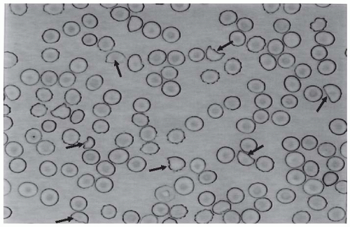
FIGURE 55.1 A peripheral blood smear from a patient with thrombotic microangiopathy. The presence of fragmented red blood cells that may acquire the appearance of a helmet (fragmented erythrocytes with the shape of a helmet are identified by the black arrows) is pathognomonic for microangiopathic hemolysis in patients with no evidence of heart valvular disease.

Evidence of renal involvement is present in all patients with HUS (by definition) and in about 25% of patients with TTP.1,12,15 Microscopic hematuria and subnephrotic proteinuria are the most consistent findings. In a retrospective study of 216 patients with a clinical picture of TTP, hematuria was detected in 78% and proteinuria in 75% of cases.15 Sterile pyuria and casts were present in 31% and 24% of cases, respectively. Gross hematuria was rare.15


Pathology

The diagnostic histologic lesions of TMA consist of widening of the subendothelial space and microvascular thrombosis. Electron microscopy best identifies the characteristic lesions
of swelling and detachment of the endothelial cells from the basement membrane and the accumulation of fluffy material in the subendothelium (Figs. 55.2 and 55.3), intraluminal platelet thrombi, and a partial or complete obstruction of the vessel lumina (Fig. 55.4).16,17,18 These lesions are similar to those seen in other renal diseases such as scleroderma, malignant nephrosclerosis, chronic transplant rejection, and calcineurin inhibitor nephrotoxicity. In HUS, microthrombi are present primarily in the kidneys, whereas in TTP they mainly involve the brain, where thrombi may repeatedly form and resolve, producing intermittent neurologic defi-cits. In pediatric patients, particularly in those younger than 2 years of age, and in those with HUS secondary to gastrointestinal infection with Stx-producing strains of E. coli , the glomerular injury is predominant.16


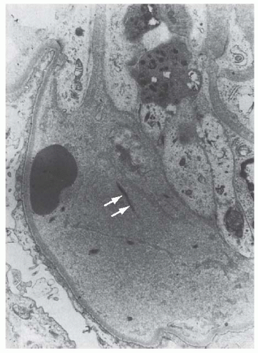
FIGURE 55.2 The detachment of an endothelial cell from the underlying glomerular basement membrane in a case of hemolytic-uremic syndrome. A red blood cell is in close contact with the glomerular basement membrane. Electron-lucent “fluffy” material and a few strands of fibrin (arrows) are present in the subendothelial space (×7,000). (Courtesy of Drs. C. L. Pirani and V. D’Agati.)

Thrombi and infiltration by leukocytes are common in the early phases of the disease and usually resolve after 2 to 3 weeks. Patchy cortical necrosis may be present in severe cases; crescent formation is uncommon. In idiopathic and familial forms and in adults, the injury mostly involves arteries and arterioles with thrombosis and intimal thickening (Figs. 55.5 and 55.6), and secondary glomerular ischemia and retraction of the glomerular tuft (Fig. 55.7). The prognosis is good in patients with predominant glomerular involvement, but it is more severe in those with predominant preglomerular injury. Focal segmental glomerulosclerosis may be a long-term sequela of acute cases of HUS and is usually seen in children with long-lasting hypertension and progressive chronic renal function deterioration.16,17,18

The typical pathologic changes of TTP are the thrombi that occlude capillaries and arterioles in many organs
and tissues (Fig. 55.8). These thrombi consist of fibrin and platelets, and their distribution is widespread. They are most commonly detected in the kidneys, the pancreas, the heart, the adrenals, and the brain. Compared to HUS, pathologic changes of TTP are more extensively distributed, probably reflecting the more systemic nature of the disease.16,17,18


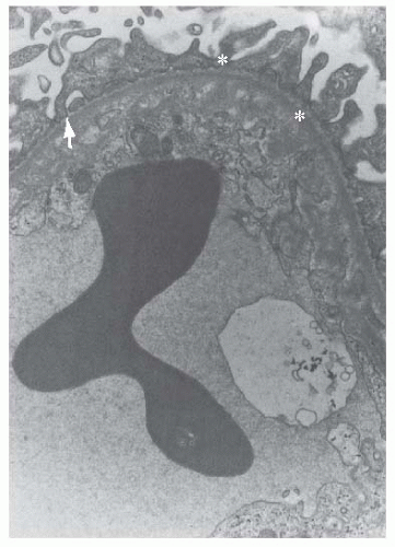
FIGURE 55.3 Electron-lucent “fluffy” material (arrow) with some electron-dense deposits (asterisks) are located between the cytoplasm of an endothelial cell and the glomerular basement membrane in a segment of glomerular capillary from a patient with hemolytic-uremic syndrome (×12,000). (Courtesy of Drs. C. L. Pirani and V. D’Agati.)


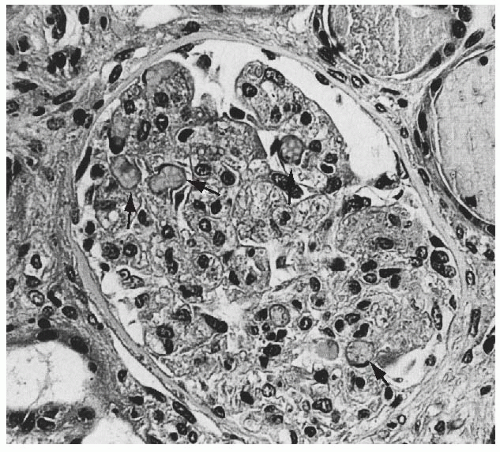
FIGURE 55.4 Swelling of the glomerular endothelial cells and occlusion of almost all capillary lumens packed with red blood cells (arrows) in a case of hemolytic-uremic syndrome (Trichrome, × 250).


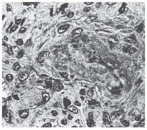
FIGURE 55.5 Thrombotic and necrotic changes in a small artery from an adult patient with hemolytic-uremic syndrome. (Trichrome, ×375.)


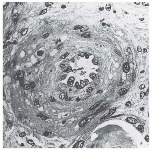
FIGURE 55.6 Occlusion of an interlobular artery with intimal swelling and myointimal proliferation in a case of adult hemolytic-uremic syndrome. (Trichrome,× 375.)


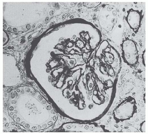
FIGURE 55.7 Ischemic glomerular lesions characterized by thickening and wrinkling of glomerular capillary walls and atrophy of the glomerular tuft in a case of adult hemolytic-uremic syndrome. (Silver, ×250.)


Mechanisms, Clinical Course, and Therapy According to Different Forms of Thrombotic Microangiopathy


Hemolytic Uremic Syndrome


Shiga Toxin-Associated Hemolytic Uremic Syndrome

Mechanisms. Stx-HUS, the most frequent form of TMA, may follow infection by certain strains of E. coli or Shigella dysenteriae, which produce powerful exotoxins (Stx).3,19 The term Shiga toxin was initially used to describe the exotoxin produced by S. dysenteriae type 1. Then, some strains of E. coli (mostly the serotype 0157:H7, but also other serotypes
such as O111:H8, O103:H2, O123, O26, and the O104:H4 strain isolated in the recent German outbreak)20,21 isolated from human cases with diarrhea were found to produce a toxin similar to the one of S. dysenteriae . After food contaminated by Stx-producing E. coli or S. dysenteriae is ingested, the toxin is released into the gut and may cause watery or, most often, bloody diarrhea because of a direct effect on the intestinal mucosa. Stx-producing E. coli closely adhere to the epithelial cells of the gastrointestinal mucosa causing the destruction of the brush border villi.22 Stxs are picked up by polarized gastrointestinal cells via transcellular pathways and translocate into the circulation,23 probably facilitated by the transmigration of neutrophils,24 which increase paracellular permeability. Circulating human blood cells, such as erythrocytes,25 platelets,26,27 and monocytes,28 express Stx receptors on their surface and have been suggested to serve as Stx carriers from the intestine to the kidney and other target organs.


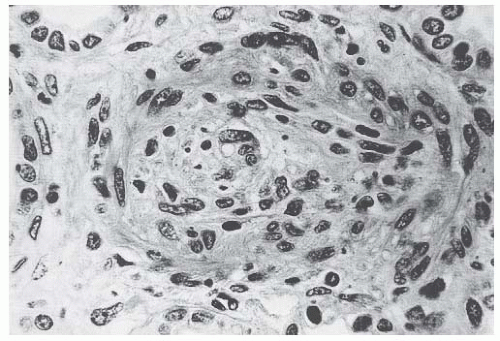
FIGURE 55.8 Marked endothelial and myointimal cell proliferation with occlusion of the lumen of an interlobular artery in a case of thrombotic thrombocytopenic purpura (Trichrome, ×375.)

The disease is caused by two distinct exotoxins, Stx-1 and Stx-2, which are almost identical to the toxin produced by S. dysenteriae type 1.29 Both Stx-1 and Stx-2 are 70-kDa AB5 holotoxins comprising a single A subunit of 32 kDa and five 7.7-kDa B subunits. Interestingly, an AB5 toxin comprising a single 35-kDa A subunit and a pentamer of 13-kDa B subunits have been isolated from a highly virulent E. coli strain (O113:H21) responsible for an outbreak of HUS, which may represent the prototype of a new class of toxins, accounting for HUS associated with strains of E. coli that do not produce Stxs.30 Despite their similar sequences, Stx-1 and Stx-2 cause different degrees and types of tissue damage, as documented by the higher pathogenicity of strains of E. coli that produce only Stx-2 than of those that produce Stx-1 alone.31,32,33 In a study in children who became infected by Stx E. coli , E. coli strains producing Stx-2 were most commonly associated with HUS, whereas most strains isolated from children with diarrhea alone or from those who remained asymptomatic only produced Stx-1.34 This is also true in mice and in baboons.35

Stx-1 and Stx-2 bind to different epitopes on the Gb3 molecules and they also differ in binding affinity and kinetics.36 Surface plasmon resonance analysis showed that Stx-1 easily binds to and detaches from Gb3, in contrast to Stx-2, which binds slowly but also dissociates very slowly, thus leaving time enough for the cell’s incorporation.36 After binding to endothelial cell receptors, the toxin is internalized in the cytosol by endocytosis37 within 2 hours38 and inhibits protein synthesis within 30 minutes (Fig. 55.9). The number of high-affinity receptors is a major determinant of susceptibility of cells to Stx.39 Therefore, cell viability and protein synthesis of endothelial cells of the kidney were reduced by 50% upon exposure to 1 pM Stx, unlike endothelial cells of the umbilical vein that were viable up to greater than 1 nM exposure to the toxin. These findings are consistent with basal levels of Stx receptors 50 times higher in the renal endothelium than in the umbilical cord endothelium.40 During internalization, the alpha subunit of the toxin dissociates from the beta subunits. Approximately 10% of the alpha subunit protein is removed in a trypsinlike process, resulting in a maximally active 27-kDa subunit enzyme. It is well established that this fragment is a direct inhibitor of protein synthesis and is responsible for the cytotoxic action of the toxin. Stx selectively inactivates 60S ribosomal subunits by removing one nucleotide in the 28S ribosomal RNA in a nucleotide-specific manner (Fig. 55.9).40

For many years it was assumed that the only relevant biologic activity of Stx was the block of protein synthesis and destruction of endothelial cells. However, the treatment of endothelial cells with sublethal doses of Stx—exerting minimal influence on protein synthesis—leads to increased mRNA levels and protein expression of chemokines, such as interleukin (IL)-8 and monocyte chemoattractant protein-1 (MCP-1) and cell adhesion molecules, a process preceded by nuclear factor-kappa B (NF- kB) activation.41 Adhesion molecules seem to play a critical role in mediating the binding of inflammatory cells to endothelium. Indeed, Stx-2 treatment enhanced the number of leukocytes adhering and migrating across a monolayer of human endothelial cells.42 Moreover, preventing IL-8 and MCP-1 overexpression by adenovirus-mediated NF-KB blocking, inhibited adhesion and the transmigration of leukocytes.41

Therefore, it can be inferred that Stx, by altering endothelial cell adhesion properties and metabolism, favor leukocyte-dependent inflammation and induce the loss of thromboresistance in endothelial cells, leading to microvascular thrombosis. Evidence for such a sequence of events has been obtained in experiments of whole blood flowing on human microvascular endothelial cells preexposed to Stx-1 at high shear stress.43 In these circumstances, early platelet activation and adhesion occurs, followed by the formation of organized endothelial P-selectin and plateletendothelial cell adhesion molecule (PECAM)-1-dependent thrombi. This offers a likely pathophysiologic pathway for microvascular thrombosis in HUS.

Evidence is also emerging that complement activation at the renal endothelial level may contribute to microangiopathic lesions in Stx-HUS. High plasma levels of complement activation products Bb and C5b-9 were recently measured in 17 children with Stx-HUS, indicating complement activation via an alternative pathway.44 Another study reported that Stx2 binds to the plasma complement regulatory protein, factor H, and may activate complement in the fluid phase in vitro.45 In a recent study, Stx-induced complement activation via P-selectin was identified as a key mechanism of microvascular thrombosis in Stx-HUS. Stx induced the expression of P-selectin on a cultured human microvascular endothelial cell surface, and P-selectin bound and activated C3 via the alternative pathway, leading to thrombus formation under flow.46 In a murine model of HUS obtained by the coinjection of Stx2 and LPS and characterized by thrombocytopenia and renal dysfunction, the upregulation of glomerular endothelial P-selectin was associated with C3 and fibrin(ogen) deposits and platelet clumps. Treatment with anti-P-selectin Ab limited glomerular C3 accumulation. Factor B deficient mice after Stx2/LPS exhibited less thrombocytopenia and
were protected against glomerular abnormalities and renal function impairment, indicating the involvement of complement activation, via the alternative pathway, in the glomerular thrombotic process in HUS mice.46


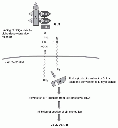
FIGURE 55.9 The binding and mechanism of action of Shigalike toxin. The B subunits of Shiga toxin molecules attach to galactose (Gal) disaccharides of globotriaosylceramide (Gb3) receptors on the membrane of monocytes, polymorphonuclear cells, platelets, glomerular endothelial cells, and tubular epithelial cells. The toxin is internalized via retrograde transport through the Golgi complex. Then the A and B subunits dissociate, and the A subunit is translocated to the cytosol. The A subunit blocks peptide chain elongation by eliminating one adenine from the 28S ribosomal RNA.

Diagnosis. Diagnosis rests on the detection of E. coli O157:H7 and other Stx-producing bacteria in sorbitol- MacConkey stool cultures. Unlike most other E coli , serotype O157:H7 and other Stx-producing bacteria do not ferment sorbitol rapidly and thus form colorless colonies on sorbitol containing MacConkey agar (SMAC). Suspect colonies can be assayed for the O157 antigen with commercially available antiserum or latex agglutination kits. Newer protocols that use SMAC that contains cefixime tellurite, other selective culture media, immunomagnetic separation, and enzymelinked immunosorbent assays to detect O157 lipopolysaccharide or Shiga toxins can further enhance detection.19 In regions where sorbitol-fermenting strains have been identi-fied,19 the use of tests that identify Shiga toxins or the genes encoding them (by PCR) is helpful for diagnosis.

Over the last 2 decades E. coli 0157:H7 and, although less frequently, other Stx-producing E. coli strains, have been responsible for multiple outbreaks throughout the world, becoming a public health problem in both developed and developing countries.19 Contaminated undercooked ground beef, meat patties, raw vegetables, fruit, milk, and recreational or drinking water have all been implicated in the transmission of E. coli . A widespread outbreak associated with spinach in North America had dramatically higher than typical rates of both hospitalization (52%) and HUS (16%), due to the emergence of a new variant of the 0157:H7 serotype that has acquired several gene mutations that likely contributed to more severe disease.47 From May through June 2011, a very large outbreak of HUS occurred in Germany and was caused by an unusual Shiga toxin- producing E. coli (STEC) strain O104:H4 through the ingestion of contaminated sprouts.48

Secondary person-to-person contact is an important way of spread in institutional centers, particularly day care centers and nursing homes. Infected patients should be excluded from day care centers until two consecutive stool cultures are negative for Stx-producing E. coli in order to prevent further transmission. However, the most important preventive measure in child care centers is supervised hand washing.

Clinical course. Following exposure to Stx E. coli , 38% to 61% of individuals develop hemorrhagic colitis and 3% to 9% (in sporadic infections) to 20% (in epidemic forms) progress to overt HUS.19,49 Stx E. coli hemorrhagic colitis not
complicated by HUS is self-limiting and is not associated with an increased long-term risk of high blood pressure or renal dysfunction, as shown by a 4-year follow-up study in 951 children who were exposed to a drinking water outbreak of E. coli O157:H7.50


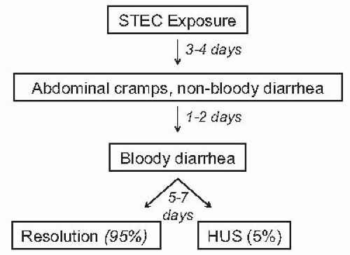
FIGURE 55.10 Timing of the events that may follow exposure to Shiga toxin-producing E. coli. HUS, hemolytic uremic syndrome.

Stx-HUS is characterized by prodromal diarrhea, followed by acute renal failure. The average interval between E. coli exposure and illness is 3 days. Illness typically begins with abdominal cramps and nonbloody diarrhea; diarrhea may become hemorrhagic in 70% of cases, usually within 1 or 2 days.51 Vomiting occurs in 30% to 60% of cases and fever in 30%. Leukocyte count is usually elevated, and a barium enema may demonstrate “thumb-printing,” suggestive of edema and submucosal hemorrhage, especially in the region of the ascending and transverse colon. HUS is usually diagnosed 6 to 10 days after the onset of diarrhea (Fig. 55.10).3 After infection, Stx E. coli may be shed in the stools for several weeks after the symptoms are resolved, particularly in children < 5 years of age.3

Bloody diarrhea, fever, vomiting, elevated leukocyte count, extremes of age, and female sex, as well as the use of antimotility agents,52 have been associated with an increased risk of HUS following an E. coli infection.19 Stx-HUS is not a benign disease. Of patients who develop HUS, 70% require red blood cell transfusions, 50% need dialysis, and 25% have neurologic involvement, including stroke, seizure, and coma.19,53,54 Although mortality for infants and young children in industrialized countries decreased when dialysis became available, as well as after the introduction of intensive care facilities, still 3% to 5% of patients die during the acute phase of Stx-HUS.53 A meta-analysis of 49 published studies (3,476 patients, mean follow-up of 4.4 years) describing the long-term prognoses of patients who survived an episode of Stx-HUS, reported death or permanent ESRD in 12% of patients and GFR below 80 mL/min/1.73m2 in 25%.54 The severity of acute illness, particularly central nervous system symptoms, the need for initial dialysis, and microalbuminuria in the first 6 to 8 months were strongly associated with a worse long-term prognosis.54,55,56

Disease presentation and outcome were particularly unusual during the STEC O104:H4 German outbreak, which since May 1, 2011 had involved more than 4,000 people in Germany, of whom 800 had progressed to HUS and 50 had died in Germany and 15 in other countries by July 20.48 The outbreak was foodborne in contaminated sprouts.21 The chain of transmission appeared to have started in Egypt with fecal contamination of fenugreek seeds by either humans or farm animals. During sprout germination, bacteria multiplied and produced large amounts of toxin and were then diffused with food provided to restaurants and consumers. Almost 90% of affected patients were adults and, compared to previous enterohemorragic E. coli (EHEC) epidemics, there was a higher prevalence of affected young and middle-aged women and an extremely high incidence of dialysis-dependent kidney failure (20% versus 6%) and death (6% versus 1%), respectively.20 The predominance of women among the case patients has been suggested to be explained by the food vehicle if women are more health conscious and thus more likely to eat sprouts.20 Conversely, severe outcome was in part explained by a lack of previous immunity to this novel phenotype and, most likely, by the exceptional virulence of the strain.57 The involved O104:H4 E. coli shared 93% of the genomic sequence of enteroaggregative E. coli (EAEC), microbes that form fimbriae that help sticking to the intestinal wall, but was also able to produce the same Shiga toxin of EHEC.20,58 Thus, this E. coli is likely the result of an acquisition of Shiga toxin- encoding phage from a preexisting Shiga toxin-producing EHEC pathogen (Fig. 55.11). Blending the two virulence factors would lead to stronger gut colonization and more toxin being released into the circulation. Moreover, although EHEC are found in the gastrointestinal tract of ruminants, EAEC have adapted to the human gut and appear to have their reservoir in humans.59 This might explain why this strain has now acquired a host of new resistances to antibiotics most commonly used in human disease that are in large part mediated by extended-spectrum beta- lactamases (ESBL).20

Therapy. The typical Stx-associated HUS treatment for children is based on supportive management of anemia, renal failure, hypertension, and electrolyte and water imbalance. Intravenous isotonic volume expansion as soon as an E. coli O157:H7 infection is suspected—that is, within the first 4 days of illness, even before culture results are available— may limit the severity of kidney dysfunction and the need for renal replacement therapy.60 Bowel rest is important for the enterohemorrhagic colitis associated with Stx-HUS. Antimotility agents should be avoided because it may prolong the persistency of E. coli in the intestinal lumen and therefore increase patient exposure to its toxin. The use of antibiotics should be restricted to the very limited number of patients presenting with bacteremia61 because, in children with gastroenteritis, the risk of HUS increases by 17-fold.62 A possible explanation is that antibiotic-induced injury to the bacterial membrane might favor the acute release of large amounts of preformed toxin. Alternatively, antibiotic therapy might give
E. coli O157:H7 a selective advantage if these organisms are not as readily eliminated from the bowel as are the normal intestinal flora. This might specifically apply to the O104:H4 strain, which has acquired a host of new resistances to antibiotics most commonly used in human diseases, such as cephalosporins, monobactams, fluoroquinolones, cotrimoxazole, tetracyclines, and aminoglycosides, which are in large part mediated by ESBL.20,63 Actually, ESBL-mediated resistances might offer to this strain a selective advantage over the normal intestinal flora upon exposure to one or more of the previous antimicrobials administered at the onset of gastrointestinal symptoms.57 Moreover, several antimicrobial drugs, particularly the quinolones, trimethoprim, and furazolidone, are potent inducers of the expression of the Stx2 gene and may increase the level of toxin in the intestine. Although the possibility of a cause-and-effect relationship between antibiotic therapy and an increased risk of HUS has been challenged by a recent meta-analysis of 26 reports,64 there is no reason to prescribe antibiotics because they do not improve the outcome of colitis, and bacteremia is only exceptionally found in Stx- associated HUS. However, when hemorrhagic colitis is caused by Shigella dysentery type 1, early and empirical antibiotic treatment shortens the duration of diarrhea, decreases the incidence of complications, and reduces the risk of transmission by shortening the duration of bacterial shedding. Thus, in developing countries where Shigella is the most frequent cause of hemorrhagic colitis, antibiotic therapy should be started early and even before the involved pathogen is identified. Whether early treatment with carbapenems or antimicrobials, such as fosfomycin, which are electively effective against ESBL-producing bacteria,65 may help prevent progression from enterocolitis to HUS in patients with evidence or suspicion of gastrointestinal infection with E. coli O104:H4 or other ESBL-producing strains may merit formal investigation.


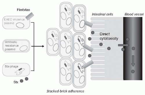
FIGURE 55.11 The hypothetical origin of the O104:H4 E. coli strain isolated from the German outbreak. An ancestral enteroaggregative E. coli (EAEC) strain with plasmids encoding for different virulence factors, including fimbriae that help stick to the intestinal cell, might have acquired the Shiga toxin phage (Stx-phage) characteristic of enterohemorragic E. coli (EHEC) and plasmids encoding for expanded- spectrum betalactamases. Close adhesion to the intestinal cell would facilitate the uptake of the Shiga toxin into the bloodstream, whereas resistance to most commonly used antibiotics would offer selective advantage over the normal intestinal flora upon antibiotic exposure.

Careful blood pressure control and renin-angiotensin system blockade may be particularly beneficial in the long term for those patients who suffer chronic renal disease after an episode of Stx-HUS. A study in 45 children with renal sequelae of HUS followed for 9 to 11 years documented that the early restriction of proteins and the use of ACE inhibitors may have a beneficial effect on the long-term renal outcome, as documented by a positive slope of 1/Cr values over time in treated patients.66 In another study, an 8- to 15-year treatment with ACE inhibitors after severe Stx-HUS normalized blood pressure, reduced proteinuria, and improved GFR.67

An oral Shiga toxin-binding agent that may compete with endothelial and epithelial receptors for Shiga toxin in the gut (SYNSORB Pk) has been developed with the rationale of limiting target organs’ exposure to the toxin (Table 55.2). However, a prospective, randomized, doubleblind, placebo-controlled clinical trial of 145 children with diarrhea-associated HUS failed to demonstrate any beneficial effect of treatment on disease outcome.68 Among newer treatments for Stx-HUS, the development of Stx-neutralizing monoclonal antibodies, including dual antibodies against Stx 1 and 2 (SHIGATEC, NCT0152199) to be given at the time of gastrointestinal infection, is the most advanced.69 Peptides impairing the ability of EHEC to survive under the acidic conditions of the gastric system could halt the disease process at even earlier stages by preventing bacterial intrusion into the gut.70 Heparin and antithrombotic agents may increase the risk of bleeding and should be avoided.

Efficacy of specific treatments in adult patients is dif-ficult to evaluate because most information is derived by uncontrolled series that may also include atypical HUS cases. In particular, no prospective, randomized trials are available to definitely establish whether plasma infusion or exchange may offer some specific benefit as compared to supportive treatment alone. However, comparative analyses of two large series of patients treated71 or not72 with plasma suggest that plasma therapy may dramatically decrease the overall mortality of Stx E. coli 0157:H7-associated HUS. These findings
lead us to consider plasma infusion or exchange suitable for adult patients, in particular in those with severe renal insuf- ficiency and central nervous system involvement.


TABLE 55.2 Specific Therapies Used in Thrombotic Microangiopathy, Dosing, and Efficacy


Therapy


Dosing


Efficacy


Antiplatelet


Anecdotal efficacy in TTP.


-Aspirin


325-1,300 mg/day


-Dipyridamole


400-600 mg/day


-Dextran 70


500 mg twice/day


-Prostacyclin


4-20 mg/kg/min


Antithrombotic


Anecdotal efficacy in HUS.


-Heparin


5000 U bolus followed by 750-1000 U/hr infusion


-Streptokinase


250,000 U bolus followed by 100,000 U/hr infusion


Shiga toxin-binding (Synsorb)


500 mg/kg per day for 7 days


Not effective in preventing or treating Stx-associated HUS.


Antioxidant (Vitamin E)


1,000 mg/sqm/day


Anecdotal efficacy in HUS.


Immunosuppressive


Probably effective in addition to plasma exchange in patients with TTP and anti-ADAMST13 autoantibodies or in aHUS with antifactor H autoantibodies and in forms associated with autoimmune diseases. Lack of evidence from controlled trials in immune-mediated HUS or TTP.


-Prednisone


200 mg tapered to 60 mg/day then 5 mg reduction per week


-Prednisolone


200 mg tapered to 60 mg/day then 5 mg reduction per week


-Immunoglobulins


400 mg/kg/day


-Vincristine


1.4 mg/sqm followed by 1 mg every 4 days


CD20 Cell-depleting (Rituximab)


375 mg/sqm per week up to CD20 depletion


Effective in treatment or prevention of TTP associated with immune-mediated ADAMTS13 deficiency resistant to, or relapsing after, immunosuppressive therapy.


Fresh frozen plasma


– Exchange


1-2 plasma volumes/day


First-line therapy for aHUS and TTP. Unproven efficacy in childhood Stx- HUS.


-Infusion


20-30 mL/kg followed by 10-20 mL/kg/day


To be considered if plasma exchange not available.


-Cryosupernatant


See plasma infusion/exchanges


To replace whole plasma in case of plasma resistance or sensitization.


-Solvent-detergent treated plasma


See plasma infusion/exchanges


To limit the risk of infections.


Liver-kidney transplant


To prevent CFH-associated HUS recurrence posttransplant. About 30% mortality risk.


Complement inhibition (Eculizumab)


600 mg weekly for the first 4 weeks 900 mg every 14 days up to 6 months


Reported efficacy in aHUS, STEC O104:H4-associated HUS, and in occasional cases of STEC O157:H7 childhood HUS.


TTP, thrombotic thrombocytopenic purpura; HUS, hemolytic uremic syndrome; Stx, Shiga toxin; aHUS, atypical hemolytic uremic syndrome; CFH, complement factor H; STEC, shiga toxin-producing E. coli.


Kidney transplants should be considered as an effective and safe treatment for those children who progress to ESRD. Indeed, recurrence rates range from 0% to 10%,73,74 and graft survival at 10 years is even better than in control children with other diseases.75

Evidence that uncontrolled complement activation may contribute to microangiopathic lesions of Stx-HUS44,45,46 provided the background for complement inhibitor therapy in three children with severe EHEC-associated typical HUS who fully recovered with the anti-C5 monoclonal antibody eculizumab.76 These encouraging results prompted nephrologists to use eculizumab therapy in HUS patients involved in the STEC O104:H4 outbreak in Germany (Table 55.2). In the setting of a controlled multicenter clinical study (EudraCT, 2011-002691-17; Clinicaltrials.gov ID: NCT01410916) 148 patients with bloody diarrhea and/or evidence of STEC/EHEC infection, microangiopathic hemolysis, thrombocytopenia, renal insufficiency, and/or central nervous system complications or thrombosis received at least an 8-week eculizumab treatment. At the inclusion, 94 patients were on dialysis therapy, 22 required ventilatory support, and 129 were receiving plasma exchange or infusion. Outcome data were reported by Dr. Rolf Stahl from the Hamburg University Medical Center during the 43rd Annual Meeting of the American Society of Nephrology held in Denver in November 2011. No patient died. At 8 weeks, platelet count and serum creatinine levels normalized in 123 and 82 patients, respectively, and no patient had persistent seizures. Dialysis, ventilatory support, and plasma therapy were no longer required. Treatment was well tolerated in all patients and no case of meningococcal infection was reported. Comparative analyses versus 108 patients who had been treated with plasma exchange but without eculizumab showed remarkably larger and faster recovery in platelet count and kidney function in those receiving eculizumab therapy. Even better outcomes were observed in patients maintained on eculizumab therapy also after the completion of the originally planned 8-week treatment period. Altogether, the data clearly showed that complement inhibition by eculizumab therapy was lifesaving and almost fully prevented the risk of renal or neurologic sequelae in patients with severe diarrhea-associated HUS secondary to O104:H4 E. coli infection. Whether and to what extent these findings can be generalized to patients with more severe forms of typical HUS associated with gastrointestinal infections with other strains of Shiga toxin-producing E. coli remains to be addressed.


Neuraminidase-Associated Hemolytic Uremic Syndrome

Mechanisms. This is a rare but potentially fatal disease that may complicate pneumonia, or less frequently, meningitis caused by Streptococcus pneumoniae .77 Neuraminidase produced by S. pneumoniae cleaves N-acetylneuraminic acid from the glycoproteins on the cell membrane of erythrocytes, platelets, and glomerular cells.78,79 Removing the N-acetylneuraminic acid exposes the normally hidden Thomsen-Friedenreich antigen (T-antigen),80 which can then react with anti-T immunoglobulin M (IgM) antibody naturally present in human plasma. This antigen-antibody reaction occurs more frequently in infants and children and causes polyagglutination of red blood cells in vitro. This is the reason why, unlike in other forms of HUS, in neuraminidase-associated HUS there is a positive Coombs test. T- anti-T interaction on red cells, platelets, and the endothelium was thought to explain the pathogenesis, whereas the pathogenic role of the anti-T cold antibody in vivo is uncertain.81 T-antigen exposure on red cells is detected using the lectin hypogaea.

Clinical course and therapy. Patients, usually less than 2 years old, present with severe microangiopathic hemolytic anemia. The clinical picture is severe, with respiratory distress, neurologic involvement, and coma. The acute mortality is about 25%. The outcome is strongly dependent on the effectiveness of antibiotic therapy. In theory, plasma either infused or exchanged, is contraindicated, because adult plasma contains antibodies against the T-antigen that may accelerate polyagglutination and hemolysis.80 Thus, patients should be treated only with antibiotics and washed red cells. In some cases, however, plasma therapy, occasionally in combination with steroids, has been associated with recovery.

Atypical Hemolytic Uremic Syndrome. Atypical HUS (aHUS) includes a number of associations and presentations. It can occur sporadically or within families. Research in the last 10 years has linked aHUS to uncontrolled activation of the complement system (Fig. 55.12 and Table 55.1).5

Familial atypical hemolytic uremic syndrome. Fewer than 20% of aHUS cases are familial. Reports date back to 1965, when Campbell and Carré described hemolytic anemia and azotemia in concordant monozygous twins.82 Since then, familial HUS has been reported in children and, less frequently, in adults. Although some were in siblings, suggesting autosomal recessive transmission, others were across two to three generations, suggesting an autosomal dominant mode.83,84 The prognosis is poor (cumulative incidence of death or ESRD, 50% to 80%).

Sporadic atypical hemolytic uremic syndrome. Sporadic aHUS encompasses cases without a family history of the disease. Triggering conditions for sporadic aHUS85 include HIV infection, anticancer drugs (e.g., mitomycin, cisplatin, bleomycin, gemcitabine), immunotherapeutic agents (e.g., cyclosporine, tacrolimus, OKT3, interferon, quinidine), antiplatelet agents (ticlopidine and clopidogrel), malignancies, transplantation, and pregnancy.12,86

De novo posttransplant HUS has been reported in patients receiving renal transplants or other organs, due to calcineurin inhibitors or humoral rejection. It occurs
in 5% to 10% of renal transplant patients who receive cyclosporine and in approximately 1% of those who are given tacrolimus.87,88,89 TMA usually develops in the first weeks posttransplant when patients are treated with high doses of the immunosuppressant. Plasma exchange, combined with dose reduction or the withdrawal of calcineurin inhibitors, achieved remission in up to 80% of patients with de novo posttransplant HUS.88


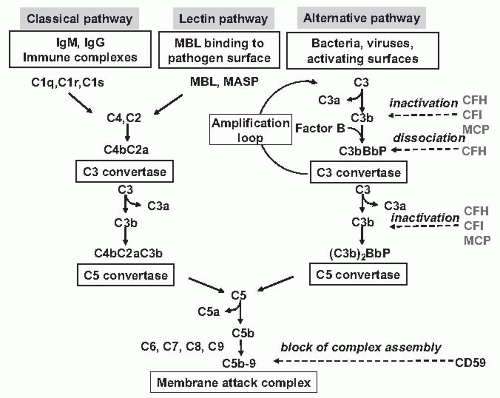
FIGURE 55.12 The Three activation Pathways fo Complement. The classical pathway is initiated by the binding of the C1 complex to antibodies bound to an antigen on the surface of a bacterial cell, leading to the formation of a C4b2a enzyme complex, the C3 convertase of the classical pathway. The mannose-binding lectin pathway is initiated by binding of the complex of mannose-binding lectin (MBL) and the serine proteases mannose-binding lectin-associated proteases 1 and 2 (MASP1 and MASP2) to mannose residues on the surface of a bacterial cell, which leads to the formation of the C3 convertase enzyme C4bC2a. The alternative pathway is initiated by the covalent binding of a small amount of C3b generated by spontaneous hydrolysis in plasma to hydroxyl groups on cell-surface carbohydrates and proteins. This C3b binds factor B to form the alternative pathway C3 complex C3bBb. The C3 convertase enzymes cleave many molecules of C3 to form the anaphylatoxin C3a and C3b, which binds covalently around the site of complement activation. Some of this C3b binds to C4b and C3b in the convertase enzymes of the classical and alternative pathways, respectively, forming C5 convertase enzymes that cleave C5 to form the anaphylatoxin C5a and C5b, which initiates the formation of the membrane-attack complex. The human complement system is highly regulated as to prevent nonspecific damage to host cells and to limit the deposition of complement to the surface of pathogens. This fine regulation occurs through a number of membraneanchored and fluid phase regulators that inactivate complement products formed at various levels in the cascade and that protect host tissues. (See Color Plate.) CFB, complement factor B; CFI, complement factor I; CFH, complement factor H; MCP, membrane cofactor protein; CD59, protectin (prevents the terminal polymerization of the membrane attack complex).

In 10% to 15% of female patients, aHUS manifests during pregnancy or postpartum.6,85 aHUS may present at any time during pregnancy but mostly in the last trimester and about the time of delivery. It is sometimes difficult to distinguish it from preeclampsia. HELLP syndrome (HEmolytic anemia, elevated Liver enzymes, and Low Platelets) is a life- threatening disorder of the last trimester or parturition with severe thrombocytopenia, microangiopathic hemolytic anemia, renal failure, and liver involvement. These forms are always an indication for prompt delivery that is usually followed by complete remission.85 Postpartum HUS manifests within 3 months of delivery in most cases. The outcome is usually poor.

About 50% of sporadic aHUS cases show no clear trigger (idiopathic HUS).


Mechanisms.

Complement abnormalities. Reduced serum levels of C3 with normal C4 in HUS patients were known since 1974.90,91 In cases of familial aHUS, serum C3 was low even during remission, hinting at genetic defects.90,92 Low C3 reflected complement activation and consumption with high levels of activated products, C3b, C3c, and C3d.93

The complement system is part of innate immunity and consists of several plasma and membrane-bound proteins
that protect against invading organisms.94 Three activation pathways—classical, lectin, and alternative pathways— produce protease complexes, termed C3 and C5 convertases that cleave C3 and C5, respectively, eventually leading to the membrane attack complex (MAC or C5b-9) that causes cell lysis (Fig. 55.12). The alternative pathway is initiated spontaneously in plasma by C3 hydrolysis, which is responsible for covalent deposition of a low amount of C3b onto practically all plasma-exposed surfaces. On the bacterial surface, C3b leads to opsonization for phagocytosis by neutrophils and macrophages. Without regulation, a small initiating stimulus is quickly amplified to a self-harming response until the consumption of complement components. On host cells, such a dangerous cascade is controlled by membrane-anchored and fluid-phase regulators (Fig. 55.12). They both favor the cleavage of C3b to inactive iC3b by the plasma serine-protease factor I (CFI, cofactor activity) and dissociate the multicomponent C3 and C5 convertases (decay acceleration activity). Foreign targets and injured cells that either lack membrane-bound regulators or cannot bind soluble regulators are attacked by complement.


TABLE 55.3 Outcome of Atypical Hemolytic Uremic Syndrome According to the Associated Genetic Abnormality

Only gold members can continue reading. Log In or Register to continue

Stay updated, free articles. Join our Telegram channel

May 29, 2016 | Posted by in NEPHROLOGY | Comments Off on Hemolytic Uremic Syndrome, Thrombotic Thrombocytopenic Purpura, and Acute Cortical Necrosis

Full access? Get Clinical Tree

Get Clinical Tree app for offline access