Fig. 1.1
(a) Lateral view of embryo in fourth week. (b) Transverse section demonstrating development of the urogenital ridge from the intermediate mesoderm (reprinted with permission, Cleveland Clinic Center for Medical Art & Photography © 2015. All Rights Reserved)
Early in the fourth week, the primitive kidneys or pronephroi appear as elevations along the cervical region of the developing embryo [1]. These transitory structures consist of epithelial buds which extend ventromedially and pronephric ducts which extend caudally to eventually open into the cloaca of the hindgut (Fig. 1.2a). By the end of the fourth week, the pronephroi have degenerated with the pronephric ducts persisting to become incorporated into the second set of kidneys as the mesonephric (Wolffian) duct (Fig. 1.2b).
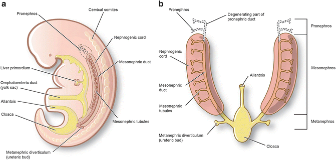

Fig. 1.2
Depiction of the nephric system transitions from the fourth to fifth week. (a) Lateral illustration of embryo in the fifth week with the development of the transitory pronephroi and pronephric ducts which open caudally into the cloaca. (b) Ventral view (reprinted with permission, Cleveland Clinic Center for Medical Art & Photography © 2015. All Rights Reserved)
During the fourth week, the mesonephroi emerge caudal to the pronephroi and function as interim structures until full development of the permanent kidneys around week 9. Along the length of the mesonephric (Wolffian) ducts (formerly pronephric ducts), mesonephric tubules begin to emerge on either side of the vertebral column extending from the upper thoracic region to mid-lumbar levels (Fig. 1.3a, b), with cranial tubules regressing by the end of week 5. The remaining tubules located in the upper three lumbar levels differentiate into cup-like pouches which extend medially to meet loops of capillaries emerging from the aorta (Fig. 1.3c). These structures are collectively referred to as a renal corpuscle and consist of the cup-like glomerular capsule and capillary plexus (Fig. 1.3d). During week 9, the mesonephric ducts regress in the female but persist in the male as part of the genital duct system including the ductus deferens, duct of epididymis, ejaculatory ducts, and seminal vesicles.
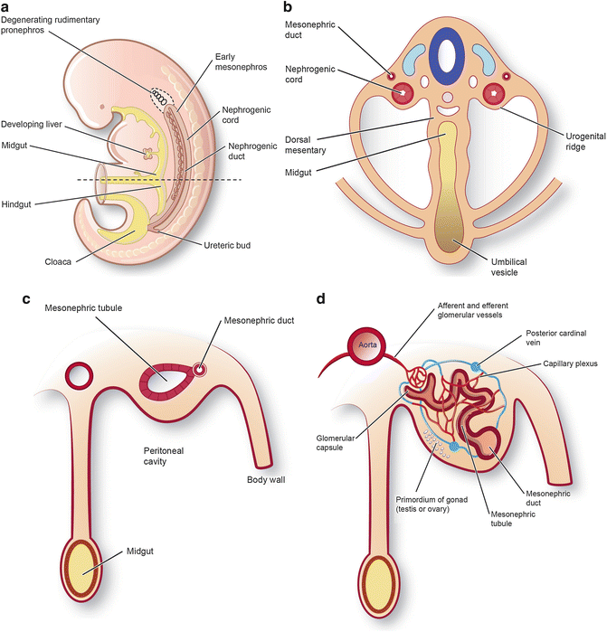

Fig. 1.3
(a) Lateral view indicating degeneration of the pronephros and emergence of the mesonephros. (b) Ventral view. (c) Sequential development of the mesonephric tubules from weeks 5 to 11. (d) The medial aspect of the mesonephric tubules extends to meet loops of capillaries emerging from the aorta (reprinted with permission, Cleveland Clinic Center for Medical Art & Photography © 2015. All Rights Reserved)
During the fifth week, the definitive kidneys or metanephroi begin to develop. Two sources give rise to the definitive kidneys: the ureteric bud (metanephric diverticulum) and the metanephrogenic blastema (metanephric mass of mesenchyme) (Fig. 1.4a). The ureteric buds emerge as outgrowths of the mesonephric ducts adjacent to their entrance into the cloaca. As they elongate, the ureteric buds invade the metanephrogenic blastema which grows from the caudal aspect of the nephrogenic cord. Penetration of the metanephric mesenchyme induces the ureteric bud to undergo a series of repetitive branchings, leading to formation of the renal collecting ducts and tubules, calyces, and primitive renal pelvis (Fig. 1.4b–d). Reciprocally, the distal end of the bud induces the metanephric mesenchyme to condense and form metanephric vesicles which will further develop into a series of metanephric tubules (Fig. 1.5a, b). The connection formed between the lumens from these S-shaped metanephric tubules with that of the ureteric duct results in the formation of the definitive uriniferous tubule (Fig. 1.5c). These uriniferous tubules will further differentiate into proximal convoluted tubules, distal convoluted tubules, and the nephron loop including descending and ascending nephron loops (Fig. 1.5d).
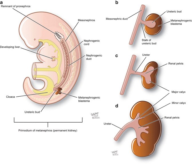
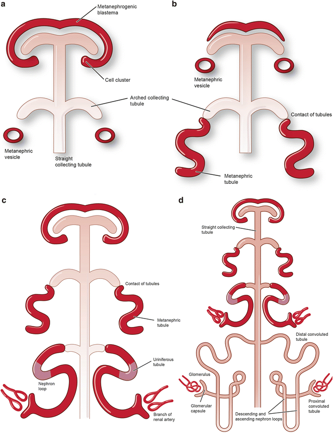

Fig. 1.4
Metanephros development from the ureteric bud and metanephrogenic blastema. (a) Lateral view of an embryo in the fifth week of development. (b–d) Sequential branching of the ureteric bud as it penetrates the metanephric mesenchyme (reprinted with permission, Cleveland Clinic Center for Medical Art & Photography © 2015. All Rights Reserved)

Fig. 1.5
Development of nephrons. (a) The distal end of the ureteric bud develops into a series of metanephric tubules. (b–d) These S-shaped structures, along with the ureteric duct, will form the uriniferous tubules (reprinted with permission, Cleveland Clinic Center for Medical Art & Photography © 2015. All Rights Reserved)
During the tenth week, the metanephroi become functional as the distal convoluted and collecting tubules begin to adjoin. In addition, the population of glomeruli further increases during this time up until week 32 of gestation. Nephrons also continue to form with approximately two million nephrons present in each kidney at term.
Between weeks 6 and 9, the kidneys withdraw from the pelvis and ascend along either side of the aorta to reach their permanent location along the posterior abdominal wall of the lumbar region (Fig. 1.6a–d). During their ascent, the kidneys’ original blood supply from the common iliac arteries disappears with branches from successive levels of the aorta taking their place. After contacting the suprarenal glands, the kidneys become fixed in their position with primary arterial supply deriving from the abdominal aorta. Caudal arterial branches may remain as accessory renal arteries; typically, these branches arise from the aorta to enter the hilum of the kidney.
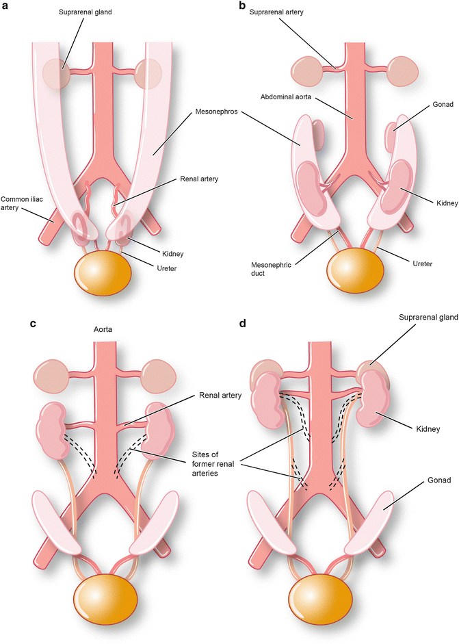

Fig. 1.6
Ventral abdominopelvic view of embryo from weeks 6 to 9. (a–d) The kidneys ascend alongside of the aorta to reach their permanent location in the lumbar region of the posterior abdominal wall (reprinted with permission, Cleveland Clinic Center for Medical Art & Photography © 2015. All Rights Reserved)
Anatomy of the Kidney
Overall Structure
The kidneys are reddish-brown bean-shaped structures located retroperitoneally along the posterior abdominal wall (Fig. 1.7). While posture and dynamic changes of the diaphragm can modify their relationship to surrounding structures, in the supine position, the kidneys typically extend from vertebral levels TXII–LIII. The right kidney occupies a position slightly lower due to its relationship with the liver superiorly. Conversely, the left kidney rests slightly closer to the midline, has a narrower profile, and is somewhat longer. On its medial surface, the kidneys are occupied by blood vessels, nerves, lymphatics, and the renal pelvis entering and exiting the concave-shaped hilum which faces anteriorly and medially.
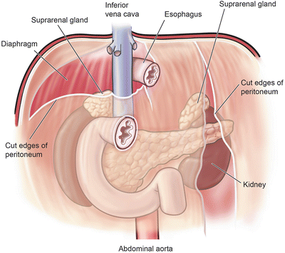

Fig. 1.7
Retroperitoneal location of the kidneys along the posterior abdominal wall (reprinted with permission, Cleveland Clinic Center for Medical Art & Photography © 2015. All Rights Reserved)
Adjacent Structures
Due to their position in the paravertebral gutters along the posterior abdominal wall, the kidneys are in contact with the surfaces of several structures directly or through an intervening layer of peritoneum. Anteriorly, the left kidney is related to the spleen, suprarenal gland, stomach, pancreas, jejunum, descending colon, and left colic flexure (Fig. 1.8a). Alternatively, the anterior surface of the right kidney is related to the liver, suprarenal gland, small intestine, right colic flexure, and descending part of the duodenum. Posteriorly, both kidneys are related to the psoas major muscle, quadratus lumborum muscle, transversus abdominis muscle, diaphragm, costodiaphragmatic recess, eleventh rib (left kidney only), twelfth rib, subcostal nerve, ilioinguinal nerve, and iliohypogastric nerve (Fig. 1.8b).
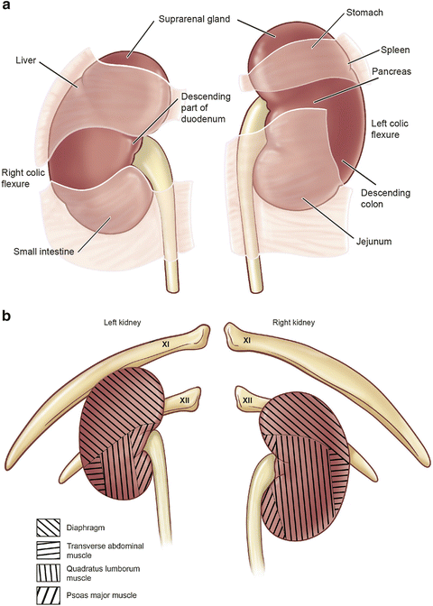

Fig. 1.8


Relationship of surrounding structures to the kidney. (a) Anterior surface. (b) Posterior surface (reprinted with permission, Cleveland Clinic Center for Medical Art & Photography © 2015. All Rights Reserved)
Stay updated, free articles. Join our Telegram channel

Full access? Get Clinical Tree


