Echoendoscopic Ultrasound–Guided Biliary Drainage

Chapter 30 Echoendoscopic Ultrasound–Guided Biliary Drainage


Echoendoscopic biliary drainage is an option to treat obstructive jaundices when endoscopic retrograde cholangiopancreatography (ERCP) drainage fails. These procedures are alternative methods to surgery and percutaneous transhepatic biliary drainage (PTBD), made possible by the continuous development and improvement of echoendoscopes and accessories. The development of linear sectorial array echoendoscopes in the early 1990s brought a new approach to diagnostic and therapeutic echoendoscopy capabilities, opening up the possibility of performing extraluminal puncture with a direct ultrasonographic view. Despite the high success rate and low morbidity of biliary drainage with ERCP, there are potential difficulties in the setting of tumor ingrowth, gut compression by tumor, periampullary diverticula, and anatomic variation. The echoendoscopic techniques for biliary access entail puncture and contrast injection into the left biliary tree. When performed from the stomach, the access is made through hepatic segment III; when performed through the duodenum, direct common bile duct puncture is performed. Diathermic dilation of the puncturing tract is required using a 6-Fr cystostome and/or balloon catheter followed by placement of a plastic or metallic stent. The technical success of hepaticogastrostomy approximates 98%. Adverse events occur in 20%: pneumoperitoneum, biliary leak, infection, and stent dysfunction. To prevent bile leakage, we have used a two-stent technique: the first stent introduced is a long uncovered metallic stent (8 or 10 cm) and inside this first stent a second fully covered stent of 6 cm is delivered to bridge the bile duct and the stomach. Choledochoduodenostomy has an overall success rate of 92%. Adverse events include pneumoperitoneum and focal bile peritonitis, occurring in 14%. Over the last 10 years, the technique has been increasingly performed in referral centers with ERCP as well as endoscopic ultrasound (EUS) expertise.


Endoscopic biliary stent placement is the most common method to treat obstructive jaundice. In 3% to 12% of cases, selective cannulation of the major papilla is unsuccessful and surgery or percutaneous biliary drainage is required. Percutaneous drainage requires dilated intrahepatic biliary ducts and the rate of adverse events approximates 25% to 30%, including peritoneal bleeding. A new technique of biliary drainage using EUS and EUS-guided puncture of the bile duct (common bile duct or left hepatic duct) is now possible.


Using EUS guidance and dedicated accessories, it is now possible to create biliodigestive anastomosis.


The aims of this chapter are:


Box 30.2


Indications for EUS Access into the Biliary Tree


Equipment


Interventional Echoendoscopes


Around 1990 the Pentax Corporation developed an electronic, convex curved, linear array echoendoscope (FG 32UA) with an imaging plane in the long axis of the device that overlaps the instrumentation plane. This echoendoscope, equipped with a 2.0-mm working channel, enabled fine-needle biopsy under EUS guidance. However, the relatively small working channel of the FG 32UA was a drawback for pseudocyst drainage since it necessitated the exchange of the echoendoscope for a therapeutic duodenoscope to insert either a stent or a nasocystic drain. To enable stent placement using an echoendoscope, the EUS interventional echoendoscopes (FG 38X, EG 38UT, and EG 3870UTK) were developed by Pentax-Hitachi. The FG 38X has a working channel of 3.2 mm, which allows the insertion of an 8.5 Fr stent or nasocystic drain, and the EG 38UT and EG 3870UTK have larger working channels of 3.8 mm with an elevator allowing the placement of a 10 Fr stent.1,2


The Olympus Corporation has also developed convex array echoendoscopes. The GF-UC 30P has a biopsy channel of 2.8 mm and is equipped with an elevator, which enables placement of a 7 Fr stent or nasocystic catheter. A new prototype, the GF UCT 30, has a larger working channel of 3.7 mm, allowing the placement of a 10 Fr stent. The main drawback of convex linear array echoendoscopes is the more limited imaging field (120 degrees using the Pentax and 180 degrees using the Olympus) produced by an electronic transducer. These instruments are coupled with the Aloka processor or with a smaller processor.


Needles and Accessories for Drainage


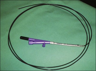
Some authors have used needle-knife catheters but the needle can be difficult to visualize endosonographically. The Zimmon needle knife (Cook Endoscopy, Winston-Salem, N.C.) has a large-gauge needle that is easier to visualize. Diathermy is usually required to penetrate a cyst, particularly when performed transgastrically (Fig. 30.1).3


In standard endosonography fine-needle aspiration (FNA), the needle is well visualized sonographically and can be used for bile duct puncture. The drawback of this needle is that the small caliber (22 or 23 gauge) only accepts a 0.018-in guidewire. Using a 19-gauge FNA needle (Cook Endoscopy), a 0.035-in guidewire can be inserted through the needle into the dilated bile duct. Cook Endoscopy has recently developed an “access needle.” However, difficulty manipulating the guidewire through this 19-gauge EUS needle is difficult when hepaticogastrostomy is performed because of “stripping” of the coating of the wire, which in turn creates risks of procedural failure and guidewire fracture within the patient.


To solve this problem we worked with Cook Endoscopy to design a special needle called the EchoTip Access Needle. This needle is unique because the stylet is sharp and it is relatively easy to insert the needle into the bile duct, the pancreatic duct, or pseudocyst. When the stylet is withdrawn, the needle left in place is smooth and the manipulation of the guidewire is relatively easy. This device is designed to decrease the possibility of wire stripping.


Tags:
Mar 11, 2017 | Posted by in GASTROENTEROLOGY | Comments Off on Echoendoscopic Ultrasound–Guided Biliary Drainage

Full access? Get Clinical Tree

Get Clinical Tree app for offline access