Disorders of Potassium and Acid-Base Metabolism in Association with Renal Disease


Disorders of Potassium and Acid-Base Metabolism in Association with Renal Disease


Mark A. Perazella

Asghar Rastegar


In this chapter, we review disturbances in potassium and acid-base homeostasis seen in patients with renal disease. Our discussion is, however, limited to disorders of potassium and acid-base homeostasis seen in (1) patients with progressive chronic kidney disease (CKD) and (2) patients with renal insufficiency and defects in the renin-aldosterone axis or in the tubular response to aldosterone. We briefly review potassium and acid-base homeostasis in healthy humans before focusing on patients with underlying renal disease. We do not, however, discuss normal renal handling of potassium and only briefly review renal handling of hydrogen ion. These two topics are extensively reviewed in Chapter 6: Tubular Potassium Transport, and Chapter 7: Renal Acid-Base Transport, respectively.


POTASSIUM HOMEOSTASIS

Potassium is the most abundant cation in the body. The distribution of potassium is such that 98% of total body potassium is intracellular, whereas only 2% is extracellular. Serum potassium is normally between 3.8 and 5.0 mEq per liter, whereas the intracellular potassium concentration is 120 to 140 mEq per liter. The high intracellular to extracellular potassium ratio (Ki/Ko) is crucial to normal cell function, because it is the major determinant of the resting membrane potential. The body is able to maintain this distribution in a highly regulated and efficient fashion through the hormonal modulation of Na-K-ATPase pump activity.1,2 Humans, as carnivorous intermittent eaters, are continuously challenged by large potassium loads. On a long-term basis, this challenge is met primarily by the renal excretion of potassium load; however, on a short-term basis, a significant amount of potassium is shifted intracellularly.3 This shift temporarily buffers the expected change in the Ki/Ko ratio until potassium intake is balanced by a comparable output. Therefore, potassium homeostasis is regulated through both extrarenal as well as renal mechanisms (Fig. 72.1).4


Extrarenal Potassium Homeostasis

The kidney is able to excrete only about 50% of the administered potassium during the first 4 hours after intravenous or oral intake of potassium. Approximately 80% of the retained potassium is shifted intracellularly, and only 20% (or 10% of the total intake) remains in the extracellular space.5,6,7 The retained potassium will be excreted completely over the next 24 hours.8 The major regulators of this internal redistribution are: (1) insulin, (2) catecholamines, and (3) mineralocorticoids. In addition to these physiologic regulators, serum potassium is also regulated by acid-base status as well as plasma osmolality. Factors that increase or decrease plasma potassium concentration are noted in Figure 72.2.


Insulin

The ability of insulin to shift potassium intracellularly has been known for over 70 years9 and has been used therapeutically for the treatment of hyperkalemia. Pancreatectomized dogs tolerate exogenous potassium loads poorly.10 This is reversed by the exogenous replacement of insulin.11,12 The partial inhibition of endogenous insulin in dogs by somatostatin infusion results in a twofold rise in serum potassium compared to controls.6 If physiologic doses of insulin were added to the somatostatin infusion, potassium tolerance returned to normal. In healthy volunteers, somatostatin infusion in the postabsorptive state led to a 50% decline in the plasma insulin concentration and a 0.5 to 0.7 mEq per liter rise in serum potassium that was reversed by a physiologic infusion of exogenous insulin.6 A similar phenomenon was observed in maturity-onset diabetic patients who have normal or increased fasting plasma insulin levels, but not in insulin-deficient juvenile diabetic patients.13

The primary sites of insulin-mediated potassium uptake include muscle and the liver, and to a lesser degree, adipose tissue.14,15 In normal volunteers on variable insulin doses, the liver is the primary site of potassium uptake during the first hour.15 However, during the second hour, despite a continued decrease in serum potassium, there is net release of potassium from the portal and splanchnic bed, indicating a shift of potassium uptake to the peripheral tissue, especially muscle.15

At the cellular level, insulin interacts with specific receptors on the plasma membrane,16 increasing the activity
of the Na-K-ATPase pump in the skeletal and heart muscle, epithelial cells of the kidney and bladder, as well as liver and fat cells.17 This results in a series of intracellular events leading to hyperpolarization of cell membranes.17 The time course for this interaction is consistent with both an increase in enzyme activity as well as the rapid recruitment of Na-K-ATPase pumps to the cellular membrane. In contrast, chronic stimulation by insulin probably increases the total number of available pump sites. This occurs through the regulation of the Na-K-ATPase pump at the transcriptional and posttranscriptional levels by inducing the synthesis of new α and β subunits.1 McDonough and Youn,18 using a potassium clamp, have recently shown that after 10 days of potassium deprivation in rats Na-K-ATPase activity decreased by more than 50% and insulin-mediated potassium shift decreased by 94%, whereas in rats deprived of potassium for only 2 days the number of pumps did not decrease, but insulin-mediated potassium shift decreased by 80%. This would indicate that insulin resistance precedes a decrease in the number of pump expression during hypokalemia. The molecular mechanism underlying this response, however, remains poorly understood.19 Several in vitro studies, including one study in humans, have shown that insulin-driven potassium uptake by both muscle and the liver is independent of glucose uptake.15,20


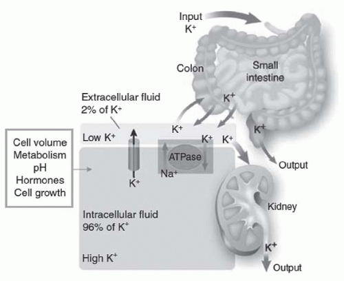
FIGURE 72.1 The distribution of potassium (K) in the body. Potassium is primarily located in cells (96%), with distribution controlled by a pump-leak mechanism involving both Na-K-ATPase and membrane potassium channels. The kidneys excrete more than 90% of the daily potassium load, and the intestines excrete the rest. (From Giebisch G, Krapf R, Wagner C. Renal and extrarenal regulation of potassium. Kidney Int. 2007;397, with permission.)


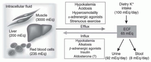
FIGURE 72.2 The distribution of potassium (K) between the intracellular and extracellular fluid compartments. Potassium distribution between the intra- and extracellular fluid is controlled by a pump-leak mechanism involving both Na-K-ATPase and membrane potassium channels. The factors noted in the figure drive potassium into or out of cells. (From Giebisch G, Krapf R, Wagner C. Renal and extrarenal regulation of potassium. Kidney Int. 2007;397, with permission.)


Catecholamines

D’Silva,21 beginning in 1934, first observed a biphasic response of plasma potassium to epinephrine injection. Plasma potassium rose during the first 1 to 3 minutes, but
with continued infusion, fell and remained lower than baseline. Other investigators have shown increased potassium tolerance in animals infused with pharmacologic doses of epinephrine22,23 despite a pancreatectomy or nephrectomy.24 Brown and coworkers25 have shown that the infusion of stress-level doses of epinephrine resulted in a decrease in serum potassium by 0.4 to 0.6 mEq per liter. Because epinephrine inhibits the renal excretion of potassium,26,27 the decline in potassium concentration is entirely accounted for by enhanced cellular potassium uptake.

Specific receptors are involved in the cellular disposal of potassium by catecholamines. Alpha stimulation in humans by phenylephrine28 significantly impairs cellular potassium tolerance, which is reversed by the α-antagonist phentolamine. This phenomenon may explain the initial rise in serum potassium after the infusion of catecholamine.26,27 β2-blockade impairs the catecholamine-induced shift of potassium into extrarenal tissues29,30 and causes hyperkalemia despite an increase in renal excretion of this ion. In normal volunteers who exercise while taking β-adrenergic blocking agents, the serum potassium level is raised 2- to 2.5-fold higher than during similar exercise performed without a β blockade.3,31 The effect of nonspecific β-blockers such as propranolol on serum potassium is mimicked by specific β2-blockers32 but not β1-blockers. Although an important role for catecholamine-stimulated uptake of potassium by muscle has been demonstrated, the role of the liver remains controversial. The effect of potassium on catecholamine levels is less clear.

At the cellular level, epinephrine binds to the β2-receptor resulting in the stimulation of adenyl cyclase and the conversion of adenosine triphosphate to cyclic 3′,5′-adenosine mono- phosphate (cAMP). It is postulated that cAMP then activates protein kinase A, which then phosphorylates the Na-K-ATPase pump, increasing its activity and promoting potassium influx into the cell and Na+ efflux.31 Binding catecholamines to the α receptor decreases cellular potassium uptake by inhibiting adenylate cyclase activity and decreasing Na-K-ATPase pump activity.32 In addition, activation of the α-1 receptor alters cytoplasmic calcium, thereby increasing intracellular calcium concentration and opening calcium-activated potassium channels, which allow potassium to exit the cell.32 Interestingly, the effect of insulin and epinephrine on plasma potassium is additive, which confirms a separate mechanisms of action.31 In insulin-induced hypoglycemia, hypokalemia is therefore due to the combined effect of both insulin and the hypoglycemia-induced rise in catecholamines.31


Mineralocorticoids

Mineralocorticoids play a major role in external potassium homeostasis by increasing its excretion by the kidney,33 colon,34 salivary,35 and sweat glands.36 However, aldosterone’s role in internal potassium homeostasis is less clear.37,38 Anephric rats adapted to high potassium intake handle an acute potassium load more efficiently than do nonadapted rats. This adaptation is lost by prior adrenalectomy and restored by exogenous mineralocorticoid replacement.39 However, Spital and Sterns40,41 observed that during the 20 hours of fasting before a nephrectomy and acute potassium loading, these rats became potassium depleted owing to marked kaliuresis resulting from high serum potassium coupled with a high aldosterone level. In adrenalectomized dogs, Young and Jackson42 have shown that plasma potassium concentration at any exchangeable potassium level was a function of aldosterone replacement dose. High-dose aldosterone in anephric rabbits delays death due to hyperkalemia.43 Similarly, baseline potassium was significantly higher in hormonally deficient adrenalectomized rats despite negative potassium balance compared to exogenously replaced controls, thus supporting a defect in the cellular uptake of potassium.5,44 This impairment was corrected by either aldosterone or epinephrine replacement. In rats, aldosterone has been shown to increase Na-K-ATPase pump activity by inducing the synthesis of new α- and β-subunits in heart and vascular smooth muscle.1 This effect presumably represents the action of aldosterone on Na-K-ATPase pump gene expression and supports a role for aldosterone in cellular potassium homeostasis. In anephric humans treated with deoxycorticosterone acetate (DOCA), spironolactone, or placebo for 3 days, the baseline potassium was similar; however, the DOCA-treated subjects showed greater tolerance to acute potassium load than did the other two groups.45 In a study of 15 patients on hemodialysis that were treated with 0.05 to 2.0 mg per day of fludrocortisone acetate, the serum K+ decreased significantly.46 Interestingly, the effect of exogenous mineralocorticoid was more pronounced in patients with a low compared to a high plasma aldosterone concentration. Low dose spironolactone (25 mg per day) was associated with an increase in a mean serum K+ concentration of 0.3 mEq per liter over 4 weeks of therapy in 15 chronic hemodialysis patients.47 In the largest study to date, serum potassium in 50 hemodialysis patients treated with 25 mg per day of spironolactone increased from baseline 4.96 to 5.16 in 2 weeks and remained stable for 6 months.48 Very low dose spironolactone (25 mg thrice weekly), however, did not increase serum K+ in hemodialysis patients,49 whereas a very high dose (300 mg per day) induced a significant rise in plasma potassium (0.5 mEq per liter) and caused hyperkalemia after 3 weeks of therapy in nine chronically hemodialyzed end-stage renal disease (ESRD) patients (three were anephric).50 In summary, these studies support a small but significant role for aldosterone in internal potassium homeostasis in anephric animals and ESRD patients.


Acid-Base Balance

The role of acid-base balance on the internal distribution of potassium51 is based on the concept that during the development of acute acidemia, the hydrogen ion enters the cell in exchange for potassium and that the reverse occurs during the development of alkalemia.51,52,53 This dynamic interrelationship has been simplified clinically to a general rule that
for each 0.1 U change in serum pH, the serum potassium changes in the opposite direction by 0.6 mEq per liter. However, the relationship between serum potassium and serum pH is much more complex and depends on the type and severity of the acid-base disorder, the anion accompanying hydrogen, the duration of acidosis, changes in plasma bicarbonate concentration independent of changes in pH and the extent of intracellular buffering, and renal adaptation as well as hormonal changes in response to the disorder.54 In addition, in clinical settings, there are often other physiologic and pathophysiologic processes that may be present, which would affect both transcellular as well as the renal and extrarenal handling of potassium. The following generalizations should therefore be used with caution.

1. On the whole, acidosis is accompanied by a greater change in serum potassium than is alkalosis.55

2. Mineral acidosis (Fig. 72.3) causes the greatest shift (0.24 to 1.7 mEq per liter for each 0.1 U in pH change), whereas organic acidosis has a much smaller effect.53,56,57 Mild mineral acidosis (a decrease in serum bicarbonate by 5 mEq per liter and an increase in hydrogen ion concentration by 0.45 nmol per liter), however, does not result in a significant change in serum potassium.58

3. Acute respiratory alkalosis paradoxically results in a small but significant rise in serum potassium (+0.30 mEq per liter with a drop in pCO2 of 16 to 22.5 mm Hg). The rise was primarily due to stimulation of α-adrenergic receptors by catecholamine.59 Chronic respiratory alkalosis, however, results in sustained hypokalemia due to a renal loss of potassium.60


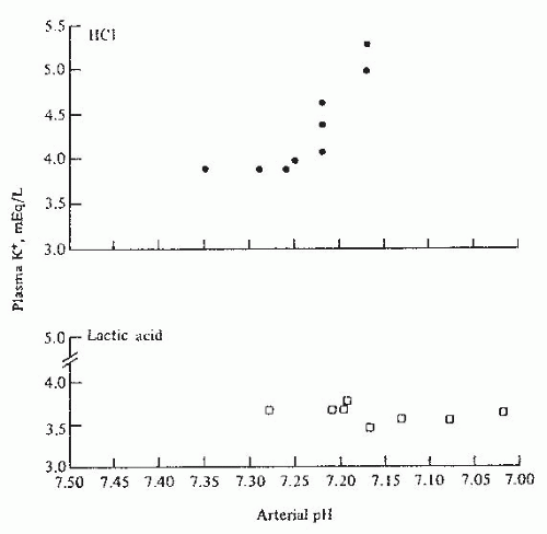
FIGURE 72.3 The effect of arterial pH on plasma potassium concentration in experimentally induced mineral acidosis (hydrochloric acid-HCl) and lactic acidosis in dogs. (From Perez GO, Oster JR, Vaamonde CA. Serum potassium concentration in acidemic states. Nephron. 1981;27:233, with permission.)

4. The amounts of potassium shifted into the cell in metabolic and chronic respiratory alkalosis are approximately similar (0.1 to 0.4 mEq per liter for each 0.1 U of pH change).

5. Acute respiratory acidosis resulting in a decrease in pH to 7.24 had no effect on serum K+.61

6. Changes in serum bicarbonate, independent of serum pH, have an inverse effect on the serum potassium concentration.

7. In chronic acidosis and alkalosis, the final serum K+ is a function of the effect of acid-base disturbance on the renal handling of potassium, as well as on the transcellular distribution of this ion. In dogs with ammonium chloride-induced acidosis, Magner and associates62 noted a fall in serum potassium below baseline by days 3 to 5, owing to severe kaliuresis.


Osmolality

The acute hyperkalemic effect of a sudden rise in plasma osmolality is probably caused by the shift of potassium-rich intracellular fluid by solvent drag.63 Clinically, this phenomenon is most commonly observed in hyperosmolar diabetic patients (Fig. 72.4), with or without ketoacidosis64,65,66,67 when insulin deficiency augments the rise in potassium. Although chronic hyperkalemia in diabetic patients is multifactorial, a sudden rise in plasma osmolality seems to play a contributory role. The infusion of hypertonic mannitol in healthy humans68 or hypertonic saline69 or hypertonic contrast media70 in patients with chronic kidney disease results in a modest rise in serum potassium (0.4 to 0.6 mEq per liter). Hyperkalemia can be severe, especially in diabetic patients with little or no
renal function facing sudden hyperglycemia.71 These clinical observations support an independent role of sudden osmolar shifts in the regulation of serum potassium.


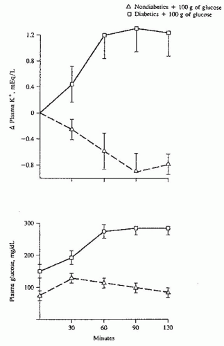
FIGURE 72.4 The effect of glucose infusion on plasma potassium and glucose concentrations in diabetics (squares) and normal subjects (triangles). The plasma potassium rises in diabetics owing to the development of hyperosmolality (hyperglycemia) but falls in normal subjects as a result of the glucose-induced release of endogenous insulin. (From Nicolis GL, Kahn T, Sanchez A, et al. Glucose-induced hyperkalemia in diabetic subjects. Arch Intern Med. 1981;141:49, with permission.)

Feedback or Feedforward Control of Potassium Homeostasis. It is well known that an increase in potassium concentration directly stimulates renal potassium excretion through an increase in potassium secretion in the collecting duct. This is accomplished by the direct stimulation of Na-K-ATPase, an increased tubular flow, and an increase in aldosterone. However, as Rabinowitz et al.72 first noted an increase in renal potassium excretion after meals in sheep was independent of change in serum potassium and aldosterone. In normal human subjects, urinary potassium excretion increased significantly 20 minutes after the ingestion of potassium salts before any change in serum potassium. Kaliuresis was more robust if potassium is ingested with meals rather than without meals or given intravenously. These and other observations support a role for a direct gut-kidney axis in potassium homeostasis favoring a feedforward rather than a feedback homeostatic mechanism (Fig. 72.5). The specific gut sensor and the gut-kidney loop remains speculative at this point. For a more detailed discussion, readers are referred to two recent reviews of this topic.73,74


POTASSIUM HOMEOSTASIS IN RENAL FAILURE

Patients with renal failure are able to maintain a near normal serum potassium concentration despite a marked decrease in glomerular filtration rate (GFR).75,76,77,78 Although hyperkalemia could be due to increased potassium intake and/or rapid shifts of potassium from the cell, renal failure is the most important cause of hyperkalemia, accounting for 77% of the cases reported by Acker and coworkers.79 In a random sample of 300 CKD patients (serum creatinine [Cr] levels
1.5 to 6.0 mg per deciliter) not receiving drugs that interfered with potassium homeostasis, 55% were noted to have hyperkalemia (K+ ≥ 5.0 mEq per liter).80 Treatment with drugs that interfere with potassium handling would be expected to further increase the development of hyperkalemia (see the following). Serum potassium rises with decreasing GFR; however, it often remains within normal range with GFR above 40 mL per minute.75 In this study, the rate of hyperkalemia ([K+] > 5.0) was 17% and was primarily limited to patients with CKD stage 4 and 5. However, under certain conditions, hyperkalemia may occur in patients with mild-to-moderate renal failure (Table 72.1). In a longitudinal study of patients with CKD, hyperkalemia ([K] > 5.5] was reported in only 8% of patients and, surprisingly, hypokalemia ([K] < 4.0) was more frequently seen in 15% of patients. Hypokalemia was not related to nutrition and was most likely secondary to the use of diuretics.78 This observation would indicate that electrolyte disturbances in patients with CKD are partly related to the underlying disease and partly to medications used in the management of concomitant comorbidities such as fluid overload and hypertension. However, it should be emphasized that the risk of hyperkalemia in patients with CKD, including those treated with renin-angiotensin-aldosterone system (RAAS) blockers, is relatively small.81


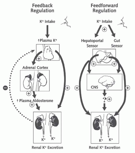
FIGURE 72.5 The integrated model of the regulation of body potassium balance: feedback and feedforward regulation. Renal potassium excretion is controlled by both feedback signals (plasma potassium concentration) and feedforward signals (liver and gut). CNS, central nervous system. (From Greenlee M, Wingo CS, McDonough AA, et al. Narrative review: evolving concepts in potassium homeostasis and hypokalemia. Ann Intern Med. 2009;150:619, with permission.)

In this section, we initially discuss total body potassium content in patients with renal failure before treatment with dialysis and then review internal and external potassium homeostasis in these patients. In the subsequent section, we discuss hyperkalemia seen in patients with renal insufficiency with a defect in the renin-angiotensin-aldosterone axis or in the tubular responsiveness to aldosterone.


TABLE 72.1 Etiologies of Hyperkalemia in Patients with Renal Insufficiency


GFR < 20 mL/min


Defects in the renin-angiotensin-aldosterone axis


Tubular defects in potassium secretion


Potassium input (e.g., rhabdomyolysis, hemolysis, severe catabolic states, gastrointestinal bleeding, exogenous potassium administration)


Shift of potassium from intracellular compartment


Drugs that interfere with renal and extrarenal potassium homeostasis


GFR, glomerular filtration rate.


Total Body and Cellular Potassium Content in Renal Failure

Total body potassium content is a reflection of the balance between potassium intake and potassium output, whereas the cellular content reflects the distribution of potassium between the intracellular and the extracellular compartments. Exchangeable potassium (Ke) in pre-ESRD patients has been generally reported as lower than normal.82 However, Berlyne and associates,83 after excluding patients with intercurrent problems (such as vomiting, diarrhea, or malnutrition), reported a normal value. It should also be noted that malnutrition is common in patients with CKD and many serum and anthropomorphic measurements of protein-energy nutritional status show progressive decline with the progression of CKD.84 As Patrick85 has pointed out, the normal range for Ke is not well defined and depends on age, sex, and the reference points used (e.g., total body weight, lean body weight, intracellular water). These reference points may be distorted in patients with CKD. The measurement of total body potassium by the use of a naturally occurring isotope (40K) also has given normal values.86

Cellular potassium content has been estimated by the use of muscle biopsy.87,88,89,90,91,92 Bergstrom and colleagues87 studied 102 patients with serum creatinine levels ranging from 4.8 to 25.0 mg per deciliter before therapy. In this and other studies, the intracellular potassium concentration was low owing to an increase in intracellular water despite normal intracellular potassium content.87,90 However, Bilbrey and coworkers93 and Montanari and coworkers92 have reported normal intracellular potassium concentrations. Importantly, the intracellular potassium content was either low or normal (but not increased) in all four studies.87,90,91,92,93 The low intracellular potassium (and high intracellular sodium content) has also been reported in erythrocytes92,94,95 and leukocytes82,96 from these patients. This bespeaks of a decrease in the number and/or the activity of the Na-K-ATPase pumps in the cell membrane. In chronic dialysis patients, the pump transport rate is higher immediately after fluid removal,97,98 and the abnormal levels of intracellular sodium and potassium in uremic patients return to normal following several weeks of dialysis.95 Because the number of pump sites inversely correlates with intracellular sodium, and a change in their number requires the production of new cells with lower intracellular sodium, the acute effect of fluid removal by dialysis may result from the removal of a volume-sensitive pump inhibitor.99 In contrast, the long-term effect of dialysis reflects the production of new cells with lower intracellular sodium and a higher number of pump sites. For a detailed discussion, refer to the article by Kaji and Kahn.99


Internal Potassium Homeostasis in Chronic Kidney Disease

The role of cellular uptake of potassium in renal failure has been studied in both humans76,77,100 and animals.101,102,103 Schon and associates103 have shown that the cellular uptake of potassium in rats with a remnant kidney is similar to that in normal rats maintained on a comparable diet but is lower than normal when both groups consume a high potassium diet. In contrast, in two different models of renal failure in rats, Bia and DeFronzo101 showed impairment in the cellular disposal of an acute potassium load. Bourgoignie and associates102 challenged chronically uremic dogs (remnant kidney model) that were adapted to different potassium intakes with an acute potassium load. Whereas the percentage of retained potassium that was shifted into the intracellular compartment was greater in normal dogs (90%), the absolute amount was significantly less than that in dogs with a remnant kidney (9.0 versus 20.5 mEq, respectively). They concluded that extrarenal cellular uptake was normal in the dogs with renal failure. Gonick and colleagues76 challenged patients with moderate renal failure with an oral potassium load. Whereas serum potassium 5 hours postchallenge was slightly higher in patients than in controls (5.2 versus 4.7 mEq per liter), this result was entirely because of a lower urinary excretion. In a study of patients with tubulointerstitial disease, the absolute amount of potassium shifted into the cell was greater in patients compared with controls, but the relative amount (expressed as a percentage of total potassium retained) was similar.100 In contrast, Kahn and colleagues77 observed a significantly greater rise in serum potassium in patients compared with controls when dietary potassium was increased by 50 mEq per day. This study77 cannot be strictly compared with others because they relied on 24-hour urinary potassium measurements, and their study reflected a long-term adaptation to a high potassium diet in patients with CKD. In hemodialysis patients, serum potassium rose significantly more in patients than in controls challenged with acute potassium load (1.06 versus 0.39 mEq per liter). However, the baseline potassium was significantly higher in patients than in controls (5.17 versus 3.59 mEq per liter), making the interpretation of this study difficult.104 More recently, Allon and colleagues105 noted a similar response in these patients with lower baseline potassium. Finally, the effect of vigorous exercise on serum potassium in hemodialysis patients was similar to the control group.106 It is reasonable to conclude that the extrarenal cellular uptake of an acute potassium load in CKD patients is near normal.

As discussed previously, internal potassium homeostasis is regulated by insulin, catecholamines, and, to a lesser extent, aldosterone. Although the serum insulin level is increased in renal failure,106,107,108 several studies provide strong support for normal insulin-stimulated potassium uptake106,108,109 by the splanchnic as well as by the peripheral tissues.109 Alvestrand and coworkers,109 using the euglycemic insulin clamp technique, demonstrated a similar uptake of potassium by both splanchnic and leg tissues in patients with CKD. The inhibition of endogenous insulin by somatostatin results in a significantly greater rise in serum potassium in uremic rats than in controls (1.0 versus 0.2 mEq per
liter at 60 minutes).110 The administration of glucose with potassium stimulates insulin secretion and attenuates the rise in potassium in patients on dialysis as well as normal controls.105

Elevated serum catecholamine levels have been reported in CKD.106,111,112 Yang and coworkers113 noted higher mean potassium in patients on propranolol. Infusion of epinephrine resulted in two different responses: In 4 of 10 patients, serum potassium did not fall; in the remaining 6, an exaggerated response was noted. The authors felt that the latter group of patients is those who have a propensity to develop hyperkalemia while on propranolol. Gifford and associates,114 using a much lower epinephrine dose, could not show a hypokalemic response in patients with ESRD. Plasma aldosterone is normal or high in most CKD patients.115,116,117,118,119 As noted, patients with ESRD who are taking DOCA, spironolactone, or placebo have similar baseline potassium levels; however, patients on DOCA can dispose an acute potassium load more promptly than the other groups.45 In addition, ESRD patients on spironolactone have a small but significant rise in serum potassium levels.48 These studies would support a minor role for aldosterone in internal potassium homeostasis in ESRD patients. In summary, extrarenal potassium homeostasis is near normal in patients with severe renal failure, although a cellular defect in potassium disposal due to abnormal response to catecholamines has been reported in a subgroup of patients on dialysis.


External Potassium Homeostasis in Severe Renal Failure


Renal Adaptation

Patients with a marked decrease in GFR are able to excrete the ingested dietary potassium load and maintain near normal potassium balance. This adaptive process is reflected by an increase in the fractional excretion of potassium (FEK) modulated by an increase in secretory rate per functioning nephron. However, this adaptive response is limited and a sudden increase in potassium intake may result in life-threatening hyperkalemia. The quantitative aspects as well as the anatomic and functional characteristics of this adaptive response are briefly reviewed herein.

In conscious dogs with a 10% remnant kidney, Schultze and coworkers119 showed that potassium excretion by the remnant kidney increased fourfold by 18 hours and approached 85% of the control value by the 7th day. Kunau and Whinnery120 and Wilson and Sonnenberg103 reported similar data in rats. In experiments by Schultze and associates,119 animals with a remnant kidney manifested an exaggerated kaliuresis following a potassium load. In contrast to these data and independent of previous potassium intake, dogs with 25% remnant kidney were only able to excrete 30% to 37% of the load in 5 hours compared with 70% to 90% in the control animals.102 There is no easy resolution to the differences in these two studies.102,119

Gonick and colleagues76 documented that human subjects with CKD were able to excrete only 20% of an oral potassium load in 6 hours compared with 46% in normal controls. Similar data were reported by Perez and colleagues100 in patients with tubulointerstitial disease. Kahn and colleagues77 demonstrated in 10 patients with stable chronic kidney disease renal adaptation to increased dietary potassium. In summary, it can be concluded that residual renal tissue is able to maintain external potassium homeostasis in the postabsorptive state. However, the initial phase of this adaptation is impaired when an acute potassium load is administered.

The nephron sites involved in this adaptation have been studied using a variety of techniques in both rats and rabbits and appear to include both the distal convoluted tubule and the collecting duct.103,119,120,121,122,123 The discrepancies reported in the literature most likely owe to interspecies and intraspecies differences as well as the anatomic definition of different distal tubular segments.

The mechanisms involved in this renal adaptation have been partially defined. In both humans115 and rodents,124 aldosterone has been shown to play an important role in the adaptive ability of the diseased kidney to maintain a normal rate of potassium excretion. This renal adaptation has been shown to be independent of dietary sodium intake.125 Schultze and coworkers119 argued that aldosterone is not important in the renal potassium adaptation that occurs following a reduction in renal mass, because uremic dogs maintained on constant aldosterone replacement maintained normal rates of potassium excretion. However, the replacement dose of aldosterone in this study was in the high pharmacologic range. Serum potassium concentration itself plays an important role in augmenting urinary potassium excretion.81 Bourgoignie and colleagues102 found a direct relationship between serum potassium and both the absolute and fractional potassium excretion (EEk). The slope of the curve relating serum potassium to the absolute rate of urinary potassium excretion was much steeper in normal dogs than in dogs with a remnant kidney. However, the slope of the curve relating serum potassium to the FEK was similar in the control and uremic dogs.

Microperfusion studies by Fine and associates122 indicate that adaptation is an inherent characteristic of the renal tubular cells of uremic animals and, once learned, it can be retained in vitro, at least for short periods of time. Schon and associates103 showed that augmented potassium excretion is associated with an increase in Na-K-ATPase in the outer medulla in animals subjected to a three-quarter nephrectomy. This increase is quite specific to this enzyme and occurs only in the kidney103 and the colon.126 Muto and colleagues127 demonstrated that an increase in peritubular [K+] increased renal potassium excretion by also enhancing K+ conductance (ROMK) and Na+ conductance (ENaC) in principal cells (Fig. 72.6). Other mechanisms may include a higher rate of potassium delivery and an increase in tubular flow rate in the distal nephron.120


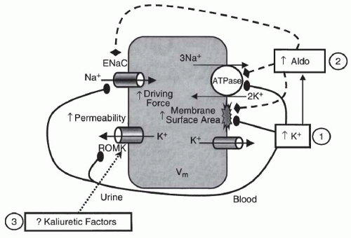
FIGURE 72.6 The major factors that regulate potassium secretion in principal cells. Sodium is reabsorbed across the luminal membrane through ENaC (epithelial sodium channels) with resultant cellular depolarization increasing the electrical driving force for potassium secretion through ROMK (potassium channels). The effects of aldosterone (Aldo) and hyperkalemia (↑K+) on potassium secretion are noted. (From Gennari FJ, Segal AS. Hyperkalemia: an adaptive response in chronic renal insufficiency. Kidney Int. 2002;62:1, with permission.)


Intestinal Potassium Excretion in Renal Failure

Patients with renal failure secrete more potassium in the stool than do normal controls.115,128,129 Net colonic secretion of potassium is increased significantly above control levels in rats with renal insufficiency.128 This increase is associated with an increase in Na-K-ATPase activity in colonic mucosa and is functionally similar to the increase seen with the administration of DOCA, glucocorticoids, or high dietary potassium.130 Although the rise in fecal potassium concentration is significant, the absolute amount of K+ lost through this route in patients with mild-to-moderate CKD is small and contributes only minimally to the external K+ homeostasis. In patients with advanced renal insufficiency (GFR < 5 to 10 mL per minute), however, up to 30% to 40% of the ingested potassium load may be excreted in the stool.129


Acid-Base Homeostasis in Renal Failure

The ability of the kidney to excrete a hydrogen ion is progressively diminished with the diminution of GFR. A significant decrease in serum bicarbonate does not usually occur until GFR falls below 25 to 30 mL per minute.75,131 Widmer and colleagues,132 in 41 ambulatory patients with CKD who had multiple electrolyte measurements over time, noted a serum bicarbonate reduction from 28 to 22 mEq per liter in patients with a moderate renal failure defined as a creatinine level of 2 to 4 mg per deciliter and a further reduction to 19 mEq per liter in patients with a creatinine level of 4 to 14 mg per deciliter. The anion gap remained unchanged in the first group and rose significantly with a further decrease in GFR. This study is criticized for the use of serum creatinine to define severity of renal failure rather than the use of a more accurate measurement of renal function. The concept of orderly progression of metabolic acidosis of renal failure from hyperchloremic to anion gap acidosis, however, occurs in the minority of patients. Wallia and colleagues133 studied the electrolyte pattern in 70 patients with ESRD just before dialytic therapy was begun. Five patterns were found: 14 patients with normal electrolytes; 14 with anion gap metabolic acidosis; 21 with hyperchloremic acidosis; 11 with mixed hyperchloremic and anion gap acidosis; and 10 with normal serum chloride, low serum bicarbonate, and normal anion gap. This last group, however, had the lowest serum sodium and therefore were relatively hyperchloremic. Therefore, among these 70 patients with ESRD, 31 (44%) had hyperchloremic acidosis, only 14 (20%) had classic anion gap acidosis, and interestingly, another 14 (20%) had normal electrolytes. Patients with an increased anion gap, however, had a slight but significantly higher serum creatinine than patients with pure hyperchloremic acidosis or with normal electrolytes (13.2 versus 10.0 versus 9.0 mg per deciliter, respectively). In addition, these two studies did not support the common impression that hyperchloremic acidosis occurs more often in patients with tubulointerstitial rather than glomerular disease.132,133 Interestingly, diabetic patients with moderately severe renal failure (GFR < 30 mL per minute) have recently been reported to have milder metabolic acidosis than nondiabetic patients with similar renal function.134

Renal tubular acidosis (RTA) defines a group of disorders characterized by the presence of metabolic acidosis out of proportion to the decrease in GFR. The hallmark of these disorders is the presence of significant metabolic acidosis with hyperchloremia and a normal anion gap. Renal tubular acidosis in patients with mild-to-moderate renal insufficiency is often associated with significant hyperkalemia and is discussed later in this chapter.


The Pathophysiology of Metabolic Acidosis in Chronic Kidney Disease

Many studies have shown that acid production in renal failure is normal, and therefore, uremic acidosis reflects a decrease in net acid excretion, defined as the difference between proton excretion in the form of titratable acid and
ammonium ion (NH4+) and bicarbonate excretion.135,136,137 Careful metabolic studies by Goodman and colleagues137 documented that patients with chronic renal failure have a daily bicarbonate deficit of approximately 13 to 19 mEq. It is notable that despite this persistent deficit, serum bicarbonate in patients with CKD after an initial drop remains stable over long periods of time.138,139 This is due chiefly to the buffering of excess hydrogen ions by bone buffers, including calcium carbonate.138


Renal Excretion of Bicarbonate

Several studies demonstrate that some patients with severe kidney disease have significant bicarbonate wasting.135,140,141,142,143,144 In an early study by Schwartz and coworkers,135 three out of four patients with renal failure had significant bicarbonaturia, which disappeared only after the fall of serum bicarbonate to below 20 mEq per liter. In a more detailed study in 17 uremic patients (serum creatinine of 5.6 to 18.9 mg per deciliter), the majority had significant bicarbonate wasting (fractional excretion of HCO3 of 0% to 17.56%) despite the presence of metabolic acidosis (serum HCO3 of 16 to 23 mEq per liter). After NH4Cl loading, serum bicarbonate decreased to below 14 mEq per liter, and bicarbonaturia disappeared in all but four patients.144 Interestingly, the bicarbonate wasting in these four patients also disappeared with the institution of a low-sodium diet.144 These two studies support the presence of a diminished maximal tubular reabsorption (Tm) for bicarbonate in the majority of patients with renal failure. Further, they demonstrate that the low Tm is partly responsive to volume status.

Arruda and colleagues143 and Wong and associates,145 working with a remnant kidney model in dogs with variable levels of volume expansion and serum bicarbonate, noted that the ratio of absolute bicarbonate to sodium reabsorption was increased in CKD. In addition, Wong and associates,145 using a micropuncture method, showed that this ratio was also higher at the beginning of the distal tubule, indicating avid bicarbonate absorption by the proximal tubule of the remnant kidney. Although absolute absorption was higher, the absolute amount of bicarbonate delivered to the distal tubule was also higher, reflecting the marked increase in filtered load per nephron owing to an increase in single nephron GFR.145 In summary, the whole kidney Tm for bicarbonate is, in general, diminished in CKD despite an absolute increase in bicarbonate resorption at the single nephron. The discrepancy in these findings may reflect the variation in the experimental designs and the role of nonvolume regulators in bicarbonate handling by the kidney.


Renal Excretion of Titratable Acid

The excretion of titratable acids chiefly reflects the amount of urinary phosphate and the urinary pH. Most CKD patients are able to maximally acidify their urine,135,146 and urine-serum PCO2, as a measure of hydrogen pump activity in the distal tubule, is normal.147 The amount of titratable acids in these patients is normal.137,138,139,140,141,142,143,144,145,146,147,148,149,150 This is primarily owing to an increase in the fractional excretion of phosphate initiated by secondary hyperparathyroidism. It should be noted, however, that urinary phosphate does decrease with severe renal failure. This reflects both a decrease in dietary phosphate as well as the effect of phosphate binders commonly used in these patients.


Renal Excretion of Ammonium

Although bicarbonaturia may contribute to metabolic acidosis, the major abnormality is a decrease in renal excretion of ammonium. Ammonium is primarily produced by the deamination of amino acids, chiefly glutamine, in the proximal tubule and, to a much lesser extent, in the loop of Henle and the distal convoluted tubule.130,131 This is reviewed in detail in Chapter 7, Renal Acid-Base Transport and will not be reviewed here. In CKD, fractional renal ammonium excretion initially increases by severalfold, thereby resulting in the maintenance of a normal absolute excretion rate.151 However, as the GFR decreases below 20 mL per minute, despite a maximal increase in fractional excretion of ammonium, the absolute excretory rate decreases significantly. Thus, progressive metabolic acidosis results. This decrease in the rate of ammonium excretion also reflects a decreased ability of the kidney to trap ammonia in the collecting duct.146 Warnock139 has suggested that the decrease in ammonia trapping in the remnant kidney model may be secondary to excess delivery of bicarbonate to the collecting duct, thereby resulting in an unfavorable environment for the diffusion and trapping of ammonia.

The role of aldosterone in ammonium excretion is complex. Aldosterone increases the rate of Na+-dependent and Na+-independent H+ secretion in the cortical and medullary collecting duct.152,153 Hypoaldosteronism is associated with a decrease in the rate of H+ secretion, whereas the ability to maintain a steep H+ gradient between urine and plasma, as measured by urinary pH and urine minus blood PCO2 in alkaline urine, is not affected.154,155 The decrease in the rate of H+ secretion is associated with a decrease in the availability of ammonium buffer in the urine that is not augmented appropriately in response to sodium sulfate infusion.156,157 Hypoaldosteronism is universally associated with a decreased potassium excretion and hyperkalemia. Hyperkalemia decreases renal ammonium excretion significantly. A decrease in accumulation of ammonium in the renal interstitium despite normal production by the proximal tubule underlies this effect.158 In the syndrome of hyperkalemic renal tubular acidosis, this mechanism probably plays the major role in the production of hyperchloremic acidosis seen early in the course of renal failure (Fig. 72.7).159 Reversal of hyperkalemia with sodium binding resin,160 mineralocorticoids,161 or low-potassium diet162 ameliorates the metabolic acidosis by increasing ammonium secretion.


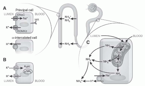
FIGURE 72.7 The factors involved in hyperkalemic acidosis. A: ENaC function at the apical surface of principal cells allows potassium secretion by ROMK (potassium channels) and the hydrogen ion by adjacent intercalated cells. B: Hyperkalemia increases intracellular pH by proton exchange, impairing the enzyme involved in ammoniagenesis. C: The process of ammoniagenesis involves deamination of glutamine, which allows ammonia to buffer the hydrogen ion in the urine. Ammonia and ammonium are reabsorbed in the medullary loop and are then excreted in the urine in the distal nephron. (From Karet FE. Mechanisms in hyperkalemic renal tubular acidosis. J Am Soc Nephrol. 2009;20:251, with permission.)

In summary, the metabolic acidosis develops universally in all patients with CKD as GFR decreases to below 20 mL per minute. The pathogenesis of this disorder is complex and reflects renal defect in both resorption as well as the generation of bicarbonate. The major mechanism, however, is in a decrease in absolute ammonia excretion despite the presence of acidosis.


HYPERKALEMIC RENAL TUBULAR ACIDOSIS OWING TO A DEFECT IN RENIN-ANGIOTENSIN-ALDOSTERONE AXIS OR TUBULAR UNRESPONSIVENESS TO ALDOSTERONE

Although a decrease in GFR may be associated with the development of significant hyperkalemia and hyperchloremic (HCA) or anion gap metabolic acidosis, this usually occurs only with severe reductions in GFR, below 15 to 20 mL per minute. However, some patients with underlying renal disease and mild-to-moderate azotemia present with striking hyperkalemia with or without HCA. The elevated serum potassium in these patients is primarily owing to a disturbance in the renin-angiotensin-aldosterone axis or to renal tubular responsiveness to aldosterone (see Fig. 72.6 and Table 72.2). Since the report by Hudson and associates,163 numerous cases have been described in which hyperkalemia with or without HCA developed in the presence of only mild-to-moderate renal insufficiency.164 The majority of these cases are diabetic or hypertensive nephropathy or chronic interstitial nephritis.165 In 1972, Schambelan and colleagues166 presented evidence linking hypoaldosteronism with hyporreninism in six patients with this syndrome. This association was verified in subsequent reports,167,168,169,170 and the entity became known as hyporeninemic hypoaldosteronism (HHA). However, it quickly became clear that a significant minority of these patients had normal renin levels. DeFronzo,171 in 1980, after reviewing 81 published cases, came to the conclusion that in 20% of cases the low plasma aldosterone levels could not be explained by renin deficiency, and therefore a primary abnormality in aldosterone synthesis had to be postulated. At the same time, some patients with sickle cell disease,172,173 systemic lupus erythematosus,174,175,176,177 and renal transplantation178,179,180 have a renal tubular secretory defect resulting in hyperkalemia despite a normal renin-aldosterone axis. Therefore, at the present time, these patients can be divided into two large categories: (1) hyperkalemia resulting from hypoaldosteronism with or without hyporreninism; and (2) hyperkalemia resulting from a primary renal tubular potassium secretory defect. One could consider this entity as a spectrum ranging from pure aldosterone deficiency with normal tubular responsiveness to severe tubular resistance with normal aldosterone secretion. Between these two extremes there are many overlapping presentations in which either the defect in the hormonal axis or the tubular responsiveness dominates. Although Table 72.2 summarizes all the hormonal or tubular defects that can lead to hyperkalemia,
often with HCA, our discussion is limited to the disturbances associated with renal insufficiency.


TABLE 72.2 Etiology of Chronic Hyperkalemia Due to Disturbances in Renal Potassium Excretion


I.


Decrease in GFR


A.


Acute renal failure


B.


Chronic kidney disease (GFR < 15-20 mL/min)


II.


Defect in renal tubular secretion of potassium


A.


Disturbance in the renin-angiotensin-aldosterone axis


1.


Hyporeninism: associated with renal insufficiency (diabetes mellitus, interstitial nephritis)


2.


Disturbance in angiotensin II activation or function (captopril, saralasin)


3.


Hypoaldosteronism


a.


With glucocorticoid deficiency (Addison disease, enzyme deficiency)


b.


Block in aldosterone synthesis (heparin, 18-methyloxidase deficiency)


c.


Primary hypoaldosteronism


B.


Tubular resistance to the action of aldosterone (renal tubular hyperkalemia)


1.


Pseudohypoaldosteronism


2.


Hyperkalemia, hypertension, and normal renal function


3.


Hyperkalemia with mild-to-moderate renal insufficiency and variable plasma aldosterone levels (sickle cell disease, systemic lupus erythematosus, renal transplant, obstructive uropathy, miscellaneous)


4.


Pharmacologic inhibition of the tubular action of aldosterone (spironolactone, eplerenone, triamterene, amiloride, pentamidine, trimethoprim) in distal nephron


GFR, glomerular filtration rate.


Hyperkalemic Renal Tubular Acidosis Owing to a Defect in Renin-Angiotensin-Aldosterone Axis

This group comprises approximately 80% of the patients with renal insufficiency and hyperkalemia.171,181,182,183 The hallmark of this group is a low plasma aldosterone concentration. The majority (80%) of this group also has low plasma renin activity (PRA) and therefore represents the classic syndrome of HHA. However, 20% have a normal PRA. Clinically and physiologically, these patients present with fairly uniform features. Several large series166,184 have defined the characteristics of these patients first summarized in a review by DeFronzo.171 These include: (1) a mean age of about 60 years, (2) the presence of diabetes mellitus in about 50%, (3) the presence of mild-to-moderate renal failure in the majority, and (4) a lack of symptoms referable to hyperkalemia in 75%. Physiologic features include: (1) low or low-normal baseline and/or stimulated aldosterone levels, (2) normal plasma cortisol, (3) low baseline and/or stimulated renin values in 80%, (4) normal aldosterone response to angiotensin or adrenocorticotropic hormone (ACTH) stimulation in the minority, (5) presence of hyperchloremic acidosis in well over 50%, and (6) a lack of significant salt wasting.

To gain an understanding of the physiologic basis of this syndrome, we initially review the defect in renin secretion and then summarize our present understanding of aldosterone deficiency in this syndrome.


Hyporreninism

At present, no single abnormality can explain the low PRA seen in 80% of these patients.171,182,183 Evidence has been presented in support of a defect in one or more physiologic regulators of renin secretion including volume, autonomic nervous system, serum potassium concentration, and prostaglandins.

Oh and colleagues,169 Perez and colleagues,185 and others186,187 have demonstrated that long-term sodium and volume depletion in these patients is associated with a significant increase in the PRA. However, comparable data in normal controls with the same degree of volume depletion were not provided. In the report of Oh and colleagues,169 after 3 to 6 weeks of salt depletion, the PRA rose into the normal range, but plasma aldosterone remained subnormal. In the study by Chan and coworkers, 8 of the 12 patients with hyporreninism responded to 2 weeks of furosemide with an increase in PRA without a similar response in plasma aldosterone.187 In a study of four patients with acute postinfectious glomerulonephritis,155 plasma renin and aldosterone concentrations were low during the acute phase, but returned to normal following recovery from acute nephritis. Interestingly, in two patients, the renin and aldosterone levels remained low during the acute phase despite an excellent response to diuretics. These two patients, however, responded appropriately to
physiologic doses of fludrocortisone. This study,155

Only gold members can continue reading. Log In or Register to continue

Stay updated, free articles. Join our Telegram channel

May 29, 2016 | Posted by in NEPHROLOGY | Comments Off on Disorders of Potassium and Acid-Base Metabolism in Association with Renal Disease

Full access? Get Clinical Tree

Get Clinical Tree app for offline access