Disorders of Phosphorus, Calcium, and Magnesium Metabolism


Disorders of Phosphorus, Calcium, and Magnesium Metabolism


Keith A. Hruska

Moshe Levi

Eduardo Slatopolsky


PHOSPHORUS

Phosphorus is a common anion ubiquitously distributed throughout the body. Approximately 80% to 85% of the phosphorus is present in the skeleton. The rest is widely distributed in the form of organic phosphate compounds that play fundamental roles in several aspects of cellular metabolism. The energy required for many cellular reactions including biosynthesis derives from hydrolysis of adenosine triphosphate (ATP). Organic phosphates are important components of cell membrane phospholipids. In the extracellular fluid (ECF), phosphorus is present predominantly in the inorganic form (Pi). The physiologic concentration of serum phosphorus ranges from 2.5 to 4.5 mg/dL (0.9 to 1.45 mmol/L) in adults.373 In serum, phosphorus exists mainly as the free ion, and only a small fraction (less than 15%) is protein bound.274,683 There is a diurnal variation in serum phosphorus of 0.6 to 1.0 mg/dL, with the nadir occurring between 8 am and 11 am.


Phosphorus Balance and Gastrointestinal Absorption

Approximately 1 g of phosphorus is ingested daily in an average diet in the United States. About 300 mg is excreted in the stool, and 700 mg is absorbed (Fig. 73.1). Most of the phosphorus is absorbed in the duodenum and jejunum with minimal absorption occurring in the ileum.204 Phosphorus transport in proximal segments of the small intestine appears to involve both passive and active components and to be under the influence of vitamin D. The movement of phosphorus from the intestinal lumen to the blood requires (1) transport across the luminal brush-border membrane of the intestine; (2) transport through the cytoplasm; and (3) transport across the basolateral plasma membrane of the epithelium. The rate-limiting step and the main driving force of absorption is the luminal membrane step.373


Intestinal Epithelial Luminal Membrane Transport

The mechanism of transport across the intestinal brush border epithelial membrane involves a sodium-phosphate (NaPi) cotransport system, NaPi-IIb.265 The NaPi cotransporters are a secondary active form of ion transport using the energy of the Na gradient from outside to inside the cell to move phosphate ion uphill against an electrochemical gradient (Fig. 73.2).

The intestinal NaPi-IIb transporter is upregulated by a low phosphate diet and 1,25-dihydroxyvitamin D3.258,314 Although low phosphate diets upregulate1,25-dihydroxyvitamin D3, studies in vitamin D-receptor (VDR) null mice indicate that the intestinal NaPi cotransport adaptation to a low phosphate diet occurs independent of vitamin D.585

Intestinal NaPi cotransport activity and NaPi-IIb protein is also regulated by several other factors—including the aging process,722 glucocorticoids,26 epidermal growth factor (EGF),723 and liver X receptor (LXR)106—that decrease intestinal NaPi transport, and estrogen724 and metabolic acidosis638 that increase intestinal NaPi transport.

Studies of phosphorus accumulation by rat intestinal brush-border vesicles have demonstrated that it is affected by the transmembrane potential, indicating that like the renal type IIa cotransporter, NaPi-IIa, the intestinal type IIb cotransporter, NaPi-IIb, is electrogenic.265 The Km(Pi) of NaPi-IIb is approximately 50 µm, similar to the renal transport protein. In contrast to the renal NaPi-IIa isoform, the intestinal NaPi-IIb cotransporter is less dependent on the pH level.


Transcellular Movement of Phosphorus

The second component of transcellular intestinal phosphorus transport involves the movement of phosphorus from the luminal to the basolateral membrane. Although little is known about the cellular events that mediate this transcellular process, evidence suggests a role for the microtubular microfilament system of intestinal cells.204 Microfilaments in the cell may be important in conveying phosphorus from the brush-border membrane to the basolateral membrane and may be involved in the extrusion of phosphorus at the basolateral membrane from the epithelial cell.


Phosphate Exit at Basolateral Membrane

Little is known about the mechanisms of phosphorus extrusion at the basolateral membrane of intestinal epithelial cells.
The electrochemical gradient for phosphorus favors movement from the intracellular to the extracellular compartment because the interior of the cell is electrically negative compared with the basolateral external surface. Therefore, the presumption has been that the exit of phosphorus across the basolateral membrane represents a mode of passive transport.321


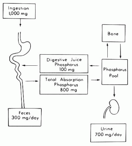
FIGURE 73.1 Summary of phosphorus metabolism in humans. Approximately 1 g of phosphorus is ingested daily, of which 300 mg is excreted in the stool and 700 mg in the urine. The gastrointestinal tract, bone, and kidney are the major organs involved in phosphorus homeostasis.


Renal Excretion of Phosphorus, Reabsorption

Most of the inorganic phosphorus in serum (90% to 95%) is ultrafiltrable at the level of the glomerulus. At physiologic levels of serum phosphorus, approximately 7 g of phosphorus is filtered daily by the kidney, of which 80% to 90% is reabsorbed by the renal tubules and the remainder is excreted in the urine (approximately 700 mg on a 1-g phosphorus diet) equal to intestinal absorption.332 As a result, adults are generally in balance between phosphorus intake and excretion (Fig. 73.1). Micropuncture studies have demonstrated that 60% to 70% of the filtered phosphorus is reabsorbed in the proximal tubule. However, there is also evidence that a significant amount of filtered phosphorus is reabsorbed in distal segments of the nephron.502 When serum phosphorus levels increase and the filtered load of phosphorus increases, the capacity to reabsorb phosphorus also increases. However, a maximum rate of transport (Tm) for phosphorus reabsorption is obtained usually at serum phosphorus concentrations of 6 mg per dL. There is a direct correlation between Tm phosphorus values and glomerular filtration rate (GFR) even when the GFR is varied over a broad range. Micropuncture studies suggest two different mechanisms responsible for phosphorus reabsorption in the proximal tubule. In the first third of the proximal tubule, in which only 10% to 15% of the filtered sodium and fluid is reabsorbed, the ratio of tubular fluid (TF) phosphorus to plasma ultrafiltrable (UF) phosphorus falls to values of approximately 0.6. This indicates that the first third of the proximal tubule accounts for approximately 50% of the total amount of phosphorus reabsorbed in this segment of the nephron. In the last two thirds of the proximal tubule, the reabsorption of phosphorus parallels the movement of salt and water. In the remaining 70% of the pars convoluta, the TF:UF phosphorous ratio remains at a value of 0.6 to 0.7, whereas fluid reabsorption increases to approximately 60% to 70% of the filtered load. Thus, in the last two thirds of proximal tubule, the TF:UF phosphorus reabsorption ratio is directly proportional to sodium and fluid reabsorption. A significant amount of phosphorus, perhaps on the order of 20% to 30%, is reabsorbed beyond the portion of the proximal tubule that is accessible to micropuncture. There is little phosphorus transport within the loop of Henle, with most transport distal to micropuncture accessibility occurring in the distal convoluted tubule. In this location, Pastoriza-Munoz et al.502 found that approximately 15% of filtered phosphorus is reabsorbed under baseline conditions in animals subjected to parathyroidectomy, but that the value falls to about 6% after administration of large doses of parathyroid hormone (PTH). The collecting duct is a potential site for distal nephron reabsorption of phosphorus.115,508,592 Transport in this nephron segment may explain the discrepancy between the amount of phosphorus delivered to the late distal tubule in micropuncture studies and the considerably smaller amount of phosphorus that appears in the final urine of the same kidney. Phosphorus transport in the cortical collecting tubule is independent of regulation by PTH. This is in agreement with the absence of PTH-dependent adenylate cyclase in the cortical collecting tubule.115


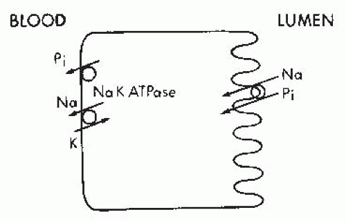
FIGURE 73.2 The apical membrane sodium-inorganic phosphate (Pi) cotransport proteins utilize the electrochemical driving force for sodium to move Pi into the cell. The electrochemical sodium gradient is maintained by active sodium extrusion across the basolateral membrane through the action of Na+-K+-ATPase.


Comparison of Superficial and Deep Nephron Transport

The contribution of superficial nephrons and deep nephrons of the kidney to phosphorus homeostasis differs.
Nephron heterogeneity in phosphorus handling has been evaluated under a number of conditions by puncture of the papillary tip and the superficial early distal tubule, with the recorded fractional delivery representing deep and superficial nephron function, respectively. Microinjection of phosphorus tracer into thin ascending and descending limbs of loops of Henle reveals that only 80% of phosphorus was recovered in the urine, whereas 88% to 100% of phosphorus was recovered when the tracer was injected into the late superficial distal tubule. It was concluded that a significant amount of phosphorus must be reabsorbed by juxtamedullary distal tubules or by segments connecting the juxtamedullary distal tubules to the collecting ducts to account for the discrepancy between the results of superficial nephron injection and juxtamedullary nephron injections. These data support an increased reabsorptive capacity for phosphorus in deep as opposed to superficial nephrons and increased responsiveness to body Pi requirements.253,254

In summary, phosphorus transport occurs in the distal nephron, particularly in the distal convoluted tubule and cortical collecting tubular system. This transport may be considerable under certain experimental conditions, but the importance of the terminal nephron system in day-to-day phosphorus homeostasis remains to be defined. It is also evident from data obtained from various micropuncture and microinjection studies that juxtamedullary and superficial nephrons have different capacities for phosphorus transport. The increased responsiveness of the deep nephrons to phosphorus intake suggests a key regulatory role for this system in phosphorus homeostasis.


Cellular Mechanisms of Phosphate Reabsorption in the Kidney

The apical membrane of renal tubular cells is the initial barrier across which phosphorus and other solutes present in the tubular fluid must pass to be transported into the peritubular capillary network. Because the electrical charge of the cell interior is negative to the exterior, and phosphorus concentrations are higher in the cytosol, phosphorus must move against an electrochemical gradient into the cell interior, whereas at the antiluminal membrane, the transport of phosphorus into the peritubular capillary is favored by the high intracellular phosphorus concentration and the electronegativity of the cell interior. Studies with apical membrane vesicles have demonstrated cotransport of Na+ with phosphate across the brush-border membrane, whereas the transport of phosphorus across the basolateral membrane is independent of that of Na+.272 The apical membrane Na+-phosphate cotransport protein (NaPi-IIa) energizes the uphill transport of phosphate across the brush-border membrane (BBM) by the movement of Na+ down its electrochemical gradient. The latter gradient is established and maintained by active extrusion of Na+ across the basolateral cell membrane into the peritubular capillary through the action of Na+-K+-ATPase (Fig. 73.2).571

Three families of NaPi cotransport proteins of the proximal tubule (types I, II, and III) have been cloned.414,472,473,636,661,709 The DNA clones encode 80- to 95-kd proteins that reconstitute Na+-dependent concentrative, or “uphill,” transport of phosphate.203,414,636 The type I cotransporter, Npt1/SLC17, is expressed predominantly in the renal proximal tubule, and it accounts for about 13% of the known NaPi cotransporter mRNA in the mouse kidney.662 Npt1 is not regulated by dietary Pi, and studies in Npt1-cRNA-injected oocytes revealed that it may function not only as a NaPi cotransporter but also as a chloride and organic anion channel.103

The type II cotransporter NaPi-II/SLC34 proteins are similar between several species including humans.472,473,661 In addition to NaPi-IIa, the predominant isoform in the renal proximal tubule, another isoform, NaPi-IIc, has been discovered.486,584,586,660 Nephron localization of NaPi-II proteins has been limited to the proximal tubule of superficial and deep nephrons (greatest in the latter, concordant with physiologic studies).472,473,661 Immunolocalization studies in renal epithelial cells demonstrated apical membrane and subapical membrane vesicle staining,472,473,661 suggesting that a functional pool of transporters is available for insertion into or retrieval from the BBM itself. This has been postulated to be a major mechanism of Pi transport regulation in response to acute changes in phosphorus, PTH, MEPE, and fibroblast growth factor 23 (FGF23) levels.33,472,473,609,661 The NaPi-II family is upregulated at message and protein levels by chronic feeding of low-Pi diets368,403 and downregulated at message and protein levels by PTH136,368,403 and dietary potassium deficiency.92

The type III NaPi cotransporters SLC20 were originally identified as retroviral receptors for gibbon ape leukemia virus (Glvr1) and rat amphotropic virus (Ram1).317 They are ubiquitously expressed, and they comprise about 1% of the known NaPi cotransporter mRNAs in the mouse kidney.662 Pit-2 protein is expressed in the apical membrane of the renal proximal tubule and the levels are regulated by dietary Pi,678 dietary potassium,92 and LXR.106

Studies of phosphorus exit across the basolateral membrane suggest that it is accompanied by the net transfer of a negative charge and occurs down a favorable electrochemical gradient via sodium-independent mechanisms.582


Proteins that Interact with the Type IIa and Type IIc Na/Pi Cotransporter Proteins

Several proteins with PDZ domains have been identified that interact with the NaPi-IIa and NaPi-IIc protein and are localized in the BBM or the subapical compartment (Fig. 73.3). PDZ domains are modular protein interaction domains that often occur in scaffolding proteins and bind in a sequencespecific fashion to the C-terminal peptide sequence or at times the internal peptide sequences of target proteins. These domains of approximately 90 amino acids are known by an acronym of the first three PDZ-containing proteins identified including the postsynaptic protein PSD-95/SAP90, the
Drosophila septate junction protein Discs-large, and the tight junction protein ZO-1.255,288,367,596


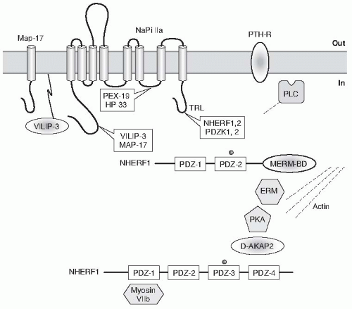
FIGURE 73.3 Interactions of NaPi-IIa with several proteins expressed in the renal proximal tubule. MAP-17 is involved in the apical location of PDZK1 (via PDZ-4) in OK cells. (From Biber J, Gisler SM, Hernando N, et al. PDZ interaction and proximal tubular phosphate reabsorption. Am J Physiol Renal Physiol. 2004;287:F871.)

PDZ domain containing proteins including NHERF-1, NHERF-2, PDZK1, CAL, and ZO-1 play an important role in: (1) the regulation of the expression and activity of renal proximal tubular BBM transport proteins including NHE-3,596,697,698,699,700 NaPiIIa,33,359 and NaPi-IIc219,677 and basolateral membrane transport proteins including Na-K-ATPase359 and Na-HCO3 cotransporter (NBC)696; (2) the regulation of the expression and activity of cystic fibrosis transmembrane conductance regulator (CFTR), a cAMP-regulated chloride channel and channel regulator389,477,540,653,686; (3) parathyroid hormone 1 receptor signaling417 and endocytic sorting of the β2-adrenergic receptor107 and platelet-derived growth factor receptor (PDGFR)313,436; (4) epithelial cell polarity and formation of tight junctions64,289; and (5) maintaining the integrity of the glomerular barrier to proteins through interactions with podocalyxin, negatively charged sialoprotein expressed on the surface of podocytes, the glomerular visceral epithelial cells.284,285,490,503,504,655

In addition to their interaction with membrane proteins and receptors, the PDZ domain-containing proteins also interact with the F-actin cytoskeleton through their interactions with the ezrin-radixin-moesin (ERM) proteins (Fig. 73.3).91,524,633 ERM proteins are typically located peripherally in the membrane and link the cytoplasmic tails of membrane proteins and receptors to the cortical actin cytoskeleton. The ERM proteins play an important role in the formation of microvilli, cell-cell junctions, and membrane ruffles and also participate in signal transduction pathways. The ERM proteins contain an F-actin binding site within their carboxy-terminal 30 residues. In addition, the ERM proteins have FERM (four-point one, ezrin, radixin, moesin) domains, which are generally located at or near the amino terminal, and act as multifunctional protein and lipid binding sites. The FERM domain of ezrin interacts strongly with NHERF-1 and NHERF-2. NHERF-1 and NHERF-2 have 2 PDZ domains and have a carboxy-terminal sequence of 30 amino acids that bind ezrin.

Using the molecular approach (yeast two-hybrid) several proteins with PDZ domains that interact with the C terminus of NaPi-IIa have been identified including: (1) NHERF-1/EBP50, (2) NHERF-2/E3KARP, (3) PDZK1/NaPi-Cap1, (4) PDZK2/NaPi-Cap2, and (5) CAL, a CFTRassociated ligand.60,68,220,221,222,263,456,516,594

Different studies suggest that apical expression of NaPi-IIa depends on the presence of NHERF-1. Expression of NaPi-IIa was reduced upon introduction of dominant-negative form of NHERF-1 in OK cells.220,221,222,263 The in vivo importance of the
NaPi-IIa interaction with NHERF-1 was also shown in a study where targeted disruption of the mouse NHERF-1 gene was associated with decreased BBM expression and increased intracellular localization of NaPi-IIa resulting in decreased renal phosphate reabsorption and renal phosphate wasting.595 On the other hand targeted disruption of NHERF-1 did not modulate the BBM expression and localization of NHE3; however, there was impaired regulation of NHE3 in response to PKA.595

In contrast to NHERF-1, targeted disruption of the PDZK1 gene failed to modulate the BBM expression of NaPi-IIa.108,334 NHERF-1, therefore, plays a critical and unique role in the renal proximal tubular apical membrane targeting of NaPi-IIa protein and maintenance of phosphate homeostasis. However targeted disruption of PDZK1modulates the BBM expression of NaPi-IIc, as compared to NaPi-IIa PDZK1, which has preferential interactions with NaPi-IIc.219,677

Recent studies have identified at least three additional proteins that may be important in the regulation of NaPi-IIa targeting and trafficking: (1) the peroxisomal protein PEX 19, a farnesylated protein that confers PTH responsiveness to NaPi-IIa297; (2) the calcium binding protein Vilip-3, a myristoleated protein that may be important in calcium dependent regulation of NaPi-IIa517; and (3) MAP 17 which may be important for apical expression of PDZK1 (Fig. 73.3).516


Factors that Affect the Urinary Excretion of Phosphorus

Of the multiplicity of factors that regulate phosphate transport in the kidney, the most important are dietary phosphate, PTH, and FG23.


Alterations in Dietary Phosphorus Intake

The mechanism by which the kidney modulates phosphorus excretion when dietary phosphorus is reduced or increased continues to be intriguing. Earlier micropuncture studies suggested that the most striking adaptive increase in phosphorus transport occurs in the proximal tubule. Later studies707 suggested that the entire nephron participates in the reduction of phosphorus excretion during dietary phosphorus deprivation. It has been shown that isolated perfused tubules obtained from rabbits that were fed a normal or low-phosphorus diet differ in their capacity to reabsorb phosphate. In normal animals, the proximal convoluted tubule (PCT) is capable of reabsorbing 7.2 ± 0.8 pmol/mL/min, whereas tubules obtained from phosphorus-deprived animals reabsorb 11.1 ± 1.3 pmol/mL/min. Conversely, animals that are fed a high phosphorus diet show reduced phosphorus reabsorption when the proximal tubules are perfused in vitro (2.7 ± 2.6 pmol/mL/min).

The effect of reduced dietary phosphorus to stimulate renal phosphorus transport is intrinsic to the renal tubular epithelium and occurs at the BBM Na+-phosphate cotransporter. The adaptation to phosphate supply by the sodium-phosphate cotransporter is biphasic.61,85,113 Incubation of cells in a low-phosphate medium result in a twofold increase in Na+-independent phosphate cotransport. The first phase of adaptation is observed rapidly (within 10 minutes) and is characterized by an increase in the Vmax of the transporter. This initial phase is independent of new protein synthesis.368,369,403 A slower phase resulting in a doubling of the phosphate transport rate, also through an increase in Vmax, occurs over several hours and is inhibited by blocking new protein synthesis.708 The adaptation to acute Pi deprivation occurs independent of de novo transcription and protein synthesis and is mediated by apical insertion of cytoplasmic NaPi-II a transporters by a microtubule dependent mechanism (Fig. 73.4).402 Secondly, through gene transcription and increased NaPi protein synthesis, additional units are produced and inserted into the brush border. Dietary Pi deprivation also induces the upregulation of NaPi-IIc and Pit-2 in the apical brush border membrane; however, the response of these two transporters is delayed and unlike NaPi-IIa may be dependent on de novo protein synthesis.677


Effects of Parathyroid Hormone on Phosphorus Reabsorption by the Kidney

Parathyroidectomy decreases urinary phosphorus excretion, whereas PTH administration increases phosphorus excretion.57,525 Micropuncture studies indicate that PTH inhibits phosphorus transport in the proximal tubule9 and in segments of the nephron located beyond the proximal tubule.502 TF:UF phosphorus ratio reaches a value of 0.6 by the S2 segment of the proximal tubule and, once achieved, this equilibrium ratio is maintained along the accessible portion of the proximal tubule. Within 6 to 24 hours after parathyroidectomy, the proximal TF:UF phosphorus ratio falls to a value of 0.2 to 0.4, indicating an increase in phosphorus reabsorption.41,43,707 TF phosphorus falls progressively with continuous fluid absorption along the length of the tubule, so by the end of the proximal tubule, the reabsorption of phosphorus is 70% to 85% of the filtered load, resulting in decreased phosphorus delivery to distal segments of the nephron. In the nonphosphorus-loaded, acutely parathyroidectomized animal, virtually all the distal load of phosphorus is reabsorbed by the distal nephron, reducing urinary phosphorus excretion to very low levels.333,358 In the phosphorus-loaded animal, the distal reabsorption of phosphorus increases until saturation is approached and urinary phosphorus excretion begins to rise. Acute administration of PTH to phosphorus-loaded parathyroidectomized dogs sharply lowers the distal reabsorption.

Administration of PTH in vivo results in decreased rates of Na+-dependent phosphorus transport in brush-border membrane vesicles isolated from the kidneys of treated rats.184,252 Intravenous infusion of dibutyryl cyclic adenosine monophosphate (cAMP) also decreased Na+-dependent phosphorus uptake in isolated brush-border vesicles, but neither PTH nor dibutyryl cAMP decreased phosphate transport when added directly to membrane vesicles.184 PTH stimulates two signaling pathways in proximal tubule cells: adenylate cyclase and phospholipase C (PLC), resulting in activation of protein
kinase A (PKA), and protein kinase C (PKC).174 The first pathway, activation of the adenylate cyclase, differs from that of PKC. Studies in OK cells show that PKA activation by PTH decreases the expression of NaPi-IIa cotransporter likely due to internalization and degradation of the transporter. Binding of PTH to its receptor leads to activation of PLC with the subsequent hydrolysis of phosphatidylinositol 4,5-bisphosphate to inositol 1,4,5-trisphosphate (IP3) and 1,2-diacylglycerol (DAG). IP3 generation leads to the release of intracellular calcium stores. DAG activates PKC.174 In addition to its direct effect on NaPi-IIa, PTH inhibits NaPi transport indirectly by inhibiting the Na+-K+-ATPase by decreasing the favorable Na+ gradient for Pi entry into the cell.546 Recent studies indicate that the activation of PKA and PKC signaling pathways by PTH also activates mitogen activated protein (MAP) kinase (MAPK) or extracellular receptor kinase (ERK1/2) which also induces inhibition of NaPi transport.32,360


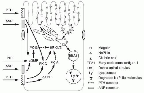
FIGURE 73.4 Mechanisms of NaPi-IIa traffic in the apical membranes of proximal tubular cells. Microvillar NaPi-IIa is retrieved in megalin containing clathrin coated vesicles. NaPi-IIa moves from the vesicles into an endosomal pool marked by EEA-1, whereas megalin is recycled through dense apical tubules back to the microvilli. The endosomal NaPi-IIa is targeted for lysosomal degradation. The process of NaPi-IIa retrieval and lysosomal degradation is stimulated by several factors (parathyroid hormone, atrial natriuretic peptide, nitric oxide) whose mechanisms of signal transduction (PK-A, PK-C, PK-G) merge in activation of ERK1/2 that modulates the process. (From Bacic D, Wagner CA, Hernando N, et al. Novel aspects in regulated expression of the renal type IIa Na/Pi-cotransporter. Kidney Int. 2004;66:S5, with permission.)

Measurement studies of in vivo renal reabsorption of phosphorus and calculations of kinetic parameters of Na+-dependent phosphorus transport in membrane vesicles isolated from the renal brush-border membranes of normal dogs, parathyroidectomized dogs, dogs fed a low-phosphorus diet, and dogs receiving human growth hormone were performed.252,281 The latter three groups of dogs had greater baseline values for absolute tubular reabsorption of phosphorus compared with normal dogs. Na+-dependent phosphate transport in BBM vesicles isolated from kidneys of these dogs was significantly increased compared with transport in brush-border vesicles from kidneys of normal dogs. Administration of PTH decreased significantly the apparent Vmax for Na+-dependent phosphorus transport in BBM vesicles isolated from kidneys of each of the four groups of dogs. The apparent Km (intrinsic binding affinity) for Na+-dependent phosphorus transport was not significantly changed by experimental maneuvers. Absolute tubular reabsorption of phosphorus measured in vivo was decreased by administration of PTH in each group of dogs with the exception of the dogs fed a low-phosphorus diet.252,281 Thus, alterations in phosphorus reabsorption measured in vivo were paralleled by alterations in Na+-dependent phosphorus transport in isolated membrane vesicles, and the administration of PTH in vivo resulted in altered transport characteristics of the isolated BBMs.

The cloning of the NaPi-IIa cotransport proteins did not completely elucidate the mechanisms of PTH action on phosphate transport. Because the phosphaturic effect of PTH can be reproduced by analogs of cAMP, the intracellular mechanism of phosphate transport regulation is thought to involve the cAMP/PKA signal pathway. However, the NaPi-IIa transport proteins are not characterized by a PKA-mediated phosphorylation site.259 Phosphorylation of BBM proteins in vitro occurs in parallel with inhibition with NaPi-IIa cotransport.252 Parathyroidectomy of rats causes a twofold to threefold increase in the NaPi-IIa protein content of BBM vesicles.319 Immunocytochemistry reveals the increase in protein exclusively in apical BBMs of proximal tubules. PTH treatment of parathyroidectomized rats for 2 hours decreased
protein levels and decreased the abundance of NaPi-IIa-specific messenger RNA (mRNA) by 31%.319 Parathyroidectomy did not affect NaPi-IIa mRNA levels. The effects of PTH were apparent within 2 hours of administration and indicate that PTH regulation of NaPi-IIa is determined by changes in the expression of NaPi-IIa protein in the renal BBMs.319

PTH decreases the NaPi-IIa protein content of the apical membrane by a endocytic retrieval pathway which is megalin and myosin VI dependent (Fig. 73.4).31,67 In megalin intact mice or rats, following treatment with PTH NaPi-IIa is internalized via clathrin-coated pits, and NaPi-IIa is then delivered to early endosomes and eventually to the lysosomes where the protein is degraded. At the present time unlike some of the other proximal tubular transport proteins or receptors, there is no evidence that the NaPi-IIa protein is present at the recycling endosomes.

PTH also regulates the NaPi-IIc protein content of the apical membrane by an endocytic retrieval pathway that is myosin VI-dependent, but the time course is delayed compared to NaPi-IIa.351


Fibroblast Growth Factor 23

Through studies of familial hypophosphatemia and tumor induced osteomalacia, a new hormone operating in a systems biology network regulating phosphate homeostasis between the skeleton and the kidney has been discovered (Fig. 73.5).307,600 The hormone is FGF23, secreted by skeletal osteocytes in response to changes in bone formation and serum phosphorus.487 The physiology is that the osteocyte monitors deposition of phosphorus into the skeletal reservoir and the saturation of the exchangeable phosphorus pool. When Pi levels increase within the pool due to decreased exit into the skeleton (bone formation) or due to increased plasma phosphorus, osteocytes secrete FGF23, which acts on the renal proximal tubule to decrease reabsorption and increase phosphorus excretion.279,604


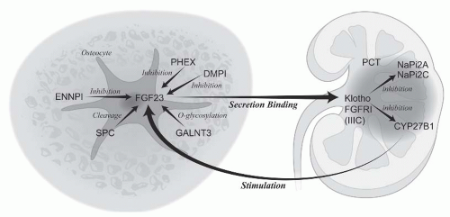
FIGURE 73.5 The skeletal-kidney endocrine axis: regulation of Pi in the exchangeable pool. FGF23 is a hormone secreted by the osteocyte to regulate proximal tubular cell Pi transport and calcitriol production. Multiple mechanisms of increased FGF23 levels cause hypophosphatemic rickets. Calcitriol stimulates FGF23 to maintain Pi levels in response to stimulation of intestinal absorption. Function of various proteins in the endocrine axis and the genetic diseases they cause are listed in Table 73.2.

FGF23 actions at the proximal tubule (PCT) have not been studied as extensively as the actions of PTH, which were described above. Studies indicate that FGF23 acts to decrease expression of NaPi-IIa and NaPi-IIc in the PCT.209,604 The actions of FGF23 on the PCT are mediated through binding to a FGF receptor—predominantly FGFR1(IIIc) and a coreceptor, Klotho.673 Signal transduction is stimulated through phosphorylation of extracellular signal-regulated kinase (ERK) and the immediate early response gene, early growth response-1 (Erg-1), a zinc finger transcription factor.212,673 A matter of current uncertainty is related to KLOTHO expression which is mainly in the renal distal tubule whereas its signaling function is in the PCT. Recent studies have shown that it is expressed in the PCT which would resolve this issue (M. Kuro-o, personal communication).

Besides inhibiting PCT renal Pi transport, FGF23 signaling inhibits PCT CYP27B1, the 25-OH cholecalciferol 1α hydroxylase, and activates 24-OH hydroxylase (CYP24R1) resulting in decreased production and increased catabolism of calcitriol.600 In addition, activation of 24-OH hydroxylase results in the high prevalence of vitamin D deficiency associated with elevations of FGF23 levels, especially in chronic kidney disease (CKD). Calcitriol increases FGF23, closing the feedback loop in the system (Fig. 73.5).


Vitamin D

Controversy still surrounds the regulatory role of vitamin D in renal phosphorus handling. Several studies have demonstrated that the chronic administration of vitamin D to parathyroidectomized animals is phosphaturic.75,467,647 Conversely, other investigators reported that vitamin D acutely stimulates proximal tubular phosphorus transport in both
parathyroidectomized and vitamin D-depleted rats.211 A unifying interpretation of these studies was hampered by the fact that the dosages of vitamin D administered and the status of the serum calcium, phosphorus, and PTH varied considerably from study to study.

Liang et al.384 administered 1,25-dihydroxycholecalciferol to vitamin D-deficient chicks and subsequently examined the transport characteristics of isolated renal tubule cells. Three hours after the in vivo administration of vitamin D, phosphorus uptake by the cells was significantly increased, whereas 17 hours after the administration of vitamin D, phosphorus uptake was reduced. The serum phosphorus concentration, however, was significantly increased at 17 hours after administration, and administration of phosphorus to vitamin D-depleted animals so their serum phosphorus levels were comparable to those of the 17-hour vitamin D-replenished group resulted in a similar decrease in phosphorus uptake.384 In response to in vitro preincubation with as little as 0.01 pm of 1,25-dihydroxycholecalciferol, renal cells isolated from vitamin D-deficient chicks demonstrated a specific increase in sodium-dependent phosphorus uptake, which was blocked by pretreatment with actinomycin D. The stimulatory effect was relatively specific for 1,25-dihydroxycholecalciferol, and kinetic analysis indicated that the Vmax of the phosphorus transport system was increased, whereas the affinity of the system for phosphorus was unaffected.384

Kurnik and Hruska346 also examined the relationship between vitamin D and renal phosphorus excretion in a normocalcemic, normophosphatemic weanling rat model fed a vitamin D-deficient diet. The animals were mildly vitamin D deficient (92 pg per mL of 1,25-dihydroxycholecalciferol versus 169 pg per mL in controls) but had no evidence of secondary hyperparathyroidism. Clearance studies performed in the basal partially vitamin D-deficient state showed an increase in both absolute and fractional phosphorus excretion compared with controls. Animals that were replenished with 1,25-dihydroxycholecalciferol and maintained on diets designed to protect against the development of hyperphosphatemia demonstrated a significant decrease in urinary phosphorus excretion. Other animals were similarly replenished with vitamin D but did not receive dietary adjustment; and in this group, both the serum phosphorus and the urinary phosphorus excretion level increased significantly. A third group was fed a normal diet and received smaller doses of 1,25-dihydroxycholecalciferol (15 pmol per g of body weight) for shorter periods, and although this dose had no effect on the serum phosphorus concentration, the phosphaturia was completely resolved.

Studies on BBM vesicles prepared from these animals revealed that in the partially vitamin D-deficient state, sodium-dependent phosphorus uptake was significantly reduced compared with control animals. Animals that were replenished with vitamin D and fed a controlled diet had a greater sodium-dependent phosphorus uptake than both vitamin D-depleted and vitamin D-replenished animals not maintained on controlled diets.

The results of this series of studies suggest that the primary action of 1,25-dihydroxycholecalciferol is to increase tubular phosphorus reabsorption. Long-term administration of vitamin D, however, represents a more complex situation, where phosphaturia may occur secondary to changes in the filtered load of phosphorus, in the body distribution of phosphorus, or in intracellular phosphorus activity.


Effects of Changes in Acid-Base Balance on Phosphate Excretion

The effect of acid-base status on the renal excretion and transport of phosphate is complex. Acute respiratory acidosis increases and acute respiratory alkalosis decreases phosphate excretion.275 These effects occur independently of PTH and plasma or luminal bicarbonate levels.275 However, other studies suggest that the effects of respiratory acid-base changes may be mediated by changes in plasma phosphate.275

Acute metabolic acidosis has minimal effects on phosphate excretion; however, the phosphaturic effect of PTH is blunted.43 Acute metabolic alkalosis causes an increase in phosphate excretion independently of PTH.206,345,448,526,530 This effect is due, in part, to volume expansion produced by the infusion of bicarbonate.448,530

Chronic acidosis increases phosphate excretion, again independent of PTH or changes in ionized Ca2+.144,234,345,509 The effect appears to be directly on the sodium-dependent phosphate transport mechanism.674 Chronic alkalosis decreases phosphate excretion, probably by the same mechanism as acidosis, operating in the opposite direction.206,535

Acute and chronic acidosis in rats decreases the proximal tubule cell luminal membrane expression of the NaPi-IIa cotransporter.18 In acute acidosis, there is rapid internalization of the transporter and the total cortical homogenate cotransporter expression is unchanged. In chronic acidosis, there are parallel changes in NaPi-IIa protein and mRNA abundance. The effects of acid-base perturbations are complex and depend on antecedent dietary intake, the chronicity of the change, and whether the change affects luminal or intracellular pH, or both.


Adrenal Hormones

Administration of pharmacologic amounts of cortisol leads to phosphaturia. Acute adrenalectomy reduces the GFR and increases the reabsorption of phosphorus in the proximal tubule. Frick and Durasene195 concluded that glucocorticoid hormones could play an important role in the regulation of fractional reabsorption of phosphorus. Indeed administration of glucocorticoids to animals has been shown to decrease proximal tubular NaPi cotransport activity and induce phosphaturia by causing parallel decreases in NaPi-IIa protein and mRNA levels.370 The inhibitory effects of glucocorticoids in NaPi transport is in part mediated by the concomitant alterations in renal proximal tubular apical BBM glycosphingolipid composition, as inhibition of glycosphingolipid synthesis prevents in part the decrease in renal NaPi cotransport activity. In contrast to glucocorticoid administration, adrenalectomy
with mineralocorticoid administration, resulting in selective glucocorticoid deficiency, is associated with increased renal proximal tubular NaPi-IIa protein expression.396


Growth Hormone

An increase in serum phosphorus and a rise in renal phosphorus transport are characteristics of growth hormone (GH) excess during the period of rapid growth in the child, during acromegaly, or during exogenous GH administration to experimental animals. Hammerman et al. reexamined this phenomenon in the BBM vesicle preparation in the dog252 and demonstrated that GH treatment resulted in an increased sodium-dependent phosphorus transport. These data reassert the importance of BBM phosphorus uptake in regulating overall renal phosphorus reabsorptive capacity. The action of growth hormone is likely mediated by insulin-like growth factor-1.114

Studies have further identified the nephron sites and mechanisms by which GH regulates renal Pi uptake.716 Micropuncture experiments were performed after acute thyroparathyroidectomy in the presence and absence of PTH in adult (14- to 17-week-old), juvenile (4-week-old), and GH-suppressed juvenile male rats. Although the phosphaturic effect of PTH was blunted in the juvenile rat compared with the adult, suppression of GH in the juvenile restored fractional Pi excretion to adult levels. In the presence or absence of PTH, GH suppression in the juvenile rat caused a significant increase in the fractional Pi delivery to the late proximal convoluted (PCT) and early distal tubule, so that delivery was not different from that in adults. These data were confirmed by Pi uptake studies into BBM vesicles. Immunofluorescence studies indicate increased BBM type IIa NaPi cotransporter (NaPi-II) expression in the juvenile compared with adult rat, and GH suppression reduced NaPi-II expression to levels observed in the adult. GH replacement in the [N-acetyl-Tyr(1)-d-Arg(2)]-GRF-(1-29)-NH(2)-treated juveniles restored high NaPi-II expression and Pi uptake. Together, these novel results demonstrate that the presence of GH in the juvenile animal is crucial for the early developmental upregulation of BBM NaPi-II and, most importantly, describe the enhanced Pi reabsorption along the PCT and proximal straight nephron segments in the juvenile rat.716


Thyroid Hormone

Because Pi is intensively used in general metabolism, Pi homeostasis should be regulated by factors controlling the rate of metabolism itself. One such factor is thyroid hormone, and its role in Pi reabsorption regulation has been extensively analyzed.45,637,729 Pharmacologic doses of T3 have been shown to increase Na/Pi cotransport in BBM vesicles from rat proximal tubules.54,729 In addition, T3 concentrations approximating the association constant (Km) of the thyroid hormone nuclear receptor also elicited a similar increase in Pi transport in opossum kidney (OK) cells.637 In both cases, the increase in transport rate was caused by an increase in the capacity of the transport system, whereas the affinity was not modified. Euzet et al.182,183 have shown an important role for T3 in the maturation of the renal Na/Pi cotransporter, which was associated with changes in both Km and Vmax, as well as in the type II Na/Pi cotransporter (NaPi-II) protein and mRNA abundance.

Sorribas et al. have determined the role of physiologic concentrations of thyroid hormone in renal phosphate transport in vivo. In addition, they also determined the potential role of thyroid hormone in impairment of phosphate reabsorption that accompanies the aging kidney.16 Their results show that chronically treated hypothyroid rats, using a physiologic dose of T3, exhibit increases in serum Pi levels, NaPi-II mRNA and protein content, and Na/Pi cotransport activity in superficial and juxtamedullary renal cortex, all these effects by means of enhanced transcription of the corresponding NaPi-II gene. The stimulatory effect of the hormone was less evident in the aging kidney, which shows a lower level of basal phosphate reabsorption. In this study, only pharmacologic hyperthyroidism was able to restore partially the level of serum Pi observed in young animals.


Epidermal Growth Factor

Epidermal growth factor (EGF) is a 53-amino acid polypeptide. The kidney is a major site of synthesis of the EGF precursor, prepro-EGF. In renal epithelial cells grown in culture, EGF has been shown to modulate sodium gradientdependent phosphate transport (Na-Pi cotransport) activity, but the directionality of the modulation in cell culture has been controversial.22,232,510 A study also determined whether EGF regulates Na-Pi cotransport activity in vivo and whether the effect of EGF to regulate Na-Pi cotransport is dependent on the developmental stage of the animal (i.e., suckling [12-dayold] vs. weaned [24-day-old] rats). This study demonstrated that proximal tubule BBMV Na-Pi cotransport activity, Na-Pi-II protein abundance, and NaPi-II mRNA abundance are higher in weaned than in suckling rats and that EGF inhibits Na-Pi cotransport activity in BBMV isolated from suckling and weaned rats by a decrease in NaPi-II protein abundance, in the absence of a change in NaPi-II mRNA.23


Aging

The aging process is associated with impairment in renal tubular reabsorption of Pi and renal tubular adaptation to a low Pi diet. In experiments using 3- to 4-month-old young adult rats and 12- to 16-month-old aged rats, it was found that there was an age-related twofold decrease in proximal tubular apical BBM Na-Pi cotransport activity, which was associated with similar decreases in BBM NaPi-II protein abundance and renal cortical NaPi-II mRNA level. Immunohistochemistry showed lower NaPi-II protein expression in the BBM of proximal tubules of superficial, midcortical, and juxtamedullary nephrons. This study also found that in response to chronic (7 days) and/or acute (4 hour) feeding of a low Pi diet, there were similar adaptive increases in BBM Na-Pi cotransport activity and BBM NaPi-II protein abundance in both young and aged rats. However, BBM Na-Pi cotransport activity and BBM NaPi-II protein abundance were still
significantly lower in aged rats, in spite of a significantly lower serum Pi concentration in aged rats. Thus, impaired expression of the type II renal Na-Pi cotransporter protein at the level of the apical BBM plays an important role in the age-related impairment in renal tubular reabsorption of Pi and renal tubular adaptation to a low Pi diet.635


Stanniocalcin

Stanniocalcin is a calcium and phosphate regulating hormone found in serum and the kidney. In teleost fish, it is produced in the corpuscles of Stannius, specialized endocrine organs closely associated with the kidneys. Stanniocalcin plays a major role in the calcium and phosphate homeostasis of fish. It inhibits calcium uptake by the gills and gut and stimulates phosphate reabsorption by the kidney.151,157,294,406,413,489,680,733

Two mammalian homologues of Stanniocalcin have been identified. Stanniocalcin 1 and Stanniocalcin 2, with seemingly opposing effects on renal phosphate transport. Stanniocalcin I induces increased gastrointestinal (GI) and renal Pi transport.295 Stanniocalcin 2 on the other hand causes inhibition of renal Pi transport by transcriptional mechanisms.295


Diuretics

Acetazolamide inhibits phosphate reabsorption by its effects on proximal tubule decreases in Na+-dependent bicarbonate transport, essential for the maintenance of the Na+ gradient. Furosemide inhibits carbonic anhydrase activity and thus decreases phosphate transport. Similar effects have been demonstrated with the administration of large doses of thiazide diuretics.246


Hypophosphatemia

Hypophosphatemia refers to serum phosphorus concentrations of less than 2.5 mg per dL. Hypophosphatemia usually results from one or a combination of the following factors (Fig. 6)328,340: (1) increased excretion of phosphorus in the urine; (2) decreased GI absorption of phosphorus; or (3) translocation of phosphorus from the extracellular to the intracellular space. The major causes of hypophosphatemia are shown in Table 73.1.


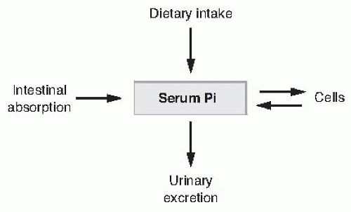
FIGURE 73.6 The major determinants of serum inorganic phosphate (Pi) concentration.


TABLE 73.1 Causes of Hypophosphatemia


I. Increased excretion of phosphorus in the urine


A. Familial


B. Primary hyperparathyroidism


C. Secondary hyperparathyroidism


D. Renal tubular defects (Fanconi syndrome)


E. Diuretic phase of acute tubular necrosis


F. Postobstructive diuresis


G. Extracellular fluid volume expansion


1. X-linked hypophosphatemia


2. Autosomal dominant hypophosphatemic rickets


3. Autosomal recessive hypophosphatemic rickets 1; autosomal recessive hypophosphatemic rickets 2


4. Oncogenic hypophosphatemic osteomalacia (TIO) – Phos


5. McCune-Albright syndrome/Fibrous dysplasia


6. Mutations in NaPi-IIa


7. Hereditary hypophosphatemic rickets with hypercalciuria


H. Posttransplant hypophosphatemia


II. Decrease in gastrointestinal absorption of phosphorus


A. Abnormalities of vitamin D metabolism


1. Vitamin D-deficient rickets


2. Familial


a. Vitamin D-dependent rickets


b. X-linked hypophosphatemia


B. Malabsorption


C. Malnutrition-starvation


III. Miscellaneous causes/translocation of phosphorus


A. Leukemia, lymphoma


B. Diabetes mellitus: during treatment for ketoacidosis


C. Severe respiratory alkalosis


D. Recovery phase of malnutrition


E. Alcohol withdrawal


F. Toxic shock syndrome


G. Severe burns


Increased Excretion of Phosphorus in the Urine

Several pathophysiologic conditions increase excretion of phosphorus in the urine. Some of these are characterized by elevated levels of circulating PTH or FGF23. Because PTH and FGF23 decrease phosphorus reabsorption by the kidney, elevations of the hormones increase urinary excretion (Table 73.1). Decreased tubular reabsorption of phosphorus may also occur without increased levels of PTH and may
be due to changes in the reabsorption of salt and water or to renal tubular defects specific for the reabsorption of certain solutes or phosphorus. Hypophosphatemia may also occur in the diuretic phase of acute tubular necrosis or in postobstructive diuresis, presumably due to a combination of high levels of PTH and decreased tubular reabsorption of salt and water.


Primary Hyperparathyroidism

Primary hyperparathyroidism is a common entity in clinical medicine.27 PTH is secreted in excess of the physiologic needs for mineral homeostasis due to either adenomas or hyperplasia of the parathyroid glands.54 This results in decreased phosphorus reabsorption by the kidney. The losses of phosphorus in the urine result in hypophosphatemia. The degree of hypophosphatemia may vary considerably among patients, because mobilization of phosphorus from bone will in part mitigate the hypophosphatemia. Moreover, if the patient ingests large amounts of dietary phosphorus, the degree of hypophosphatemia observed may be mild. Because these patients also have elevated levels of serum calcium, the diagnosis is made relatively easy in most cases by the finding of elevated levels of immunoreactive PTH.


TABLE 73.2 Inherited Disorders of Phosphate Homeostasis Cause Hypophosphatemic Rickets or Hyperphosphatemia and Are Components of a Bone Kidney Endocrine Axis


Protein/Gene


Function


Disease


FGF23


Hormone regulating phosphate excretion and calcitriol production


ADHR: excess tumoral calcinosis: deficiency


PHEX


Unclear, inhibits FGF23 secretion/production


XLH


DMPI


Matrix protein, inhibits FGF23 secretion/production


ARHR1


ENNPI


Produces pyrophosphate in osteocyte/osteoblast extracellular fluid/inhibits FGF23 secretion/production


ARHR2: homozygous infantile calcific arteriopathy: homozygous


GALNT3


O-glycosylation of FGF23, deficiency increases SPC mediated FGF23 cleavage


Tumoral calcinosis


KLOTHO


FGF23 co-receptor


Tumoral calcinosis


Hyperphosphatemia


Early senescence


NaPi2c/SLC34A3


Proximal tubule phosphate transport protein


HHRC


CYP27BI


Produces calcitriol which stimulates FGF23 production


Vitamin D dependent rickets


NaPi2a/SLCH34A1


Proximal tubule phosphate transport protein


Nephrolithiasis


FGF23, fibroblast growth factor 23; ADHR, autosomal dominant hypophosphatemic rickets; PHEX, phosphate regulating gene with homologies to endopeptidases on the X chromosome; XLH, x-linked hypophosphatemia; DMPI, dentin matrix protein 1; ARHR1, autosomal recessive hypophosphatemic rickets 1; ENNPI, ectonucleotide pyrophosphatase 1; ARHR2, autosomal recessive hypophosphatemic rickets 2; GALNT3, N- acetylglucosaminyltransferase 3; SLC34A3, solute carrier family 34A3; HHRC, hereditary hypophosphatemic rickets with calciuria; CYP27BI, cytochrome P450 family 27 subfamily B polypeptide I.


Secondary Hyperparathyroidism

Although secondary hyperparathyroidism is present in most patients with chronic renal disease, hyperphosphatemia rather than hypophosphatemia occurs in such patients because of decreased phosphorus excretion in the urine resulting from the fall in GFR. However, certain conditions characterized by malabsorption of calcium from the GI tract may produce hypocalcemia, leading to development of secondary hyperparathyroidism.224 The elevated levels of PTH will decrease phosphorus reabsorption by the kidney, resulting in hypophosphatemia. Thus, patients with GI tract abnormalities resulting in calcium malabsorption and secondary hyperparathyroidism will have low levels of serum calcium and phosphorus. In these patients, the hypocalcemia is responsible for the increased release of PTH. In addition, decreased intestinal absorption of phosphorus as a result of the primary GI tract disease may also contribute to the decrement in the levels of serum phosphorus. In general, these patients have
urinary losses of phosphorus that are out of proportion to the hypophosphatemia in contrast to patients with predominant phosphorus malabsorption and no secondary hyperparathyroidism in whom urinary excretion of phosphorus is low.


Familial Hypophosphatemia

Studies of hereditary (Table 73.2), and acquired (oncogenic hypophosphatemic osteomalacia [OHO] and McCune-Albright syndrome) renal phosphate wasting disorders have led to the identification of novel genes involved in the regulation of renal Pi transport and calcitriol synthesis.70,87,536,537,561,579,659 The discovery of these genes has established a bone-kidney axis responsible for maintaining phosphate homeostasis (Fig. 73.5).


X-linked Hypophosphatemia

X-linked hypophosphatemia (XLH) is a common cause of rickets with a prevalence of approximately 1 in 20,000. It is inherited in an X-linked dominant manner. Manifestations of XLH include short stature, bone pain, tooth abscesses, calcification of tendon insertions, ligaments, joint capsules, and lower extremity deformities. However, with genetic sequencing causing reclassification of some presumed XLH patients into autosomal dominant hypophosphatemic rickets (ADHR) or autosomal recessive hypophosphatemic rickets (ARHR) categories, the clinical manifestations may change. For example, posterior longitudinal ligament ossification may be a manifestation mainly of ARHR2.379 XLH is characterized by hypophosphatemia, decreased reabsorption of phosphorus by the renal tubule, decreased absorption of calcium and phosphorus from the GI tract, and varying degrees of rickets or osteomalacia. Patients with this disorder exhibit normal to reduced levels of 1,25-dihydroxycholecalciferol despite hypophosphatemia and reduced Na-phosphate transport in the proximal tubule. The message levels of NaPi-IIa are reduced by 50% in the PCT of Hyp mice similar to the reduction in apical membrane vesicle NaPi-IIa protein levels.663

The gene responsible for XLH was designated PHEX for PHosphate regulating gene with homology to Endopeptidases on the X chromosome.3 PHEX is a member of the M13 family of type II cell surface zinc-dependent metallopeptidases which includes neprilysin, endothelin-converting enzymes 1 and 2, KELL, and DINE/X-converting enzyme. The mouse PHEX DNA sequence is highly homologous to that of humans and the inactivating mutations of PHEX have been identified in the mouse homologues of XLH, Hyp and Gy mice. More than 180 mutations in the PHEX gene have been shown to result in XLH. PHEX is expressed predominantly in osteoblasts, osteocytes, and odontoblasts, but not in the kidney. The hypophosphatemia and rickets of XLH is produced by excess FGF23 in the circulation.307,726 How PHEX inactivation variably increases FGF23 secretion is currently unknown (Fig. 73.5). However, the finding that homozygous ablation of FGF23 in the Hyp background produced the phenotype of FGF23 deficiency (hyperphosphatemia, elevated calcitriol, and vascular calcification) and loss of the Hyp phenotype demonstrates that FGF23 is causative of hypophosphatemia in Hyp and XLH.619 The initial thought that FGF23 was a PHEX substrate84 proved untrue.47,244,392,393 In fact, FGF23 has recently been shown to be cleaved by subtilisin-like protein convertases (SPC) (Fig. 73.5). SPCs are a seven-member family of calcium-dependent serine proteases responsible for the processing of peptide hormones, neuropeptides, adhesion molecules, receptors, growth factors, cell surface glycoproteins, and enzymes. Instead PHEX, most likely through the actions of unidentified PHEX substrates or other downstream effectors, regulates FGF23 secretion as part of a hormonal axis between bone and kidneys that controls systemic phosphate homeostasis and mineralization.47

From a therapeutic point of view, the combination of neutral phosphate and 1,25-dihydroxycholecalciferol has led to an improvement in the bone disease of patients with XLH and in the Hyp mice.225,676 The administration of phosphorus in X-linked hypophosphatemia is usually divided into four doses, with the total amount ranging between 1 to 4 g per day. Pharmacologic doses of 1,25-dihydroxycholecalciferol on the order of 1 to 3 µg per day may be necessary to correct the skeletal alterations. 1,25-dihydroxycholecalciferol does not correct the increased fractional excretion of phosphate. The enthusiasm for this regimen is tempered by a high incidence of nephrocalcinosis and occasional renal failure.196,225,676


Autosomal Dominant Hypophosphatemic Rickets

The clinical presentation of ADHR is similar to XLH. However, ADHR exhibits male to male transmission, consistent with autosomal dominant inheritance, and is characterized by incomplete penetrance and variable age of onset. Adults typically complain of bone pain, fatigue, and/or weakness, and can present with stress fractures or pseudofractures. Renal phosphate wasting and inappropriately normal serum calcitriol levels are the most typical laboratory findings.

The gene responsible for ADHR is a member of the FGF family, FGF23.4 FGF23 is a 251 amino acid peptide secreted by osteocytes and processed to amino- and carboxy-terminal peptides at a consensus pro-protein convertase (furin) site. In four unrelated ADHR families missense mutations have been identified in FGF23 in the proteolytic cleavage site (R176Q, R179W, and R179Q) that interfere with peptide processing and result in gain of function of FGF23.603,712,713

Administration of wild-type FGF23 or FGF23 expressing the ADHR mutations in the furin cleavage site (R176Q, R179W, or R179Q) to rats and mice has been shown to induce hypophosphatemia, increased urinary phosphate excretion via inhibition of the type IIa NaPi cotransport protein, and decreased 1,25 (OH)2D3 levels.36,572,586,600 Chronic administration of FGF23 and increased expression of FGF23 (FGF23 transgenic mice) has also been shown to induce osteomalacia and decreased 1,25 (OH)2D3 levels via decrease in 25-hydroxyvitamin D 1α-hydroxylase mRNA.35,353,604
In vitro studies in OK cells have demonstrated that FGF23 inhibits Na/Pi cotransport activity and type IIa NaPi cotransport protein abundance by the MAPK signaling dependent pathway.725

In contrast, targeted ablation of FGF23 in mice results in significantly increased serum Pi levels with increased renal Pi absorption. These mice also have increased serum 1,25 (OH)2D3 levels that is due to increased expression of renal 25-hydroxyvitamin D 1α-hydroxylase (1α-OHase).601 Another study with homozygous ablation of FGF23 in mice revealed that these mice have marked hyperphosphatemia, increased serum 1,25 (OH)2D3 levels, growth retardation, increased total body bone mineral content but decreased bone mineral density of the limbs, and excessive mineralization in soft tissues, including in the heart, vasculature, and kidneys.619


Autosomal Recessive Hypophosphatemic Rickets

Genetic studies have led to the discovery of autosomal recessive inheritance familial hypophosphatemic rickets. The genes involved are dentin matrix protein 1 (DMP1) in ARHR1, and ectonucleotide pyrophosphatase/phosphodiesterase 1 (ENPP1) in ARHR2 (Fig. 73.5).188,400,401 Circulating levels of FGF23 are elevated in ARHR1 and 2, and FGF23 is thought to be the basis of the hypophosphatemia and rickets. The clinical picture of ARHR resembles that of ADHR and XLH. Therefore, genetic studies may be needed in the future to establish molecular etiology of hypophosphatemic rickets.


Oncogenic Hypophosphatemic Osteomalacia or Tumor-Induced Osteomalacia

Tumor-induced osteomalacia (TIO) is an acquired disorder of renal phosphate wasting with clinical and biochemical features similar to XLH and ADHR. This disorder is characterized by hypophosphatemia associated with tumors. It was described initially in association with benign mesenchymal tumors; however, other reports have emphasized the association of this syndrome with malignant tumors.501,565 The other characteristics of this syndrome are increased phosphate excretion, low plasma 1,25-dihydroxycholecalciferol concentrations, and osteomalacia. All of the biochemical and pathologic abnormalities disappear when the tumor is resected.

The tumors associated with this syndrome have been found to secrete substances that inhibit the renal tubular reabsorption of phosphate and suppress 25-hydroxycholecalciferol 1α-hydroxylase activity. The TIO substances include FGF23, MEPE, sFRP4, and others.53 FGF23 has been cloned from the tumors of patients who have presented with TIO and FGF23 is the most prevalent factor causing both the impaired renal Pi reabsorption resulting in hypophosphatemia and also the decreased serum 1,25 (OH)2D3 levels.150,602,648 Several studies have shown increased serum levels of FGF23 and/or immunohistochemical detection of FGF23 in patients who present with TIO and the serum levels to normalize after the resection of the tumors.193,354,479,656,688 sFRP-4 has been detected in patients with TIO and it has been shown to inhibit Pi transport in OK cells and also in normal rats by PTH-independent mechanisms.53 sFRP-4 antagonizes Wnt signaling but at this time the role of the Wnt pathway in regulation of renal Pi transport or 25-hydroxyvitamin D 1α-hydroxylase has not been established.

MEPE is exclusively expressed in osteoblasts, osteocytes, and odontoblasts and is markedly upregulated in murine XLH (Hyp) osteoblasts and TIO tumors (Fig. 73.5).24,90,300,561,562 The recombinant human-MEPE has been shown to result in dose-dependent inhibition of renal Pi reabsorption, phosphaturia, and hypophosphatemia.564 In addition, human-MEPE dose dependently inhibited BMP-2 mediated mineralization of a murine osteoblast cell line (2T3) in vitro.563

A protease-resistant carboxy-terminal MEPE peptide containing the acidic serine-aspartate rich motif (ASARM) peptide has been shown to play a role in the inhibition of the mineralization (minhibin). PHEX prevents proteolysis of MEPE and release of ASARM. In XLH mutated PHEX may, therefore, contribute to the increased ASARM peptide seen in that disorder.563 Recent studies using surface plasmon resonance (SPR) indicates that MEPE binds to PHEX via the MEPE-ASARM motif which can provide a molecular basis for the inhibition of bone mineralization in XLH subjects and Hyp mice.563 The potential role of ASARM in regulation of renal Pi transport or 25-hydroxyvitamin D 1α-hydroxylase, however, remains to be determined.

In contrast to the potential role of MEPE and ASARM to inhibit bone mineralization, disruption of MEPE gene in mice results in increased bone mass, resistance to ageassociated trabecular bone loss, increased mineralization apposition rate, and accelerated mineralization in ex vivo osteoblast cultures.233 These mice, however, have normal serum Pi levels, perhaps due to normal FGF23 and PHEX expression.


McCune-Albright Syndrome and Fibrous Dysplasia

McCune-Albright syndrome (MAS) is characterized by the clinical triad of polyostotic fibrous dysplasia (FD), café au lait skin pigmentation, and endocrine/metabolic disorders. The endocrine disorders include autonomous secretion of various hormones such as GH, thyroid hormone, cortisol, estradiol, and testosterone. Rickets and osteomalacia due to hyperphosphaturic hypophosphatemia are prominent components of the syndrome.14,58,59,134,140,146,386,387,443,551,569,704

The disorders of the syndrome share in common excessive function of cells whose actions are normally regulated by hormones that induce cAMP generation. The molecular basis for the phenotype is an activating mutation of GNAS1 which encodes the GSα protein (α component of
the stimulatory heterotrimeric guanosine triphosphate binding protein, GS) in cells from affected tissues from patients with the syndrome.14,58,59,134,140,146,386,387,443,551,569,704 Kidney tissue, presumably proximal tubule, from patients has been reported to contain cells with the mutation.

A study using a combination of real-time polymerase chain reaction (RT-PCR), in situ hybridization, enzyme-linked immunosorbent assay (ELISA) of media conditioned by normal and FD stromal cells and trabecular bone cells, and measurements of FGF23 in the serum has determined that FGF23 is expressed in FD tissues and osteogenic cells and that high levels of circulating FGF23 correlate with renal Pi wasting in FD/MAS patients.550


Mutations in the Type IIa NaPi Cotransporter (SLC34A1)

Epidemiologic studies suggest that genetic factors confer a predisposition to the formation of renal calcium stones or bone demineralization. Low serum phosphate concentrations due to a decrease in renal phosphate reabsorption have been reported in some patients with these conditions, suggesting that genetic factors leading to a decrease in renal phosphate reabsorption may contribute to them. Prie and colleagues investigated if mutations in the gene coding for the main renal sodium-phosphate cotransporter (NaPi-IIa) may be present in patients with these disorders. Twenty patients with urolithiasis or bone demineralization and persistent idiopathic hypophosphatemia associated with a decrease in maximal renal phosphate reabsorption were studied. The coding region of the gene for NaPi-IIa was sequenced in all patients. The functional consequences of the mutations identified were analyzed by expressing the mutated RNA in Xenopus laevis oocytes. Two patients, one with recurrent urolithiasis and one with bone demineralization, were found to be heterozygous for two distinct mutations. One mutation resulted in the substitution of phenylalanine for alanine at position 48, and the other in a substitution of methionine for valine at position 147. Phosphate-induced current and sodium-dependent phosphate uptake were impaired in oocytes expressing the mutant NaPi-IIa. Coinjection of oocytes with wild-type and mutant RNA indicated that the mutant protein had altered function. This study, therefore, concluded that heterozygous mutations in the NaPi-IIa gene may be responsible for hypophosphatemia and urinary phosphate loss in persons with urolithiasis or bone demineralization.520

A follow-up study by Virkki and colleagues recreated the two mutants, expressed them in Xenopus oocytes, and analyzed their kinetic behavior by two-electrode voltage clamp. They also performed coexpression experiments where they injected mRNA for wild-type (WT) and mutants containing an additional S462C mutation, enabling complete inhibition of cotransport function with cysteine-modifying reagents. Finally, WT and mutant NaPi-IIa as C-terminal fusions to green fluorescent protein (GFP) in opossum kidney (OK) cells was expressed They found in oocyte expression experiments that Pi-induced currents were reduced in both mutants, whereas Pi and Na affinities and other transport characteristics were not affected. The amount of cotransport activity remaining after cysteine modification, corresponding to WT activity, was not affected by coexpression of either mutant. Finally, GFP-tagged WT and mutants were expressed at the apical membrane in OK cells, showing that both mutants are correctly targeted in a mammalian cell.679 This, therefore, suggests that the heterozygous A48F and V147M mutations cannot explain the pathologic phenotype observed by Prie and colleagues. In this regard Prie and colleagues reported mutations in NaPi-IIa interacting PDZ domain containing protein NHERF1 that result in renal phosphaturia.312,372,518,519 In addition Magen and Skorecki and colleagues have also reported a loss of function mutation in NaPi-IIa in ARHR with renal Fanconi syndrome.415


Mutations in the Type IIc NaPi Cotransporter (SLC34A3)

Hereditary hypophosphatemic rickets with hypercalciuria (HHRH) is an autosomal form of hypophosphatemic rickets.666 The gene involved in HHRH is NaPi-IIc (SLC34A3). The disease is characterized, and differs from other forms of hereditary hypophosphatemic rickets and/or osteomalacia, by increased serum levels of 1,25-dihydroxyvitamin D, increased GI calcium absorption, and hypercalciuria. Some of the NaPi-IIc mutations cause mistargeting of NaPi-IIc protein and uncoupling of NaPi cotransport activity.50,301,371,658


Renal Tubular Defects

Several conditions characterized by single or multiple tubular defects have been described in which phosphorus reabsorption is decreased. In the Fanconi syndrome,559 patients excrete not only increased amounts of phosphorus in the urine but also increased quantities of amino acids, uric acid, and glucose, resulting in hypouricemia and hypophosphatemia. Rare familial forms of hypercalciuria are often associated with one or more of the components of the Fanconi syndrome including hypophosphatemia or hyperphosphaturia.210,394,665 Interestingly, these familial syndromes, Dent disease, and its variants have been found to be caused by a mutation in the CLCN5 chloride channel,394,578 which is an intracellular vesicular channel, perhaps related to the vesicles that harbor the NaPi cotransport proteins.242,304,305,331 There are other conditions in which an isolated defect in the renal tubular transport of phosphorus has been found, for example, fructose intolerance, which is an autosomal-recessive disorder.278 After renal transplantation, an acquired renal tubular defect may be responsible for the persistence of hypophosphatemia in some patients.366,556


Diuretic Phase of Acute Tubular Necrosis

Most patients with acute renal failure develop secondary hyperparathyroidism and hyperphosphatemia during
the oliguric phase. During the recovery phase of acute renal failure, the combined occurrence of a profound diuresis, secondary hyperparathyroidism, and continued use of phosphate binders may lead to severe hypophosphatemia. This hypophosphatemia is usually short lived, and serum phosphorus levels return to within the normal range as the diuretic phase of acute tubular necrosis subsides.


Postobstructive Diuresis

A marked phosphaturia may develop in some patients after relief of urinary tract obstruction. This phosphaturia may be severe enough in a few patients to lead to hypophosphatemia.185


Extracellular Fluid Volume Expansion

Expansion of the ECF volume by the administration of solutions containing sodium increases the urinary excretion of phosphorus. An important mechanism by which ECF volume expansion produces phosphaturia consists of a fall in ionized calcium and subsequent release of PTH.42 This condition is probably of minor importance in clinical medicine, and restoration of the ECF volume to within the normal range results in the return of phosphorus reabsorption to physiologic levels.


Posttransplant Hypophosphatemia

Posttransplanthypophosphatemia, a commondisorder, is well described in the literature. Although described mainly in patients following renal transplantation,56,208,234,245,458,488,492,500,555,574,643,687 posttransplant hypophosphatemia also occurs in patients undergoing bone marrow transplantation.143,538 In all reports, the decrease in serum Pi concentration was associated with an increase in urinary phosphate excretion and a significant decrease in the measured or derived ratio of maximal rate of renal tubular transport of phosphate to glomerular filtration rate (TmPi/GFR).684 In addition to the impairment in renal tubular phosphate reabsorption, evidence indicates that intestinal phosphate absorption is impaired in transplant patients.186,366,429,557

The mechanism for posttransplant hypophosphatemia has not been fully elucidated, but it is linked to disordered regulation of renal tubular reabsorption of Pi. As discussed earlier, PTH leads to a reduction in the expression of type II Na/Pi cotransport at the BBMs, which accounts for the phosphaturic action of PTH. Given this property of PTH, it has been postulated that increased PTH activity during chronic renal failure (CRF) may be the major mechanism responsible for maintaining Pi balance during CRF. According to this hypothesis, posttransplant hypophosphatemia has been attributed to persistent hyperparathyroidism (HPT), that is, incomplete involution of hyperplastic glands produced by renal failure prior to transplantation would cause hypophosphatemia and increased phosphaturia during the early posttransplant period.262 Several studies, however, have documented that protracted HPT cannot account for the phenomenon of posttransplant hypophosphatemia since it can be seen in transplant patients with normal PTH levels. Moreover, transplant recipients failed to decrease Pi excretion in the urine even when PTH was suppressed by calcium infusion.555 In addition, the phosphaturia following kidney transplantation could not be ascribed to the effects of nephrectomy or to the influence of immunosuppressive drugs.555

A study by Green et al. determined that a non-PTH humoral mechanism accounted for the entity of posttransplant hypophosphatemia.237 The factor, however, had characteristics different from FGF-23, sFRP-4, and MEPE, phosphatonins discussed earlier.237 However, recent studies continue to focus on PTH and FGF23 as the causes of posttransplant hypophosphatemia in the early posttransplant period.


Decrease in Gastrointestinal Absorption of Phosphorus


Abnormalities of Vitamin D Metabolism

Vitamin D and its metabolites play an important role in phosphorus homeostasis.236 Vitamin D promotes the intestinal absorption of calcium and phosphorus and is necessary to maintain the normal mineralization of bone. Dietary deficiencies of vitamin D increase the amount of osteoid tissue in the skeleton and decrease normal mineralization. Bone mineralization is a complex process that is not completely understood. Normally, the osteoblast is responsible for laying down normal collagen that is well organized and distributed in a lamellar fashion. Between the recently deposited collagen and the old bone, there is an area called the mineralization front. Initially, amorphous calcium phosphate is deposited in the mineralization front and eventually matures into hydroxyapatite [Ca10(PO4)6(OH)2]. Thus, the osteoid tissue changes into bone. Optimal mineralization requires the following: (1) normal bone cell activity; (2) normal supply of minerals; (3) the appropriate pH level (7.4 to 7.6); (4) normal synthesis and composition of the matrix; and (5) control of inhibitors of calcification.

The appositional growth rate in normal bone is about 1 µm per day and complete mineralization of the osteoid requires 13 to 21 days. Thus, the thickness of the osteoid usually does not exceed 20 µm. Less than 20% of the surface of the bone is normally covered by osteoid. When a biopsy is performed in a healthy subject who has previously ingested two doses of tetracycline separately and 3 weeks apart, one usually detects two fluorescent rings or bands, indicating the locations of the mineralization front. In a patient with osteomalacia, usually a single band, no band, or an irregular and spotty uptake of tetracycline is seen. In rickets or osteomalacia, there is a quantitative and qualitative defect in bone mineralization.


Vitamin D-Deficient Rickets

Diets deficient in vitamin D lead to the metabolic disorder known as rickets when it occurs in children or osteomalacia when it appears in adults.480 Vitamin D deficiency in childhood results in severe deformities of bone because of rapid
growth. These deformities are characterized by soft loose areas in the skull known as craniotabes and costochondral swelling or bending (known as rachitic rosary). The chest usually becomes flattened, and the sternum may be pushed forward to form the so-called pigeon chest. Thoracic expansion may be greatly reduced with impairment of respiratory function. Kyphosis is a common finding. There is remarkable swelling of the joints, particularly the wrists and ankles, with characteristic anterior bowing of the legs, and fractures of the “greenstick” variety may also be seen. In adults, the symptoms are not as striking and are usually characterized by bone pain, weakness, radiolucent areas, and pseudofractures. Pseudofractures represent stretch fractures in which the normal process of healing is impaired because of a mineralization defect. Mild hypocalcemia may be present; however, hypophosphatemia is the most frequent biochemical alteration. This metabolic abnormality responds well to administration of small amounts of vitamin D.


Vitamin D-Dependent Rickets

These are recessively inherited forms of vitamin D-refractory rickets. The conditions are characterized by hypophosphatemia, hypocalcemia, elevated levels of serum alkaline phosphatase, and, sometimes, generalized aminoaciduria and severe bone lesions. Currently, two main forms of vitamin D-dependent rickets have been characterized. The serum concentrations of 1,25-dihydroxycholecalciferol serves to differentiate the two types of vitamin D-dependent rickets.

Type I vitamin D-dependent rickets is associated with reduced calcitriol levels. It is caused by a mutation in the gene converting 25(OH)D to 1,25-dihydroxycholecalciferol, the renal 1α-hydroxylase enzyme.178,202 This condition responds to very large doses of vitamin D2 and D3 (100 to 300 times the normal requirement of physiologic doses), or to 0.5 to 1.0 µg per day of 1,25-dihydroxycholecalciferol.

Type II vitamin D-dependent rickets is characterized by end-organ resistance to 1,25-dihydroxycholecalciferol. Plasma levels of 1,25-dihydroxycholecalciferol are elevated. This finding, in association with radiographic and biochemical signs of rickets, implies resistance to 1,25-dihydroxycholecalciferol in the target tissues. Cellular defects found in patients with vitamin D-resistant rickets type II are heterogeneous, providing in part an explanation for the different clinical manifestations of this disorder. Among the cellular defects are (1) decreased number of cytosolic receptors, (2) deficient maximal hormonal binding, (3) deficient hormone binding affinity, (4) normal hormonal binding but undetectable nuclear localization, and (5) abnormal DNA binding domain for the 1,25-dihydroxycholecalciferol receptor.385

Numerous studies119,187,264,266,419,445,544,632 have demonstrated that hereditary type II vitamin D-resistant rickets is a genetic disease affecting the vitamin D receptor (VDR). Defects in the hormone binding domain119,187 and the DNA binding domain266,419 have been defined. In addition, several cases of human vitamin D-resistant rickets have been studied and no abnormality in the coding region of the VDR has been found,264 suggesting a defect elsewhere in the hormone action pathway. An unexplained feature of this disease in adolescents is the tendency for calcium levels to normalize and for the radiographic abnormalities of rickets to improve, thus giving the appearance that they outgrow the disease. Human vitamin D-resistant rickets as a genetic defect in the VDR varies significantly from other genetic diseases of steroid hormone receptors caused by resistance to thyroid hormone, androgens, and estrogens.445,544,632 For example, individuals heterozygous for VDR mutations are apparently completely healthy. Secondly, no dominant negative mutations, which are prominent in thyroid hormone resistance, have been identified as a cause of human vitamin D-resistant rickets. Thus, much remains to be learned from the genetic analysis of this disease. The treatment of this condition requires large pharmacologic doses of calcium, which overcome the receptor defects and maintain bone remodeling.266 Studies in mice with targeted disruption of the VDR gene, an animal model of vitamin D-dependent rickets type II, confirm that many aspects of the clinical phenotype are due to decreased intestinal ion transport and can be overcome by adjustments of dietary intake.382


Malabsorption

Because most of the absorption of phosphorus from the GI tract occurs in the duodenum and jejunum, gastrointestinal tract disorders such as celiac disease, tropical and nontropical sprue, and regional enteritis may decrease the absorption of phosphorus.224 Phosphorus malabsorption has also been described in patients who have undergone surgical bypass procedures for morbid obesity. The degree of hypophosphatemia varies among patients with intestinal malabsorption, being extremely mild in some and severe in others.


Malnutrition

Most of the phosphorus ingested in the diet is present in protein, particularly meat, cheese, milk, and eggs. In many parts of the world where protein consumption is extremely low, hypophosphatemia occurs predominantly in children. Overall growth is retarded and a series of metabolic abnormalities are present.324


Administration of Phosphate Binders

Certain compounds, mainly aluminum salts (aluminum hydroxide, aluminum carbonate gel) and calcium carbonate, are used in the treatment of hyperphosphatemia.598 However, when these compounds are given in excess, they may produce profound hypophosphatemia. These gels trap phosphorus in the small intestine and increase the amount of phosphorus in the stool. Patients ingesting large amounts of phosphate binders and not followed closely may develop phosphate depletion. With time, such individuals may develop severe weakness, bone pain, and osteomalacia.


Miscellaneous Causes of Hypophosphatemia

Major reviews of the causes of hypophosphatemia in hospitalized patients309,352 attributed most instances to intravenous administration of carbohydrate. However, many other causes were found, including diuretic usage, hyperalimentation, alcoholism, respiratory alkalosis, and use of phosphate binders.55 A 31% incidence of hypophosphatemia was seen in patients admitted to a general medical ward, and a further fall in serum concentrations occurred in all patients with acute alcoholism between the second and fifth day after admission to a medical ward.570 Hypophosphatemia is also seen frequently during treatment of diabetic ketoacidosis.587 When diabetic patients develop ketoacidosis, they usually have an increase in phosphate excretion in the urine; however, the serum phosphate level may be slightly elevated due to acidosis. During the administration of insulin, there is a rapid decrease in the level of glucose with translocation of phosphate from the extracellular to the intracellular space, resulting in hypophosphatemia.

Acute respiratory alkalosis decreases urinary phosphate excretion but produces marked hypophosphatemia.465 In contrast, patients who receive sodium bicarbonate excrete large amounts of phosphate in the urine; however, the hypophosphatemia that may develop is only moderate in nature. It has been postulated that in respiratory alkalosis, there is an increase in the intracellular pH level with activation of glycolysis and increased formation of phosphate-containing sugars, leading to a precipitous fall in the concentration of serum phosphorus. The mild hypophosphatemia that may be seen during administration of sodium bicarbonate is probably secondary to increased renal phosphate excretion due to a decrease in ionized calcium and release of PTH, as well as to the consequences of ECF volume expansion.

In addition, new clinical disorders have been identified in which hypophosphatemia is an important aspect of the pathologic condition. Marked hypophosphatemia has been associated with acute leukemia or with lymphomas in the leukemic phase.8,435,730 These individuals typically present with hypophosphatemia, normocalcemia, and no evidence of excess PTH activity. Urinary phosphate concentration is typically extremely low. Although kinetic studies have not been performed in this setting, the facts that serum phosphate concentration correlates with a growth phase of the tumors and that hyperphosphatemia is seen when cells are destroyed by chemotherapy or radiotherapy strongly suggest that serum phosphorus was initially used in the rapid growth of new cells. Because these patients are often severely ill and under treatment with glucose infusions, as well as antacids and other drugs known to induce hypophosphatemia, they may be at great risk of developing severe acute phosphorus depletion.

Another clinical condition in which hypophosphatemia has been a prominent feature is the toxic shock syndrome. Chesney et al.124 described 22 women with this disorder who showed hypocalcemia and hypophosphatemia as prominent manifestations. Whether respiratory alkalosis or staphylococcal sepsis-induced release of substances were responsible for acute phosphorus shifts into cells is unknown. Lindquist et al.363 studied in a prospective fashion the importance of hypophosphatemia in patients with severe burns. In 33 patients studied for 2 weeks after injury, transient hypophosphatemia was seen in the second to tenth day in all these individuals. Five of seven patients who died from complications of the terminal injury had severe hypophosphatemia. Because urinary phosphorus excretion was not increased, tissue uptake seems to be the predominant mechanism responsible for the hypophosphatemia. Levy378 reported the occurrence of severe hypophosphatemia during the rewarming phase in a profoundly hypothermic patient. In this individual, urinary excretion of phosphorus was minimal, suggesting that a shift of phosphate into the cells occurred as a result of rewarming. Finally, the development of hypophosphatemia resulting from refeeding clinically starved patients has been emphasized. Silvis et al.612 showed that the classic phosphorus-depletion syndrome, consisting of paresthesias, weakness, seizures, and hypophosphatemia, can occur in individuals who receive oral caloric supplements after a prolonged period of starvation. To further evaluate this issue, they performed studies in normal dogs that had been starved or had received normal diets and found that the infusion of calories through an intragastric catheter to previously starved animals resulted in a fall in serum phosphorus concentration from an average of 4.8 mg per dL to 1.6 mg per dL. Nearly 50% of starved animals developed clinical signs of phosphate depletion after oral refeeding. Weinsier and Krumdiek reported two patients who developed the phosphorus-depletion syndrome in association with cardiopulmonary decompensation following overzealous hyperalimentation after prolonged caloric deprivation.701


Clinical and Biochemical Manifestations of Hypophosphatemia

The manifestations of hypophosphatemia are presented in Table 73.3. It has been suggested that the clinical manifestations of hypophosphatemia and severe phosphorus depletion are related to disturbances in cellular energy and metabolism. Studies have examined the effects of phosphate depletion on cellular energetics and other components of cell function. A study of glycolytic intermediates and adenine nucleotides during insulin treatment of patients with diabetic ketoacidosis emphasized the important effects of insulin-induced cellular phosphate depletion on cell metabolism.337 These results demonstrated that the reduced level of 2,3-diphosphoglycerate (2,3-DPG) seen during insulin treatment of diabetes is due to intracellular phosphorus depletion, producing a decrease in glyceraldehyde 3-phosphate dehydrogenase activity rather than inhibition of the phosphofructokinase enzyme system. Ditzel163 has suggested that repeated transient decreases in red cell oxygen delivery due to reduced 2,3-DPG with insulin-induced hypophosphatemia could contribute over many years to the microvascular
disease seen in diabetic patients. Patients with mild degrees of hypophosphatemia are usually asymptomatic. However, if hypophosphatemia is severe—that is, if serum phosphorus levels are less than 1.5 mg per dL—a series of hematologic, neurologic, and metabolic disorders may develop. In general, the patients become anorectic and weak, and mild bone pain may be present if the hypophosphatemia persists for several months (Table 73.3).


TABLE 73.3 Clinical and Biochemical Manifestations of Marked Hypophosphatemia


I. Cardiovascular and skeletal muscle


A. Decreased cardiac output


B. Muscle weakness


C. Decreased transmembrane resting potential


D. Rhabdomyolysis


II. Carbohydrate metabolism


A. Hyperinsulinemia


B. Decreased glucose metabolism


III. Hematologic alterations


A. Red blood cells


1. Decreased adenosine triphosphate (ATP) content


2. Decreased 2,3-DPG


3. Decreased P50


4. Increased oxygen affinity


5. Decreased lifespan


6. Hemolysis


7. Spherocytosis


B. Leukocytes


1. Decreased phagocytosis


2. Decreased chemotaxis


3. Decreased bactericidal activity


C. Platelets


1. Impaired clot retraction


2. Thrombocytopenia


3. Decreased ATP content


4. Megakaryocytosis


5. Decreased lifespan


IV. Neurologic manifestations


A. Anorexia


B. Irritability


C. Confusion


D. Paresthesias


E. Dysarthria


F. Ataxia


G. Seizures


H. Coma


V. Skeletal abnormalities


A. Bone pain


B. Radiolucent areas (X-ray)


C. Pseudofractures


D. Rickets or osteomalacia


VI. Biochemical and renal manifestations


A. Low parathyroid hormone levels


B. Increased 1,25(OH)2D3


C. Hypercalciuria


D. Hypomagnesemia


E. Hypermagnesuria


F. Hypophosphaturia


G. Decreased glomerular filtration rate


H. Decreased Tm for bicarbonate


I. Decreased renal gluconeogenesis


J. Decreased titratable acid excretion


K. Increased creatinine phosphokinase


L. Increased aldolase


From Slatopolsky E. Pathophysiology of calcium, magnesium, and phosphorus. In: Klahr S, ed. The Kidney and Body Fluids in Health and Disease. New York: Plenum Press; 1983:269, with permission.


Cardiovascular and Skeletal Muscle Manifestations

Severe cardiomyopathy with decreased cardiac output has been described in patients and animals with severe hypophosphatemia.484,731 Studies revealed that the resting muscle membrane potential fell, sodium chloride and water content of the tissue increased, and potassium content decreased in severe hypophosphatemia.205 These values returned to within the normal range after phosphate was administered. Skeletal muscle weakness and electromyographic abnormalities are associated with chronic hypophosphatemia and phosphate depletion. Dogs that were fed low-phosphate diets for several months developed changes in muscle, rhabdomyolysis, and characteristic increases in their levels of creatinine kinase and aldolase in blood.329 Rhabdomyolysis has been observed in alcoholic patients with hypophosphatemia.330 Knochel et al.329 showed that myopathy associated with phosphate depletion in dogs did lead to changes in cell water content, sodium concentration, and transmembrane potential difference. Kretz et al.341 examined the possibility that changes in calcium transport in the sarcoplasmic reticulum of muscle were responsible for the clinical myopathy seen in acute phosphate depletion. Despite significant hypophosphatemia and a reduction in muscle phosphorus concentration, they found no significant changes in the rate of calcium uptake of calcium-concentrating ability in vesicles prepared from muscle sarcoplasmic reticulum of phosphate-depleted rats. Thus, the role of altered transcellular calcium
movements in phosphate-depleted tissues is yet to be completely resolved.


Effects on Carbohydrate Metabolism

Hyperinsulinemia and abnormal glucose metabolism suggesting insulin resistance have been described in phosphate depletion. DeFronzo and Lange155 have used the glucose and insulin clamp technique to study the kinetics of glucose metabolism in patients with various chronic hypophosphatemic conditions including vitamin D-resistant rickets. When glucose was infused to maintain constant glycemia at 125 mg per dL, hypophosphatemic individuals required 36% less glucose to maintain these glycemic levels than controls. Also when euglycemia was achieved by combined insulin and glucose infusion, the hypophosphatemic individuals required 40% less glucose to maintain euglycemia than controls. Insulin catabolism was apparently unaffected in these hypophosphatemic individuals. These data indicate that hypophosphatemia is associated with impaired glucose metabolism in both hyperglycemic and euglycemic patients.


Hematologic Manifestations

Hematologic abnormalities of hypophosphatemia are a major manifestation of this syndrome.388,668 In addition to defects in affinity of oxyhemoglobin leading to generalized tissue hypoxia, there may be increased hemolysis.298,327 Quantitative and functional defects have also been described in platelets and leukocytes.142 These defects lead to diminished platelet aggregation and abnormalities in chemotaxis and phagocytosis of white blood cells. The latter may contribute to the increased risk of gram-negative sepsis reported in hypophosphatemic patients.549 This is of particular concern in immunosuppressed patients receiving phosphate-poor alimentation through a central venous line.


Neurologic Manifestations

Manifestations at the level of the central nervous system, resulting in generalized anorexia and malaise or more severe disturbances such as ataxia, seizures, and coma, have been described in hypophosphatemia.404,405,522 Neuromuscular abnormalities include paresthesias and weakness, the result of both myopathic changes and diminished nerve conduction.72


Skeletal Abnormalities

The skeletal abnormalities associated with hypophosphatemia, particularly in vitamin D-resistant rickets, may be quite marked. In addition, bony abnormalities, including osteomalacia and pathologic fractures, have been described in antacid-induced phosphate depletion,37,139 as well as in hypophosphatemic patients undergoing hemodialysis who did not receive phosphate binding gels.11 A rheumatic syndrome resembling ankylosing spondylitis also has been reported in hypophosphatemic patients.464


Gastrointestinal Disturbances

These manifestations include anorexia, nausea, and vomiting.405 It has been speculated that hypophosphatemia in the alcoholic patient may further impair hepatic function through hypoxic insult.


Renal Manifestations

There is decreased phosphorus excretion and decreased tubular reabsorption of calcium, magnesium, bicarbonate, and glucose.130,165,181,227,228,230 The renal conservation of phosphorus occurs early in the syndrome and is the result of a primary increase in the tubular reabsorption of the anion and a decrease in the GFR and consequently in the filtered load of phosphorus.230,468 This mechanism results in complete renal conservation of phosphorus, with net losses representing only a small fraction of total body phosphorus stores.405 The increase in phosphorus reabsorption seen with phosphorus depletion is independent of several hormones known to influence phosphorus transport under other circumstances, including PTH, vitamin D, calcitonin, and thyroxine.640 The possibility that serum phosphorus concentration per se (or intracellular phosphorus) may in some manner regulate its absorption along the nephron seems plausible. Hypercalciuria of enough magnitude to produce a negative calcium balance is seen commonly in hypophosphatemic patients. Several factors contribute to this increase in calcium excretion including increased calcium mobilization from bone, enhanced GI tract calcium absorption, and inhibition of renal tubular calcium reabsorption.130,165,181,227,228,230 These effects appear to be independent of PTH activity and may be the result of a direct effect of phosphate on these transport processes.


Acid-Base Disturbances

Renal bicarbonate wasting, diminished titratable acid excretion, and decreased ammoniagenesis have been reported in hypophosphatemia.165,485 However, these defects are counterbalanced to some extent by the mobilization of alkali from bone. Thus, steady-state pH may be near normal at the expense of skeletal buffers.181


Differential Diagnosis of Hypophosphatemia

In general, the cause of hypophosphatemia can be determined either from the medical history or from the clinical setting in which it occurs. When the cause is in doubt, measurement of the urinary phosphorus excretion level may be helpful. If the urinary phosphorus concentration is less than 4 mg per dL when the serum phosphorus level is less than 2 mg per dL, renal losses may be excluded.256 Of the three major extrarenal causes including diminished phosphorus intake, increased extrarenal losses (GI tract), and translocation into the intracellular space, the last is the most common, particularly in the hospitalized patient.309,364
When the urinary phosphorus excretion level is high, the differential diagnosis includes hyperparathyroidism, a primary renal tubular abnormality, or vitamin D-dependent or -resistant renal rickets. Measurements of serum calcium, PTH, and vitamin D and its metabolites, as well as urinary excretion of other solutes (glucose, amino acids, and bicarbonate), will usually elucidate the underlying disturbance that is responsible for the hypophosphatemia.


Treatment of Hypophosphatemia

There are several general principles that apply to the treatment of hypophosphatemic patients. As with any predominantly intracellular ion (e.g., potassium), the state of total body phosphorus stores, as well as the magnitude of phosphorus losses, cannot be readily assessed by measurement of the concentrations in serum. In fact, under conditions in which a rapid shift of phosphorus has resulted from glucose infusion or hyperalimentation, total body stores of phosphorus may be normal, although with diminished intake and renal losses, there may be severe phosphorus depletion. Furthermore, the volume of distribution of phosphorus may vary widely, reflecting in part the intensity and duration of the underlying cause.364

In clinical situations in which hypophosphatemia is to be expected (e.g., glucose infusion or hyperalimentation in the alcoholic or nutritionally compromised patient during treatment of diabetic ketoacidosis), careful monitoring of the concentration of serum phosphorus is crucial. In these situations, addition of phosphorus supplementation to prevent the development of severe hypophosphatemia may prove very helpful. Certainly, other contributing causes of hypophosphatemia in this setting should be identified and treated. This is particularly true of the use of phosphate binding antacids (aluminum and magnesium hydroxide) for peptic ulcer disease, which may be replaced by aluminum phosphate antacids (Phosphagel) or cimetidine (Tagamet). It is now generally recommended that hyperalimentation solutions contain a phosphorus concentration of 12 to 15 mmol per L or 37 to 46.5 mg per dL, in order to provide an appropriate amount of phosphorus in the patient in whom renal impairment is absent.364 Phosphorus supplementation during glucose infusion or during the treatment of ketoacidosis is usually withheld until the serum phosphorus levels decrease to less than 1 mg per dL. Phosphorus may be given orally to these patients and others with mild asymptomatic hypophosphatemia in the form of skim milk, which contains 0.9 mg per mL, Neutra-phos (3.3 mg per mL), or phosphorus soda (129 mg per mL). However, intestinal absorption is quite variable, and diarrhea often complicates the oral administration of phosphate-containing compounds. For these reasons, parenteral administration is usually recommended in the hospitalized patient. If oral therapy is permissible, Fleet Phospho-Soda may be given at a dosage of 60 mmol daily in three doses (21 mmoL per 5 mL or 643 mg per 5 mL). A convenient method is to provide the phosphorus together with potassium replacement in these patients. Addition of 5 mL of potassium phosphate (K phosphate) into 1 L of intravenous fluid provides 22 mEq of potassium and 15 mmol (466 mg) of phosphorus.364 However, because potassium losses may greatly exceed the phosphorus deficit, the repletion of potassium should not be totally linked to phosphorus therapy. In patients with severe phosphate depletion, it is difficult to determine the magnitude of the total deficit of phosphorus and to calculate a precise initial dose. It is usually prudent to proceed with caution and repair the deficit slowly. The most frequently recommended regimen is 0.08 mmol per kg of body weight (2.5 mg per kg body weight) given over 6 hours for severe but uncomplicated hypophosphatemia and 0.016 mmol per kg of body weight (5 mg per kg of body weight) in symptomatic patients.364 Parenteral administration should be discontinued when the serum phosphorus concentration is greater than 2 mg per dL.

Calcium administration may be needed during phosphate repletion to prevent severe hypocalcemia. Calcium must not be added to bicarbonate- or phosphate-containing solutions because of the potential precipitation of calcium salts. Intravenous infusion of calcium gluconate or calcium chloride may be given until tetany abates. In addition to hypocalcemia, metastatic calcification, hypotension, hyperkalemia, and hypernatremia are potential side effects of parenteral infusion of phosphorus. These problems can be prevented by judicious use of therapy and frequent monitoring of serum electrolyte concentrations.


Hyperphosphatemia

Hyperphosphatemia is said to occur when the serum phosphorus concentration exceeds 4.6 mg per dL in adults. In children, serum levels of phosphorus of up to 6 mg per dL may be physiologic. The most frequent cause of hyperphosphatemia is decreased excretion of phosphorus in the urine as a result of a fall in the GFR. However, increases in serum phosphorus concentration can also occur as a result of increased entry into the ECF due to excessive intake of phosphorus, increased release of phosphorus from tissue breakdown, and release of phosphorus from the skeletal reservoir through bone resorption. The major causes of hyperphosphatemia are listed in Table 73.4.


Decreased Excretion of Phosphorus in Urine


Decreased Renal Function

In progressive kidney failure, phosphorus homeostasis is maintained by a progressive increase in phosphorus excretion per nephron.624,625 As a result of increased phosphorus excretion per nephron, it is unusual to see marked hyperphosphatemia until GFRs decrease to less than 25 mL per minute.624,625 Under physiologic conditions with a GFR of 120 mL per minute, a fractional excretion of 5% to 15% of the filtered load of phosphorus is adequate to maintain phosphorus homeostasis. However, as renal insufficiency progresses and the number of nephrons decreases, fractional excretion of phosphorus may increase to as high as 60% to
80% of the filtered load. This progressive phosphaturia per nephron serves to maintain the concentration of phosphorus within normal limits in plasma as renal disease progresses. The decrease in phosphate reabsorption per nephron is stimulated by increased PTH and FGF23 levels. However, when the number of nephrons is greatly diminished, if the dietary intake of phosphorus remains constant, phosphorus homeostasis can no longer be maintained and hyperphosphatemia develops. This usually occurs when the GFR falls to less than 25 mL per minute. As hyperphosphatemia develops the filtered load of phosphorus per nephron increases, phosphorus excretion rises, and phosphorus balance is reestablished but at higher concentrations of serum phosphorus, PTH, and FGF23. Hyperphosphatemia is a usual finding in patients with far-advanced renal insufficiency unless phosphorus intake in the diet has decreased through dietary manipulations or the patient is receiving phosphate binders such as calcium carbonate, sevelamer, or lanthanum carbonate that decrease the absorption of phosphate from the GI tract.568 In patients with acute kidney injury (AKI), hyperphosphatemia is a common finding.430 The degree of hyperphosphatemia in patients with acute renal failure varies considerably. It is quite marked in patients with renal insufficiency secondary to severe trauma or nontraumatic rhabdomyolysis.335 Hyperphosphatemia in CKD and AKI directly stimulates osteocyte, osteoblast, odontoblast, and vascular smooth muscle cell signaling that results in gene transcription of RUNX2 and osterix.434 In vascular smooth muscle cells of neointimal atherosclerotic plaques and cardiac valves, stimulation of RUNX2 and osterix produce matrix calcification akin to bone formation.590 In mineralizing vascular smooth muscle cells, Pi is a signal stimulating molecule, and the sodium-dependent Pi transport protein PIT1 may be the phosphorus sensing receptor.381 Thus, hyperphosphatemia is related to vascular calcification and both of these are cardiovascular risk factors in CKD.69,398 Cardiovascular mortality is extremely high in CKD and hyperphosphatemia and vascular calcification account for much of this.192,630


TABLE 73.4 Causes of Hyperphosphatemia


I. Decreased renal excretion of phosphate


A. Renal insufficiency


1. Chronic


2. Acute


B. Hypoparathyroidism


C. Pseudohypoparathyroidism


1. Type I


2. Type II


D. Abnormal circulating parathyroid hormone


E. Acromegaly


F. Tumoral calcinosis


G. Administration of bisphosphonates


II. Increased entrance of phosphorus into the extracellular fluid


A. Neoplastic diseases


1. Leukemia


2. Lymphoma


B. Increased catabolism


C. Respiratory acidosis


III. Increased intake and gastrointestinal absorption of phosphorus


A. Pharmacologic administration of vitamin D metabolites


B. Ingestion and/or administration of phosphate salts


IV. Miscellaneous


A. Cortical hyperostosis


B. Intermittent hyperphosphatemia


C. Artifacts


From Slatopolsky E. Pathophysiology of calcium, magnesium, and phosphorus. In: Klahr S, ed. The Kidney and Body Fluids in Health and Disease. New York: Plenum Press; 1983:269, with permission.


Decreased or Absent Levels of Circulating Parathyroid Hormone

Hypoparathyroidism is characterized by low or absent levels of PTH, low levels of serum calcium, and hyperphosphatemia.498 The most common causes of hypoparathyroidism result from injury to the parathyroid glands, or their blood supply during thyroid, parathyroid, or radical neck surgery. Idiopathic hypoparathyroidism is a rare disease. Because PTH normally inhibits the renal reabsorption of phosphorus, its absence leads to an elevation in the Tm for phosphorus and a decrease in the excretion of the anion in the urine. Balance is reestablished when the serum phosphorus concentration rises to 6 to 8 mg per dL. At this concentration of serum phosphorus, the filtered load of phosphate is increased, exceeding the Tm for phosphorus reabsorption, and a new steady-state is reestablished. Patients with hypoparathyroidism are easily diagnosed by the findings of a low level of serum calcium, hyperphosphatemia, and undetectable levels of circulating immunoreactive PTH. After several years of hypoparathyroidism, other signs may become manifest such as cataracts and bilateral symmetrical calcification of the basal ganglia on X-ray films of the skull. The most striking symptoms in patients presenting with hypoparathyroidism are related to an increase in neuromuscular excitability resulting from a decrease in the levels of ionized calcium in serum. Some patients may not develop hypocalcemia and severe tetany, but increased neuromuscular excitability may be demonstrated by contraction of facial muscles in response to stimulus over the facial nerve (Chvostek’s sign) or by carpal spasm (Trousseau’s sign) occurring 2 or 3 minutes after inflating a blood pressure cuff around the arm above systolic blood pressure. In other patients, psychiatric disturbances,
paresthesias, numbness, muscle cramps, and dysphagia may be presenting symptoms.


Pseudohypoparathyroidism

This is a relatively rare condition characterized by end-organ resistance to the action of PTH.13 Characteristically, the kidney and skeleton do not respond appropriately to the action of PTH. Some patients with pseudohypoparathyroidism (PHP) may have specific somatic characteristics such as short stature, round face, short metacarpal bones and phalanges, and some degree of mental retardation. Biochemically, these patients, like those with hypoparathyroidism, have low concentrations of serum calcium and hyperphosphatemia. However, there are two important points in the differential diagnosis. First, in most patients with PHP, the circulating levels of immunoreactive PTH are elevated, whereas in patients with true hypoparathyroidism PTH levels are low or absent. Second, patients with PHP do not respond to the administration of exogenous PTH with phosphaturia. Patients with true hypoparathyroidism demonstrate a heightened phosphaturic response to administration of exogenous PTH. Two major types of PHP have been described. In type I, patients fail to increase the excretion of cAMP or phosphate in the urine in response to the administration of exogenous PTH. PHP type Ia is due to defects in the guanosine triphosphate (GTP) binding protein, G(the alpha subunit of the heterotrimeric stimulatory G protein), which is a product of the GNAS gene locus, whereas PHP type Ib is due to methylation defects in the imprinted GNAS cluster.425 In other patients, there is an increase in cAMP in response to the administration of exogenous PTH but no phosphaturic response. This condition has been termed PHP type II.169

May 29, 2016 | Posted by in NEPHROLOGY | Comments Off on Disorders of Phosphorus, Calcium, and Magnesium Metabolism

Full access? Get Clinical Tree

Get Clinical Tree app for offline access