Fig. 26.1
Combined endo-laparoscopic polypectomy. Laparoscopic manipulation of the bowel wall allows invagination of the bowel wall (right) facilitating polypectomy
Indications
Current indications for CELS include large benign colon polyps or polyps in a difficult anatomic location that are unable to be removed by colonoscopic snare polypectomy. In addition, a similar polyp that has been incompletely removed via traditional endoscopic techniques may be considered for CELS. Patients should have a preoperative colonoscopic biopsy that is benign, although polyps with high-grade dysplasia can be included. If patients have other polyps, they should be able to be removed colonoscopically or with CELS technique. CELS should not be performed on patients with a known polyposis syndrome. Finally, relative contraindications for CELS would include a history of multiple previous abdominal surgeries or polyps that are too close to the ileocecal valve.
Preoperative Planning
A complete history and physical examination should be done including past medical and surgical history. If the patient has a history of multiple abdominal operations, then CELS may not be feasible. Generally, if the colonoscopy has been done elsewhere, it is important to obtain both the colonoscopy and pathology report, and frequently the pathology slides themselves for internal review. If the polyp is on the left side, it is often useful to evaluate the area in the office with a flexible sigmoidoscope to determine the exact location, polyp characteristics, and feasibility of CELS.
Patients should undergo a preoperative workup as they would for any other abdominal procedure including blood work, electrocardiogram, and chest X-ray. Patients should receive a full mechanical bowel preparation the day prior to the procedure in order to aid in visualization of the polyp. When discussing the procedure, the patient should be informed that colonoscopic polypectomy would be attempted; however, if the polyp cannot be resected endoscopically or if there are findings suspicious for malignancy, then laparoscopic colectomy will need to be performed. In addition, patients should be made aware that even if CELS is successful in completely removing the polyp, it is possible that the final pathology may reveal a malignancy and that they may require a bowel resection at a later date.
Procedure (Video 26.1)
Setup
After the induction of general anesthesia, Venodyne boots, a nasogastric tube, and a Foley catheter are placed. The patient is positioned in modified lithotomy, ensuring the legs are abducted and placed in padded yellow fin stirrups to facilitate the insertion and manipulation of the colonoscope during the operation. Both arms are tucked at the sides, and the hands and wrists are padded. All equipment should be available to perform colonoscopic polypectomy as well as laparoscopic and open colectomy (though only opened as needed) (Table 26.1). Subcutaneous heparin and intravenous antibiotics are given prior to incision.
Table 26.1
Equipment needed for CELS
Adult or pediatric colonoscope with monitor (CO2 insufflation if available) |
Indigo carmine diluted 50 % with injectable saline |
Endoscopic injector needle |
Endoscopic snare |
Endoscopic Roth net® (US Endoscopy, Mentor, OH) |
Suction trap |
Bovie cautery |
Laparoscopic monitors |
High-definition, flexible-tip laparoscope |
Trocars: 5 mm × 4, 10 mm × 1, and 12 mm × 1 |
Laparoscopic bowel graspers and scissors |
Laparoscopic needle driver |
Laparoscopic energy device (surgeon preference) |
Micro-laparoscopic (3 mm) instruments if available |
Laparoscopic linear stapler (with appropriate loads) |
Endo Catch bag (Covidien, Norwalk, CT) |
Wound protector |
Polysorb or vicryl sutures |
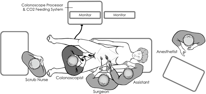
Laparoscopic monitors will be placed depending on the location of the lesion. For right colon polyps, monitors are placed on the patient’s right side and toward the head of the bed (Fig. 26.2). For left colon lesions, the monitors are placed at the patient’s left and toward the foot of the bed. For transverse colon or flexure lesions, the monitors are placed at the head of the bed as the surgeon may stand between the patient’s legs (as will the endoscopist).


Fig. 26.2
Patient positioning and room setup for a right-sided CELS procedure
Endoscopic equipment may vary. Surgeons may prefer to use pediatric versus an adult colonoscope. In addition, we feel it is a prerequisite to have CO2 colonoscopy available in the operating room. Simultaneous performance of laparoscopy and colonoscopy with room air can present technical challenges. Insufflation using room air can significantly obscure the laparoscopic view and compromise exposure. For institutions where this is not possible, a technique of laparoscopically clamping the terminal ileum to minimize bowel distention during laparoscopy has been described, but we have found that colonic distention alone still is a major impediment to this method [3, 4]. Since 2003, our group has been performing colonoscopy with the use of CO2 insufflation during laparoscopy. Because the bowel absorbs CO2 gas approximately 150 times faster than room air, there is minimal unwanted dilation of the colon and excellent simultaneous endoscopic and laparoscopic visualization. We have previously demonstrated that intraoperative CO2 colonoscopy is safe during laparoscopy and can be used to avoid excessive bowel dilation during CELS procedures [9, 17]. Therefore, if available, it is preferred to have CO2 for insufflation during colonoscopy.
Procedure Steps
Endoscopy
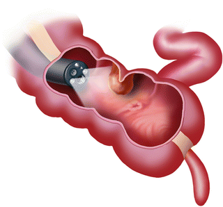
After the abdomen is prepped and draped in a sterile fashion, CO2 colonoscopy is performed to locate the lesion (Fig. 26.3). We then use dilute indigo carmine solution (50 % dilution of indigo carmine with injectable saline solution) to mark the area directly under and surrounding the polyp.

Fig. 26.3
CO2 colonoscopy to determine lesion location. With permission from Yuko Tonohira
Port Placement
Initial access: A periumbilical incision is made and the fascia is entered sharply. A 5 mm port is placed and pneumoperitoneum is established. A 5 mm, high-definition, flexible-tip laparoscope is preferred for better visualization. The abdomen is explored and the site that was previously marked is located.
Secondary trocars: Depending on the location of the lesion, typically two 5 mm trocars may be placed. For right colon lesions, trocars can be placed in the left lower quadrant and suprapubically. For left colon lesions, trocars can be placed in the right lower quadrant and suprapubically. For transverse colon lesions, trocars can be placed on both sides in both the lower and upper quadrants. If available, micro-laparoscopic (3 mm) instruments are used.
Optional trocars: A 5–12 mm port may be needed for a stapler if a colonoscopic-assisted laparoscopic wall excision is anticipated.
GelPort: For CELS, a hand port is not necessary. However, if converting to a segmental or formal colectomy, then some may elect to place a GelPort™ for hand-assisted laparoscopy.
Mobilization
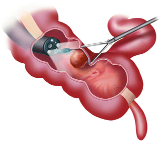
For laparoscopic-assisted colonoscopic polypectomy, the lesion is located by the endoscopist, and its position is confirmed by laparoscopic visualization with the use of transillumination and/or by endoscopic visualization during laparoscopic manipulation of the colon (Fig. 26.4). This maneuver can also expose areas that were not previously visualized because of mucosal folds or segmental kinks of the colon. The location of the polyp in relation to the peritoneum is important. Polyps that are located on the retroperitoneal side or mesenteric side require lateral mobilization of the colon for adequate exposure.

Fig. 26.4
Laparoscopic manipulation of the bowel wall helps to put the polyp in ideal position for endoscopic removal. With permission from Yuko Tonohira
If the polyp is in a difficult location (i.e., at a flexure or near the mesenteric border of the colon) and this area cannot be manipulated, the colon will need to be mobilized. This is done as in any laparoscopic procedure. We prefer to use an energy device along the line of Toldt and carried in the native planes. Once the colon is mobilized adequately, the polyp can then be manipulated.
Stay updated, free articles. Join our Telegram channel

Full access? Get Clinical Tree


