Fig. 16.1
Hartmann’s procedure. With permission from Cleveland Clinic Foundation
Preoperative Planning
Proper patient selection is crucial to preoperative planning, and patients should be both medically fit and able to tolerate laparoscopy. All patients should undergo a detailed history and physical examination, including a thorough review of their surgical history. Such preparation is especially important if the original stoma creation was performed in a different center from where the reversal will be performed. Physical examination can reveal important hints regarding the potential severity of intra-abdominal adhesions. A soft abdomen, with good anterior abdominal wall mobility as determined by bimanual examination, is usually indicative of a more favorable anatomy. On the other hand, a massive midline scar that is found to be sunken and possessing minimal mobility on bimanual examination is classically associated with underlying dense adhesions and restriction of the abdominal wall to accommodate. Patients in the latter group are usually not good candidates for the laparoscopic approach.
Preoperative colonoscopy and flexible sigmoidoscopy is especially recommended for high-risk and IBD patients or those who had a previous bowel perforation due to diverticulitis. Flexible sigmoidoscopy reveals useful information regarding the length of the distal segment (i.e., a rough estimation of above or below the promontory) and thus facilitates operative planning. As part of the preoperative preparation, patients with a colostomy should undergo a mechanical bowel preparation, ensuring the distal stump is clear of stool with one or two enemas. In our practice, however, patients undergoing ileostomy closure with bowel anastomosis are not usually given a mechanical bowel preparation. Preoperative intravenous antibiotics are given within 30–60 min of the incision time, to ensure adequate concentration at the outset, and later readministered in cases taking longer than 3–4 h. Deep venous prophylaxis should include the use of sequential compression devices as well as chemical prophylaxis (preoperative heparin).
Procedure
Setup
After informed consent is obtained, IV induction is given, followed by endotracheal intubation. A Foley catheter and an orogastric tube are placed. The patient is routinely placed in the modified lithotomy position (Fig. 16.2), which allows access to the anus. This position also allows an intraoperative CO2 colonoscopy to be performed with ease, when required. The lithotomy position also provides additional space for the surgical team, especially when operating in the upper quadrants of the abdomen, by standing between the patient’s legs (Fig. 16.3). Padded stirrups or yellow fins are used, and attention is given to preventing peroneal nerve injury. Both arms are tucked at the patient’s sides. A gel pad on the operating table can provide additional decubitus support and stability against gravity with tilting. Additionally, we prefer to secure patients on the operating table with a strong tape placed over the chest to prevent patients from sliding during steep Trendelenburg and right or left tilt. For closure after Hartmann’s procedure, the operating surgeon stands on the patient’s right side, with the assistant either on the opposite or same side, as needed. Two monitors are placed on both sides of the table.
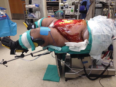
Fig. 16.2
Modified lithotomy position
Fig. 16.3
Room setup for a laparoscopic stoma reversal. With permission from Cleveland Clinic Foundation
Procedure Steps
Laparoscopic Reversal of Colostomy After Hartmann’s Procedure
Port Placement
Initial access: Usually, the colostomy site can be taken down first. An incision is made at the mucocutaneous junction, and the colostomy is freed from the surrounding attachments. A purse-string suture is placed in the proximal bowel and the anvil is secured. This technique also helps to prevent stool or mucous spillage from the end of the colon (Fig. 16.4). After the colostomy has been completely mobilized, the bowel segment proximal to the anvil is returned to the abdomen and peritoneal access is gained. However, it is often necessary to place the anvil in the proximal bowel after adequate mobilization is performed. If so, the proximal colon can be closed with sutures or staples prior to returning it to the abdomen. In this approach, sealing can be achieved in different ways: Our general preference is to use Alexis bundle wound protectors with “a cap” (Alexis laparoscopic system with Kii Fios First Entry, Applied Medical, Rancho Santa Margarita, CA), which helps maintain the pneumoperitoneum (Fig. 16.5). A 5–12-mm port is situated in the middle of the cap, which also enables a laparoscopic approach before and after specimen retrieval. This approach thus converts the stoma site into an additional working port, with 12-mm trocar. This port can subsequently be used as an access port for endoscopic staplers as well as a port site where specimens can be removed when necessary. Additionally, the operating surgeon can utilize this trocar by standing between the patient’s legs and take down the splenic flexure when needed. Alternatively, single-port access laparoscopic Hartmann’s reversal can be performed. When using this approach, a similar circumferential incision around the colostomy is made, followed by disconnection of the stoma from the mucocutaneous border. After the proximal bowel with the anvil is returned to the abdomen, a single-port access device (GelPOINT, Applied Medical, Rancho Santa Margarita, CA) is inserted into the abdomen (Figs. 16.6 and 16.7).
Fig. 16.4
Initial port placement and peritoneal access. With permission from Cleveland Clinic Foundation
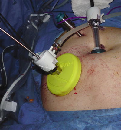
Fig. 16.5
Alexis bundle wound protectors with “a cap” and trocar
Fig. 16.6
Single-port access laparoscopic Hartmann’s reversal. With permission from Cleveland Clinic Foundation
Fig. 16.7
Side view of the single-port access device. With permission from Cleveland Clinic Foundation
Alternatively, an open Hasson technique may be used and access to the peritoneal cavity gained through an incision just above the umbilicus, in cases where initial stoma takedown is not preferred.
After insufflation, two 5-mm trocars (Fig. 16.8) can be placed under direct vision on the right side, lateral to the rectus. A minimum of a fist-sized distance is left between the 5-mm ports. This room provides superior freedom to each of the instruments during dissection and manipulation.
Fig. 16.8
Port sites for straight laparoscopic or hand-assisted stoma reversal. With permission from Cleveland Clinic Foundation
Hand-assisted approach: The utilization of the hand-assisted approach allows the surgeon the ability to assess the intraoperative findings prior to committing to the costs of opening the laparoscopic equipment. A hand port can be created in the suprapubic position, and via the open incision, the presence of adhesions and the state of the pelvis can be assessed (Fig. 16.9). If favorable, the laparoscopic equipment can be opened and the case can proceed. If conditions are not favorable, the incision can be extended and the case approached in an open fashion.
Fig. 16.9
Hartmann’s procedure with implantation of the rectal stump above the fascia. With permission from Cleveland Clinic Foundation
Establishment of pneumoperitoneum: This step is achieved via insufflation through either the umbilical port or colostomy side port. Following adequate pneumoperitoneum, the camera is inserted first through the colostomy side port, and then, according to the density of adhesions, more space is created through further adhesiolysis or with the insertion of additional trocars as described above. All four quadrants of the abdomen are then explored and any abnormalities noted.
Optional trocars: A 5-mm trocar may be placed in the left paramedian position, lateral to the edge of the rectus in the left upper quadrant. This placement can assist both with sigmoid retraction and small bowel adhesiolysis on the right side of the abdomen.
Mobilization of the Proximal Colon
Once the adequate pneumoperitoneum is established and the peritoneal cavity is adequately assessed, the proximal colon needs to be mobilized. The decision to perform a lateral-to-medial or medial-to-lateral approach is dependent upon the surgeon’s preference and the intraoperative conditions. A lateral-to-medial dissection is usually satisfactory for mobilizing the remaining descending colon. In cases where, during the initial operation, perforectomy alone (i.e., resection of the perforated segment only) was performed and a long distal sigmoid colon was left behind, a more extensive descending colon mobilization will be required. At this stage the distal sigmoid colon will be mobilized down to the rectum. We generally prefer a medial-to-lateral approach; however, depending on the comfort level of the surgeon, a lateral approach can also be utilized. The superior rectal/inferior mesenteric vessels can be identified and ligated after the left ureter is visualized and preserved. Ligation of the inferior mesenteric vein just below the level of the pancreatic body gives additional mobility to the proximal colon segment. Once the colon is adequately mobilized, it will need to be exteriorized for resection and insertion of the stapling anvil. The colon can be exteriorized via the stoma site or a suprapubic incision.
Mobilization of the Hartmann’s Pouch and Rectum
The goals of this step are to achieve visualization of the pelvis and mobilize both the descending colon and the Hartmann’s pouch. Laparoscopic adhesiolysis may be required, in order to free the left lower quadrant small bowel of adhesions. Additional mobilization of the descending colon or rectal stump may be required. The goal here is to create a tension-free anastomosis; however, any remaining sigmoid colon on the colostomy or rectal stump must be resected if the Hartmann’s procedure was initially performed for perforated diverticulitis. Resection should be extended all the way down to the top of the rectum. In these cases, a formal splenic flexure takedown may be required before a tension-free reach and anastomosis can be achieved.
In our experience, we prefer to implant the rectal stump above the fascia and just under skin, at the lower aspect of the incision, at the time of the primary operation. This practice makes finding the rectal stump significantly easier, and the potential for rectal scarring and small bowel adhesion formation around the stump itself is minimized (Fig. 16.9).
Resection of the Distal Sigmoid Colon
Distal division of the sigmoid colon: If the distal sigmoid colon is not resected during the index operation, this step must be completed, since this area is usually described as the “high-pressure zone” and recurrent diverticulitis attacks may be observed if this bowel segment is left behind. Therefore, a distal division point is chosen where the taenia splays, ensuring that transection is performed on the rectum. The bowel is then transected at this point, utilizing a laparoscopic linear cutting stapler. Endocutter stapler can be introduced either through the right lower quadrant or the stoma side port. Usually, one firing of the stapler is satisfactory to staple and cut across the bowel at this level of the rectum, provided the mesentery is meticulously prepared.
Extra-corporealization of the distal sigmoid colon: Once the distal portion of the bowel is transected, the resected bowel segment is removed from the previous colostomy side or Pfannenstiel incision, through the wound protector (Fig. 16.10). If the remaining rectal stump is short and previous resection of the bowel was performed right at the top of the rectum, further stapling may not be required. In these circumstances we usually prefer to use “rectal sizers” and make sure the stump is adequately mobilized from the pelvic adhesions. This step also allows us to advance the circular stapling gun easily to the end of the bowel.