Fig. 8.1
“Asymmetric baseball diamond” laparoscopic port positioning for total abdominal colectomy (With permission from Mayo Clinic)
Operative Steps (Video 8.1)
Right Colon
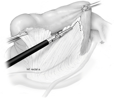
The right or left colon can be approached first. We prefer to start with the right side and use a modified medial-to-lateral approach to the colonic mobilization. The patient is placed in steep Trendelenburg with the right-sided tilt. The small intestine is swept out of the pelvis and placed into the left upper quadrant. The ileum, or a leaf of ileal mesentery near the cecum, is grasped and retracted up and slightly left which exposes the areolar plane between the distal ileal mesentery and the retroperitoneum. Using hot scissors, the peritoneum overlying this indentation just anterior to the iliac artery is incised and carried proximally and medially exposing the retroperitoneum. This dissection can be carried as far as the ligament of Treitz, if needed. This exposes the plane between Toldt and Gerota’s fascia and, when in this avascular plane, blunt dissection is performed in a medial-to-lateral fashion across to the underside of the ascending colon mesentery and superiorly to the inferior border of the duodenum (Fig. 8.2). It is critical during this dissection to stay cleanly between Toldt and Gerota’s fascia as this avoids unnecessary bleeding and decreases the risk of injury to retroperitoneal structures. If significant bleeding is encountered, the surgeon is likely not in the correct plane. The ureter and gonadal vessels should be clearly visible at this point. The dissection is then continued inferiorly and laterally toward the cecum. The appendix is grasped and the peritoneum incised laterally to separate the cecal attachments from the pelvic sidewall (Fig. 8.3). Care must be taken to stay just next to the bowel to avoid injury to retroperitoneal structures. The final step in fully mobilizing the right colon is to grasp the colon and to pull it medially so that the lateral line of Toldt is placed under tension and can be easily incised. Very little dissection is required at this point to join the previous medial dissection plane. Once complete, this mobilization provides excellent visualization of the right colon mesenteric vessels (Fig. 8.4). The vessels to the right colon are taken by opening a plane between the ileocolic and right colic arteries (Fig. 8.5). The vessels can either be taken close to the colon or more proximally close to the superior mesenteric artery and vein. High ligation of these vessels is done in IBD patients who have dysplasia in the right colon and in patients with known malignancy (Fig. 8.6). In benign cases, the vessels are taken where easy and convenient.
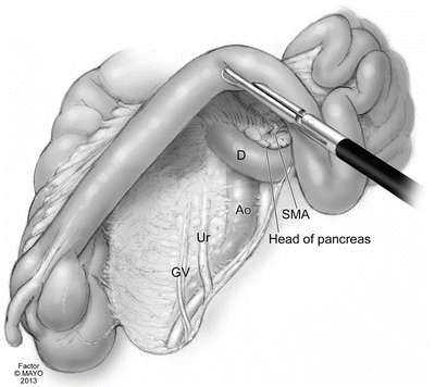
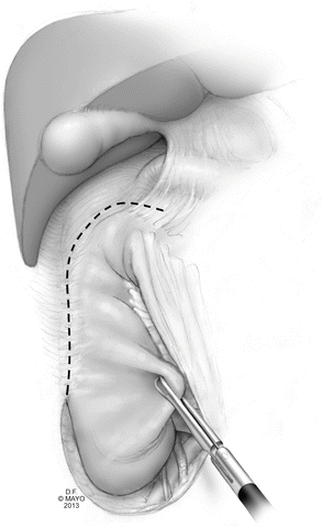
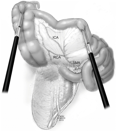
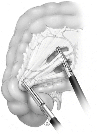
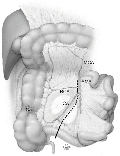

Fig. 8.2
Retroperitoneal exposure during medial-to-lateral dissection of the right colon (D duodenum, Ao aorta, SMA superior mesenteric artery, Ur ureter, GV gonadal vessels) (With permission from Mayo Clinic)

Fig. 8.3
Dissection plane for separating the lateral attachments of right colon from the sidewall (With permission from Mayo Clinic)

Fig. 8.4
Posterior view of right colon after complete mobilization (RCA right colic artery, ICA ileocolic artery, SMA superior mesenteric artery) (With permission from Mayo Clinic)

Fig. 8.5
Ligation of the right colon mesentery (With permission from Mayo Clinic)

Fig. 8.6
Plane for high ligation of mesentery near the SMA (MCA middle colic artery, RCA right colic artery, ICA ileocolic artery, SMA superior mesenteric artery, Ca cancer) (With permission from Mayo Clinic)
Transverse Colon and Hepatic Flexure
The patient is then placed level (side-to-side) and in a slight reverse Trendelenburg position. The omentum is addressed first. In most cases it is preserved and taken off the transverse colon and hepatic and splenic flexure regions by pulling downward to the transverse colon and upward to the omentum. Caution must be taken when taking the omentum off the splenic flexure as it is sometimes adherent to the splenic capsule, and too much tension may result in splenic capsule avulsion and significant bleeding (Fig. 8.7). The hepatic flexure can be mobilized from the left by taking down the hepatocolic ligaments, exposing the plane between Gerota’s fascia, identified by its pale white appearance and the transverse mesocolon (Fig. 8.8). Downward traction of the colon at the hepatic flexure toward the pelvis greatly facilitates this portion of the dissection as this motion puts the hepatocolic ligaments under maximal tension. Ultimately, the avascular plane around the flexure is fully developed bluntly all the way to the right abdominal side wall, forming a tunnel, and the transverse mesocolon is separated from the gastrocolic, duodenal, and pancreatic attachments (Fig. 8.9). This plane ultimately joins the dissection done when mobilizing the right colon earlier. At this point, the right and left branches of the middle colic are easily exposed and can be taken (Fig. 8.10). Once the left branch of the middle colic artery is taken, we address the sigmoid and left colon.
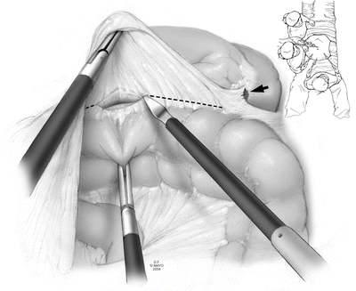
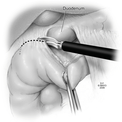
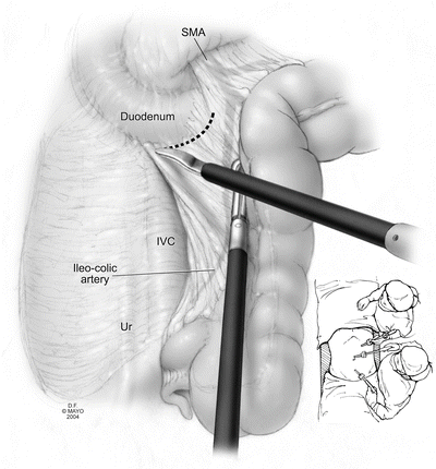
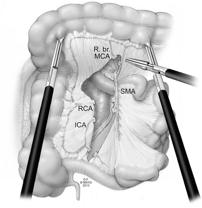

Fig. 8.7
Separation of the omentum from the distal transverse colon mesentery during splenic flexure mobilization (With permission from Mayo Clinic)

Fig. 8.8
Excision of the hepatocolic ligament during mobilization of the proximal transverse colon at the hepatic flexure, exposing the duodenum below (With permission from Mayo Clinic)

Fig. 8.9
Exposure of right retroperitoneal structures with downward traction of hepatic flexure after completion of mobilization (SMA superior mesenteric artery, Ur ureter, IVC inferior vena cava) (With permission from Mayo Clinic)

Fig. 8.10
Ligation of major colic arteries and mesentery of right colon (R. br. MCA right branch of middle colic artery, RCA right colic artery, ICA ileocolic artery, SMA superior mesenteric artery) (With permission from Mayo Clinic)
Sigmoid Colon, Left Colon, and Splenic Flexure
For this portion of the operation, the patient is placed in steep Trendelenburg with the patient’s left side tilted maximally up. The small bowel is placed in the right upper quadrant, exposing the left colon mesentery. The left colon is grasped and pulled medially, putting the lateral line of Toldt under maximal tension (Fig. 8.11). The sigmoid and left colon are then fully mobilized to the midline of the abdomen in a lateral-to-medial fashion. Again, it is critical during this point of the dissection to stay cleanly between Toldt and Gerota’s fascia. This exposes the retroperitoneal structures (gonadal vessels and ureter) and avoids injury (Fig. 8.12). The mobilization is then carried proximally to the splenic flexure. The splenic flexure is taken down enough to visualize the mesentery for a safe, tension-free vessel ligation. The splenocolic, phrenocolic, and pancreaticocolic ligaments are identified and incised for full splenic flexure mobilization as needed to ensure safe vessel ligation (Fig. 8.13a, b).
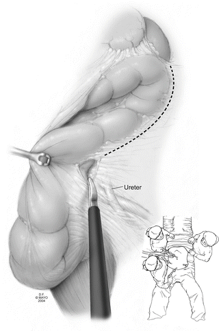
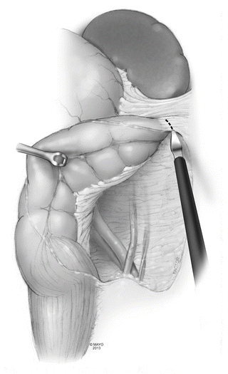
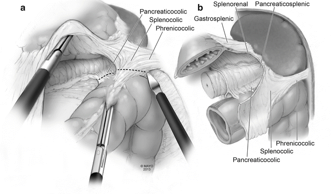

Fig. 8.11
Dissection plane for separating the lateral attachments of left colon from the sidewall (Ur: ureter) (Inset: position of surgeon and assistants around patient) (With permission from Mayo Clinic)

Fig. 8.12
Exposure of retroperitoneal structures during lateral mobilization of left colon

Fig. 8.13
(a) Exposure of splenocolic ligament; (b) Splenic flexure recess with associated ligaments (With permission from Mayo Clinic)
Once full mobilization of the left colon and splenic flexure is completed, the decision of where to transect the colon must be made. If there is concern for a high-risk rectal stump, as in the case of a patient with fulminant colitis, we transect the sigmoid at a point that leaves enough length so it can be brought up to the suprafascial position at the suprapubic port site or lower part of the incision if one is made (Fig. 8.14). If the stump is low risk for leak, or if ileorectostomy is going to be performed, transection should be at the top of the rectum. In either case, we typically preserve the superior rectal artery to the rectal stump.
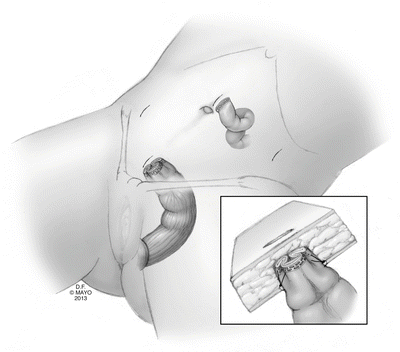

Fig. 8.14
Creation of a suprafascial sigmoid mucous fistula (With permission from Mayo Clinic)
For patients who will get an ileostomy and who have a high-risk distal bowel stump, the mid-sigmoid is transected with the Endo GIA stapler (60 mm load) after upsizing the right lower quadrant 5 mm port to a 12 mm port (Fig. 8.15a, b). Alternatively, the rectum can be transected transabdominally if a Pfannenstiel extraction port is planned. The sigmoid colon is then grasped and retracted medially and anteriorly up toward the abdominal wall, and a vessel-sealant device is then used to ligate all remaining mesentery until the site of the planned colon transection is reached proximally (Fig. 8.16).
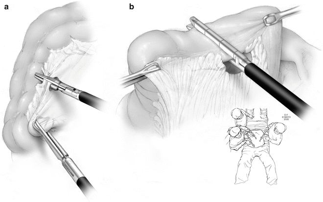
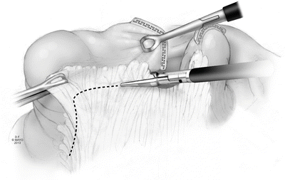

Fig. 8.15
(a) Ligation of marginal artery in sigmoid colon mesentery; (b) Intracorporeal transection of sigmoid colon with laparoscopic stapler to create a rectal stump (inset: position of surgeon and assistants around patient) (With permission from Mayo Clinic)

Fig. 8.16
Ligation of proximal sigmoid mesentery with vessel sealer after transection (With permission from Mayo Clinic)
For patients undergoing ileorectostomy, a window in the mesentery at the level of the distal sigmoid is created by taking the marginal artery. The mesentery is then taken from this point inferiorly to the top of the rectum, staying close the colon (Fig. 8.17). This preserves the superior rectal artery blood supply to the rectal stump and significantly decreases the risk of sympathetic nerve injury. Once the top of the rectum is adequately cleared of mesentery, the right lower quadrant port can be upsized and the 60 mm Endo GIA stapler is used to transect the rectum with a single firing (Fig. 8.18). The sigmoid colon is then grasped and retracted medially and superiorly, and all the remaining mesentery proximally from this point up to the transected mesentery in the transverse colon done earlier is taken (Fig. 8.19).

