Clinical Aspects of Renal Transplantation


Clinical Aspects of Renal Transplantation


Alexander C. Wiseman

James E. Cooper

Laurence Chan


INTRODUCTION

Since the first successful renal transplant over 50 years ago,1 more than 500,000 patients with renal failure have had their lives prolonged with renal allografts. Renal transplantation is associated with improved longevity compared to dialysis2 with increased quality of life,3 and currently is the preferred treatment modality for eligible patients with chronic kidney disease (glomerular filtration rate [GFR] <20 mL per minute).

The progressive increase in the incidence and prevalence of severe chronic kidney disease (CKD) has led to a parallel increase in the number of patients waiting for a transplant. This increase substantially outpaces the supply of available organs (Fig. 82.1). The reported average waiting time for a deceased donor kidney transplant (DDKT) is more than 3.5 years (2009 Scientific Registry of Transplant Recipients [SRTR] Annual Report Table 5.2). For this reason, efforts have been made to increase living kidney donor transplantation. Unfortunately, living donation rates in the Unites States have not increased in recent years, and modest growth in kidney transplantation has occurred as a result of increased deceased donor use.4,5 Worldwide, rates of kidney transplantation and use of living donors for kidney transplant vary widely due to societal differences in the perception of transplantation and of organ donation following brain death or cardiac death (Fig. 82.2).

Patient and graft survival after kidney transplant are affected by a large number of variables (Table 82.1). Primary factors include: age, sex, and race of the recipient and donor; type of donor (living versus deceased, expanded criteria versus standard criteria); tissue compatibility; prior sensitization to human leukocyte antigens (HLA); original renal disease, pretransplant health status, and concomitant extrarenal disease of the recipient; adherence of the recipient; donor factors, such as age, cold ischemia time, and nephron dosing effect; and choice of immunosuppressive agents (Figs. 82.3 and 82.4). Short-term outcomes have improved substantially over the past 15 years, with 1-year graft survival averaging 90% to 94% and patient survival averaging 94% to 97%; however, improvements in long-term graft survival have been more difficult to achieve. An analysis by Hariharan et al.6 of graft survival for all 93,934 renal transplantations performed in the United States between 1988 and 1996, suggested that the estimated half-life for grafts from living donors increased steadily from 12.7 to 21.6 years, and that for deceased donor grafts increased from 7.9 to 13.8 years. However, in this analysis graft survival was calculated upon projected, not actual, graft survival. A later analysis of graft outcomes from 1988 to 1995 demonstrated that actual graft survival demonstrated far less improvement in graft half-life of 6.0 to 8.0 years for deceased donor grafts.7 A recent analysis of transplants from 1989 to 2009 suggests slow improvements in graft survival over time, primarily in higher risk transplants (the expanded criteria donor, described later) (Table 82.2). For living donor kidney transplants, the estimated graft half-life did not change appreciably for transplants performed from 1989 to 2005 (11.4 years to 11.9 years).8 With greater understanding of the causes of graft loss, it is hoped that this will translate into better outcomes in renal transplantation with improved long-term graft and patient survival. In the subsequent sections of this chapter, we will discuss each of the factors influencing outcomes of renal transplantation, the recipient and donor evaluation prior to transplantation, immunosuppressive drugs, posttransplantation management, and complications.


PATIENT SELECTION AND PRETRANSPLANT EVALUATION


General Philosophy in Recipient Selection

In general, patients with CKD stage IV through V (GFR <30 mL per minute) should be presented with information regarding dialysis modalities and transplantation. Patients who express an interest in undergoing kidney transplantation should be fully evaluated by the transplant team. This is typically as an outpatient during a clinic visit; however, some centers may provide this on an inpatient basis. Early referral prior to the onset of dialysis should be encouraged, because
the degree of dialysis time prior to transplant has been associated with poorer graft survival following transplant.9


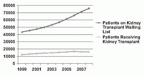
FIGURE 82.1 Counts of patients on the renal transplant waiting list and counts of renal transplants by year in the United States from 1999 to 2008. (From Scientific Registry of Transplant Recipients 2009 Annual Data Report, Ann Arbor MI, Tables 5.1a, 5.1b, 5.4, 5.4d, with permission.)

There are few absolute contraindications to kidney transplantation, and many of these contraindications are relative (Table 82.3). The current protocols for a transplant evaluation focus on ensuring the safety of undergoing surgery and the ability to assume the risks of immunosuppressive therapy in order to optimize successful kidney transplant outcomes. This evaluation is tailored to the individual candidate’s risk for complications, and includes consideration of the patient’s age, diabetes, and heart disease status. These factors are also associated with a higher risk of death in the general population and in patients with end-stage renal disease (ESRD) treated by dialysis.


Age

The adolescent patient (age 12 to 17) and the elderly recipient (age >65) have poorer graft survival than other age groups (SRTR Annual Report Table 5.8c). In the former, this is due primarily to difficulties in medication adherence, whereas in the latter, this is due to complications of immunosuppressive therapy leading to death or to nontransplantrelated complications, in particular, cardiovascular disease in the elderly leading to death.10,11


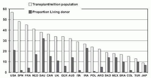
FIGURE 82.2 Transplantation rates for countries reporting more than 500 kidney transplants in 2006 (count of new renal transplants per million total population) and number of transplants arising from living donors. SPN, Spain; FRA, France; NED, Netherlands; SAU, Saudi Arabia; CAN, Canada; UK, United Kingdom; GER, Germany; AUS, Australia; ITA, Italy; IRA, Iran; POL, Poland; ARG, Argentina; SKO, South Korea; MEX, Mexico; BRA, Brazil; COL, Columbia; TUR, Turkey; JAP, Japan. (Data from Horvat LD, Shariff SZ, Garg AX. Global trends in the rates of living kidney donation. Kidney International 2009; 75:1088-1098.)

In the United States, national kidney allocation policy prioritizes pediatric candidates to ensure a minimum of waiting time to promote the beneficial effects of transplantation, including growth and development. The challenge of ensuring appropriate medical support for young people with solid organ transplants as they move into adultcentered services has become a topic of significant interest in the field of organ transplantation. Proceedings from a consensus conference of the major transplant societies has outlined the need for collaborative transitional care between pediatric and adult providers and the need for research in this area.12


TABLE 82.1 Factors Influencing the Outcome of Renal Transplantation


Immunologic


Nonimmunologic


Immunosuppressive protocol


Delayed graft function/ischemic time


Matching for HLA


Medication adherence


Sensitization


Cardiovascular disease


Rejection


Recipient age


Nephron dose/donor and recipient gender


HLA, human leukocyte antigens.


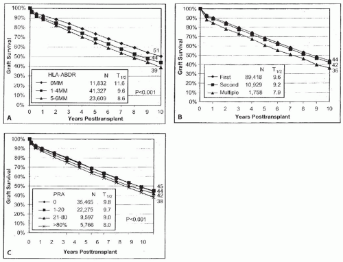
FIGURE 82.3 Graft survival related to immunologic factors. A. Effects of human leukocyte antigen (HLA) mismatches (MM) on the survival of first diseased donor transplants. Graft survival rates declined as the number of HLA-A, HLA-B, and HLA-DR MMs increased. The difference in graft half-life between the best and the worst matched grafts was 3 years (11.6 years vs. 8.6 years). B. Graft survival among first and repeat deceased donor transplant recipients. The differences between first and subsequent transplants have diminished, with no significant differences in graft survival. C. Graft survival related to HLA sensitization. HLA sensitization remains a significant risk factor for graft loss. Highly sensitized recipients (panel reactive antibody [PRA] >80%) have a lower long-term graft survival than patients with PRA <20%. Data are from the United Network for Organ Sharing Scientific Renal Transplant Registry, 1996-2005. (From: Cecka J, Terasaki P, eds. Clinical Transplants 2008. Los Angeles, CA: UCLA Tissue Typing Laboratory; 2008:1-18, with permission.)

With the improvements in perioperative management and immunosuppressive strategies, advanced age itself is no longer a contraindication to renal transplantation. Based on a retrospective analysis of wait-listed patients >70 years old from the SRTR, 1990 to 2004, elderly transplant recipients had a 41% lower overall risk of death compared with wait-listed candidates.13 These benefits also extend to selected patients over age 80.14 Older patients may have better immunologic survival despite the higher mortality from cardiovascular disease. One explanation may be an age-related change in immunologic function that confers less alloreactivity with aging, as suggested by a registry analysis that demonstrated that acute rejection rates significantly fell with advancing recipient age.15 Recipients older than 65 years demonstrated significantly elevated numbers of memory T cells, whereas counts for naive T cells were significantly reduced.16 For this reason, many centers advocate the use of lower immunosuppression in elderly patients. In summary, kidney transplantation can now be safely and successfully performed in selected elderly patients but requires comprehensive screening of underlying cardiovascular disease and occult malignancy.


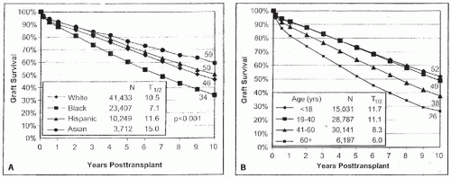
FIGURE 82.4 Graft survival rates of deceased donor kidney transplants are related to donor and recipient factors. A. The effect of recipient race on graft survival. The race of the recipients was a significant factor in the outcome of the first deceased donor transplants. Asian patients had the highest graft survival rates—59% at 10 years, respectively—whereas blacks had the poorest survival rates—34% at 10 years. B. The effect of donor age on graft survival. Donor age remains one of the most important factors in deceased donor kidney transplant graft survival. Data are from the United Network for Organ Sharing Scientific Renal Transplant Registry, 1996-2005. (From: Cecka J, Terasaki P, eds. Clinical Transplants 2008. Los Angeles, CA: UCLA Tissue Typing Laboratory; 2008:1-18, with permission.)


Obesity

Obesity alone is rarely an absolute contraindication to transplantation, yet it is a well-defined risk factor. Lower graft survival rates, higher postoperative mortalities, and complications have been demonstrated in patients with a body mass index (BMI) greater than 35 kg per square meter.17,18 The large body size is also a risk factor for progression and

subsequent premature failure due to the physiologic changes that have been linked to nephron hyperfiltration.18 Although weight reduction is important for obese dialysis patients before proceeding to transplantation, often patients will regain weight following transplantation and mandatory weight loss pretransplant may not substantially improve longer term outcomes.19


TABLE 82.2 Actual Transplant Half-Life for Transplants Performed in 1997, and Projected Transplant Half-Life for Transplants Performed in 20049


Transplant Subgroup


Actual Graft Half-Life, 1997 Transplants


Actual Graft Half-Life, 1997 Transplants


Projected Graft Half-Life, 2004 Transplants


Projected Graft Half-Life, 2004 Transplants


All recipients


African American recipients


All recipients


African American recipients


All deceased donor transplants


8.2 yr


6.3 yr


8.8 yr


7.1 yr


SCD


8.9 yr


6.8 yr


9.7 yr


7.7 yr


ECD (first transplant)


5.1 yr


4.4 yr


5.9 yr


5.4 yr


Living donor


12.0 yr


8.7 yr


14.2 yr


10.8 yr


SCD, Standard criteria donor; ECD, Expanded criteria donor.


TABLE 82.3 Contraindications to Transplantation


Absolute


Relative


Active infection


Renal disease with high recurrence rate


Disseminated malignancy


Urologic abnormalities


Extensive vascular disease


Active systemic illness


High risk for perioperative mortality


Ongoing substance abuse


Persistent coagulation abnormality


Uncontrolled psychosis


Informed patient refusal of consent


Refractory nonadherence


Prior Kidney Transplantation

Renal allograft failure is now one of the most common causes of ESRD, accounting for about 30% of patients awaiting renal transplantation. Graft survival of a second transplant is decreased compared to that of the first, but outcomes have improved over time (Fig. 82.3).7 Evaluation of a potential recipient for a repeat allograft requires careful attention to the reason for the graft failure, such as nonadherence with immunosuppressive medications, recurrent renal disease, or high alloreactivity with high panel reactive antibody (PRA) titers. These patients may also manifest complications of prior immunosuppressive therapy and, as such, should be screened for complications associated with these medications, such as infection and malignancy.20 No controlled, prospective studies have been performed to determine the best method for tapering or withdrawal of immunosuppression following renal allograft failure, with some suggestion that nephrectomy after graft loss may improve patient survival and rates of retransplantation.21 Most centers have adopted a policy of immediate withdrawal of immunosuppression combined with preemptive nephrectomy for patients with early allograft failure. However, this practice is less common for patients with late graft failure. A longer taper of immunosuppression may permit the maintenance of some residual renal function while on dialysis. Further studies are needed to determine the optimal means of immunosuppression withdrawal or nephrectomy in patients who return to dialysis.


Underlying Renal Diseases

It is most important to assess the cause of the potential recipient’s renal failure. The primary pathologies leading to renal failure are expected to influence outcome depending on the etiologic mechanisms, propensity for recurrence, and status of the immune system.


Diabetes Mellitus

Although patients with diabetes are at a higher risk for post-transplant complications primarily related to their pre-transplant comorbidities, kidney transplantation is the treatment of choice for otherwise eligible patients due to their high mortality rate while on dialysis.22 In particular, patients with type 1 diabetes (T1DM) enjoy the highest net mortality benefit of transplantation compared to dialysis following receipt of a simultaneous pancreas kidney transplant (SPK) when compared to other kidney transplant recipients.23 Patient survival and pancreas graft survival rates continue to improve, with data from U.S. centers demonstrating 95% and 86% 1-year patient and pancreas graft survival, and 85% and 70% 5-year patient and pancreas graft survival, respectively.4

With the increase in use of living donors for kidney transplantation, solitary pancreas transplant after kidney transplant (PAK) is often considered for patients with T1DM. Although this offers the benefit of timely kidney transplant, ideally prior to the need for hemodialysis, this strategy requires two separate survival procedures, two different HLA-mismatched organs, and the risks inherent to surgery. Pancreas allograft survival is worse as a PAK than SPK likely due to the additional immunologic factors of a second organ and the lack of use of renal function changes as a surrogate marker of pancreas function changes. Recommendations for patients with T1DM approaching kidney failure should be tailored to the individual’s circumstance, and should include an assessment of the following: (1) can a living donor be identified; (2) is the patient (and transplant program) willing to accept a higher risk of early death and possibility of pancreas graft loss (˜2% and ˜15% in the first year, respectively) when considering SPK versus living donor kidney transplant; (3) how debilitating are the patient’s diabetes-related quality-of-life issues and achieved level of glycemic control; and (4) what is the expected waiting time for an SPK in the patient’s geographic region.24 In general, SPK appears to offer advantages over kidney transplantation alone with respect to long-term survival if the waiting time for a deceased donor is not excessive and dialysis time can be minimized (perhaps to less than 6 months). For those patients who are unable to wait for SPK, a living donor kidney transplant followed by a later pancreas transplant appears to be associated with better kidney graft function with a risk of mortality that is similar to living donor kidney transplant alone.25 One suggested algorithm for patients with T1DM and CKD considering their transplant options is provided in Figure 82.5.

Another treatment option in development for the patient with T1DM is pancreatic islet cell transplantation (ICT). In experienced centers, ICT can achieve insulin independence in 80% to 90% of recipients at 1 year; however, <30% have remained insulin free after 5 years.26 At present, this therapy should still be considered experimental, because
the long-term graft survival is unknown and the risks of immunosuppression and HLA sensitization must be weighed against the benefits of normalization of blood glucose.


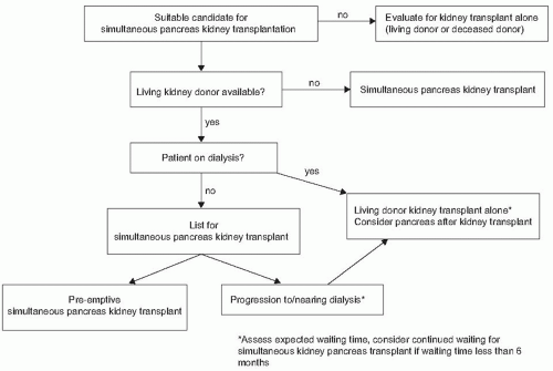
FIGURE 82.5 Proposed algorithm for type 1 diabetic patients requiring transplant. (Adapted from Wiseman AC. Simultaneous pancreas kidney transplantation: a critical appraisal of the risks and benefits compared with other treatment alternatives. Adv Chronic Kidney Dis. 2009;16(4):278-287, with permission.)

Recurrence of the diabetic nephropathy in T1DM recipients is a late and slowly developing complication. An examination of biopsy specimens early after transplantation indicates that there are few glomerular pathologic abnormalities other than frequent afferent and efferent arteriosclerosis. Glomerular basement immunoglobulin G (IgG) deposition is seen <2 years after transplantation, but the onset and progression of glomerular basement membrane thickening and mesangial expansion only occurs after 2 years, and the typical nodular glomerular hyalinosis is rarely seen in these patients. Long-term follow-up has shown that recurrent nephropathy progresses to ESRD with the same time course as primary type I diabetic nephropathy. The mean time to recurrent ESRD is estimated to be 15 to 20 years. Therefore, recurrence of the lesion is not a barrier to long-term renal graft survival in diabetic recipients. The frequency and natural history of recurrence in type II diabetic recipients remain to be elucidated.


Metabolic and Congenital Disorders

Results of renal transplantation in the metabolic and congenital disorders causing end-stage renal failure such as Alport syndrome, amyloidosis, cystinosis, familial nephritis, gout, and cystic disease are similar to those of the more common causes of end-stage renal failure with the exception of primary hyperoxaluria, sickle cell, and Fabry disease, as discussed in detail in the following paragraphs.


Primary Hyperoxaluria

Although often presenting in childhood, inherited deficiencies in alanine:glyoxalate aminotransferase (AGT) levels or function may present in the young adult as calcium oxalate nephrolithiasis, nephrocalcinosis, renal failure, and systemic oxalate deposition. Registry analyses generally favor combined liver-kidney transplantation (to correct the AGT defect and promote long-term kidney graft survival), but occasionally, patients may have a functional AGT deficiency that is pyridoxine sensitive, and 5 to 10 mg/kg/day of pyridoxine may decrease oxalate levels to a level that is acceptable to consider kidney transplantation alone.27,28 To reduce the chance of oxalate accumulation, dialysis treatment or kidney transplantation should be considered when the GFR approaches 20 mL per minute. Aggressive dialysis schedules should be implemented before transplantation to deplete the oxalate metabolic pool. Medical therapy with pyridoxine, neutral phosphate, and magnesium should be given after transplantation to reduce oxalate deposition and recurrence (Fig. 82.6).


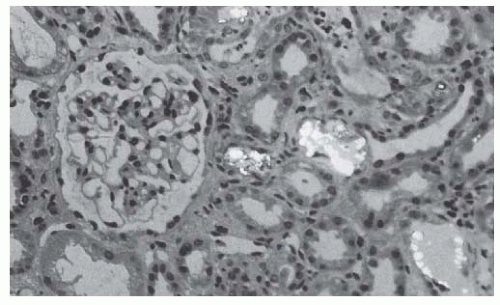
FIGURE 82.6 A renal biopsy specimen from a transplanted kidney showing calcium oxalate deposition in a patient with primary hyperoxaluria and a recurrence of oxalosis.

Unlike primary hyperoxaluria, secondary oxalosis is due to excessive intake or absorption of oxalates from the diet. Secondary oxalosis is seen primarily in fat malabsorption, short bowel syndromes after gastrointestinal surgery, and high-oxalate diets. For these patients, consideration should be given to reanastomosis of gastric bypass, hydration, and dietary restriction of oxalates. Good allograft function can be achieved when attention is paid to reduce the oxalate excretion load.29


Cystinosis

Cystine stones recur after transplantation, but have little effect on graft function.30 Renal transplantation has been recommended as a preferred therapy in children with ESRD due to cystinosis. The systemic effects of cystine accumulation, including corneal crystallization and retinal degeneration, leading to blindness, progress after renal transplantation but can be reduced with chronic cysteamine therapy.31


Sickle Cell Disease

The autosomal recessive conditions of sickle cell disease and sickle cell trait may be complicated by a variety of renal abnormalities, which may eventually lead to ESRD.32 The North American Pediatric Renal Transplant Cooperative Study (NAPRTCS) reports favorable outcomes in pediatric patients with patient survival of 89%, and graft survival at 12 and 24 months posttransplant of 89% and 71%, respectively.33 A second registry analysis demonstrates comparable short-term but diminished long-term outcomes compared to other causes of ESRD.34 The importance of recurrence after transplantation is difficult to determine because of the relatively nonspecific nature of sickle cell nephropathy.


Fabry Disease

Fabry disease is an X-linked disorder of glycosphingolipid metabolism due to a ceramide trihexosidase. Fabry nephropathy does not recur in the allograft, and transplantation provides superior outcomes to Fabry patients on dialysis.35 Graft survival at 5 years is comparable to patients with other causes of ESRD, but with a higher risk of death.36 Enzyme replacement therapy with agalsidase alfa is well tolerated in patients with Fabry disease following renal transplantation, but data regarding an impact on survival has yet to be determined.37 Transplantation is considered the optimal mode of renal replacement therapy for otherwise eligible patients with Fabry disease.


Amyloidosis

Recurrent nephrotic syndrome and graft failure can occur in primary and secondary amyloidosis. Although transplantation is uncommonly performed for patients with primary amyloid light chain (AL) amyloidosis, in selected patients kidney transplantation has been shown to be successful.38 Often, the treatment for this disorder requires chemotherapy and autologous stem cell transplantation. The decision to perform stem cell transplant before or after kidney transplant has been debated; reports of living donor kidney transplant followed by stem cell transplant have demonstrated favorable results.39 Without definitive treatment, the recurrence of renal AL amyloidosis is common following kidney transplant.40 Familial Mediterranean fever (FMF), rheumatoid arthritis, and osteomyelitis are the most common causes of secondary amyloidosis. FMF is an autosomal recessive disorder that occurs in Sephardic Jews, Armenians, Turks, and Arabs of the Levant. In Israel, amyloidosis constitutes 6% of all patients on dialysis, compared to 0.6% in Europe. Although there has been a higher early mortality rate in the transplanted patients in the past, the incidence of rejection episodes is lower than in patients without amyloidosis. Reduced immunosuppression has decreased postoperative mortality and morbidity. Colchicine at 1 to 2 mg per day dramatically relieves the symptoms and reduces the incidence of attacks in FMF, and interleukin (IL)-1 receptor antagonism is an increasingly attractive treatment alternative.41


Alport Syndrome

Dialysis and transplantation pose no particular problems for patients with Alport syndrome. Recurrent disease has not been well documented. Improvement or stabilization of deafness after renal transplantation has occasionally been reported. There is a 3% to 5% risk of developing de novo antiglomerular basement membrane (anti-GBM) nephritis after transplantation, typically occurring within the first year and resulting in graft loss.42


Polycystic Kidney Disease

Autosomal dominant polycystic kidney disease is responsible for approximately 4% to 12% of ESRD cases in the United States and Europe. Native kidney removal is only required if the kidneys are massive due to polycystic disease or there is associated persistent infection or severe hypertension. Embolization of native kidneys prior to transplant may be a
less invasive treatment strategy in the future.43 Occasionally, patients with severe liver cysts will require combined liver-kidney transplantation, primarily due to symptoms related to cyst volume and the impact on nutritional status.44 Screening for cerebral aneurysms prior to transplant is generally directed toward those with a family history or with new onset headaches.45


Glomerulonephritis

Almost all types of glomerulonephritis have been reported to recur after transplantation. There is, however, much variation between the various types of glomerulonephritis with regard to the frequency of recurrence, the clinical course, and the prognosis. The overall incidence of recurrence is less than 10% to 20% and recurrent disease accounts for less than 2% to 4% of all graft failures (Table 82.4).46


Focal Segmental Glomerulosclerosis

Recurrent focal sclerosis may be seen early after transplantation, presenting with nephrotic-range proteinuria and a rapid decline in renal function. Histologically, the features on light microscopy that permit categorization are focal and segmental sclerosis, affecting a small number of glomeruli, often those in the deep juxtamedullary cortex. The development of foot-process fusion can be immediate after transplantation and precede glomerular segmental sclerosis by weeks to months (Fig. 82.7). The frequency of recurrence is about 20% in adults and may be as high as 40% in children. When stringent definitions of primary focal segmental glomerulosclerosis (FSGS) are applied (i.e., the nonfamilial inheritance pattern from patient history and documented biopsy-proven disease), the recurrence rate approaches 50%.47 Patients presenting with rapid progression of renal disease from the time of diagnosis of nephrotic syndrome to ESRD have higher risk for recurrence. If a transplant patient suffers graft loss because of recurrent FSGS, there is ˜50% risk of subsequent allograft failure within 5 years of a second transplantation. With increasing understanding of the genetic causes of FSGS (e.g., podocin and nephrin mutations) a more tailored approach to FSGS may be possible in the future, with avoidance of living donors with similar genetic risk.48


TABLE 82.4 Recurrent Disease in Renal Allografts


Disease


Approximate Recurrence Rate (%)


Graft Loss Due to Recurrence


Primary Glomerulonephritis


Membranous


10%-30%


Uncommon


FSGS


30%-60%


Common


HUS


20%-50%


Common


Type I MPGN


20%-30%


Common


Type II MPGN


80%-100%


Common


HSP


15%-50%


Uncommon


IgA nephropathy


30%-50%


Uncommon


Anti-GBM


Rare


Uncommon


ANCA associated


20%


Common


Systemic Disease


Hyperoxaluria


80%-100%


Common


Cystinosis


50%-100%


Uncommon


Fabry disease


Rare


Common


Sickle cell disease


Rare


Common


Diabetes type I


100%


Uncommon


SLE


<10%


Uncommon


FSGS, focal segmental glomerulosclerosis; HUS, hemolytic uremic syndrome; MPGN, membranoproliferative glomerulonephritis; HSP, Henoch-Schonlein Purpura; IgA, IgA nephropathy; GBM, anti-glomerular basement membrane disease; ANCA, antineutrophil cytoplasmic antibody associated vasculitis; SLE, systemic lupus erythematosis.


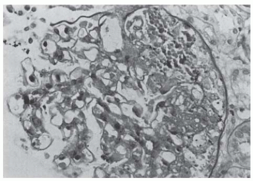
FIGURE 82.7 A renal biopsy specimen of a transplanted kidney showing recurrence of focal segmental glomerulosclerosis. (Periodic acid-Schiff stain, magnification ×250.)

Treatment for recurrent FSG remains disappointing. Heavy proteinuria and nephrotic syndrome are usually resistant to steroids.49 Cyclosporine (CsA) or other immunosuppressants do not seem to prevent recurrence. In many cases, the rapidity of recurrence immediately post-transplant strongly suggests the presence of a circulating factor in primary FSGS that is toxic to the glomerular epithelial cell/podocyte interface. It has been shown that sera from some patients with FSGS increases the permeability of isolated glomeruli to albumin.50 Recently, this circulating factor has been suggested to be urokinase receptor (uPAR) potentially derived from circulating neutrophils.51 Use of a regenerating protein adsorption
column or plasma exchange can reduce protein excretion in patients with recurrent FSGS in the transplant. More prolonged remissions have been achieved using plasma exchange that is initiated promptly after the onset of proteinuria or the combination of plasma exchange and cyclophosphamide. These prolonged beneficial results have also been reported in children treated with plasma exchange and cyclophosphamide.52


Antiglomerular Basement Membrane Disease

Based on histology and fluorescence studies, anti-GBM disease is associated with >50% recurrence rate in the allograft. However, only 25% of patients with biopsy-proved IgG staining along the capillary wall have evidence for clinical disease activity. Furthermore, graft failure due to recurrent disease is less common, estimated at <5%.53 Although engraftment during the presence of anti-GBM antibodies has been reported to be successful, many transplant centers still prefer serologic quiescence of anti-GBM antibody production for 6 to 12 months before proceeding with transplantation to reduce the risk for recurrent anti-GBM disease. Despite delaying transplantation to allow anti-GBM antibody to fall, recurrence has been reported.54


Hemolytic Uremic Syndrome (HUS)

Typical (diarrhea-associated) hemolytic uremic syndrome (HUS) does not recur in the transplant, although atypical (nondiarrheal) aHUS has a high recurrence rate that usually leads to graft loss.55 aHUS is the clinical manifestation of complement dysregulation, either via complement deficiencies or autoantibodies. Recurrence rates of 80% to 100% have been reported for factor H or I deficiencies, whereas membrane cofactor protein (MCP) deficiency does not usually recur. The recurrence rate may be higher in recipients of living-related transplants, those of an older age at the onset of HUS, those with a short duration between disease onset and ESRD or transplantation, who use living related donors, and, to a lesser degree, in those who had been administered calcineurin inhibitors (CsA or tacrolimus).56 There is no treatment for recurrent HUS that has been proven to be consistently successful. Salicylates, dipyridamole, plasma infusion, and plasma exchange have been shown to be of limited benefit. However, case reports of successful treatment and prevention of recurrent HUS in kidney transplant with the anti-C5a antibody eculizumab have generated encouraging results.57,58 In preparation for transplant, patients with suspected aHUS should be screened at a minimum for factor H, I, and MCP deficiencies to aid in prognosis and in potential peritransplant treatment with plasma exchange and/or eculizumab. CsA and tacrolimus have both been associated with altered coagulation mechanisms and the development of de novo HUS in renal transplant recipients, particularly in combination with sirolimus.59 These agents should, therefore, be used with caution in patients whose original kidney disease was due to HUS.


IgA Nephropathy/Henoch-Schönlein Purpura

In many parts of the world, IgA nephropathy (IgAN) is the most common type of glomerulonephritis. Although histologic recurrence of IgAN is common (up to 75%), its presentation is often clinically mild, and graft loss specifically due to IgAN is uncommon (<5%).46 Patients with IgAN have at least comparable if not better graft survival rates than those with other diseases.60

The closely related Henoch-Schönlein purpura (HSP) has been reported to recur with similar frequency and outcomes as IgAN.61 Clinically, recurrent HSP or IgAN can be severe with crescentic glomerulonephritis, nephrotic syndrome, graft failure, and variable recurrence of purpura.62 To reduce recurrence, the delay of engraftment is recommended for at least 6 to 12 months after the skin lesions of HSP have resolved.


Membranoproliferative Glomerulonephritis Type I and Type II

Type I and type II membranoproliferative glomerulonephritis (MPGN) can recur posttransplant and can negatively impact long-term graft survival.63 Type II MPGN may recur at a higher frequency (60% to 100%) than type I MPGN (15% to 30%).64,65 The early development of nephrotic syndrome and persistent microscopic hematuria from the time of transplantation are clinical markers suggesting recurrence rather than rejection. Levels of serum C3 do not accurately predict recurrences. Specific disease-targeted therapy is not well defined, except in the case of MPGN type II with known complement factor deficiency (factor H or I) in which plasma exchange is warranted.66


Membranous Nephropathy (MN)

Graft survival for patients with membranous nephropathy (MN) is similar to the general transplant population despite a recurrence rate of up to 40%.67,68 MN can also present as a primary de novo condition in allograft recipients.69 Recurrent MN with nephrotic syndrome generally occurs earlier, at an average of 10 months compared with de novo MN, which is usually seen about 18 to 20 months after transplantation (Fig. 82.8). Rituximab has been shown in initial reports to be of benefit in proteinuria regression and renal function stabilization.68,70


Systemic Lupus Erythematosus (SLE)

Recurrence of clinically significant systemic lupus erythematosus (SLE) is relatively rare following transplant.71 Similarly, the reactivation of other nonrenal manifestations of SLE after transplantation is extremely infrequent and is often controlled by the immunosuppressive medications when it occurs.72 Recurrence is not predictable with serologic monitoring. However, there should be no systemic disease activity prior to transplantation.73 Recurrences can be successfully treated with steroids, mycophenolate mofetil, or chlorambucil.


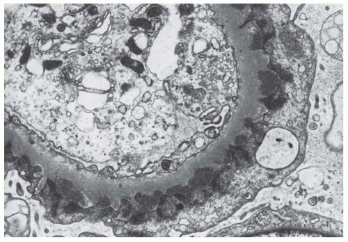
FIGURE 82.8 An electron micrograph of de novo membranous glomerulonephritis.


Antineutrophil Cytoplasmic Antibody-Associated Small Vessel Vasculitis

Patients with antineutrophil cytoplasmic antibody (ANCA)-related vasculitis have graft survival rates comparable to nondiabetic transplant populations.74 As a relapsing and remitting disease, its recurrence rate following transplant is ˜20%, which is slightly less than those remaining on dialysis.75 ANCA titers do not appear to be predictive of recurrence posttransplant, thus transplantation can be reasonably pursued once clinical remission is achieved.76 Recurrences are not prevented by baseline transplant immunosuppression, but can be treated successfully by adding cyclophosphamide and by increasing the steroid dose, together with decreasing or discontinuing some transplant medications.


Progressive Systemic Sclerosis (Scleroderma)

Transplant outcomes for patients with scleroderma are worse than in other diseases but are better than their wait-listed counterparts on dialysis.77 Recurrence in the graft can occur within the first few months after transplantation. Recurrent scleroderma renal crisis in the allograft may be preceded by systemic features of scleroderma, such as the progression of diffuse skin thickening, new onset anemia, and cardiac complications.78 The current recommendation for transplantation is that the patient should be clinically stable with an absence of visceral progressive systemic sclerosis activity prior to transplantation. Patients with early diffuse scleroderma should be closely monitored for new onset hypertension and should be treated continuously with angiotensin-converting enzyme (ACE) inhibitors. The majority of patients with scleroderma will improve generally after transplantation with a loss of Raynaud syndrome and improvement of the skin condition.77 Therefore, transplantation is justified if the patient has not been severely debilitated by the systemic effects of scleroderma.


Interstitial Disease


Chronic Pyelonephritis

Chronic pyelonephritis is a diagnosis that has been frequently used for nonspecific interstitial nephritis, not necessarily caused by bacterial infection. The presence or history of significant urinary infection is important to identify. Because of the risk of residual foci of infection that may predispose a patient to bacteremia or may seed the urinary tract and transplant kidney, pretransplant nephrectomy may be indicated in these patients.


Analgesic Nephropathy

Patients with analgesic nephropathy need to be identified because cessation of the use of nephrotoxic analgesics is essential for these patients. Kidney function may improve after cessation of the use of analgesics, and damage to the allograft is a significant risk if this use persists. There is an increase in the incidence of transitional cell carcinoma of the urinary tract in patients with analgesic nephropathy.


GENERAL EVALUATION

This assessment should include not only a complete medical evaluation and a determination where possible of the underlying disease causing renal failure, but also a careful surveillance for problems that might arise following transplantation (Table 82.5).79

A careful physical examination should be performed to identify coexisting cardiovascular disease, infection, and malignancy. Additional examinations should assess pulmonary reserve, gastrointestinal (GI) disease, and genitourinary (GU) disease, as indicated by the patient’s history. A psychosocial assessment should be performed to screen for potential barriers to successful transplantation.


TABLE 82.5 Pretransplantation Recipient Medical Evaluation


1.


History and physical examination


2.


Social and psychiatric evaluation


3.


Determine primary kidney disease activity and residual kidney function


4.


Dental evaluation


5.


Laboratory studies


Complete blood cell count and blood chemistry


HBsAg


HIV


Antibodies to cytomegalovirus and Epstein-Barr virus


HLA typing and antibodies screening


Urine analysis and urine culture


6.


Chest X-ray


7.


Electrocardiogram


8.


Special procedures for selected patients


Abdominal ultrasound of gallbladder


Upper gastrointestinal study or endoscopy


Barium enema or colonoscopy


Purified protein derivative (PPD) skin test for tuberculosis


Cardiac stress testing


Angiogram: coronary


Cystourethrography


9.


Consults (optional)


Psychiatric


Gynecology evaluation and mammography (for female >40 yr)


Urologic assessment (voiding cystourethrography, cystoscopy, or urodynamic studies in patients with vesicoureteric reflux, neurogenic bladder, bladder neck obstruction, or strictures)


HBsAg, hepatitis B surface antigen; HLA, human leukocyte antigen; PPD, purified protein derivative.


The laboratory evaluation should include routine hematologic tests to detect leukopenia or thrombocytopenia, liver function tests to identify patients in whom the metabolism of immunosuppressive agents may be abnormal, complete hepatitis and HIV profiles, viral titers, and urinalysis when possible.

In general, there are few absolute contraindications to transplantation (Table 82.2). Conditions excluding a patient from renal transplantation may include the presence of severe ischemic heart disease, the presence of persistent infection, or untreated cancer. When a patient has had previous curative therapy for cancer, it is generally thought appropriate to wait at least 2 years with proven freedom from recurrence before proceeding with transplantation, although individual tumor types and patient circumstances may shorten this waiting time.80


Cardiovascular Evaluation

Cardiovascular disease is a major cause of morbidity and mortality for the patient with CKD and ESRD, whether the patient remains on dialysis or chooses to have a kidney transplant.81,82 Risk factor assessment and modification should be pursued. Patients considered at high risk for heart disease (for patients with CKD, men >45 years and women >55, those with an abnormal ECG, history of DM or of prior ischemic heart disease) should undergo further investigation with a stress test and/or coronary angiography.83 Up to 50% of asymptomatic diabetic transplant candidates have significant coronary artery disease, which may be missed on stress testing.84 Thus, some centers consider angiography as the initial screening test for this subgroup. Although most centers will intervene on identified asymptomatic coronary lesions either with stenting or coronary artery bypass grafting, no randomized trial has clarified the value of this preemptive strategy, and in the nontransplant scenario (major vascular surgery), intervention has not been shown to be of benefit.85,86 Additional assessment of peripheral arterial disease should be considered in those with known atherosclerotic disease, diabetes, and poor femoral or peripheral pulses on exam.


Hepatitis Screening


Hepatitis B Virus

Patients should undergo routine screening for the hepatitis B surface antigen (HBsAg), surface antibody, and core antibody pretransplant. Because of the poor conversion rate in patients with ESRD, a hepatitis B vaccination of patients should be given early in the course of progressive renal failure.87 Previously vaccinated patients who are HBsAg-negative should be tested annually for antihepatitis B virus (HBV) antibodies and should receive booster vaccinations when the titer decreases to < 10 mIU per milliliter. No known loss of graft function has occurred as a result of active vaccination with the hepatitis B vaccine.88 Given the success of antiviral therapy (lamivudine, entecavir, tenofovir, and adefovir) against hepatitis B, chronic HBV infection is not a contraindication to transplantation.89 Pretransplant management should include a liver biopsy to determine the degree of underlying liver disease and risk of progressive liver failure after transplantation. Patients with decompensated cirrhosis and ESRD should be evaluated for a combined liver-kidney transplant rather than kidney or liver transplant alone because of the high mortality risk associated with cirrhosis in this population.90 To minimize the risk of viral replication and progressive liver disease, HBsAg-seropositive kidney transplant recipients should be treated with antiviral therapy at the time of transplantation, irrespective of their HBV-DNA level.


Hepatitis C Virus (HCV)

The prevalence of antihepatitis C virus (HCV) antibody positivity in kidney transplant recipients is estimated to be between 6% and 46% depending on the transplant center and/or country.91 Although HCV-related liver disease can worsen
after transplantation in the setting of chronic immunosuppression, the survival benefit of transplantation over dialysis outweighs this risk. Transplant candidates who are HCV+ with detectable RNA and no clinical stigmata of cirrhosis should undergo liver biopsy to determine histologically the degree of underlying liver disease. In those with cirrhosis, combined liver-kidney transplantation should be considered (Fig. 82.9). In those without cirrhosis, antiviral therapy should be considered to minimize the risk of developing posttransplant complications.92 Goals of therapy are not only to avoid progressive liver disease, but also to avoid the extrahepatic complications such as the development of new onset diabetes after transplantation (NODAT) or glomerulonephritis that may occur in HCV infected renal transplant recipients.93 A 48-week course of pegylated interferon (IFN)-α and ribavirin is often used in non-CKD populations. Unfortunately, in the setting of CKD, rapid accumulation of ribavirin can occur, which can lead to significant hemolysis. In the setting of CKD, pegylated IFN-α is associated with a high rate of adverse effects that lead to discontinuation of this therapy with no demonstrable benefit in sustained viral response (SVR) over nonpegylated IFN-α. Therefore, in patients on dialysis, monotherapy with nonpegylated IFN-α for 24 to 48 weeks is suggested as first-line therapy, with viral response rates as high as 70% to 80%, with the average SVR of 30% to 40%.


HIV Screening

All patients should be screened for HIV prior to transplantation. Successful transplantation in HIV individuals is now common.94 Current disease-specific inclusion criteria for transplantation include an undetectable viral load, CD4 T-cell count >200 cells per milliliter, in addition to other features from the medical history including absence of multidrug-resistant fungal infection, history of malignancy, or progressive multifocal leukoencephalopathy. Unique considerations in the management of the patient with HIV following transplant include the potential for significant drug interactions between protease inhibitors, nonnucleoside reverse transcriptase inhibitors and calcineurin inhibitors and mammalian target of rapamycin (mTOR) inhibitors, and the surprisingly high rate of acute rejection encountered in HIV+ transplant recipients.95 For these reasons, management is often coordinated with infectious disease consultation at experienced transplant centers.


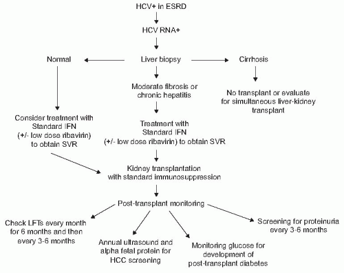
FIGURE 82.9 An algorithm for pre- and posttransplant management of HCV+ patients. ESRD, end-stage renal disease; IFN, interferon; SVR, sustained viral response; HCC, hepatocellular carcinoma. (Adapted from Huskey J, Wiseman AC. Chronic viral hepatitis in kidney transplantation. Nat Rev Nephrol. 7(3):156-165, with permission.)


Malignancy Screening

Patients with no history of malignancy should be screened using age-appropriate guidelines developed for the general population. Additionally, screening for renal cell carcinoma via ultrasound is gaining attention given its increased prevalence in patients with end-stage kidney disease and following a transplant.96,97 Patients with a history of malignancy should be disease free prior to transplantation. Generally, it is recommended that patients should have a disease-free interval of 2 to 5 years prior to transplantation, due to the increase in malignancy risk ascribed to immunosuppressive medications following the transplant. However, with advances in treatment options for patients with various forms of malignancy, it is often difficult to ascribe a specific waiting period following successful treatment. The Canadian Society of Transplantation
has published consensus guidelines that attempt to take into consideration a number of more common clinical circumstances, but these must continue to be reviewed in the context of emerging data.80 Oncology referral and discussion of expected disease-free survival is an important part of the evaluation process for those with a history of malignancy.


Infection

Patients should be free of active infection prior to transplantation. Appropriate immunizations against influenza, pneumococcus, hepatitis B, and, when appropriate, varicella, should be performed prior to transplant. Patients in areas with high prevalence rates of tuberculosis and those with an abnormal chest X-ray suggesting granulomatous disease should undergo purified protein derivative (PPD) testing. If the PPD is nonreactive (as is common in patients with renal failure) or if the patient has a history of BCG vaccination, IFN-γ release assay testing may be of benefit in the diagnosis of latent tuberculosis.98


Additional Pretransplant Evaluation Considerations


Gastrointestinal Evaluation

In patients with symptomatic cholelithiasis, a cholecystectomy should be performed to eliminate the risk of possible sepsis after transplantation. Patients with diabetes and asymptomatic gallstones seen with ultrasonography (˜20% to 30% prevalence) may also benefit from pretransplant elective cholecystectomy.99 A colonoscopy should be performed for patients >50 years of age to screen for colon cancer. Those with known colonic disease, especially those with diverticulitis, should be evaluated with a barium enema and a colonoscopy and, if appropriate, should be treated with surgical resection prior to transplantation.


Genitourinary Evaluation

An accurate evaluation of the lower urinary tract function prior to transplantation is important to minimize postoperative urologic complications. The original renal disease must be clearly defined. Any history of prior bladder surgery, repeated urinary infections, and current reports of urine cultures should be obtained. A voiding cystourethrogram should be performed if there is clinical or historical evidence of a bladder or ureteric abnormality. Cystoscopy and urodynamic studies should be performed in patients with evidence of bladder dysfunction. Urologic operations are necessary either to correct or improve obstructive lesions or sometimes to provide a conduit in the presence of a neurogenic bladder or a previous cystectomy.


Immunologic Evaluation

The human HLA system—encoded on the short arm of chromosome 6—encodes antigens that play a major role in host immune responses. The importance of these antigens in the practice of organ transplantation became evident when immunologic mechanisms were found to be responsible for immediate allograft destruction in early attempts at kidney transplant between non-HLA identical pairs.100 Antibodies against HLA antigens are formed as a result of pregnancy, transfusions, and prior organ transplantation and have the potential to cause hyperacute, acute, or chronic antibody-mediated allograft rejection (AMR).101 A landmark study by Drs. Patel and Terasaki in 1969 described a complement-dependent cytotoxicity assay (CDC) for anti-HLA antibodies that was highly predictive of hyperacute graft rejection.102 The CDC assay screens for donor-directed complement fixing antibodies in the sera of recipients via in vitro mixing studies with donor lymphocytes, and became the first routinely used cross-match technique in organ transplantation.

Although CDC cross-matching revolutionized the pre-transplant immunologic evaluation and has remained in use for 5 decades, it is associated with limited sensitivity and requires a subjective visual assessment of cell lysis. Flow cytometry (FCXM) was introduced in the 1980s as a method for screening recipient sera for donor-directed HLA antibodies with up to a threefold higher sensitivity compared to CDC. FCXM involves the incubation of donor T and B lymphocytes with recipient sera, allowing for the binding of any donor-directed HLA antibodies that may be present. After the addition of a fluorochrome-conjugated secondary (anti-IgG) antibody, anti-HLA antibody strength is measured by mean fluorescence intensity (MFI) or channel shift. Positive FCXM has been shown to be predictive of rejection and graft loss.103 In addition to a more sensitive antibody detection, FCXM involves independent testing of B and T lymphocytes and thus allows for further characterization of HLA antibodies as specific to antigens belonging to either class I (present on all nucleated cells) or class II (present only on antigen-presenting cells such as B cells).

More recently, the practice of pretransplant antibody screening was again revolutionized by the introduction of solid phase testing using antigen-coated microbeads (SAB).104 This assay, unlike the cell-based CDC and FCXM, uses microparticle “beads” coated with a single HLA antigen peptide incubated with recipient sera and a fluorochrome-conjugated secondary antibody. The strength of antibody binding is again determined by MFI; however, the identification of exact antigen specificities is now possible with anti-HLA antibodies further characterized as donor specific (DSA) or not. The presence of pretransplant DSA detected by single antigen beads (SAB) has been associated with an increased risk of antibody-mediated rejection (AMR) in multiple reports, however the antibody strength (MFI) that correlates with poor graft outcomes remains a matter of debate.

In addition to HLA and blood group ABO typing, most patients undergo a final cross-match prior to the kidney transplant in order to minimize the risk of hyperacute and acute AMR. There is considerable center-to-center variation in the cross-match technique and it includes CDC, FCXM, SAB, or any combination of the three. In general,
contraindications to transplant include a positive CDC cross-match or T-cell FCXM.105 A number of transplant centers in the United States have forgone the CDC method in favor of FCXM and SAB analysis, tests that offer improved sensitivity at the likely expense of decreased specificity. For example, although a positive CDC cross-match has remained an absolute contraindication to transplant, the clinical implications of a weak FCXM or low level antibodies detected by SAB are less clear and are currently a matter of intense clinical research. Thus, the evolution of cross-match techniques has resulted in increasing protection against early AMR at the expense of potentially withholding the transplant in patients with clinically irrelevant antibodies detected by sensitive assays.

Although pretransplant cross-matching serves to minimize the risk of early AMR between a recipient and a particular donor, the overall level patient sensitization helps to estimate the likelihood of positive cross-match with the general population. Patients with high levels of circulating anti-HLA antibodies are regarded as sensitized and of higher immunologic risk. Sensitization is quantified by the degree of PRA, or more recently by calculated PRA (cPRA). Historically, PRA has been determined by complement-dependent cytotoxicity mixing studies of recipient sera with a panel of lymphocytes derived from the general population, where positive reactions in 50% of samples would correspond to a PRA of 50%. It should be noted that the degree of sensitization has no bearing on the outcome of a cross-match between recipient and an individual donor, serving instead to estimate the probability of a positive cross-match between recipient and any given potential donor in the general population. As increasing levels of PRA correspond to decreasing numbers of donors to which the recipient will have a negative cross-match, sensitized patients wait much longer for transplants and are transplanted at a lower rate per year. Strategies aimed at desensitizing patients with either high PRA or positive cross-matches to potential living donors using plasmapheresis, intravenous immunoglobulin (IVIG), and anti-B cell agents bortezomib and rituximab have been met with variable success and are associated with high rates of posttransplant AMR.106,107

In October 2009, the United Network for Organ Sharing (UNOS) implemented a strategy replacing conventional PRA measurements with cPRA, a measure of sensitization based on unacceptable antibody levels as determined by SAB analysis.108 Potential transplant patients are screened for antibodies against HLA antigens by SAB assays at various intervals while on the waiting list, with antigens to which the patient has significant levels of antibody listed as “unacceptable” by the transplant center. The cPRA is determined by entering the patient’s unacceptable antigens into a formula that calculates the relative frequency of these antigens in the general population. Using this strategy, patients are able to undergo a “virtual” cross-match with prospective donors, taking into account both the donor HLA profile and the previously listed unacceptable antigens for the recipient. Final cross-matching is then performed only if the virtual cross-match is negative. An initial analysis of virtual cross-matching shows improved organ allocation efficiency and improved access to transplantation for sensitized patients on the waiting list compared to prior eras.109


DONOR SELECTION


Live Kidney Donation

Living donor transplants comprise about 35% of all transplants performed in the United States (Fig. 82.2), whereas their proportion is much less (10% to 15%) in Europe and Australia, and much higher in the Middle East.110 Outcomes of related versus unrelated donor kidney transplants are comparable, with the exception of the 2-haplotype HLA matched living related donor transplant, and are superior compared to kidney transplants from a deceased donor.4 This is due to a number of factors that include the minimization of cold ischemia time and the risk of delayed graft function, as well as the benefits imparted by the opportunity to perform a detailed history and medical assessment of the donor.

The initial series of tests for a potential living donor include ABO blood group and HLA tissue typing, which can be completed at a brief outpatient visit. The living donor not only needs a thorough medical evaluation, with particular attention to renal function and the urinary tract, but also a renal angiography or magnetic resonance angiography to identify vascular or anatomic variation of the kidneys or the collecting systems (Table 82.6). It is important to ascertain that both kidneys are of normal size and configuration and that a donor kidney with a single renal artery can be obtained. Several long-term follow-up studies have not revealed any adverse problems for the living donor with a single kidney and support the judicious use of the live kidney donor.111,112 The donor surgical mortality risk is 3.1 per 10,000 donors, and life expectancy in the donor remains unaffected.112 Although compensatory hyperfiltration occurs in the remaining kidney, the achieved glomerular filtration rate is typically 70% of baseline after 2 to 4 weeks. Blood pressure appears to increase by ˜5 mm 5 to 10 years from donation over pre-transplant values, adjusted for blood pressure increases with aging.113 Black donors appear to have a greater risk of hypertension than white donors, thus it may be reasonable to have more stringent blood pressure thresholds for the black potential kidney donor.114 Women who may desire pregnancy following kidney donation should be counseled that current observational data suggest a similar rate of fetal loss, preeclampsia, gestational diabetes, and gestational hypertension compared to the general population, but higher rates of each of these parameters compared to pregnancies that had occurred in donors prior to donation.115,116

Efforts to increase transplantation rates have led to the consideration of living donors with mild medical conditions and from extended social relationships from the potential recipient (Table 82.7). The nondirected kidney donor, an individual who contacts transplant centers wishing to
donate a kidney for purely altruistic reasons, provides an opportunity to benefit individuals who may have an incompatible donor or individuals without a living donor option.117 Paired exchange programs have been developed to identify two potential donors who wish to donate to a family or friend but are unable to due to blood group incompatibility or a positive cross-match. Two such donors and their prospective recipients are then paired, with donor A donating to recipient B and donor B donating to recipient A. When an altruistic donor is introduced to paired exchange programs, it may result in significant opportunity for transplantation of a number of incompatible pairs.118,119 Another circumstance is the matched donor in which a prospective recipient pays a monthly fee to a coordinating site, which presumably has access to a list of potential parties interested in donating their kidney. In the United States, assurances required from these donor/recipient circumstances must include the lack of monetary benefit for the donor (altruism). The U.S. Organ Transplantation Act of 1984 (HR5580, Title II) makes it a federal crime to engage in organ sale and commerce. Other countries have eliminated the waiting list with the use of monetary incentives for living unrelated donation, a topic that continues to be debated worldwide.120,121


TABLE 82.6 Suggested Evaluation Process for Potential Living Donors


Donor screening


Educate patient regarding deceased and live donation


Take family and social history and screen for potential donors


Review ABO compatibilities of potential donors


Tissue type and cross-match ABO-compatible potential donors


Choose primary potential donor with patient and family


Educate donor regarding process of evaluation and donation


Donor evaluation


Complete history and physical examination


Comprehensive laboratory screening to include complete blood count, chemistry panel, HIV, very low-density lipoprotein, hepatitis B and C serology, cytomegalovirus, glucose tolerance test (for diabetic families)


Urinalysis, urine culture, pregnancy test


Protein, 24-hr urine collection


Creatinine, 24-hr urine collection


Chest radiograph, cardiac stress test for patients >50 years of age


Helical computed tomography urogram


Psychosocial evaluation


Repeat cross-match before transplantation


TABLE 82.7 Exclusion Criteria for Living Kidney Donors


Age < 18 or >65-70 yr


Significant medical illness (e.g., cardiovascular/pulmonary diseases, recent malignancy)


History of recurrent kidney stones


History of thrombosis or thromboembolism


Psychiatric contraindications


Obesity


Hypertension (> 140/90 mm Hg or necessity for medication)


Proteinuria (>250 mg/24 hr)


Microscopic hematuria


Abnormal glomerular filtration rate (<80 mL/min)


Diabetes (abnormal glucose tolerance test or hemoglobulin A1c)


Urologic/vascular abnormalities in donor kidneys


Deceased Kidney Donation

Deceased kidney donors can be classified as those donors who are deceased by brain death (DBD) or those who are deceased by cardiac death (DCD). The criteria for the diagnosis of brain death have been well defined in most Western countries, although the requirements vary little from country to country (Table 82.8). Protocols exist that vary from country to country regarding DCD donation, but generally involve a waiting period of 5 minutes following the declaration of death prior to organ procurement.

DBD donors have been subcategorized as standard criteria donors (SCD) or expanded criteria donors (ECD). ECD donors are defined based on the presence of variables
that increased the risk for graft failure by 70% compared with an SCD kidney and include donors over the age of 60, or donors between the ages of 50 to 59 with two of three additional criteria: (1) cerebrovascular accident as a cause of death, (2) prior diagnosis of hypertension, or (3) terminal serum creatinine greater than 1.5 mg per day. The rationale for making the distinction between SCD and ECD was to allocate kidneys efficiently to those in greatest need (those at greatest risk for mortality while on dialysis).122 The survival benefit of ECD transplant over dialysis is present across all candidates, but in particular is of benefit to those with diabetes over the age of 40 or who are in regions with waiting times for a transplant of > 1,350 days.5


TABLE 82.8 Medical Evaluation of the Potential Deceased Donor


I.


Diagnosis of death


A.


Preconditions


1.


Positive diagnosis of brain death (irremediable structural brain damage)


2.


Planned withdrawal of cardiopulmonary support for irreversible conditions


B.


Exclusions


1.


Primary hypothermia (<33°C)


2.


Drugs


3.


Severe metabolic or endocrine disturbances


C.


Tests


1.


Absent brainstem reflexes


2.


Apnea (strictly define)


II.


No preexisting renal disease


III.


No active infection tests:


A.


HBsAg; five antibodies to cytomegalovirus and hepatitis C virus


B.


HIV antibodies


C.


HIV antigen in high-risk patients


HBsAg, hepatitis B surface antigen.


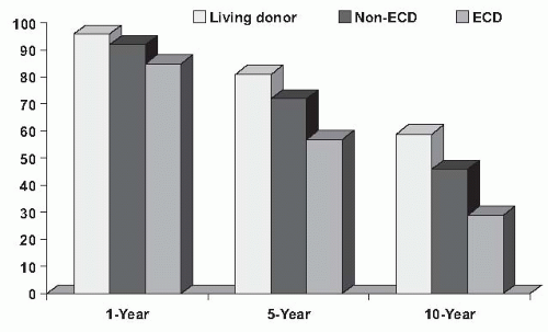
FIGURE 82.10 One, 5-, and 10-year kidney graft survival from living donors (LD), standard criteria donors (non-SCD), and expanded criteria donors (ECD). (Adapted with permission from 2009 OPTN/ UNOS Annual Report, Tables 5.10a, b, d.)

DCD donors can be controlled (with a planned withdrawal of cardiopulmonary support following a consent for donation) or uncontrolled (a cardiopulmonary death in a medical setting with rapid perfusion of organs, prior to consent). The latter is practiced in countries in which there are national policies of presumed consent.123 The additional warm ischemia time that occurs during the DCD procurement process results in higher rates of delayed graft function and primary nonfunction, but with comparable long-term graft survival to SCD kidney transplants.124 Figure 82.10 summarizes the most recent graft survival data from the United States by type of organ.

For all organ donors, there should be no evidence of primary renal disease and no generalized viral or bacterial infection. Biopsies are often performed to determine glomerulosclerosis in cases in which there is a question of suitability for transplant, but this has not consistently demonstrated a predictive value for graft function or longevity.125 Screening for hepatitis B, C, and HIV infection is performed to exclude donors, although in the case of hepatitis C reactivity, these donors may be used for selected recipients with chronic hepatitis C infection with good results.126 Epstein-Barr virus (EBV) and cytomegalovirus (CMV) testing is performed to assess the risk of transmission and posttransplant complications for the recipient.


THE TRANSPLANT OPERATION-DONOR PROCUREMENT


Living Donor Nephrectomy

A living donor nephrectomy can be performed via either an open or a laparoscopic approach. The open approach entails a flank incision by an open nephrectomy. The approach to the kidney, typically the left kidney because this has the longer renal vein, may be either below or through the bed of the 12th rib using a retroperitoneal approach, or rarely via an anterior transperitoneal approach using a midline incision. Care
must be given to retraction of the kidney during its removal to avoid traction injury of the renal artery and dissection in the hilum of the kidney, particularly between the ureter and the renal artery, which should be avoided to prevent damage to the ureteric blood supply. Furthermore, in removing the ureter down to the brim of the pelvis, care should be taken to leave an adequate amount of periureteric tissue. A living donor nephrectomy for transplantation can also be performed by laparoscopic approach.127 This approach results in less postoperative surgical pain, a shorter hospital stay, and a quicker recovery than the standard open donor nephrectomy (Table 82.9). The laparoscopic techniques have been rapidly adopted worldwide; an analysis from Australia/ New Zealand transplant centers upon the introduction of the laparoscopic technique in 1997 through 2004 demonstrates comparable rates of technical failure, delayed graft function, and graft survival to an open nephrectomy, with a conversion rate to open procedures of 6%.128 This conversion rate is much higher than that reported for experienced centers of 1%.129


TABLE 82.9 Advantages and Disadvantages of Laparoscopic Nephrectomy


Advantages


Less postoperative pain


Minimal surgical scarring


Rapid return to fill activities and work (approx. 4 weeks)


Shorter hospital stay


Magnified view of renal vessels


Disadvantages


Longer operative time, impaired early graft function, graft loss or damage during “learning curve”


Pneumoperitoneum may compromise renal blood flow


Tendency to have shorter renal vessels and multiple arteries


Added expense of specialized instrumentation


Deceased Donor Nephrectomy

Currently, most kidneys will be removed as part of a multiple-organ procurement procedure in which not only the kidneys are removed, but also the liver and heart and, occasionally, the lungs and pancreas. There are two basic approaches to a deceased donor nephrectomy. In one, each kidney is removed individually with a patch of aorta via an anterior approach, whereas in the other, which is the more satisfactory technique, both kidneys are removed en bloc with the appropriate segment of the aorta and vena cava. The dissection of the vessels and the kidneys can then be completed after hypothermic perfusion and storage. In situ perfusion may be performed in both cases before and during removal.


Renal Preservation

The effective preservation of the kidney is an integral part of a kidney transplantation program and has evolved on the basis of known principles of preservation because of a need for longer storage of kidneys.130 The ability to preserve kidneys provides time for tissue typing and cross-matching and the selection of the most appropriate recipients for a particular donor on the basis of matching, as well as the preparation of the patients selected, who often may need dialysis before transplantation, and, finally, the transport of the kidneys to a center where an appropriately matched recipient may be awaiting a transplant.

There are two methods of preservation: simple cold storage in ice after flushing with a hypothermic solution to give a renal core temperature of 0°C and a more complicated approach of continuous perfusion of the kidney with an oxygenated colloid solution. The simple cold storage approach is most commonly used, and provides adequate preservation for at least 24 to 30 hours. The kidneys are initially flushed free of blood with a cold solution via the aorta and renal artery while the kidney is in situ. Many different flushing solutions have been used; currently the most common preservation solutions in use in the United States are Viaspan (University of Wisconsin [UW] solution or Belzer solution) and Custodiol (histidine-tryptophan-ketoglutarate [HTK]) (Table 82.10).131 Drugs, metabolites, and other agents have been used to enhance the effects of cold preservation. The aim of these maneuvers is to reduce the incidence of posttransplant acute tubular necrosis.

In the absence of any warm ischemia, which is generally the case with a brain-dead donor on a ventilator, the immediate function can be obtained in most kidneys with up to 24 hours of preservation and even after 48 hours of preservation in some patients. However, from 24 hours onward, most kidneys will have a significant period of delayed function ranging from 1 day to several weeks and there will be a significant incidence of primary nonfunction. Because 18 to 36 hours is an adequate time for most units and also allows time for transport of kidneys within a region or country, there has been widespread adoption of the simple cold storage (CS) technique for preservation.

Compared to the more traditional CS technique, machine perfusion (MP) involves placing kidneys from a deceased donor on a perfusion device that provides either continuous or pulsatile flow of a hypothermic solution through the renal vasculature.132 In theory, by eliminating toxic metabolic byproducts and providing nutrients and oxygen, MP may protect deceased-donor kidneys from peritransplant ischemia/reperfusion injury that is responsible for the majority of clinically significant delayed graft function (DGF), an event independently associated with acute rejection and poor graft survival.133 In recent years, the use
of kidneys from less traditional donors has been on the rise, including ECD and DCD, both of which are associated with significantly higher rates of DGF.134,135 As a result, a number of recent clinical trials have studied preservation methods in an attempt to demonstrate improved rates of DGF in deceased donor kidney transplants.


TABLE 82.10 Contents of Commonly Used Cold Preservation Solutions


Custodiol (HTK)


UW


Sodium (mM)


15


30


Potassium (mM)


10


120


Magnesium (mM)


4


5


Histidine (mM)


198


Tryptophan (mM)


2


Alpha-ketoglutarate


n/a


Mannitol (mM)


30


Sulfate (mM)


5


Phosphate (mM)


25


Lactobionate (mM)


100


Raffinose (mM)


30


Adenosine (mM)


5


Allopurinol (mM)


1


Glutathione (mM)


3


Insulin (units/L)


100


Dexamethasone (mg/L)


8


Hydroxyethyl starch (g/L)


50


HTK, histidine-tryptophan-ketoglutarate; UW, University of Wisconsin.


Most recent clinical trials have shown improved DGF rates with the use of MP compared to CS. For example, in the largest prospective randomized controlled trial to date, Moers et al.136 studied the outcomes of 336 kidney pairs, where 1 kidney per pair underwent MP and the other CS, and reported both lower rates of DGF and improved 1-year allograft survival in the MP group. Subsequent prospective extensions of this trial demonstrate lower DGF rates for both ECD and DCD, improved 1-year graft survival for ECD, but comparable 1-year graft survival for DCD kidneys with MP versus CS transplants.137,138 In contrast, a UK-based paired kidney analysis of DCD kidneys undergoing either MP or CS failed to show any difference in terms of DGF rates or 1-year graft survival.139 Despite these mixed results, MP likely results in modestly less DGF for deceased donor kidney transplants of any type compared to CS. Whether the increased cost associated with MP can be offset by improved long-term graft outcomes has yet to be clarified.


THE TRANSPLANT OPERATION-RECIPIENT SURGERY

The surgical technique of renal transplantation is standardized.140 In cadaver transplantation, the kidney must first be inspected to ensure that it is suitable for transplantation before undertaking the operation. This procedure should be carried out in the operating room on a sterile back table. The procedure is to remove the unnecessary fatty tissue and to prepare the donor vessels. In small pediatric donors, both kidneys can be used en bloc for transplantation in adults.

The transplanted kidney is implanted in the retroperitoneal space in either the right or left iliac fossa through an oblique incision extending from the suprapubic area to a point just above and medial to the anterior superior iliac crest. For transplantation, after failed transplants in both iliac fossae, a lower midline intraperitoneal approach should be used.

The iliac vessels should be carefully dissected141 and the lymphatics ligated to prevent lymphocele formation. The donor renal vein is anastomosed end to side to the external iliac vein. The renal artery is anastomosed to the external or common iliac artery end to side using a cuff of aorta as a patch for the anastomosis, or it is anastomosed end to end to the internal iliac artery, which has been previously ligated and divided. The end-to-side anastomosis using a cuff of aorta is the simpler anastomosis; it is the most appropriate one to use in cadaver transplantation when the renal artery is provided with a cuff of aorta. The end-to-end anastomosis to the internal iliac artery is technically more demanding and should only be used in living donor kidney transplantation.

Implantation of the ureter in the bladder is performed in one of two ways.142 The first is to anastomose the spatulated end of the ureter mucosa to the dome of the bladder drawing muscle over the anastomosis to provide a tunnel. The second technique is to bring the ureter through the lateral wall of the bladder and down through a 2- to 3-cm submucosal tunnel and out in the vicinity of the patient’s own ureteric orifices at the trigone, where it is anastomosed mucosa to mucosa. The success of the first technique is greater than the second. Preventive antibiotics with appropriate broadspectrum activity should be given with the premedication, in particular to protect against the possibility of infection being transmitted with the transplanted kidney.


General Postoperative Management and Follow-up

Routine postoperative observations should include the monitoring of vital signs, fluid intake, and urine output. A postoperative hematuria is usually transient. The Foley
catheter is generally left in these patients for 3 to 4 days because of high urine outflow rates that occur during this time in order to prevent overdistension of the bladder. This is particularly important in diabetic patients who frequently have neurogenic bladders and can have extremely large bladder volumes before they develop an urge to micturate. Catheters should also be carefully monitored for obstruction and irrigated under sterile conditions if occluded by a clot.

Immediate function of the transplanted kidney makes postoperative management of the patient in the first few days much simpler than if the kidney is not functioning. The patient, particularly in the case of a living related transplant, may have a massive diuresis in the first 48 hours, and, for this reason, hourly monitoring of the urine output and a central venous line are essential to balance the fluid requirements appropriately. A very basic regimen, at least for the first few hours, is to replace fluid at the rate of the last hour’s output plus 50 mL per hour of IV fluid. This can then be modified according to the kidney function and the central venous pressure.

Within 48 hours, particularly with a functioning kidney, the patient’s restored sense of well-being is quite remarkable and most patients can get out of bed on the second postoperative day. Provided that no complications ensue and that any early rejection episode can be dealt with satisfactorily with appropriate treatment, these patients are ready to leave the hospital by the 3rd to 5th or 6th postoperative day.

Only gold members can continue reading. Log In or Register to continue

Stay updated, free articles. Join our Telegram channel

May 29, 2016 | Posted by in NEPHROLOGY | Comments Off on Clinical Aspects of Renal Transplantation

Full access? Get Clinical Tree

Get Clinical Tree app for offline access