Chronic Tubulointerstitial Nephritis


Chronic Tubulointerstitial Nephritis


Allison A. Eddy

Ikuyo Yamaguchi


Chronic tubulointerstitial nephritis (TIN) encompasses a vast array of chronic kidney diseases that share a primary pathologic process that begins at the level of the tubules and their surrounding interstitial space. In addition, it is now recognized that chronic tubulointerstitial disease is the final common pathway that causes progressive renal functional loss in all chronic kidney disease (CKD), whether it begins in the tubulointerstitium or in other renal compartments. Due to the importance and unique clinicopathologic features of many diseases that cause chronic TIN, many are discussed in greater detail in other chapters. The present chapter provides a general overview and discussion of this entire group of disorders, with an effort to highlight shared and unique features of each. For the purpose of presentation, chronic TIN is divided into subcategories (Table 57.1). One of the most common causes of chronic TIN in developed countries is chronic renal allograft rejection, which is discussed in Chapter 81.


NORMAL TUBULOINTERSTITIAL ARCHITECTURE

Renal tubules constitute the largest component of the renal parenchyma, estimated at 80% to 90%, which explains why disrupted tubular integrity and function plays such an important role in renal functional decline. Most of the tubules in the renal cortex are proximal tubules. The peritubular region is occupied by the vasculature and the interstitial space. The glomerular efferent arterioles branch to form the peritubular capillary network, which serves the vital role of delivering oxygen to support tubular cell metabolic and transport functions. The extravascular peritubular compartment, known as the interstitium, is typically inconspicuous, especially in the renal cortex. However, stromal cells and extracellular matrix proteins residing in the interstitium play a key role during renal development and in polarizing the renal response to injury toward regeneration or chronic sequelae. Residing within the interstitium are two important cell populations. The most abundant are fibroblasts, well recognized for their role of synthesizing extracellular matrix proteins. These proteins (primarily fibronectin; fibrillar; collagens I, III, and VI; and proteoglycans) provide a structural framework for nephrons and the vascular network. The functional heterogeneity of interstitial fibroblasts is increasingly recognized, even within normal kidneys.1 A subset is specialized to synthesize erythropoietin (Fig. 57.1),2 whereas others are pericytes closely opposed to peritubular capillaries.3 Present within the medulla is a unique population of lipid-laden interstitial cells which are thought to be a source of prostaglandins involved in blood pressure control.4 The second interstitial cell population consists of myeloid cells that are derived from bone marrow cells and are slowly and continuously replenished. This group of cells also has functional heterogeneity. The majority appear to be MCH class II positive dendritic cells, whereas others are scavenger-type macrophages. In normal kidneys the myeloid cells are thought to serve surveillance functions to protect the kidney from noxious materials and foreign invaders. They become actively engaged in renal responses to injury. Interstitial myeloid cells rarely proliferate in situ; the interstitial inflammatory response that characterizes many acute and chronic kidney diseases is dependent upon the recruitment of lymphohematopoietic cells from the circulation.

Ongoing studies are attempting to answer the question of whether a pluripotent stem cell also resides within the renal interstitium.5 It has been suggested that the renal medulla is a niche for kidney stem cells. These slowing dividing cells can be identified as “label-retaining cells” using detectable thymidine markers. There are conflicting data about the ability of these cells to proliferate and migrate to the site of injury and participate in renal regeneration.6,7 It has also been proposed that specialized progenitor cells may reside within tubules, but this remains unproven.


HISTOPATHOLOGIC FEATURES OF CHRONIC TIN

The histologic hallmark of chronic TIN is an increase in the fractional volume of the interstitial space caused by an expansion of extracellular matrix proteins—the defining feature
of interstitial fibrosis or scarring. This abnormal matrix is comprised of both a greater abundance of normal interstitial matrix proteins and the de novo appearance of additional matrix proteins. Interstitial fibrosis is accompanied by irreversible tubular damage, ranging from abnormally dilated (ectatic) tubules to atrophic tubules surrounded by abnormally thickened and wrinkled tubular membranes to complete tubular drop-out (often leaving behind the signature “atubular” glomeruli). In parallel, peritubular capillaries are also lost. In some diseases such as chronic allograft rejection, an abnormal process of interstitial lymphangiogenesis has been described, but its specificity and functional significance remain unclear.


TABLE 57.1 Chronic Tubulointerstitial Nephritis Classification


PRIMARY TUBULOINTERSTITIAL KIDNEY DISEASES


Genetic diseases


Familial TIN (MCKD2)


Uromodulin mutations
Renin mutations
Unknown mutations


Nephronophthisis (NPHP)


Isolated kidney disease
Associated with extrarenal manifestations


Polycystic kidney diseases (PKD)


Autosomal dominant
Autosomal recessive
Syndromic
Others


Metabolic disorders


Cystinosis
Oxalosis
Mitochondrial cytopathies
Methylmalonic acidemia


Immunologic diseases


TIN most common kidney manifestation


Sjögren syndrome
IgG4-related disease
Sarcoidosis
TINU
Renal allograft rejection
Anti-TBM nephritis


TIN usually associated with glomerulonephritis


Systemic lupus erythematosus
ANCA+ vasculitis
Anti-GBM nephritis
Cryoglobulinemia
Membranoproliferative glomerulonephritis
IgA nephropathy
Others


Chronic nephrotoxicity


Drugs


Calcineurin inhibitors
Analgesic nephropathy
Lithium


Herbs


Aristolochic acid
Others


Heavy metals


Lead Cadmium


Chronic metabolic disorders


Hypercalcemia/hypercalciuria
Hyperphosphatemia/hyperphosphaturia
Hyperuricemia/hyperuricosuria
Hypokalemia


Congenital abnormalities


Dysplasia
Obstruction


CHRONIC KIDNEY DISEASE-ASSOCIATED TIN


Proteinuria-associated TIN


Chronic kidney disease universal progression pathway


SEQUELAE TO ACUTE TUBULOINTERSTITIAL INJURY


Acute kidney injury


Ischemia-reperfusion injury


Acute interstitial nephritis/nephrotoxicity


Infections


Bacterial (systemic, pyelonephritis, xanthogranulomatous pyelonephritis)
Mycobacteria
Viral
Fungal
Parasitic


Drugs


Proton pump inhibitors
Chemotherapeutic drugs
Antimicrobial drugs
NSAIDs


Hematologic disorders


Leukemia
Lymphoma
Multiple myeloma
Sickle cell disease


MCKD2, medullary cystic kidney disease type 2, familial juvenile hyperuricemic nephropathy; TINU, tubulointerstitial nephritis with uveitis; anti-TBM, anti-tubular basement membrane; anti-GBM, anti-glomerular basement membrane; NSAIDs, nonsteroidal anti-inflammatory drugs.


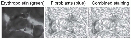
FIGURE 57.1 Peritubular fibroblasts produce erythropoietin. Using a mouse line that was genetically engineered to express green fluorescent protein-labeled erythropoietin, and kidney Cre-labeled fibroblasts that are detectable by beta galactosidase staining (blue), peritubular fibroblasts are identified as the source of erythropoietin. Loss of this function explains why anemia may be more severe in patients with chronic kidney disease due to chronic tubulointerstitial nephritis. (From Asada N, Takase M, Nakamura J, et al. Dysfunction of fibroblasts of extrarenal origin underlies renal fibrosis and renal anemia in mice. J Clin Invest. 2011;121:3981, with permission.) (See Color Plate.)

The other important histopathologic feature that typifies chronic TIN is a significant change in interstitial cellularity. Unlike normal kidneys, the interstitial space becomes populated by transformed fibroblasts that are recognized by their expression of α-smooth muscle actin (α-SMA), a protein typically associated with smooth muscle cells. Known as “myofibroblasts,” these cells are considered the primary source of extracellular matrix proteins that generate the fibrotic or scarred interstitium. The second important
change in interstitial cellularity is the appearance of an infiltrate of mononuclear cells. These cells are primarily of bone marrow origin.


TABLE 57.2 Histopathologic Features of Acute and Chronic Tubulointerstitial Nephritis


Features


Acute


Chronic


Tubules


Epithelium


Necrosis


Atrophy


Basement membrane


Disrupted


Thickened


Shape


Preserved


Dilated


Interstitiuma


Cell infiltrates


Lymphocytes (CD4+ T cell dominant)
Eosinophils in early stage


Monocytes and macrophages
Lymphocytes


Myofibroblasts


Minimum


Increased


Edema


+ + +


+


Fibrosis


+


+ + +
(Collagen and other matrix protein deposits)


Vasculature


Peritubular capillaries


Preserved


Reduced density
De novo lymphatic vessels


Large vessels


Minimum


Variesb


Glomeruli


None to minimal change


Periglomerular fibrosis Focal or global glomerulosclerosis


a The severity of the changes is given as an estimate with + for minimal to + + + as severe.

b Pathologic changes may suggest a primary process such as atherosclerosis, scleroderma, thromboembolic disease, vasculitis, or chronic allograft rejection.


A frequent challenge of a new histopathologic diagnosis of chronic TIN is the lack of clarity of the initiating disease process. For some disease entities, specific diagnoses are made using other criteria: imaging for cystic kidney disease and anatomic genitourinary anomalies, the presence of extrarenal manifestations (autoimmune diseases, metabolic disorders, hematologic diseases, and congenital hepatic fibrosis), a positive family history, or history of exposure to a drug or toxin that is known to cause chronic TIN. In the absence of these diagnostic clues, it may not be possible to determine the primary etiology, as the renal histologic findings of many chronic TINs overlap. When more specific diagnostic criteria are available, they are discussed under the specific disease entities that are reviewed later.

Another potential diagnostic dilemma is the difficulty of differentiating acute and reversible TIN from chronic progressive TIN. Early diagnosis is important for certain disease etiologies, such as an exposure to nephrotoxins or development of treatable autoimmune diseases, for which a delayed diagnosis may be too late for injury reversal. Many of the tubulointerstitial disorders have a variable clinical course that spans the spectrum from acute to chronic and reversible to progressive. The tissue repair process itself may lead to pathologic fibrosis. Frequent regional variations in the TIN process mean that the degree of acute and chronic TIN may vary considerably from one tissue sample to another. The primary histologic findings that are used in an effort to differentiate acute from chronic TIN are summarized in Table 57.2.


CLINICAL MANIFESTATIONS AND LABORATORY ABNORMALITIES

Clinical manifestations of chronic TIN tend to be subtle. Patients with TIN may present with symptoms related to their primary diseases. They often also have nonspecific constitutional symptoms of chronic kidney disease such as fatigue, loss of appetite, nausea, vomiting, and sleep disturbance. In general, tubular dysfunction develops proportionally as glomerular filtration rate (GFR) declines. However, primary TIN diseases may present more prominent tubular dysfunction in the early stage compared to glomerular or vascular diseases. Proximal tubule dysfunction is characterized by inability to reabsorb filtered bicarbonate, glucose, amino acids, and phosphate in varying combinations, resulting in acidosis, glucosuria, phosphaturia, and aminoaciduria, as in
Fanconi syndrome. Low molecular weight proteins such as β2-microglobulin may not be properly reabsorbed, leading to tubular proteinuria. Distal tubular dysfunction manifests as renal sodium wasting, hyperkalemia, and nonanion gap metabolic acidosis. Collecting duct dysfunction leads to renal concentrating defects including features of diabetes insipidus and countercurrent exchange washout resulting in polyuria. Most TIN affects multiple sites of the nephron simultaneously, but to varying degrees. Hypertension, severe proteinuria, and edema are not usually characteristic of TIN in the early stage, but may develop later as progressing chronic kidney disease (CKD) with glomerular sclerosis. In addition to tubular dysfunction, anemia may be found disproportionally compared to the change in GFR if erythropoietin-producing peritubular cells (Fig. 57.1) are damaged early in the disease. Bone disease may also be prominent, as a result of chronic phosphate wasting caused by proximal tubular dysfunction.


FIGURE 57.2 Interstitial fibrosis: detection and correlation with renal functional loss. The fibrotic or scarred interstitium contains several extracellular matrix proteins, the most abundant being fibrillar collagens such as collagen III (A,B). Routine renal biopsy staining with Masson trichrome reacts with collagen to produce a green-blue color (C). Quantitative pathologic research studies often use picrosirius red staining, which is specific for cross-linked collagen fibrils (polarized image shown in D). The key structural change that underlies the loss of renal function in all chronic kidney diseases is tubular atrophy (upper graph) which is closely associated with interstitial fibrosis severity (lower graph). (A and B are from Jones CL, Buch S, Post M, et al. Pathogenesis of interstitial fibrosis in chronic purine aminonucleoside nephrosis.Kidney Int. 1991;40:1020. Upper graph is from Mackensen-Haen S, Bohle A, Christensen J, et al. The consequences for renal function of the interstitium and changes in the tubular epithelium of the cortex and medulla in various diseases. Clin Nephrol. 1992;37;70. Lower graph is from Schainuck LI, Striker GE, Cutler RE, et al. Structural-functional correlations in renal diseases: Part II: the correlations.Human Pathol. 1970;1:631. All are reproduced with permission.) (See Color Plate.)


INTERSTITIAL FIBROSIS: THE FINAL COMMON PATHWAY TO CHRONIC KIDNEY DISEASE

In both native and transplanted kidneys, progressive fibrosis of the renal interstitium is the predominant final common pathway of renal destruction, regardless of the etiology of the original kidney disease.8 Fibrotic injury is not limited to extracellular matrix accumulation, but also results in the subsequent loss of tubules and peritubular capillaries. Histopathologically, interstitial volume and reduced tubular epithelial cell density closely correlate with the loss of renal function and predict long-term outcomes (Fig. 57.2). The pathogenic process leading to fibrosis can be initiated by a variety of insults, including chronic tubular, glomerular, and vascular disease. Chemokines and chemoattractants such as monocyte chemoattractant protein 1 (MCP-1), complement
component C3, and osteopontin (OPN) activate capillary endothelial cells, leading to increased capillary permeability, recruitment of leukocytes into the interstitium, and activation of myofibroblast precursors. Inflammatory macrophages secrete diverse proinflammatory and profibrotic products that perpetuate injury and promote scarring. This process unleashes a cascade of inflammatory and fibrogenic signals within the interstitium. Some key molecules including fibrinogen, complement components C3a and C5a, tissue plasminogen activator (tPA), and oxidized albumin may arrive by leakage from the plasma, whereas others, such as the major fibrogenic factor transforming growth factor β (TGF-β), connective tissue growth factor (CTGF), platelet-derived growth factor (PDGF), tumor necrosis factor α (TNF-α), endothelin-1, angiotensin-II, placental growth factor, and angiopoietin-2, appear to be produced locally. Together, these factors activate fibroblasts, and promote their transformation into α-SMA positive myofibroblasts. The activated myofibroblasts produce collagen I, collagen III, fibronectin, and other matrix proteins, which accumulate in the interstitial space. The fibrotic process culminates in death of tubular cells and peritubular capillaries, leading to ablation of the entire nephron (Fig. 57.3).9,10,11,12


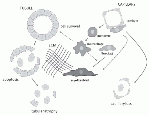
FIGURE 57.3 Schematic summary of the key cellular events contributing to the pathogenesis of chronic tubulointerstitial nephritis. Inflammatory macrophages are primarily recruited from the circulating pool of peripheral blood monocytes, under the direction of chemotactic signals derived from endothelial cells and damaged tubules and facilitated by increased capillary permeability. Functionally distinct macrophage subpopulations either propagate injury or promote tissue repair (including fibrosis) by releasing a variety of cytokines, growth factors, and other soluble products. A population of myofibroblasts appears de novo in the interstitium where they synthesize the majority of the extracellular matrix (ECM) proteins responsible for interstitial scarring. The primary origin of the myofibroblasts is still controversial; resident interstitial fibroblasts and capillary pericytes are considered most likely during active fibrogenesis. Through a variety of mechanisms, including hypoxia and oxidant stress, interstitial capillaries disappear and tubular epithelia undergo apoptotic death in parallel with progressive fibrosis.


Key Mechanisms


Tubular Epithelial Cells

The renal tubules account for approximately 80% of the total kidney volume. Tubular epithelial cells may be injured by immunologic, mechanical, chemical, genetic, or ischemic insults, which stimulate synthesis of inflammatory cytokines, cause functional perturbations, and/or lead to necrotic or apoptotic cell death. In acute kidney injury (AKI), tubular epithelial cells proliferate and replace damaged cells, restoring the architecture of the tubules. However, in CKD, complete tubular regeneration fails due to incomplete repair and persistent inflammation, leading to endoplasmic reticulum (ER) stress, loss of cytoskeletal integrity and polarity, and tubular barrier dysfunction, ultimately resulting in irreversible atrophic changes. The failure of tubular restoration is a critical turning point for CKD. Tubular atrophy may leave behind intact, atubular glomeruli, which are nonfunctional nephrons.13


Injured tubular epithelial cells often play a direct role in renal inflammation by secreting proinflammatory cytokines and growth factors including TNF-α, MCP-1, TGF-β, and RANTES. The production of growth factors may be cell cycle stage-specific, as it has been shown that acute kidney injury induced by ischemia/reperfusion leads to cell cycle arrest in the G2/M phase, followed by the release of growth factors such as TGFβ-1 and CTGF. These factors activate c-Jun N-terminal kinase (JNK) signaling, which promotes fibrosis.14

The fate of tubular epithelial cells is a critical determinant of nephron regeneration, and several mechanisms can direct each cell toward death by necrosis or apoptosis, or toward survival and proliferation. Tubular cell apoptosis is a common feature of CKD, and is known to be triggered by TGF-β1, TNF-α, Fas, p53, caspases, ceramide, and reactive oxygen species. Apoptosis can also be stimulated by the downregulation of survival factors such as epidermal growth factor (EGF) and vascular endothelial growth factor (VEGF). An important step in cell survival following injury is the removal of damaged proteins and organelles by cell-mediated autophagy. In this process, an autophagosome is formed from the ER membrane, engulfs intracellular deposits, and delivers the contents to lysosomes for degradation. There is evidence of enhanced autophagy in obstructed tubules and AKI, which is thought to promote recovery; failure of autophagy may lead to apoptosis and prevent recovery of tubular epithelial cells.15,16

When the tubular damage is controlled, the tubules can regenerate. The origin of the new tubular epithelial cells has been a topic of debate. The current prevailing view is that proliferation of surviving tubular cells is sufficient to account for the recovery, without compelling evidence that renal and/or extrarenal progenitor cells are incorporated. However, soluble factors released by bone marrow-derived cells are thought to facilitate the repair process.7,17,18


Inflammatory Cells

Infiltration of the interstitium by inflammatory cells is an integral component of the fibrogenic response (Fig. 57.4). One of the most important inflammatory cell types is the macrophage, which primarily originates from circulating monocytes. Resident dendritic cells have limited proliferative capacity. Macrophages are functionally heterogeneous and have the potential to secrete a vast repertoire of soluble mediators, including proinflammatory and profibrotic cytokines. Inflammatory monocytes undergo differentiation in response to cytokines and typically become polarized into one of two distinct phenotypes.19,20,21 This polarization process has been extensively investigated in mice, where classically activated “M1” macrophages are generated by exposure to interferon-γ and lipopolysaccharide. The M1 cells produce proinflammatory cytokines that propagate tissue injury. Alternatively activated “M2” macrophages are generated by exposure to interleukin-4 (IL-4) and IL-13. The M2 cells synthesize anti-inflammatory cytokines that promote tissue repair; however, this repair response may also lead to fibrosis. These differential functions have mainly been characterized using in vitro studies. In vivo, macrophage phenotypes are more diverse, and macrophages appear to switch phenotypes in response to different stimuli and microenvironments.19,21,22 The role of the lymphocytes (which are typically present and may even outnumber interstitial macrophages) and the resident dendritic cells in renal fibrosis is still not clear.23 It has been hypothesized that chronic
renal injury may expose neoantigens that trigger a secondary antigen-driven immune response to propagate injury, but this hypothesis has been difficult to test using experimental models. Candidate neoantigens have not been identified, and thus this paradigm remains hypothetical.


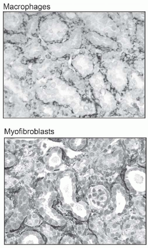
FIGURE 57.4 Interstitial cell mediators of chronic tubulointerstitial nephritis (TIN). Interstitial hypercellularity is a feature of chronic TIN, characterized by the presence of two distinct cell populations: lymphohematopoietic cells, with macrophages in particular (shown by CD68 staining in the upper photomicrograph) known to play an important pathogenic role, and myofibroblasts (shown by alpha smooth muscle actin staining in the lower photomicrograph). Limited human biopsy data show a significant relationship between interstitial myofibroblast density and renal prognosis. (The upper image is from Yamaguchi I, Tchao BN, Burger NL, et al. Vascular endothelial cadherin modulates renal interstitial fibrosis. Nephron Exp Nephrol. 2011; 120:e20, with permission.)


Myofibroblasts

Interstitial myofibroblasts serve a pivotal role in renal fibrosis by synthesizing extracellular matrix (ECM) proteins such as collagen I, collagen III, and fibronectin that accumulate within the renal interstitium. Myofibroblasts contain contractile stress fibers and express α-SMA (Fig. 57.4). Myofibroblasts are essential for wound healing and tissue remodeling. During wound healing, they are activated, migrate within the damaged tissue, proliferate, and secrete ECM in response to inflammatory factors such as TGF-β1. Once healing is complete, myofibroblasts disappear by apoptosis. However, in chronic fibrosis myofibroblasts persist and lead to pathologic tissue remodeling, ultimately impairing organ function.24

The origin of renal interstitial myofibroblasts is a topic of great interest and some controversy. Myofibroblasts are functionally heterogeneous, depending to some extent on their local environment and perhaps on their origin. Most myofibroblasts appear to be derived from intrarenal cells, which may include resident interstitial fibroblasts, pericytes, or perivascular cells within the adventitia of arterioles and arteries.3,25 Myofibroblasts might also be derived from epithelial-mesenchymal transition (EMT)26 and endothelial-mesenchymal transition (EndMT),27 although both of these events appear to be delayed until the advanced phase of renal fibrosis when basement membranes are destroyed. Other origins could include bone marrow-derived circulating fibroblasts or fibrocytes. Recent cell lineage tracing studies in genetically engineered mice support the view that myofibroblasts are rarely derived from tubular epithelial cells or fibrocytes; rather, they represent transformed interstitial fibroblasts, perivascular progenitor cells, and pericytes.3 TGF-β and CTGF produced by injured tubular cells and inflammatory interstitial cells not only stimulate fibroblast proliferation and transformation to myofibroblasts, but also induce fibroblast epigenetic changes that influence cell survival. For example, TGFβ-induced hypermethylation of RASAL1, an inhibitor of the Ras oncoprotein, results in prolonged fibroblast activation and kidney fibrosis.28

Multiple growth factors such as TGF-β1, PDGF, fibroblast growth factor 2, and CTGF are known to stimulate fibroblast activation and extracellular matrix production, whereas hepatocyte growth factor and bone morphogenetic protein 7 are antifibrogenic. Numerous studies have investigated the downstream signaling cascades leading to fibrogenesis, which are too numerous to describe here.8,11,12,29,30 Epigenetic mechanisms of regulation such as inhibition of DNA methylation and control of mRNA stability and translation by microRNA are also thought to play important roles in renal fibrosis.28

The processes of myofibroblast activation and apoptosis are of considerable interest as potential targets for antifibrotic therapy. TGF-β inhibition would appear to be an ideal strategy, but the complex effects of this multifunctional growth factor have presented challenges. Recent studies have focused on its downstream intracellular signals such as SMAD3, which activates microRNA-21, stimulating matrix production and fibrosis.31 The renoprotective effects of renin-angiotensin system (RAS) blockade are thought to be mediated at least in part by TGF-β inhibition. Other celltargeted strategies currently under investigation are aimed at inhibiting the formation of scar-forming myofibroblasts by profibrotic cytokines, promoting myofibroblast apoptosis, and/or inhibiting myofibroblast function (e.g., cell contraction or interactions via specific integrins).32,33


Capillary Changes, Hypoxia, and Oxidant Stress

The renal interstitium is perfused by an intricate network of peritubular capillaries that serve the vital role of oxygen delivery to metabolically active tubular epithelial cells. Peritubular capillary endothelial cells (ECs) undergo apoptosis during CKD, leading to capillary loss, and propagation of tissue hypoxia and oxidant stress. Based on several studies in animal models and human chronic kidney diseases, it is known that peritubular capillaries disappear in association with progressive interstitial fibrosis and tubular atrophy (Fig. 57.5).34,35,36,37 Although the sequence of events connecting capillary loss to fibrosis and impaired tubular function is poorly characterized, it has been suggested that interstitial hypoxia caused by arteriolar vasoconstriction and/or peritubular capillary regression is a primary event in CKD.38

Under normal conditions, ECs are quiescent and turn over slowly. Vessel stability depends on cell-cell and cell-matrix interactions, normal levels of growth and angiogenic factors, and shear stress from blood flow. However, during kidney injury shear stress is altered, interactions between ECs change, cell-matrix interactions are disrupted, and growth and angiogenic factors are produced, including TGF-β, angiopoietin 2, and in some models VEGF.39 As a consequence, ECs enter an activated state characterized by hyperpermeability, expression of leukocyte adhesion molecules, release of cytokines and growth factors, and enhanced cell migration and proliferation. EC activation is crucial for host defense and repair but may lead to dysfunctional changes including reduced production of nitric oxide (NO), a chronic proinflammatory state, and apoptotic EC cell death leading to capillary rarefaction.40,41 Chronic hypoxia is a significant component of the pathogenetic process in interstitial fibrosis, in part because oxygen demand is actually increased above the high basal level during inflammation and tubular epithelial cell regeneration. The distortion and loss of peritubular capillaries establishes a vicious cascade, with worsening hypoxia propagating inflammation and fibrosis, with further nephron loss and renal functional decline.


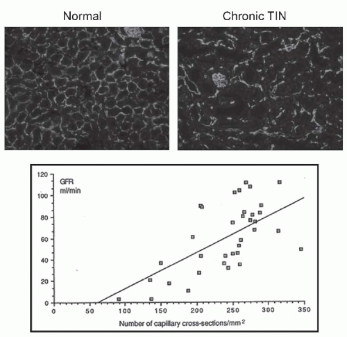
FIGURE 57.5 Interstitial capillary rarefaction is a feature of chronic tubulointerstitial nephritis (TIN). Using CD31 as an endothelial cell marker, the decrease in interstitial capillary cell density in chronic TIN is illustrated by the photomicrographs. In a study of human kidney biopsies, the extent of capillary loss was shown to correlate with the decline in glomerular filtration rate. (The photomicrographs are from Yamaguchi I, Tchao BN, Burger NL, et al. Vascular endothelial cadherin modulates renal interstitial fibrosis.Nephron Exp Nephrol. 2011;120:e20 and the graph is from Serón D, Alexopoulos E, Raftery MJ, et al. Number of interstitial capillary cross-sections assessed by monoclonal antibodies: relation to interstitial damage. Nephrol Dial Transplant. 1990;5:889, both with copyright permission.)


Matrix Accumulation

During fibrosis, the interstitial space is expanded by the accumulation of native and novel extracellular matrix (ECM) proteins. Expansion of the interstitial matrix appears to be the consequence of both increased matrix protein synthesis by myofibroblasts and decreased degradation by intracellular and extracellular connective tissue proteases. The expanded interstitium may include a gelatinous matrix of glycosaminoglycans (heparan sulfate, dermatan sulfate, chondroitin sulfate) and hyaluronic acid, an early scaffold rich in fibronectin, a fibrillar network of collagens (mainly types I, III and VI), and the presence of a variety of other extracellular matrix proteins (basement membrane collagens IV and V, collagens VII and XV, tenascin), laminin, proteoglycans (aggrecan, versican, decorin, fibromodulin, biglycan, perlecan), and various glycoproteins (thrombospondin, tenascin, hensin, vitronectin, secreted protein acidic and rich in cysteine [SPARC]). In addition to their structural effects, many of these matrix proteins elicit important effects on neighboring cells and molecules. For example, SPARC may inhibit cellular adhesion and proliferation, and also stimulates TGF-β expression and collagen I and fibronectin synthesis. Thrombospondin also activates TGF-β expression.

An unresolved question is the identity of the matrixdegrading proteases that maintain the status quo in normal kidneys despite ongoing collagen synthesis; it is also unclear why these mechanisms are perturbed during fibrogenesis.42 For example, in normal mouse kidneys, approximately 20% of the kidney collagen is newly synthesized over a 2-week period yet total kidney collagen content does not increase, indicating that a similar rate of collagen degradation is going on at the same time.43 The metalloproteinases (MMPs) are known to be important for extracellular matrix degradation and were long considered lead candidates for renal matrix homeostasis. MMP-2 and MMP-9 are abundant in the kidney and degrade collagen IV. However, paradoxically, MMP-2 and MMP-9 do not attenuate but accelerate interstitial fibrosis in experimental models. The serine proteases
urokinase-type plasminogen activator (uPA), tPA, and plasmin have been investigated as alternative candidates, but tPA and plasmin were found to promote fibrosis and uPA had no effect, despite the fact that the inhibitor PAI-1 is a potent fibrosis-promoting molecule. The latter effect may best be explained by PAI’s ability to enhance macrophage and myofibroblast recruitment in the interstitium. The urokinase receptor (uPAR) attenuates myofibroblast recruitment and fibrosis, and acts in conjunction with its coreceptor LDL receptor-related protein (LRP) to regulate fibroblast proliferation and extracellular signal-regulated kinase (ERK) signaling.44 Recent studies have focused on the role of an uPAR coreceptor, uPAR-associated protein (uPARAP), also known as the mannose receptor 2 (Mrc2) and Endo180. This receptor is expressed by interstitial macrophages and myofibroblasts and serves as a collagen endocytic receptor that delivers interstitial collagens to lysosomes for degradation by cathepsins.43 Renal fibrosis is significantly worse in mice with genetic Mrc2 deficiency.


PRIMARY DISEASES ASSOCIATED WITH CHRONIC TIN


Genetic Renal Diseases


Familial Juvenile Hyperuricemic Nephropathy/Medullary Cystic Disease Type 2

Medullary cystic disease type 2 (MCKD2) is a rare form of autosomal-dominant chronic TIN that is now known to be caused by a mutation on chromosome 16p12 involving the gene that encodes uromodulin (UMOD) (also known as Tamm-Horsfall protein).45,46,47 Approximately 60 distinct mutations have been identified. UMOD expression is restricted to the thick ascending limb of the loop of Henle and the early distal convoluted tubule. UMOD is the most abundant normal urinary protein, with levels reported in the range of 50 mg per day. Although its function is still under active investigation, UMOD is known to form a water-impermeable barrier on the surface of these cells. It may also regulate cell membrane function, based on recent evidence that it associates with cilia, lipid rafts, and sodium transporters such as ROMK2.48 It is also thought to inhibit stone formation. The UMOD mutation results in the production of a misfolded, aberrantly trafficking protein that is trapped in the endoplasmic reticulum (ER), leading to reduced urinary levels (hence the description of MCKD2 as a “UMOD storage disease”) (Fig. 57.6). Such accumulation is thought to cause ER stress, which leads to renal tubular cell death. Most patients first seek medical attention with gout symptoms between 15 and 40 years of age, caused by hyperuricemia (present in ˜70% of the patients) due to a reduced fractional excretion of uric acid—these patients are found to also have CKD.47 Some patients have mild urinary concentrating deficits, which may contribute to the genesis of hyperuricemia. The renal biopsy shows nonspecific changes of tubular atrophy, interstitial fibrosis, and mild interstitial inflammation but no unique diagnostic features. Small cysts are detected by renal ultrasound in one third of the patients. Treatment with allopurinol may slow the progression of kidney disease, and RAS blockade may decrease production of the abnormal UMOD protein. Although most patients develop end-stage renal disease (ESRD), the rate of progression is highly variable, with ESRD developing between 30 and 60 years of age.


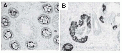
FIGURE 57.6 Familial chronic tubulointerstitial nephritis associated with mutations in the uromodulin (UMOD) gene, which encodes a protein normally expressed on the apical membrane of the thick ascending limb of the loop of Henle (shown on left). These mutations result in an abnormal UMOD protein that is trapped within the endoplasmic reticulum (right photograph). (From Dahan K, Devuyst O, Smaers M, et al. A cluster of mutations in the UMOD gene causes familial juvenile hyperuricemic nephropathy with abnormal expression of uromodulin. J Am Soc Nephrol. 2003;14:2883, with permission.)

Familial juvenile hyperuricemic nephropathy type 2 has been reported as a distinct genetic entity (autosomal dominant) caused by a mutation in the REN gene encoding renin.46,49 The mutations impair translocation of the nascent preprorenin protein into the ER, resulting in reduced or abolished renin biosynthesis and secretion. It has been suggested the mutant preprorenin may be toxic to juxtaglomerular
cells, causing additional damage to the RAS and leading to nephron dropout and progressive renal failure. Patients typically present with early-onset anemia due to low erythropoietin production, mild hyperkalemia, and low-normal blood pressure. A history of gout in some affected family members should raise the suspicion of this disorder. Plasma renin, aldosterone, and erythropoietin levels are low; fractional urate excretion is reduced; and kidney biopsies show chronic TIN, although none of these findings alone confirms the diagnosis. Mutational analysis of the REN gene is required. Treatment is supportive. CKD typically develops in the third or fourth decade and progresses slowly.


Nephronophthisis: Associated Ciliopathies

Nephronophthisis (NPHP) is a group of autosomal-recessive disorders that share chronic progressive TIN and genetic mutations in genes encoding proteins that localize to primary tubular cell cilia (reviewed in greater detail in Chapter 15).50,51,52 Clinically the patients have been subdivided into four groups based on the presence or absence of extrarenal manifestations and the causative genetic mutation. All patients with renal involvement share a form of chronic tubulointerstitial disease that typically progresses to ESRD before adulthood (median age 13 years). It is estimated that NPHP may account for 5% to 10% of pediatric patients with ESRD. The name nephronophthisis means “disintegration of nephrons” and epitomizes the histologic findings, which include nonspecific tubular atrophy with tubular basement membrane thickening and/or disruption, interstitial inflammation, and fibrosis. Small corticomedullary cysts may be present, especially with more advanced disease. These cysts and small echogenic kidneys may be detected by renal ultrasonography. Clinically, most patients have polyuria, polydipsia, and anemia but are otherwise asymptomatic until manifestations of renal failure develop in the second decade of life. The causative gene has been identified in ˜30% of cases, the most common (20%) being a homozygous deletion in nephrocystin 1 (NPHP1), which encodes a protein involved in ciliary function in collecting duct cells. An estimated 10% to 15% of the NPHP patients have extrarenal involvement. The most common is retinitis pigmentosa (Senior-Loken syndrome). Others include cerebellar ataxia (Joubert syndrome) and oculomotor apraxia (Cogan syndrome), as well as several rarer genetic syndromes. Treatment of the kidney disease is symptomatic. The kidney disease does not recur after kidney transplantation.


Polycystic Kidney Diseases

Genetic disorders associated with polycystic kidney disease (PKD) are reviewed in Chapter 16. They are mentioned here to emphasize the importance of interstitial inflammation and fibrosis to disease progression. The progression of CKD is not simply a matter of total cyst volume expanding to mechanically compress adjacent renal parenchyma; the disease is also associated with damage to otherwise normal noncystic nephrons as a consequence of chronic TIN. The degree of renal fibrosis in patients with PKD is closely associated with the rate of progression to ESRD, just as it is in all CKD.53 Studies by Grantham et al.54 in the 1990s first suggested a potential pathogenetic link between renal cysts and interstitial inflammation and fibrosis. Many macrophage innate immune response genes are upregulated in cystic mouse kidneys.55 Polycystin-1-deficient tubular cells have been shown to stimulate macrophage migration and to secrete monocyte chemoattractant protein-1 and the chemokine CXCL16.56 Inflammatory cytokines are also present in cystic fluid. This inflammatory cell response has been implicated in both cystogenesis and interstitial fibrosis. Anti-inflammatory therapy such as corticosteroids or depletion of monocytes significantly attenuates interstitial inflammation and the rate of renal functional decline in animal cystic kidney disease models (Fig. 57.7).56,57 Taken together, these data suggest that epithelial cell changes precede and drive the interstitial inflammatory response in patients with PKD.58 Possible protective tubulointerstitial effects should be taken into consideration as potential beneficial mechanisms when evaluating new drug therapies such as mammalian target of rapamycin inhibitors and vasopressin receptor antagonist.


Genetic Metabolic Disorders

Several severe metabolic disorders that present during infancy and childhood are known to cause CKD via disease processes that primarily involve the tubulointerstitial compartment.

Cystinosis. Cystinosis is an autosomal recessive disorder caused by a mutation in the lysosomal membrane protein cystinosin (CTNS).59 The estimated incidence is 1 in 100,000 to 200,000 live births. Affected children are normal at birth but develop clinical complications due to renal Fanconi syndrome, which typically brings them to medical attention before 2 years of age with failure to thrive and a history of excessive thirst, polyuria, recurrent vomiting, constipation, and episodes of dehydration. The children may already have evidence of rickets due to renal phosphate wasting. Although this is a systemic disorder, the kidney is the first organ affected and CKD progresses rapidly over the first decade if untreated. In addition to this classical presentation in infancy (nephropathic infantile form), a less aggressive renal disease has been reported but is much less common, accounting for <5% of cases (nephropathic juvenile form). Cystine is a dimeric amino acid formed by the oxidation of two cysteine residues, which become linked by a disulfide bond. Cysteine is a product of normal protein turnover, and the cystine dimer is normally recycled via the lysosomal cystinosin transporter (Fig. 57.8). In its absence, abnormal levels of cystine accumulate within lysosomes, often forming cystine crystals and leading to significant cellular damage. It is thought that the renal proximal tubules are an early target of injury due to their high rate of urinary protein uptake and processing. By 1 year of age, pathognomonic ocular corneal crystals can be detected by slit lamp examination. The
diagnosis is typically confirmed by the presence of elevated cystine levels in peripheral blood leukocytes, measured in a reference laboratory. Since the cystinosis gene (CTNS) was cloned in 1998, over 90 mutations have been reported in the United States and northern Europe—approximately 40% have a homozygous 57 kb deletion.


FIGURE 57.7 Interstitial inflammation is a pathogenic feature of polycystic kidney disease. In a mouse model of autosomal dominant polycystic kidney disease, macrophages (green) are seen lining cystic spaces (upper left). When macrophages were experimentally depleted (upper right), renal parenchyma was better preserved (lower left), and kidney function estimated by blood urea nitrogen levels was significantly better in the macrophage depleted (—) mice, shown in the lower right graph. (From Karihaloo A, Koraishy F, Huen SC, et al. Macrophages promote cyst growth in polycystic kidney disease. J Am Soc Nephrol. 2011;22:1809, with permission.) (See Color Plate.)

Human renal pathologic studies and recent studies in a mouse model indicate universal changes in renal tubules. The classical findings of a “swan neck deformity” occur as a consequence of proximal tubular cell atrophy, emphasizing the early and severe involvement of this nephron segment in cystine-associated injury. Progressive CKD is characterized by chronic TIN together with nonspecific chronic glomerular changes. The primary pathogenesis of target organ injury is thought to be due to lysosomal cystine accumulation, which perturbs several cellular functions, leading to altered energy metabolism, oxidant stress, and tubular cell death by apoptosis. However, many aspects of the kidney injury are not completely understood, such as the failure of the early and severe tubular transport defects to improve with cysteamine therapy.60 In the cystinosin knockout mice, a mild renal phenotype despite high kidney cystine levels and the benefit of bone marrow transplantation suggest that mechanisms beyond renal tubular toxicity are involved.61

Medical therapy includes a combination of water, mineral, and electrolyte replacement therapy; nutritional support; and specific therapy with the amino thiol drug cysteamine. Cysteamine lowers lysosomal cystine levels via a disulfide exchange reaction with cystine, generating a cysteine-cysteamine product that can exit via an alternative transport system (Fig. 57.8). If therapy is started at a young age and leukocyte cysteine levels are maintained in the target range, kidney survival can be significantly prolonged; however, most patients still develop ESRD by the second or third decade of life.62 The disease does not recur in a renal allograft. Most children require a gastrostomy tube in order to maintain fluid and electrolyte balance and to achieve normal growth. A minority of the patients may also require growth hormone therapy. Extrarenal manifestations are universal and may include skin and hair hypopigmentation, hypothyroidism (70% within the first decade), and eye involvement
with photophobia that requires treatment with topical cysteamine. Neuromuscular involvement and pancreatic insufficiency often develop in the older patients.


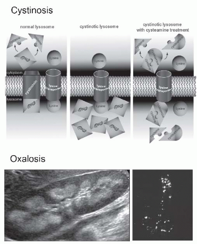
FIGURE 57.8 Inherited metabolic diseases cause chronic tubulointerstitial nephritis. The upper drawing illustrates the abnormal function of the lysosomal membrane in patients with autosomal recessive cystinosis due to a mutation in the CTNS gene that encodes the cystine transporter cystinosin. In the absence of cystinosin, cystine accumulates in lysosomes and contributes to some of the associated tissue pathologies. An elevated peripheral blood leukocyte cystine level is diagnostic. The drug cysteamine provides an alternative pathway for cystine exit from lysosomes by forming a cysteine-cysteamine dimer that is transported by an alternative (system c) lysosomal transporter. Though not yet definitively identified, it has been suggested that system c is the lysine transporter. (From Wilmer MJ, Schoeber JP, van den Heuvel P, et al. Cystinosis: practical tools for diagnosis and treatment.Pediatr Nephrol. 2011;26:205, with permission.) Primary infantile oxalosis is associated with aggressive chronic tubulointerstitial nephritis due to the deposition of calcium oxalate in renal tubules and the interstitium, which may be detected as nephrocalcinosis on the renal ultrasound (lower left) or the actual deposits can be visualized by polarized light microscopic examination of kidney tissue (lower right). (See Color Plate.)

May 29, 2016 | Posted by in NEPHROLOGY | Comments Off on Chronic Tubulointerstitial Nephritis

Full access? Get Clinical Tree

Get Clinical Tree app for offline access