(1)
Al Agouza, Cairo, PO, Egypt
Nomenclature
Caudal regression syndrome (CRS), sirenomelia, sacral agenesis syndrome, caudal dysplasia, and phocomelic diabetic embryopathy
Historical Note
The term “syndrome of caudal regression” was first used by Duhamel to describe a spectrum ranging from simple sacral agenesis to severe lower limb anomalies, including fusion as “sirenomelia,” often with imperforate anus and major malformations of the anus and rectum, omphalocele, and anomalies of the genitourinary system, but sacrococcygeal agenesis or hypoplasia was the constant feature [1].
But partial or complete absence of the sacrococcygeal bones was first described in 1852 by Hohl; in 1857, over the next 10 years, about 50 additional cases were published singly or in small series. In 1959, Blumel and colleagues reported 50 more cases, many of which included other malformations such as meningoceles, anal and bowel anomalies, and abnormalities of the lower limbs often associated with dysfunction of the bowel and urinary bladder [2].
Sirenomelia, also a syndrome of sacral agenesis in which the fused lower extremities cause the fetus to resemble a mermaid (Figs. 15.1, 15.2, and 15.3), was previously thought to be the most severe form of caudal regression and is currently discussed in most literature involving caudal regression.
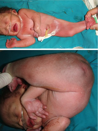
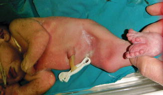
Figs. 15.1 and 15.2
A mermaid baby with absence of the phallus, urethral opening, or anal canal
Fig. 15.3
Sirenomelia with fusion of both lower limbs
Incidence
It is an uncommon malformation seen in 0.1–0.25:10,000 of normal pregnancies. However, its occurrence in infants born to diabetic mothers is about one in 350 live births, association first shown by Blumel et al. suggesting 200 times increase. However, other etiologic factors are presumably involved, as demonstrated by the rare incidence of caudal regression syndrome compared to diabetes; this anomaly is more common in boys (2.7 boys/1 girl) [3].
Type I: partial or total unilateral sacral agenesis (Fig. 15.4)
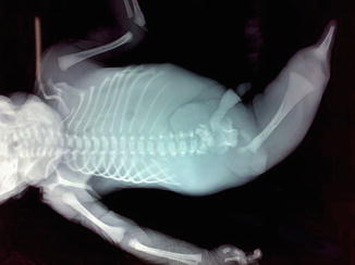
Fig. 15.4
Sirenomelia with single lower limb bone and sacral agenesis
Type II: partial bilateral, symmetrical sacral agenesis
Type III: total sacral agenesis with variable lumbar anomaly and iliac wings attached to the last lumbar vertebrae (Fig. 15.5)
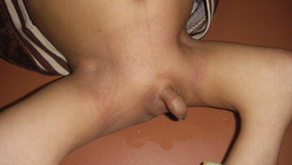
Fig. 15.5
A child with caudal regression, flat buttocks, and small funnel pelvis
Type IV: total sacral agenesis +/− lumbar anomaly and iliac wings fissioned behind the last vertebrae, if they are present
Mild form: types I and II, coccyx agenesis without functional repercussions.
Major form: types III and IV, systematic sequelae are present with neurologic impairment. Perinatal death is frequent. Thoracic vertebral involvement renders the child incompatible with life (Figs. 15.5, 15.6, and 15.7).
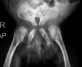
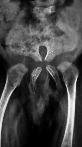
Fig. 15.6
Pelvic x-ray of the previous case with absent sacral vertebrae and coccyx; fused iliac bones give the heart-shaped appearance
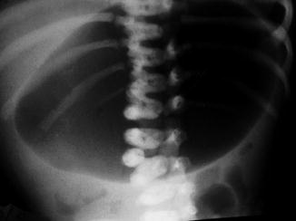
Fig. 15.7
Minimal vertebral anomaly in the form of incomplete fusion of the lumbar vertebra
Variable proximal, mainly lumbar, vertebral anomalies associating CRS range from incomplete fusion of the vertebral body (Fig. 15.8) to complete separation of the hemivertebra (Fig. 15.9).