(1)
Al Agouza, Cairo, PO, Egypt
The female urethra is a short tubular structure 3–5.5 cm in length; it is supported by the pubocervical fascia posteriorly. The sphincters are neither well developed nor as strong as in the male. Adequate function depends to a great extent on the support of this fascia. The urinary meatus lies in the middle of the vaginal vestibule and is constantly exposed to bacterial contamination from the vagina and even the rectum; therefore, urethritis and cystitis are more frequent in the female than in the male. The muscular coats of the female urethra are continuous with those of the bladder. There is an outer and inner longitudinal layer with a circular layer between, which is involuntary and more efficient around the proximal third. There is a voluntary sphincter around the middle third derived from the urogenital trigone. It is lined with stratified squamous variety in the lower portion and transitional type above. Paraurethral glands are numerous; the best known and largest are the compound racemose glands of Skene; its ducts empty into the floor of the urethra on each side just within the meatus. The upper glands have been considered an analogue of the prostate gland. The accessory urethral glands including the lesser vestibular or paraurethral glands (Skene) and urethral glands arise from the urogenital sinus from endodermal (epithelial) buds growing into the urethral mesenchyme. The paired greater vestibular glands (Bartholin) form in the 12th fetal week and empty into the vaginal vestibule. Embryological vestigial structures (Wolffian vestiges): In early fetal life, the mesonephric Wolffian and paramesonephric ducts coexist, the mesonephric ducts regress in a female, but its remnants typically persist. Their importance relates to the potential for the development of pathology. The Gartner’s ducts are paired remnants of the mesonephric duct that may give rise to Gartner’s duct cysts and are typically located in the broad ligament.
11.1 Urethral Prolapse
Definition
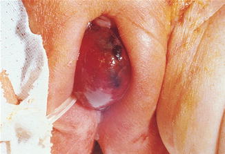
Urethral prolapse is the circular eversion of the urethral mucosa through the urethral meatus with vascular congestion and possible strangulation. Most authors believe that urethral prolapse is restricted to the terminal urethra (Fig. 11.1).
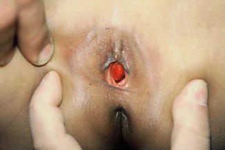
Fig. 11.1
Urethral prolapse in a young girl
Incidence
Urethral prolapse is relatively uncommon and has a bimodal age distribution. It occurs almost exclusively in black girls younger than 10 years, with an average age at presentation of 4 years.
Historical Background
Solinger described urethral prolapse for the first time in a prepubertal black girl in 1732 [1].
Etiology
One report of urethral prolapse in identical twins suggests that heredity may play a role. The exact cause of urethral prolapse remains unknown; however, several theories have been proposed. These theories may be divided into congenital or acquired defects.
Congenital Defects
Weak pelvic floor structures such as inadequate pelvic attachments and urethral hypermobility
Intrinsic abnormalities of the urethra (e.g., an abnormally patulous urethra, a wide urethra, redundant mucosa)
Neuromuscular disorders, urethral malposition, submucosal weakness, or deficient elastic tissue
Acquired Defects
Physiological hypoestrogenemia
Trauma as in cases of sexual abuse or masturbation
Debility
Malnutrition
Bad hygiene
Weakened attachment between the inner longitudinal and outer circular-oblique smooth muscle layers of the urethra. Separation of the two muscle layers, coincident with episodic increases in intra-abdominal pressure as a result of chronic coughing or constipation, which may predispose to urethral prolapse
Urethral prolapse may be misdiagnosed as a cause of urogenital bleeding in girls. The correct initial diagnosis has been reported in only few cases. The prolapse appears as a doughnut-shaped mass protruding through the vulva that encircles the urethral meatus; the mucosa is an edematous, friable rosette of bright red cyanotic tissue that may be infected, ulcerated, necrotic, or gangrenous.
Classification
Prolapse was graded from I to IV depending on the extent of prolapse and degree of inflammation [2]. Prolapse may be complete or incomplete, although complete prolapse is more common (Fig. 11.2).
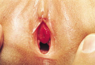
Fig. 11.2
Minimal urethral prolapse
Differential diagnosis includes prolapsed ectopic ureterocele, prolapsed bladder, prolapsed urethral polyp, and ectopic ureter.
Clinical Picture
Urethral prolapse usually is an incidental finding during routine examination. The most common presentation is vaginal bleeding associated with a paraurethral mass. Symptomatic children present with bloody spotting on their underwear or diapers, hematuria is uncommon, and voiding disturbances: as the affected girls may report dysuria, urinary frequency, or introital pain. Children may report genital pain if the prolapsed mucosa becomes very large or if thrombosis and gangrene have developed. Furthermore, acute urinary retention secondary to urethral prolapse has been reported in a young girl.
Management
Medical therapy or conservative measures are indicated in small noncomplicated prolapse; these include local hygiene with sitz baths and topical antibiotic, steroid, or estrogen creams, along dealing with relieving the possible predisposing causes. Simple manual reduction and urethral catheterization for 1–2 days have been effective in minor cases of urethral prolapse; however, recurrence rates are high.
Surgical Therapy
If medical therapy does not rapidly reduce the prolapse, surgery is the treatment of choice. The Kelly–Burnham technique involves excising the prolapsed mucosa over an indwelling Foley catheter and closure of the incision by approximating the normal urethral mucosa to the introital mucosa with interrupted absorbable sutures, without any pulling down of more urethra than the part already prolapsed because shortening the urethra may lead to urinary incontinence. A modification of the Kelly–Burnham technique involves placing absorbable stitches in 4 quadrants of the prolapsed mucosa. Incise each quadrant between the holding sutures up to the mucocutaneous junction. Excise the prolapsed urethra in quadrants, followed by immediate approximation of the mucocutaneous junction with absorbable sutures [3] (Fig. 11.3).
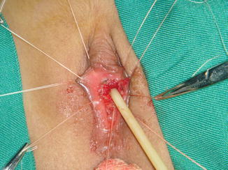
Fig. 11.3
Surgically removed urethral prolapse
Other modalities for treatment of this anomaly had been reported like cautery, fulguration, and cryosurgery to destroy or incise prolapsed tissue, with less effective results.
11.2 Urethral Caruncle
Definition
It is a small, fleshy outgrowth of the distal edge of the urethra. The tissue of the caruncle is soft, smooth, friable, and bright red and initially appears as an eversion of the urethra. Urethral caruncles are generally small, single, and sessile, but they may be pedunculated and grow to be 1 to 2 cm in diameter.
Etiology
Urethral caruncles are believed to arise from an ectropion of the posterior urethral wall. The growth of the caruncle is secondary to chronic irritation or infection; it is reported in girls who had continuous urine dripping as in cases of urinary incontinence secondary to meningomyelocele.
Histologically, the caruncle is composed of transitional and stratified squamous epithelium with a loose connective tissue. Often the submucosal layer contains relatively large dilated veins. Caruncles are frequently subdivided by their histological appearance into papillomatous, granulomatous, and angiomatous varieties. This separation is based on the most prominent component (surface epithelial, vascular, and inflammatory, respectively); but this distinction has no apparent clinical relevance. The mixed inflammatory infiltrate and rich vascularity, combined with the clinical setting, should establish the correct diagnosis. They are often secondary infected, producing ulceration and bleeding [5].
Clinical Picture
The symptoms associated with urethral caruncles are variable. Many cases are asymptomatic, whereas others experience dysuria, frequency, and urgency. Sometimes the caruncle produces point tenderness after contact with undergarments. Ulcerative lesions usually produce spotting on contact more commonly than hematuria.
The differential diagnosis of urethral caruncles includes prolapse of the urethral mucosa and urethral and paraurethral cysts. The diagnosis of a urethral caruncle could be established by biopsy under local anesthesia (Fig. 11.4).
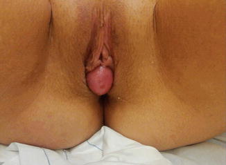
Fig. 11.4
Urethral caruncle in an adolescent girl
Management
Initial therapy is topical estrogen and avoidance of irritation. If the caruncle does not regress or is symptomatic, it may be destroyed by cryosurgery, laser therapy, fulguration, or operative excision. Following operative excision, a Foley catheter should be left in place for 48–72 h. Follow-up is necessary to ensure that the patient does not develop urethral stenosis.
11.3 Urethral Polyps
A urethral polyp is an irregularity existing at birth. It is usually a benign lesion composed of fibrous or fibroepithelial tissue but may include some smooth muscle, probably arising from a prolapsing urothelium that has evolved into a polyp; morphologically, congenital urethral polyp is covered by a urothelium that may be inflamed and ulcerated or exhibit squamous metaplasia [6].
Incidence
Urethral polyps arising from the anterior urethra are rarely encountered in the pediatric age group and are even rarer in the females. Scarcely reported in English literature, their exact incidence is not known [7].
Clinical Picture
This urethral polyp may be presented as a small mass coming out of the urethra, blood in the urine, and/or difficulty urinating, but it may be asymptomatic for years or present with features of urinary obstruction, mass, or “vaginal” bleeding. They are important in the differential diagnosis of interlabial masses in female children. Patients usually come to clinical attention between the ages of 3 and 9 years, but may rarely present during infancy or adulthood. For this reason, it has been suggested that congenital urethral polyp is secondary to a poorly understood congenital defect in the urethral wall.
Investigations
Urethral polyps are diagnosed with cystoscopy and/or a voiding cystourethrogram (VCUG).
Treatment
Surgery is indicated for relief of symptoms and differentiation from malignant lesions such as a sarcoma or a papilloma. The urethral polyp needs a simple excision and fulguration of the base. No recurrences have been reported [8].
11.4 Prolapsed Ectopic Ureterocele
Ectopic ureteroceles are usually associated with duplex kidneys, and large ectopic ureteroceles associated with the upper pole of the duplex kidney are prone to prolapse. The prolapsed ureterocele is usually cystic and covered with pink bladder mucosa.
11.4.1 Clinical Picture
Prolapse of a large ureterocele through the urethral orifice should be considered in the differential diagnosis of all interlabial masses in infants and children. Ureteroceles are cystic dilations of the distal ureter, which are located in the bladder or urethra and may prolapse through the urethral meatus as reddened or even necrotic mucosal surfaces. A prolapsed ureterocele, unlike urethral prolapse, does not present a symmetrical orifice but rather presents an asymmetrical protrusion through the urethra. Catheterization alongside the prolapse may locate the lumen of the urethra. Prolapse of a ureterocele may be associated with a palpable distended bladder or flank mass (hydronephrosis) (Fig. 11.5).