Fig. 1
Transperineal ultrasound. Technical approach (left panel; Figures a, c) and correspondent findings (right panel, Figures b, d). With patient position is dorsal or left lateral position, the transducer is placed directly on perineal body. Transducer is pressed posteriorly and angled cranially-to-caudally, to obtain a cross sectional view, mainly in women (a, b) or placed on the perineum to obtain a long axis view (c, d). In both sections, internal anal sphincter (IAS or i) and external anal sphincter (EAS or e) appears as a hypoechoic ring and as a tissue of mixed echogenicity like in anorectal ultrasound. P: probe; R: rectum; arrow: ano-rectal angle
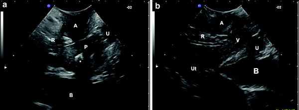
In this way, it is possible to obtain high-resolution images of the anal canal, the anal sphincters, pubo-rectal muscle, recto-vaginal and anovulvar septum, urinary bladder, prostate and vagina. Internal anal sphincter is the terminal condensation of the circular rectal smooth muscle. It is seen as an approximately 3-mm thick hypoechoic symmetric band (Fig. 1) sometimes demarcated from heterogeneous subepithelial tissues medially. The surrounding mixed-echogenicity striated external sphincter is slightly thicker, extends approximately 1 cm beyond the internal anal sphincter (Fig. 1). It is a circular structure, shorter anteriorly in women. The deep part of the external anal sphincter is fused with or intimately related to the puborectalis muscle. Anteriorly it is closely related to the superficial transverse muscle of the perineum and perineal body. The external anal sphincter shows a fibrillar pattern of fine parallel hyperechoic lines in proximal third of the anal canal (deeper part of external anal sphincter) which become more homogeneous at distal third of the anal canal (superficial or subcutaneous part of external anal sphincter). Besides anal sphincter complex transperineal ultrasound allows the simultaneous visualisation of the distal part of the urinary bladder and prostate and proximal art of the urethra in men, and uterus, vagina and entire urethra in women (Fig. 2).