Nonneoplastic Diseases of the Prostate and Seminal Vesicles


Nonneoplastic Diseases of the Prostate and Seminal Vesicles


Peter A. Humphrey


PROSTATE


Introduction

Nonneoplastic diseases of the prostate are common afflictions of men. Prostatitis and benign prostatic hyperplasia (BPH) in particular are highly prevalent clinical disorders. About one in nine men aged 25 to 80 have clinical prostatitis-like symptoms.1,2 Moderate to severe lower urinary tract symptoms, which are clinical hallmarks of BPH, are found in one-quarter of men in their 50s, one-third of men in their 60s, and about one-half of men 80 years or older.3 Histologic detection of inflammation and BPH (nodular glandular and/or stromal hyperplasia) in prostatic tissues is even more common. In contrast, nonneoplastic diseases of the seminal vesicles are distinctly uncommon to rare. In this chapter, benign nonneoplastic conditions of the prostate and seminal vesicles are surveyed, with a focus on pathologic diagnosis and differential diagnosis, with clinicopathologic correlations.

Nonneoplastic diseases of the prostate are also important since several nonneoplastic lesions can be misdiagnosed as prostate cancer, and are therefore pseudoneoplasms or benign mimickers of prostate cancer (Table 8-1). Foremost amongst these histologic benign mimickers are prostatic atrophy, atypical adenomatous hyperplasia (AAH) (adenosis), basal cell hyperplasia, and inflammatory atypia.4, 5, 6, 7 In the seminal vesicle a classical pitfall is the misinterpretation of normal nuclear atypia as malignancy, and in particular, prostatic carcinoma.


Function

The prostate is one of the male sex accessory tissues, which include the prostate, seminal vesicles, and bulbourethral glands. The main function of the prostate is to form secretions that constitute one-half to two-thirds of the 3 mL volume of the ejaculate.8 The biologic role of many biochemical substances found in the semen is unknown. A well-defined function has been established for several enzymes that participate in the clotting and lysis of the seminal vesicle clot. One of these enzymes is prostate specific antigen (PSA), which is a serine protease and esterase that cleaves semenogelin, a protein involved in clotting. Additional substances found in high concentration include potassium, zinc, citric acid, spermine, amino acids, prostaglandins, and a number of enzymes. These secretory products enhance fertility by promoting sperm viability and motility. An antimicrobial role has been suggested for zinc, spermine, and proteases. Physically, by mass effect, musculature, and location, the prostate may contribute to urine flow from the bladder. The prostate is not essential for life. Removal of the entire gland by surgery (radical prostatectomy) can be accomplished without consequences. In some men, there may be complications of incontinence and impotence, the latter being due to damage to periprostatic neurovascular bundles. Failure of the gland to form, as seen in 5α-reductase deficiency9 and testicular feminization,10 is not lethal.


Normal Embryology, Anatomy, and Histology


Embryology

The prostate is derived from the urogenital sinus and is first recognizable at 9 to 10 weeks of development.11 Testosterone from the embryonic testis stimulates ingrowth of endodermal buds into urogenital sinus mesenchyme. It is thought that there is a reciprocal interaction of epithelium and mesenchyme during development by which, under the influence of androgens, urogenital sinus mesenchyme induces urogenital sinus epithelium to undergo prostatic ductal morphogenesis and differentiation.12 The differentiating prostatic epithelium in turn signals the urogenital mesenchyme to differentiate
into smooth muscle cells that closely surround the epithelial ducts. This dynamic dance of tissue remodeling entails coordinated temporal and spatial processes of ductal budding, branching morphogenesis, cellular proliferation, and secretory function.13 By 13 weeks some 70 primary ducts are present. Three stages of development from 20 weeks’ gestation to 1 month of age have been delineated.14 The bud stage at 20 to 30 weeks’ gestation exhibits solid cellular buds at the end of ducts, with spindled cells in the center and columnar cells at the periphery. The bud-tubule stage at 31 to 36 weeks shows small collections of cellular buds and acinar structures (Figs. 8-1 and 8-2). The third stage is characterized by more distinct lobular organization of acinotubular clusters. Squamous metaplasia of prostatic ducts and urethra is a common finding in the fetal prostate. This metaplasia is gradually lost after birth.


Table 8-1 ▪ PSEUDONEOPLASMS OF THE PROSTATE


Atrophy*


Atypical adenomatous hyperplasia (adenosis)*


Basal cell hyperplasia*


Cribriform hyperplasia


Nephrogenic metaplasia (adenoma)


Verumontanum mucosal gland hyperplasia


Squamous metaplasia


Urothelial metaplasia


Radiation atypia


Prostatitis (especially xanthogranulomatous)


Malacoplakia


Endometriosis


Postoperative spindle cell nodule


Atypical stromal cells


Extramedullary hematopoiesis


Cowper glands


Paraganglia in prostate


Benign glands adjacent to nerves and skeletal muscle


Signet ring change in stromal nodule


Crowded benign glands


Seminal vesicle/ejaculatory duct epithelium


* Most likely to be misdiagnosed as carcinoma.


The immunophenotype of fetal prostatic epithelial cells is similar to the rare adult transiently amplifying epithelial cells, which show coexpression of basal and luminal cytokeratins, high proliferation, and lack of p27 expression.15

Molecular pathways thought to be important in prostate organogenesis and morphogenesis include those regulated by androgens, hedgehog, p63, Nkx3.1, Sox9, Noggin, fibroblast growth factor receptor, forkhead transcription factor, Notch, and BMP7. Most of this work, with a few exceptions,16 has been done in rodent model systems. Interestingly, some of these molecules have been implicated in human prostate cancer biologic behaviors.17 The definitive identification of prostatic epithelial stem cells involved in prostate gland development has not yet been realized, but cell populations enriched for cells exhibiting stem cell characteristics express the stem cell markers CD133(+), alpha2beta1(hi), CD44, and Sca-1 along with embryonic stem cell factors including Oct-1, Nanog, Sox2, and nestin.18


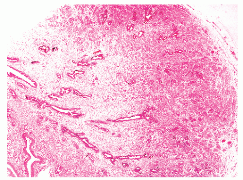
FIGURE 8-1 ▪ Fetal prostate at 33 weeks with dominance of stroma and immature epithelium.

The seminal vesicles, epididymis, vas deferens, and ejaculatory ducts are formed from Wolffian (mesonephric) ducts under the influence of testosterone. It has been proposed that the central zone of the prostate is also of mesodermal Wolffian duct origin.11 In this model the prostate gland is of dual embryonic derivation.


Anatomy

From birth to age 10 to 12 years there is an infantile resting phase where prostate size remains stable at about 1 to 1.4 g. During this time there is continual duct formation, solid budding at the periphery, and branching morphogenesis.13 From
12 to 18 years of age there is a pubertal maturation period with a significant increase in gland size, to 11 to 18 g by age 18. This period is characterized by androgen-driven gland branching, and differentiation of immature prostatic epithelium into adult-type basal and secretory cells.


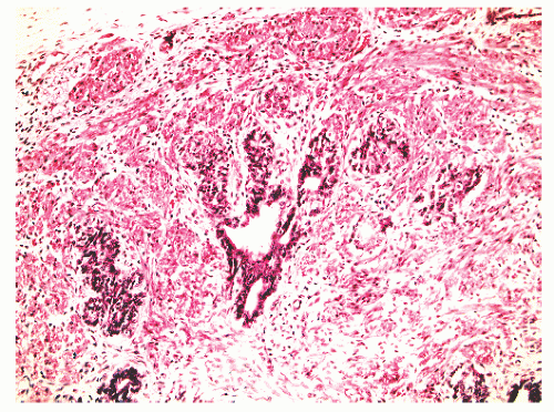
FIGURE 8-2 ▪ Fetal prostate at 33 weeks with small glands and solid cellular buds.


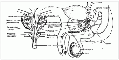
FIGURE 8-3 ▪ Anatomical relationships of prostate from the frontal (left) and coronal (right) perspectives. TZ, transition zone; CZ, central zone; PZ, peripheral zone; AFS, anterior fibromuscular stroma. (From Timms BG. Anatomical perspectives of prostate development. In: Naz RK, ed. Prostate: basic and clinical aspects. New York: CRC Press; 1997:36, figure 4-A).

The adult prostate gland surrounds the urethra immediately below the base of the urinary bladder and is located posterior to the inferior symphysis pubis, superior to the urogenital diaphragm, and anterior to the rectum (Figs. 8-3 and 8-4). It has a shape like a truncated cone or a top lying on its side. It measures 5 cm × 4 cm × 3 cm and weighs 20 g from ages 20 to 50, with an increase to 30 g from ages 60 to 80.

Historically, the prostate was thought to have anywhere from two to five lobes.19,20 The five-lobe model was utilized in the first half of the 20th century, until 1954, when Franks21 forwarded the concept of an outer gland, where carcinomas arose, and a periurethral inner gland. Further modeling evolution occurred when McNeal developed the three-zone model, which defines the central zone, the transition zone, and the peripheral zone (Fig. 8-4). It may be difficult in some cases to grossly and microscopically recognize a sharp demarcation between these zones, but these zones do have structural differences, and disease differentially affects these zones (Table 8-2). Carcinoma tends to arise in the peripheral zone (although about 20% of carcinomas do arise in the transition zone) and BPH is typically a transition zone process, while the central zone is remarkably resistant to disease. The prostatic urethra is formed at the bladder neck, turns anteriorly 35 degrees at its midpoint (the urethral angle), and exits the prostate at the apex, where it is continuous with the membranous urethra. The urethral angle can vary between 0 degree and 90 degrees and divides the urethra into proximal (so-called preprostatic) and distal (so-called prostatic) segments. The transition zone, which normally makes up 5% of prostate gland volume, wraps around
the proximal urethra (Fig. 8-4). The main ducts of this zone drain into posterolateral recesses of the urethra at a point just proximal to the urethral angle. Some of the more medial ducts penetrate through the thick smooth muscle bundles of preprostatic sphincter. Also found in the smooth muscle of the preprostatic sphincter are minute ducts and abortive acinar arrangements of periurethral glands, which constitute only about 1% of the total glandular volume of the prostate. The central zone makes up about 25% of the entire prostate gland and is a posteriorly situated cone-like structure with the base of the cone projecting toward the base of the bladder. The paired ejaculatory ducts (Fig. 8-5) run through the central zone from the seminal vesicles to their exit at the posterior urethral protuberance, known as the verumontanum (or colliculus seminalis) (Fig. 8-6). Within the verumontanum is a cul-de-sac, the prostatic utricle, located between the ejaculatory ducts (Fig. 8-7). It may also lie deep to the ejaculatory ducts or may be embedded as a tiny cavity in the prostatic parenchyma.22 The ducts from the central zone drain into the urethra via orifices positioned just next to the ejaculatory duct orifices on the verumontanum. The peripheral zone of the prostate gland is the bulk of the posterior, lateral, and apical portion of the prostate gland and accounts for 70% of the total gland volume. The ducts of this zone empty into the posterior urethral recesses or grooves in a double row from the verumontanum to the prostatic apex. Finally, a patch of nonglandular tissue called the anterior fibromuscular stroma is present anteriorly over the prostate and extends from the bladder neck to the apex of the prostate.


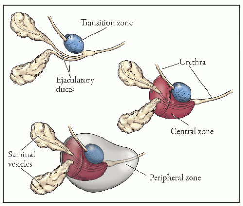
FIGURE 8-4 ▪ Anatomical relationships of the adult prostate. The prostate has three zones: central zone (CZ, through which the ejaculatory ducts course), transition zone (TZ, site of benign prostatic hyperplasia nodules), and the peripheral zone (PZ), where most carcinomas arise. (Modified from Cancer: Principles and Practice of Oncology, 4th edition, Lippincott Williams and Wilkins).


Table 8-2 ▪ COMPARISON OF THE THREE ZONES OF THE PROSTATE


Peripheral Zone


Transition Zone


Central Zone


Percentage of normal prostate


70%


5%


25%


Embryologic origin


Urogenital sinus


Urogenital sinus


Wolffian duct


Anatomic location


Posterior, lateral, and apical


Around proximal urethra


Posterior cone-like structure


Inflammation


Common


Common


Rare


Hyperplasia (BPH)


Uncommon


Common


Rare


PIN


Common


Uncommon


Rare


Percentage of cases of adenocarcinoma


70%


20%


5%


Gland:stroma ratio


1:1


1:1


2:1


Epithelium


Irregular small acini


Irregular small acini


Larger, darker, complex glands


Stroma


Loose fibromuscular


Compact fibromuscular


Compact fibromuscular


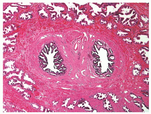
FIGURE 8-5 ▪ Paired ejaculatory ducts that run through the central zone of the prostate.


Histology/Immunohistology


Epithelium

Microscopically, the normal adult prostate is a branching duct-acinar glandular system embedded in a dense fibromuscular stroma (Fig. 8-8). Zonal architectural differences can be appreciated. Normally, the peripheral zone ducts and acini are evenly distributed but are irregular in size and shape. Normal transition zone glands are similar to those of the peripheral zone. Central zone glands are more densely arranged than peripheral and transition zone glands. The epithelial-stromal ratio in the central zone is 2:1, compared
with 1:1 for the peripheral and transition zones. Also, central zone glands are larger and display intraluminal projections with fibrovascular cores. Central zone epithelium displays tall columnar cells with eosinophilic cytoplasm, a prominent basal cell layer, and on occasion complex intraluminal architecture such as Roman bridge and cribriform formations (Fig. 8-9). These findings can be misdiagnosed as atypia or prostatic intraepithelial neoplasia (PIN).23 Central zone epithelium is distinctive in its selective expression of pepsinogen II, lactoferrin, and lectin-binding sites.7 Central zone epithelium also differs from epithelium in other zones in proteomic profile.24


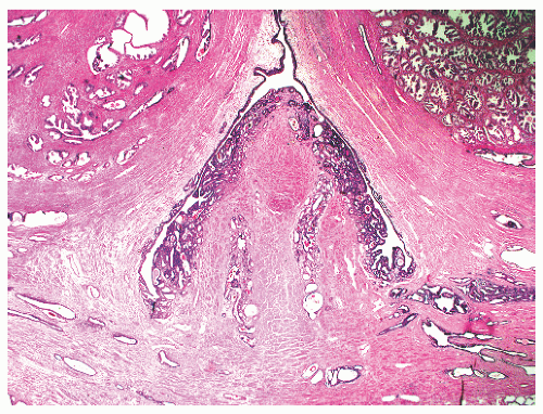
FIGURE 8-6 ▪ Verumontanum of prostate—a mound of tissue protruding into the urethral lumen.


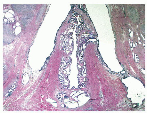
FIGURE 8-7 ▪ Utricle of the prostate in the verumontanum.

The normal epithelium of the prostate is classically defined as having two cell layers: a luminal or secretory cell layer and a basal cell layer (Fig. 8-10). A third cell type in normal prostatic epithelium is the neuroendocrine cell, which is rare (Box 8-1). The secretory or luminal cells of the normal prostate glandular epithelium make up the bulk (73%) of the epithelial volume. The secretory cells are cuboidal to columnar with nuclei positioned in the basal to midportion of the cell. Cleared cytoplasm is a hallmark of normal prostatic secretory cells because of the presence of a large number of small clear secretory vacuoles. The central zone secretory cell cytoplasm is somewhat denser because it contains a smaller number of theses vacuoles.7 Mucin is not usually seen in H&E-stained sections of normal glands, but histochemical stains for mucins reveal neutral mucins (with periodic acid-Schiff [PAS] with diastase positivity), whereas neoplastic glands demonstrate neutral and acidic mucin staining. Pigment is common in the cytoplasm of normal prostatic secretory epithelial cells. These granules are 1 to 3 µm in diameter, are apical or subnuclear, and are yellow-brown to gray-brown to blue by
hematoxylin and eosin (H&E) staining (Fig. 8-11).25 The histochemical staining characteristics, including positivity for Fontana-Masson, PAS with diastase, Congo red, Luxol fast blue, and Oil-Red-O, and autofluorescence of the pigment are consistent with lipofuscin. This lipofuscin pigment can also be found in the cytoplasm of seminal vesicle and ejaculatory duct epithelium, where it is more abundant, coarser, and more refractile. The nuclei of normal prostatic secretory epithelial cells are small and round with fine, evenly dispersed chromatin. Nucleoli usually are not evident or are pinpoint in size. Nuclei in the central zone usually are larger than those in the peripheral zone and also appear crowded. This results in a pseudostratified nuclear appearance in the central zone and along with the aforementioned denser cytoplasm may yield a false impression of PIN.23 Normal secretory cells are immunoreactive for pan-cytokeratins, cytokeratins 8 and 18, PSA (Fig. 8-12), prostate-specific acid phosphatase (PSAP) (Fig. 8-13), and the androgen receptor. Of diagnostic significance, immunostains for α-methylacyl-coenzyme A racemase (alpha methylacyl CoA racemase [AMACR]; also known as P504S), a marker for prostatic neoplasia, can be focally positive in secretory cells in normal glands, but this staining should be focal and noncircumferential within normal and benign glands (Fig. 8-14).26 Additional prostate markers that have been utilized in diagnostic immunohistochemistry to assess for prostate versus nonprostate carcinoma include prostate-specific membrane antigen (PSMA), prostein (P501S) (Fig. 8-15), proPSA, and NKX3.1. In normal prostatic tissues PSMA is weakly expressed in benign luminal cells.27,28 P501S is strongly expressed in benign luminal cells, with prominent dot-like Golgi complex staining29,30 (Fig. 8-15). Two different proPSA molecular forms differ in expression in benign glands, with strong or moderate/diffuse [−5/−7]proPSA expression and negative or weak [−2] proPSA expression.31 NKX3.1 immunostains show intense labeling of most secretory luminal cell nuclei, with weak staining of basal cell nuclei.32


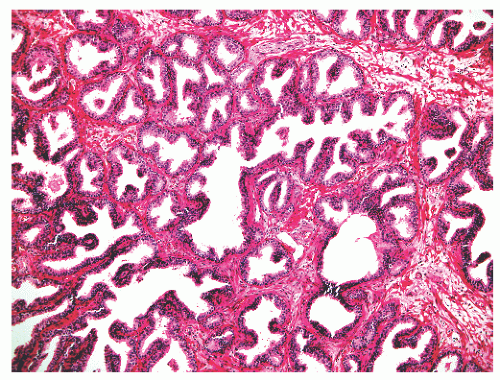
FIGURE 8-8 ▪ Normal prostate with complex glands embedded in a fibromuscular stroma.


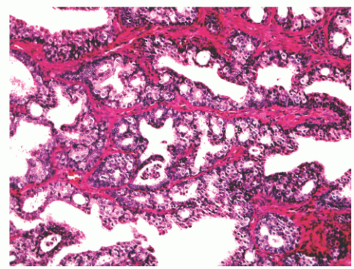
FIGURE 8-9 ▪ Normal central zone epithelium with cribriform growth and Roman bridge formation.


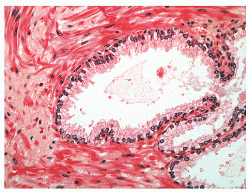
FIGURE 8-10 ▪ Normal prostatic epithelium with a secretory luminal cell layer and basal cell layer.


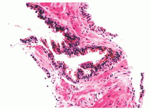
FIGURE 8-11 ▪ Lipofuscin pigment in benign prostatic epithelium.


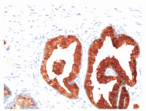
FIGURE 8-12 ▪ Prostate-specific antigen immunoreactivity in normal prostatic epithelium.

The basal cell layer separates the secretory cells from the basement membrane and is nearly continuous (Figs. 8-10, 8-16, and 8-17). Basal cells in the prostatic epithelium often appear as rounded or oblong cells, but can also be flattened, spindled, cuboidal, and triangular.33 Basal cells have a scant amount of dense cytoplasm and small, hyperchromatic nuclei. The immunophenotype of basal cells is distinctive, is different
from luminal secretory cells, and can be of diagnostic utility. The most commonly employed basal cell-specific antibodies are those that react with p63 and high-molecular-weight cytokeratins, especially 1, 5, 10, and 14 that are detected by mouse monoclonal antibody 34βE12 (also known as CK903).26 A cocktail consisting of 34βE12 and p63 antibodies slightly improves the detection of basal cells (Fig. 8-12).34 Immunostaining of basal cells can also be achieved using antibodies directed against cytokeratins 5/6.26 It should be noted that discontinuity or even focal lack of basal cell staining may be seen in a minority of entirely normal glands.35 The precise function of basal cells is unsettled, but they represent the proliferative component of prostatic epithelium. Stem cells may reside within the basal cell population.


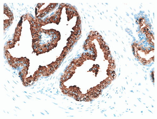
FIGURE 8-13 ▪ Prostate-specific acid phosphatase immunoreactivity in normal prostatic epithelium.


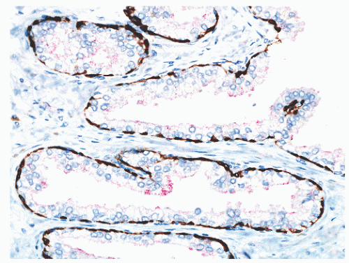
FIGURE 8-14 ▪ Focal, noncircumferential AMACR immunoreactivity in benign prostatic epithelium. Red, AMACR signal; brown, 34βE12/p63 cocktail antibody binding to basal cells.

Neuroendocrine (endocrine-paracrine) cells constitute a third population of cells in normal prostatic epithelium.36 These cells are a small minority at about 0.4% of the total adult prostatic epithelial cell population. This is a terminally differentiated, postmitotic cell population that variably expresses androgen receptor and the secretory products PSA and PSAP. The keratin expression pattern is more like luminal than basal cells. These neuroendocrine cells are usually recognizable only by histochemical (Fontana-Masson argentaffin and Churukian-Schenk argyrophil) or immunohistochemical staining. By immunohistochemical detection of chromogranin and neuron-specific enolase, these cells may be found throughout the prostate but are at highest concentrations in the periurethral region and prostatic ducts.37 A variety of neuroendocrine markers, including chromogranin, neuron-specific enolase, serotonin, thyroid-stimulating hormone-like peptide, calcitonin, bombesin, gastrin-releasing peptide, somatostatin, parathormone-related protein, and neurotensin, have been identified in these cells by immunohistochemistry.36 These immunostains highlight the highly
interdigitating nature of these cells, which exhibit slender, cytoplasmic dendritic process that may be up to 200 µm long. Neurosecretory granules of varying sizes are found in these cells by electron microscopy. The functional significance of neuroendocrine cells in the normal prostate is not clear, but it seems likely that the neuroendocrine products of these cells influence neighboring cells through the elaborate dendritic processes via a paracrine effect. Autocrine and endocrine influences may also be in operation.


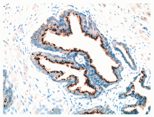
FIGURE 8-15 ▪ Prostein (P501S) immunoreactivity in normal prostatic epithelium. Note granular perinuclear cytoplasmic pattern.


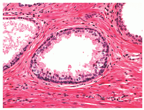
FIGURE 8-16 ▪ Continuous prominent basal cell layer in a benign prostatic gland.


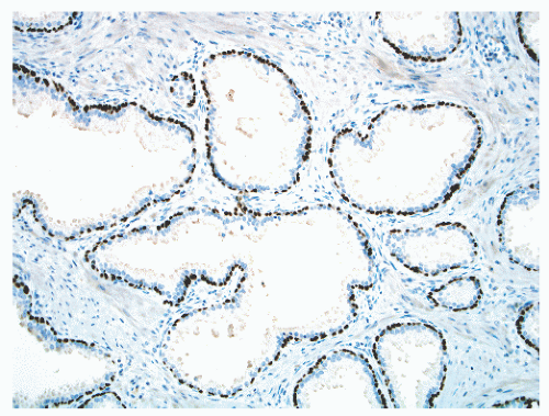
FIGURE 8-17 ▪ Continuous basal cell layer in normal prostatic epithelium, as highlighted by a p63 immunohistochemical stain.

Urothelium is another type of epithelial cell normally found in the prostate. In addition to lining the prostatic urethra, urothelium also normally lines major prostatic ducts (Fig. 8-18). This ductal lining is variable in extent from one man to the next. The urothelium in the prostatic ducts differs from such lining elsewhere in the urinary tract in that an umbrella layer of cap cells is lacking and is replaced by secretory cells that are PSA-positive. The urothelial cells are PSA-negative. Recognition of urothelium usually is straightforward: this is a multilayered epithelium with rounded to elongated nuclei, some of which possess nuclear grooves. A degree of cytoplasmic clearing usually is evident. When urothelium involves acini in the peripheral aspects of the prostate gland, the designation urothelial metaplasia is applied.38 Here, unlike the normal ductal urothelial epithelial lining, there is often an associated acute or chronic inflammatory cell infiltrate.38

Intraluminal contents of normal prostatic glands include shed and degenerating epithelial cells, corpora amylacea, and calculi. Corpora amylacea are extremely common in the normal prostate and are found in up to 78% of benign prostates.39 They are inspissated secretions that often assume a concentrically lamellar appearance40 like rings in a tree (Fig. 8-19). They are mainly rounded and may vary widely in size and shape (Fig. 8-20). Corpora amylacea usually are pink to purple but may be yellow-gold to orange, particularly in verumontanum glands. Although corpora amylacea usually have a concentrically lamellar structure, one may uncommonly observe a radiating or starburst pattern within corpora amylacea. Sometimes corpora amylacea can be seen in atrophic glands, with only a thin atrophic epithelial lining surrounding them, and in spaces without an obvious epithelial lining, or they can be seen in stroma, with or without associated giant cells. They often calcify and probably contribute to the formation of prostatic calculi. Prostatic corpora amylacea possess many cellular constituents, including RNA, DNA, lipid, mucopolysaccharide, and protein41,42; by electron microscopy they show interwoven fibrils arranged in rings.43 Prostatic calculi are also extremely common, being detectable in 75% to 100% of men by ultrasonography.44 They are typically found in central, large prostatic ducts. These stones vary in size from microscopic to 4 cm in size and grossly are usually multiple, round or ovoid, and brown, with variable white or gray areas.39 Prostatic stones mainly contain calcium phosphate, but calcium oxalate, carbonate-apatite, and hydroxyapatite may also be present. Prostatic calculi are most often seen as incidental findings in the setting of inflammation and BPH. The diagnostic impact of corpora amylacea, microcalcifications, and calculi is that they are all more common in benign glands, but their presence does not rule
out malignancy. Corpora amylacea have been reported in 13% of carcinomas,45 and calculi are found associated with 6% of prostate cancers.46


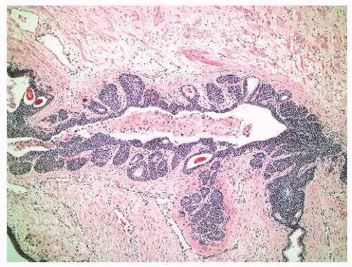
FIGURE 8-18 ▪ Urothelium lining a normal prostatic duct.


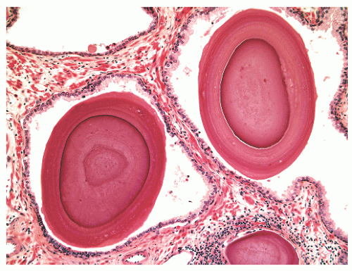
FIGURE 8-19 ▪ Corpora amylacea.


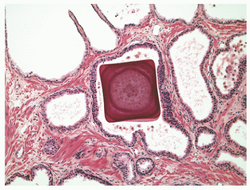
FIGURE 8-20 ▪ Corpus amylaceum with square appearance.


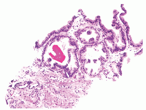
FIGURE 8-21 ▪ Crystalloid in lumen of benign gland.

Other intraluminal materials are, conversely, rare in normal gland lumina but are more common in neoplastic processes. These intraluminal materials include pink amorphous acellular secretions, intraluminal wispy blue mucin (also known as blue-tinged mucinous secretions), and crystalloids. Identification of these secretions, mucin, or crystalloids47 in benign prostatic tissue from needle biopsy (Fig. 8-21) is not an indication for rebiopsy.

Nonprostatic epithelium that can be seen associated with all prostatic tissue samples includes ejaculatory duct and seminal vesicle epithelium and colorectal epithelium. This last epithelium is frequently seen in needle biopsies of the prostate and is often of colonic glandular type, but squamous epithelium can also be observed. Distorted rectal tissue can potentially be confused with prostatic adenocarcinoma due to intraluminal blue mucin, prominent nucleoli, mitotic activity, and the immunoprofile of negative 34βE12 and p63 immunoreactivity, with positive staining for alpha-methylacyl CoA racemase.48


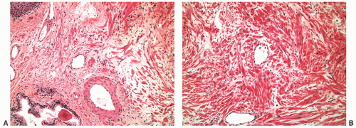
FIGURE 8-22 ▪ Normal peripheral zone stroma (A) compared to denser normal transition zone stroma (B).


Stroma

Other structures of the prostate include the capsule, preprostatic sphincter, striated sphincter, anterior fibromuscular stroma, and intraglandular stroma, including smooth muscle cells, fibroblasts, vasculature, and nerves.11 There are zonal differences in stromal density. The normal peripheral zone stroma is fibromuscular and loosely woven (Fig. 8-22A), whereas normal transition zone stroma is more compact, with interlacing smooth muscle bundles (Fig. 8-22B). Rarely, adipocytes can be seen in the prostate,49,50 including in needle biopsy.51 For practical purposes, carcinoma in fat should be viewed as extraprostatic extension.50


“Capsule”

The outer prostatic “capsule” is actually a band of concentrically placed fibrovascular tissue that is inseparable from prostatic stroma11,52 and surrounding fasciae. Outside the prostate, this band is also continuous with pelvic fascia and rectovesical fascia (of Denonvilliers). Denonvilliers fascia is a sheath between the rectum and prostate that covers the posterior aspect of the prostate and seminal vesicles.

The fibromuscular band (“capsule”) of the prostate is 0.5 to 3 mm thick and is incomplete, being absent at the apex.52, 53, 54 It is composed mainly of transversely disposed bundles of smooth muscle and collagen. Smooth muscle bundles course between the gland and periepithelial stroma inside the prostate gland, without obeying any sort of boundary (Fig. 8-23). Also, the concentration of smooth muscle fibers in the band and prostatic stroma is identical. Glands from the prostate approach this band, but the border between the outer limit of epithelium and the band is not often clearly definable under the light microscope. Posteriorly, the band fuses with Denonvilliers fascia and does not clearly separate prostate from seminal vesicle.

Posterolaterally, peripheral gaping blood vessels have been used as a demarcation landmark between intraprostatic
and extraprostatic tissue, but such vessels clearly can extend into the substance of the prostate and therefore are unreliable as a capsular marker. Anteriorly and anterolaterally, the band blends with the pelvic fascia. Apically, the band is no longer present, and instead there is a jumble of smooth muscle, skeletal muscle, and fibroelastic fibers. Here, it is important to know that the urethral striated sphincter (rhabdosphincter) is in direct continuity with the prostate, and indeed skeletal muscle fibers can normally be found, at an anterior and apical location, within the substance of the gland itself (Fig. 8-24).55 At the bladder neck, a capsular separation of bladder and prostate also does not exist; rather, there is a fusion of smooth muscle bundles (Fig. 8-25).


FIGURE 8-23 ▪ Fibromuscular sling or “capsule” surrounding the prostate is ill defined.


Smooth Muscle, Fibroblasts, Extracellular Matrix, and Basement Membrane

The stroma in the prostate gland consists chiefly of smooth muscle cells and fibroblasts, with ramifications of blood and lymph vessels as well as nerve bundles and axons.11 The extracellular stromal matrix of the prostate is primarily collagen of types I and III, complex polysaccharides, and glycosaminoglycans such as dermatan sulfate, heparin, chondroitin, and hyaluronic acid.8 Fibronectin and tenascin polypeptides are also found in prostatic stroma.56,57 Abutting prostatic glands is a think, delicate basement membrane, that, at 100-nm thickness, is too small to be seen in H&E-stained sections.


FIGURE 8-24 ▪ Benign prostatic glands admixed with skeletal muscle in needle biopsy tissue.


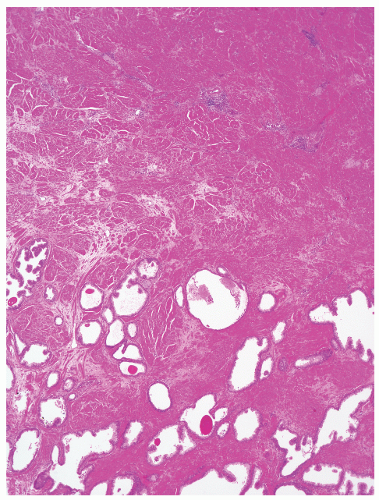
FIGURE 8-25 ▪ Boundary between prostate (below) and urinary bladder neck (top): absence of a capsule.


Nerves

The prostate gland has a rich nerve supply, with sympathetic and parasympathetic innervation arising from the pelvic plexus.58 These nerves run with branches of the capsular artery and penetrate the prostate, where parasympathetic fibers course to acini and stimulate secretion and where sympathetic fibers cause contraction of outer band “capsular” and intraprostatic smooth muscle. Innervation of the peripheral zone and posterior “capsule” is significantly higher than that of the transition zone and anterior “capsule.”59 Histologically, nerves are seen in the periprostatic neurovascular bundle, in the outer fibromuscular band, and in the prostate itself. Ganglia and paraganglia (Fig. 8-26) are usually found outside the gland but may also be found in the outer fibromuscular band and within the prostate itself. Paraganglia are most often located in or adjacent to the lateral neurovascular bundles.60 They should not be confused with “hypernephroid” prostatic carcinoma. Because ganglia and paraganglia can be found inside the prostate and in the outer fibromuscular
band, carcinoma invading around these structures should not necessarily be equated with extraprostatic extension of malignancy. Another diagnostic pitfall is the relationship of normal and benign prostatic glands to intraprostatic nerves. Benign prostatic glands can abut nerves61, 62, 63 (Fig. 8-27) and can even be found within nerves.63,64 Such glands should not be considered automatically malignant, and a constellation of histomorphologic traits should be used in diagnosis, not solely the physical proximity of prostatic glands and nerves.


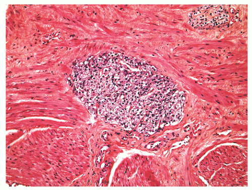
FIGURE 8-26 ▪ Paraganglion in smooth muscle tissue.


Lymphatics and Blood Vessels

The blood supply to the prostate is from the inferior vesical artery. By light microscopy, intraglandular blood vessels are easily seen in prostatic stroma, but capillaries adjacent to normal glands are not so readily appreciated on H&E sections, probably because a large number have closed lumina.65 There is a network of intraprostatic lymphatics that drain principally into obturator and then internal iliac lymph nodes. A minor amount of drainage occurs through the presacral group and external iliac nodes. An uncommon site of flow is through periprostatic or periseminal vesicle lymph nodes, which are found in about 4% of radical prostatectomy specimens.66


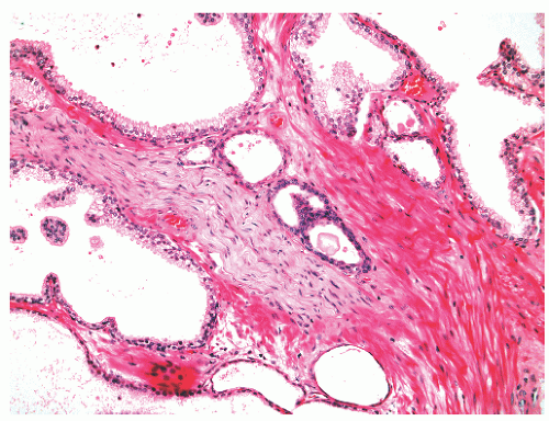
FIGURE 8-27 ▪ Benign perineural glands.


Periprostatic Adipose Tissue

Outside the prostate in posterolateral sites one can find neurovascular bundles and abundant adipose tissue, although the amount and distribution of periprostatic adipose tissue in radical prostatectomy specimens is quite variable. Assessment of this periprostatic adipose tissue for invasion by carcinoma is essential for pathologic staging. One can also find periprostatic adipose tissue in some needle biopsy cases.


Cowper Glands

Cowper bulbourethral glands are extrinsic to the prostate gland but are discussed here because these normal anatomic structures can be misdiagnosed as prostatic adenocarcinoma67,68 (Table 8-1). They are located in the urogenital diaphragm, just inferior to the prostate gland and lateral to the membranous urethra. These small structures measure on average 10 mm × 6 mm × 5 mm in adult men. Cowper glands can be incidentally and inadvertently sampled by needle biopsy directed at the prostate gland,67 by transurethral resection of the prostate,69 and by cystoprostatectomy.70 The incidental sampling in needle biopsy is a rare event, with an incidence of 0.006%.67 Microscopically, Cowper glands are compound tubuloalveolar glands composed of lobules of acini, admixed with excretory ducts and ductules (Fig. 8-28). Skeletal muscle fibers are seen associated with Cowper gland in all needle biopsy cases.67 The acinar lumens are small to occluded. Cuboidal to columnar pale-staining mucinous cells form the acini, whereas more flattened hybrid mucinous-ductal cells line the ducts and ductules. The mucinous cells are distended, with bluish,
foamy cytoplasm, and possess small, bland, basally situated nuclei. An acinar basal cell layer often is not apparent on H&E-stained sections. Immunohistochemical studies have produced mixed findings. In particular, studies have found luminal cells in the acini to be PSA positive67,70 or PSA negative68 and PSAP positive70 or PSAP negative.68 Also unsettled is whether the basal epithelial cells express high-molecularweight cytokeratins recognized by antibody 34βE12. The acini can be negative with 34βE12 immunolabeling.68 In most cases the basal cells are highlighted by antibodies to smooth muscle actin (SMA), which can be useful in the differential diagnosis with prostatic adenocarcinoma, although Cowper glands can usually be recognized by examination of H&E-stained slides without use of immunostains.


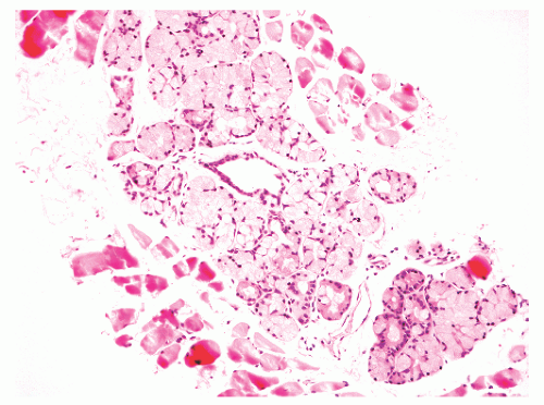
FIGURE 8-28 ▪ Cowper glands with mucinous acini, central duct, and surrounding skeletal muscle tissue.


Malformations

Clinically manifest developmental abnormalities of the prostate itself are rare and include agenesis, hypoplasia, cystic change, and abnormal persistence and hyperplasia of mesonephric remnants. Ectopic prostatic tissue is another form of maldevelopment.


Agenesis and Hypoplasis

Agenesis and hypoplasia of the prostate occur in several conditions including 5α-reductase deficiency,10 testicular feminization,71 and prune belly syndrome.72 Congenital 5α-reductase deficiency results in a rare form of pseudohermaphroditism in which patients have ambiguous external genitalia and small or undetectable prostates. In males with testicular feminization, the androgen receptor is defective, and the prostate is entirely absent. In the prune belly syndrome the prostate is hypoplastic and may not be grossly evident.72 Histologically, there is a pronounced reduction in epithelial elements, a reduction in smooth muscle fibers, and increased fibrous tissue. Concomitantly, the seminal vesicles can be rudimentary or absent and the vas deferens segmentally atretic. The prostatic urethra is markedly dilated in these cases.


Congenital Cysts

Congenital cysts in the prostate region may be classified as utricular (of endodermal origin),73 müllerian duct cysts (which are of mesodermal origin), ejaculatory duct cysts, vas deferens cysts, and seminal vesicle cysts.

Müllerian duct cysts and utricular cysts are uncommon midline structures that rarely produce clinical symptoms. The embryologic origin of the prostatic utricle is controversial, with suggested contributions from müllerian duct and urogenital sinus, with recent evidence suggesting a urogenital sinus origin.74 The incidence of utricular cystic dilatation (congenital or postinflammatory) is estimated at 7% (5/70) in newborns and young infants and 1% (7/678) in adults.75 Utricular dilatation and cyst formation are associated with intersex abnormalities and hypospadias, whereas true müllerian duct cysts are not.76 Considering the two together, the median age at presentation is 26 to 39 years, and symptomatic patients present most often with hematospermia, recurrent testicular or pelviperineal pain, and irritative lower urinary tract symptoms such as frequency, dysuria, and urgency.73,77 These cysts may also be associated with infertility due to ejaculatory duct obstruction.73 Aspiration fluid or portions of the cyst obtained by transurethral resection “unroofing,” or a complete cyst, removed by open surgery, may be received in the anatomic pathology laboratory.78 Grossly, incidentally detected utricular cysts are 2 to 3 cm in greatest dimension, with a smooth inner wall.75 The ejaculatory ducts are displaced laterally, with compression of their lumens. Microscopically, the epithelium lining the cystic cavity has been described as columnar or cuboidal (32% of cases), although it may be stratified squamous (19%), urothelial (10%), or devoid of epithelium (19%).73,75 The cyst walls are composed of smooth muscle or fibrous tissue. In 25% of cases, acini with columnar epithelium have been noted in the cyst wall near the connection with the urethra. Aspiration fluid should not reveal any sperm (unlike the ejaculatory duct cyst). The fluid has been characterized as clear75 or whitish to brown,73 with old blood and cellular debris.79 Stones have been found in these cysts. Treatment depends on symptoms, cyst size, and location. Options range from observation for asymptomatic patients to aspiration to surgery.73,77 A rare complication in these cysts is the occurrence of malignancy. A few cases of ductal adenocarcinoma, 80, 81, 82 squamous cell carcinoma,83 and clear cell adenocarcinoma84 arising in müllerian duct or utricular cysts have been reported.

Congenital seminal vesicle cysts and ejaculatory duct cysts are also in the differential diagnosis of cystic change in the prostatic region. Congenital ejaculatory duct cysts are rare; in one series where vesiculography was performed in infertile men, the incidence was 0.6% (1/158).85 Minor congenital anatomic variations of the ejaculatory duct include caudal formation of ejaculatory ducts within the central zone (18% of cases), abnormal posterior penetration of the ejaculatory ducts at the rectal surface (12%), abnormally large muscle bundles in the duct sheath (6%), and ductal dilatation (7%).86


Mesonephric Remnants

As a developmental anomaly, rare examples of mesonephric remnants have been recognized in the prostate, where they may assume pseudoneoplastic status due to florid hyperplasia87, 88, 89, 90 (see Hyperplasia section below). The incidence of nonhyperplastic mesonephric remnants in the prostate is unknown.


Prostatic Tissue Ectopia

Ectopic benign prostatic tissue in males has been found in a number of extraprostatic locations including testis, epididymis, processus vaginalis, urinary bladder, penile urethra,
seminal vesicle, root of the penis, subvesical space, retrovesical space, pericolic fat, anal submucosa, perirectal fat, urachal remnant, and spleen.91, 92, 93, 94, 95, 96, 97, 98, 99, 100, 101, 102, 103 The most frequently cited type of prostatic ectopia is in the prostatic urethra in the form of prostatic urethral polyps. However, because prostatic epithelium normally can line the urethra in the verumontanum region, one could argue that these polyps do not represent ectopia.

Prostatic tissue has been reported in females in the posterior wall of the distal urethra, in paraurethral Skene glands,104 in ovarian hilar mesonephric rests,105 in ovarian teratomas,106 and in the vagina, vulva, and cervix.104,107In the vagina, tubulosquamous polyps may display prostatic differentiation, as substantiated by PSA and PSAP immunostains.104,107 A single case of ovarian teratoma with prostatic tissue with morphologic features of prostatic carcinoma has been published.106

Mature-appearing prostatic tissue has also been rarely reported in neoplasms, including a retroperitoneal lipoma108 and a testicular teratoma.109

Ectopic prostatic tissue usually presents as an incidental finding but can manifest clinically as a mass110 or as urinary frequency, voiding difficulty, dysuria, or hematuria. Although these patients could conceivably present with serum PSA elevation due to ectopic prostate, this has not been reported. By cystoscopy, ectopic prostatic tissue in the bladder may be confused with cystitis cystica.92 Benign adenomatous110 and malignant111 changes have been described in ectopic prostatic tissue. The precise mechanisms for the formation of ectopic prostatic tissue are not definitely established. An embryologic remnant of the ventral portion of the cloaca is favored, and urothelial metaplasia has also been forwarded. For the lower female genital tract, a developmental anomaly, metaplasia of preexisting endocervical glands, and derivation from mesonephric remnants have been raised as possibilities.94 The presence of prostatic tissue in ovarian teratomas could be caused by induction by locally-produced androgen or tissue-specific genomic imprinting, but this is speculative.


Inflammation and Infection

Inflammation of the prostate (prostatitis) and infection of the prostate are common clinical problems. It is important for histopathologists to appreciate the National Institutes of Health’s (NIH’s) definition of prostatitis,112 and to not necessarily equate histologic evidence of prostatic inflammation (“histologic prostatitis”) with clinical prostatitis. Infections of the prostate are most often bacterial and are diagnosed clinically. Histopathologic detection of inflammatory cells in the prostate is common, but histologic identification of specific infectious agents in prostatic tissues is rare.


Prostatitis: Introduction

Clinical prostatitis is a major health problem and has been considered to be the third most important disease of the prostate gland after BPH and cancer.113 In the United States it accounts for 2 million annual doctor visits and 25% of all office visits for genitourinary-related complaints.114 The estimated community-based prevalence of clinical prostatitis is 10% to 16%,113,115 which is of a magnitude similar to that of diabetes and ischemic heart disease.116 Compared with BPH and prostate cancer, prostatitis is far more likely to affect younger men (18 to 50 years of age).


From a strict definitional standpoint, prostatitis is inflammation of the prostate. However, clinical use of the term prostatitis is not confined to cases with inflammation. Conversely, when histopathologists see inflammation in, for example, a prostate needle biopsy, a diagnosis of prostatitis, although histologically correct, does not usually correlate with the clinical concept of prostatitis (Box 8-2).

In the 1999 NIH consensus statement, prostatitis and prostatitis-like symptoms were classified into four broad categories (Table 8-3).112 This is now the most widely accepted classification system. Note that only category IV has a tissue-based component in the clinical diagnosis. Category III prostatitis, known as chronic prostatitis/chronic pelvic pain syndrome, is by far the most common form of prostatitis, comprising 90% of all prostatitis cases.117 These patients
present with pelvic pain localized to the prostate, perineum, or urethra, and a variable degree of voiding abnormalities such as urinary frequency, and sexual dysfunction.118 The etiology is unknown and optimal treatment has not been established.118 Acute bacterial prostatitis (category I) (usually due to Escherichia coli), is rare, being diagnosed in 0.02% of all prostatitis patients and chronic bacterial prostatitis affects 5% to 10% of patients with chronic prostatitits.119 Acute bacterial prostatitis is usually readily diagnosed clinically by the sudden onset of urogenital and often systemic symptoms, such as fever, chills, irritative voiding symptoms, and pain in the lower back, rectum, and perineum, along with bacteriuria.120 Treatment is with systemic antibiotic therapy.120 Patients with chronic bacterial prostatitis (category II), in contrast, experience prolonged or recurrent symptoms and relapsing bacteriuria. Treatment is more difficult and requires selection of an antibiotic with properties that allow for penetration into the prostate.120 Prostate biopsy is contraindicated in patients with acute bacterial prostatitis due to the risk of septicemia and prostate biopsy for patients with chronic prostatitis/chronic pelvic pain syndrome is currently a research tool only.121 Asymptomatic inflammatory prostatitis (category IV) is an incidental finding of unknown clinical significance, except that such inflammation can be associated with an elevated serum PSA.121


Table 8-3 ▪ NIH CLASSIFICATION OF PROSTATITIS SYNDROMES


Category I


Acute bacterial prostatitis


Category II


Chronic bacterial prostatitis


Category III


Chronic abacterial prostatitis/chronic pelvic pain syndrome


Category IIIa


Inflammatory chronic pelvic pain syndrome


Category IIIb


Non-inflammatory chronic pelvic pain syndrome


Category IV


Asymptomatic inflammatory prostatitis


Histologic Acute Inflammation

An acute inflammatory cell infiltrate made up of neutrophils is common in prostatic tissue samples acquired for reasons not related to clinical prostatitis. In needle biopsy tissue taken to rule out prostate cancer, up to 50% of cases harbor acute inflammation,122 while 20% to 98% of transurethral and open prostatectomy cases have acute inflammatory infiltrates.123, 124, 125 Microscopically, the acute inflammation is often intraluminal (Fig. 8-29) and varies in extent from a few scattered neutrophils to microabscesses. Neutrophilic infiltrates in the prostate can be associated with intraluminal necrotic debris, duct rupture, and epithelial alterations, especially atrophy and hyperplasia. Squamous metaplasia in the prostate can also be acutely inflamed, but neoplastic proliferations— PIN and carcinoma—rarely exhibit neutrophilic infiltrates. The main differential diagnostic difficulty with neutrophilic infiltrates is the distinction of reactive, inflammatory nuclear atypia from prostatic adenocarcinoma. The diagnostician should be aware that prominent nucleoli (of >1 µm) are detectable in almost one-half of cases of acutely inflamed atrophy.126 Special studies generally are of limited value in assessing histologic acute inflammation of the prostate. The one special study that can be diagnostically beneficial is immunohistochemical staining to confirm basal cell presence (using p63 and/or 34βE12 immunostains) in acutely inflamed, reactive, benign glands, but this is not usually necessary. One need not apply histochemical stains for organisms to sections of prostate with acute inflammation.


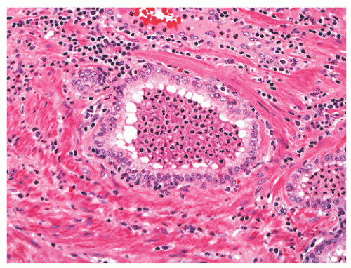
FIGURE 8-29 ▪ Intraluminal acute inflammation in a benign gland with reactive nuclear atypia, including visible nucleoli.


Histologic Chronic Inflammation

Chronic inflammatory cell infiltrates, consisting of lymphocytes, histiocytes, and plasma cells, are extremely common in all types of prostatic tissue samples, being found in the majority of needle biopsy and prostatectomy cases.121,123,124

Microscopically, several patterns of chronic inflammatory cell involvement of benign prostatic tissue have been described.124,127,128 Lymphocytic and plasmacytic chronic inflammatory cell infiltrates, in contrast to acute inflammation, tend to be stromal-based and periglandular. Admixture with other inflammatory cell types is common. In tissue from the peripheral zone chronic inflammation is often associated with atrophy (Fig. 8-30A and B).

In hyperplastic TURP tissue, a very common pattern seen in 85% of TURP chips is dilated glands containing cell debris, foamy macrophages, neutrophils, and pink proteinaceous material, associated with surrounding periglandular lymphocytes and plasma cells (Fig. 8-31).124 A second highly common arrangement is a periglandular, lymphoplasmacytic infiltrate in the absence of intraluminal inflammatory cells. Another morphologic presentation is a more diffuse stromal extension by the lymphocytes and plasma cells. Lymphoid aggregates and even lymphoid follicles (Fig. 8-32) may be seen. In the glandulocentric infiltrates of lymphocytes or plasma cells, direct extension of lymphocytes into benign prostatic epithelium commonly occurs, sometimes with perilymphocytic clearing that allows for recognition of intraepithelial lymphocytes. Scattered single lymphocytes can also be noted, especially in nodules of stromal hyperplasia (stromal nodules). Additionally, perivenular lymphocytic cuffs can be observed; it is thought that these venules are an avenue of entry for the lymphocytes into the prostate gland.129

Mast cells, eosinophils, and macrophages are cell types that can also be identified in histologic prostatitis. Mast cells are difficult to discover in prostatic tissues without
histochemical stains. They increase in number with age130 but are not associated with any specific disease process.130,131 Eosinophils can nonspecifically accompany other inflammatory cells in histologic prostatitis (Fig. 8-33). The designation of eosinophilic (allergic) prostatitis should be reserved for those rare patients with a hypersensitivity disorder (such as asthma), peripheral eosinophilia, and large numbers of eosinophils admixed with granulomas.132, 133, 134, 135, 136 Macrophages can be found in dilated glands surrounding ruptured ducts and acini and in granulomatous prostatitis.


FIGURE 8-30 ▪ Chronic inflammation associated with atrophy in needle biopsy tissue (A) and in the whole gland (B).

Benign prostatic epithelium in the setting of chronic histologic inflammation may display a range of reactive alterations. Secretory epithelial cells can lose cytoplasmic volume, some of the cells become cuboidal instead of columnar, and the cytoplasm becomes more dense and eosinophilic rather than clear to granular.124,129 This lends an atrophic appearance to these glands. Basal cell hyperplasia is often involved by lymphocytes.137,138 Urothelial metaplasia, nephrogenic adenoma, and squamous cell metaplasia in the prostate are also often associated with inflammation. Reactive nuclear atypia in the chronically inflamed epithelium generally is mild128 and is regularly manifested as crowded, enlarged nuclei with small nucleoli. These nucleoli are usually small, but in inflamed basal cell hyperplasia and in some inflamed atrophic or squamous metaplastic cells larger nucleoli may be seen.


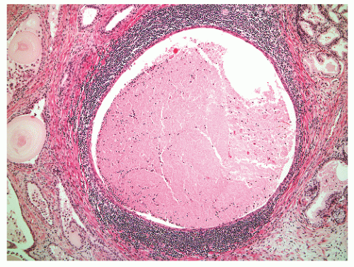
FIGURE 8-31 ▪ Dilated benign prostatic gland with surrounding band of chronic inflammatory cells.

Stromal alterations are less well described in chronic prostatic inflammation, but stromal sclerosis can definitely occur,128 particularly in response to gland rupture.

Inflammation in the prostate involves neoplastic epithelium less often than benign epithelium.129 Rarely is highgrade PIN involved by abundant inflammation,139 whereas a minor lymphocytic infiltrate is common in prostatic carcinoma.140 Of note, when urothelial carcinoma of the urethra or urinary bladder extends into the prostate, it can exhibit an exuberant peritumoral inflammatory and sclerotic response.


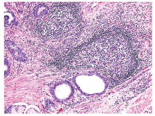
FIGURE 8-32 ▪ Follicular prostatitis.


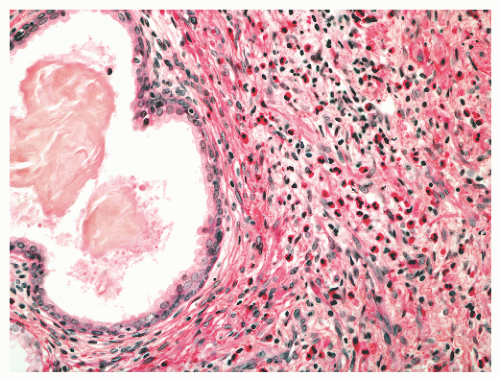
FIGURE 8-33 ▪ Eosinophils in nonspecific inflammation in the prostate.

Benign lymphocytes in the prostate may be mistaken for malignant cells of epithelial or hematopoietic types. Arrangement of lymphocytes in cords, linear arrays, or sheets may cause confusion with high-grade Gleason pattern 4 or 5 adenocarcinoma, prostatic small cell carcinoma, or lymphoma at low magnification. Higher-power examination will allow for identification of small bland lymphocytic nuclei. Lymphocytes with crush/distortion artifact or thermal damage may on occasion be difficult to distinguish from carcinoma, especially when such distorted lymphocytes are present around nerves or at the margin of radical prostatectomy tissue sections. Thermal damage at TURP can induce signet ring change in benign lymphocytes,141 mimicking signet ring carcinoma.


Granulomatous Prostatitis

Granulomatous prostatitis is a distinctive form of prostatitis that can be mistaken for carcinoma clinically,142, 143, 144 radiologically (by ultrasound),145,146 and histopathologically.147 The incidence of granulomatous prostatitis in all prostatic samples ranges from 0.4% to 4%.124,128,148,149 The incidence in needle biopsy is <1%.148 Most patients are 50 to 70 years of age (mean = 62 years; range = 18 to 86 years) and present with irritative or obstructive voiding symptoms, fever, and chills. Consistent with this presentation, most patients had a urinary tract infection 1 to 8 weeks before diagnosis. Clinical concerns for malignancy are raised in granulomatous prostatitis because of an elevated total serum PSA, a low percentage free PSA, hypoechoic lesions on ultrasound, and a hard, fixed, indurated prostate gland on digital rectal examination.144,146,148

Granulomatous prostatitis can be classified based on cause (Table 8-4). The most common cause of granulomatous prostatitis is so-called nonspecific granulomatous prostatitis, which accounts for about three quarters of granulomatous prostatitis cases. In needle biopsy, granulomatous prostatitis is categorized as nonspecific in 77% of cases, infectious in 18% of cases, and indeterminate in 4% of cases.148 The infectious agent here is bacille Calmette-Guerin (BCG), used in therapy for urothelial carcinoma. In larger tissue samples, postbiopsy and postresection granulomas are more common, being diagnosed in 25% of all granulomatous prostatitis cases.149 The other listed causes of granulomatous prostatitis in Table 8-4 are uncommon to rare.


Table 8-4 ▪ GRANULOMATOUS PROSTATITIS: CLASSIFICATION


I.


Nonspecific (due to duct/acinar rupture)


Usual type


Xanthogranulomatous


Nodular histiocytic prostatitis


Xanthoma


II.


Infectious


A.


Bacterial


Tuberculosis


Brucellous


B.


Spirochetal


Syphilis


C.


Fungal


Coccidioidomycosis


Cryptococcosis


Blastomycosis


Histoplasmosis


Paracoccidioidomycosis


Candidiasis


Aspergillosis


D.


Parasitic


Schistosomiasis


E.


Viral


Herpes zoster


III.


Postbiopsy/resection


IV.


Malakoplakia


V.


Systemic granulomatous disease


Allergic (eosinophilic)


Sarcoidosis


Wegener granulomatosis


VI.


Foreign body


Teflon


Hair


Modified from reference Roberts RO, Lieber MM, Bostwick DG, et al. A review of clinical and pathological prostatitis syndromes. Urology 1997;49:809.


Nonspecific Granulomatous Prostatitis

Nonspecific granulomatous prostatitis is termed nonspecific because it is not considered secondary to a specific agent but rather it is viewed as representing a foreign body-type response to prostatic secretions, with associated duct or acinar rupture.150,151 Yet some patients have positive urine cultures (mainly for E. coli), such that some of this response may also be caused by bacterial products. Impairment of drainage, perhaps secondary to BPH, and infection seem to
be pathogenetic components.144,151 The serum PSA is often significantly elevated, with a mean of around 13 ng/mL (range 0.4 to 114 ng/mL).148,152


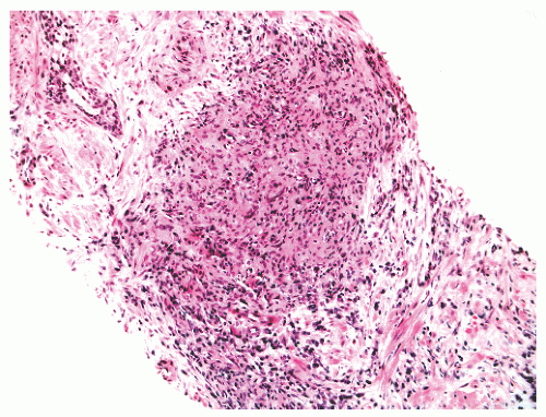
FIGURE 8-34 ▪ Nonspecific granulomatous prostatitis that is noncaseating with lobular pattern.

Grossly, nonspecific granulomatous prostatitis appears as small, firm, yellow granular nodules.144 Microscopically, the usual type of nonspecific granulomatous prostatitis is a lobulocentric noncaseating granulomatous inflammatory cell infiltrate (Fig. 8-34). The inflammation is centered on ducts and glands, which exhibit a variable degree of destruction. Liquefactive necrosis has been reported in the center of the inflammatory infiltrate,132 although other series do not allow necrosis.149 Necrosis raises the possibility of specific infectious granulomatous prostatitis and is not usually seen in nonspecific granulomatous prostatitis. The granulomas can be focal or diffuse. In needle biopsy, more than one core is involved in most cases.148 The lobulocentricity of the granulomas can be appreciated in some needle biopsy cases, where smaller granulomas are present, but in other cases the granulomatous inflammation can appear more sheet-like because of partial sampling of larger granulomas. In TURP and simple prostatectomy tissues, the granulomas can be extremely focal, can form a discrete mass, or can be a diffuse spread of fused nodules. The cellular infiltrate in usual nonspecific granulomatous prostatitis is mixed, with epithelioid histiocytes, lymphocytes, neutrophils, eosinophils, plasma cells, and multinucleated giant cells (Fig. 8-35). The giant cells can be of foreign body, Langhans, and Touton types.153 They can be difficult to detect in needle biopsies. Lymphocytic infiltrates, sometimes with lymphoid follicle formation, surround the granulomatous nodules. Perinodular stromal fibrosis and scarring can ensue in some cases.144

The spectrum of morphologic abnormalities in nonspecific granulomatous prostatitis also includes “xanthogranulomatous prostatitis,” “nodular histiocytic prostatitis,” and “prostatic xanthoma.” The first two designations are applied when the epithelioid histiocytes possess a cleared to vacuolated cytoplasm (due to lipid accumulation).153, 154, 155, 156 In prostatic xanthoma there is a solitary, microscopic aggregate of foamy histiocytes (Fig. 8-36).157,158 This is a rare entity that is usually an incidental finding in peripheral zone tissues removed to evaluate for BPH or carcinoma. Only one patient had hyperlipidemia.


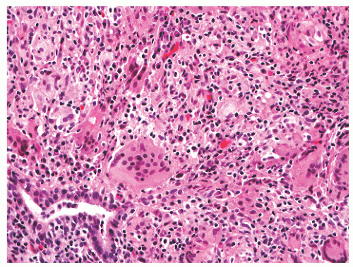
FIGURE 8-35 ▪ Nonspecific granulomatous prostatitis with a mixed inflammatory cell infiltrate comprising lymphocytes, plasma cells, histiocytes, and multinucleated giant cells.

Sheets or cords of epithelioid histiocytes in nonspecific granulomatous prostatitis or xanthoma can masquerade as poorly differentiated carcinoma or carcinoma with hormonal treatment effect.148,149,158 This is particularly problematic when histiocytes predominate in nonspecific granulomatous prostatitis, without significant numbers of giant cells, plasma cells, lymphocytes, eosinophils, or neutrophils. Also, the epithelioid histiocytes can display prominent nucleoli, which can cause concern for malignancy. An immunohistochemical marker panel can be helpful in defining the differentiation status of the atypical epithelioid cells. Negative
immunoreactions for pan-cytokeratin, PSA, and PSAP, with immunopositivity for CD68 and lysozyme in the epithelioid cells are useful in establishing a diagnosis of granulomatous prostatitis in this context.147,148,158


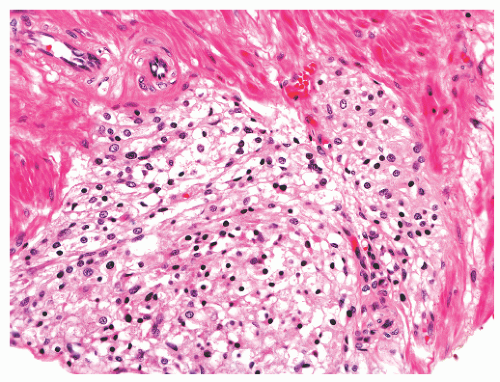
FIGURE 8-36 ▪ Xanthoma of prostate consisting of a small aggregate of foamy histiocytes.


Infectious Granulomatous Prostatitis

Specific infectious types of granulomatous prostatitis include bacterial, fungal, parasitic, and viral prostatitis (Table 8-4).


BCG Granulomatous Prostatitis

The most common type of infectious granulomatous inflammation is that due to BCG therapy for urothelial carcinoma (usually of the bladder). In fact, every case of infectious granulomatous prostatitis in one needle biopsy series was caused by BCG treatment.148 Whereas only 10% of men treated with intravesical BCG for superficial urinary bladder carcinoma develop clinically symptomatic BCG prostatitis,159 a substantial percentage (80% to 100%) of patients have histologic evidence of BCG granulomatous prostatitis.159, 160, 161 Grossly, the cut surfaces of prostate glands in radical cystoprostatectomies from patients treated with BCG show multiple, firm, white nodules or soft, yellowish-gray nodules with granular centers, central caseation, and focal cavitation.160 In exceedingly rare cases abscess formation can occur.162 The large, confluent granulomas can measure up to 1.6 cm in greatest dimension.160 Microscopically the granulomas, particularly the larger ones, are of the typical caseating, tuberculoid type (Fig. 8-37). Smaller (<1 mm) granulomas composed predominantly of histiocytes can lack giant cells and caseous necrosis. Indeed, in needle biopsy up to one-quarter of BCG granulomatous prostatitis cases lack caseous necrosis.148 The smaller granulomas are periglandular (Fig. 8-38), often with intraluminal protrusion. With a history of previous BCG treatment, it is not necessary to stain sections with Ziehl-Neelsen for acid-fast organisms. If the stain is performed, 40% to 77% of cases demonstrate mycobacteria in the granulomas.160,163


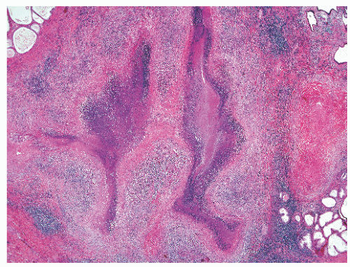
FIGURE 8-37 ▪ BCG granulomatous prostatitis with large geographic areas of caseating granulomas.


FIGURE 8-38 ▪ BCG granulomatous prostatitis with small periglandular epithelioid granuloma without caseation.


Infectious Granulomatous Prostatitis: Non-BCG Types

Mycobacterial prostatitis due to Mycobacterium tuberculosis or atypical species is rare,164, 165, 166, 167, 168 with an incidence of 3% of all examples of granulomatous prostatitis.165 The spread to the prostate is hematogenous, usually from active pulmonary disease. The prostate is nodular to palpation. Grossly, yellow to grayish-yellow nodules are located in the peripheral zone.167 Larger lesions exhibit caseation, but caseation is minimal in the “small yellow tubercle” granulomas.167 The granulomas are similar to those in BCG granulomatous prostatitis, including lack of caseous necrosis in some examples in needle biopsy. Presentation as an incidental finding in TURP chips has also been a mode of diagnosis.164 For patients without a history of BCG treatment and those in whom tuberculosis is clinically suspected, special stains for acid-fast bacilli and fungi should be performed.

Isolated case reports of prostatic brucellosis144 and prostatic syphillis169 indicate that these organisms can also elicit a granulomatous prostatitis.

Fungal, parasitic, and viral granulomatous prostatitis due to specific organisms is also rare.170, 171, 172, 173, 174, 175, 176, 177, 178, 179 Predisposing conditions for prostatic fungal infection include immunosuppression, prolonged antibiotic use, diabetes mellitus, malignancy, and an indwelling bladder catheter.177,180,181 Even in disseminated fungal infections, the prostate is uncommonly invaded. Fungi detected in the prostate include Cryptoccocus,178,182,183 Paracoccidioides,184 Coccidioides (Fig. 8-39A and B),172,173 Histoplasma,185 Blastomyces,186 Candida,170,176,177 and Aspergillus.149 Among cases of granulomatous prostatitis, fungal granulomatous prostatitis accounted for 1 in 200 (0.5%) of cases.133 In patients with the aforementioned predisposing conditions, and granulomatous prostatitis or necrotizing granulomas in the absence of BCG therapy, special histochemical stains for fungal organisms and acid-fast bacilli should be
performed. PCR specific for cryptococcal 18S rDNA from fixed prostate tissue was performed in one case.183


FIGURE 8-39Coccidioides immitis granulomatous prostatitis (A) with spherules highlighted by Gomori methenamine-silver histochemical stain (B).

Granulomatous prostatitis due to parasitic infection can occur in schistosomiasis. Eggs of Schistosoma (usually haematobium) are deposited in the prostate of about 20% of African men with schistosomiasis, but this rarely gives rise to prostate-related symptoms.175

Necrotizing, focally granulomatous prostatitis has been reported in two patients in association with herpes zoster infection.174


Postbiopsy and Postresection Granulomatous Prostatitis

The incidence of this kind of granulomatous prostatitis is highly variable, depending on the type of tissue sample. Amongst cases of granulomatous prostatitis, the incidence in needle biopsy was 0%,148 whereas in another series that included TURP chips, the incidence was 25%.149 This is so because this granulomatous process tends to be central, involving periurethral transition zone tissue, which is readily sampled by TURP, as opposed to involvement of the peripheral zone, which is most often the target of needle biopsies. Amongst all cases of prostatitis, it made up 7.1%.128 Patients were 51 to 87 years of age and had undergone TURP 7 days to 3 years previously.132,187, 188, 189, 190 Two patients had a history of needle biopsy of only the prostate.190 Patients present for repeat TURP (or initial TURP in the needle biopsy patients) because of persistent prostatitism, urinary obstruction or retention, and hematuria. It is noteworthy that TURP does not always seem to elicit such a granulomatous response, as 12 of 23 patients in one repeat TURP series191 did not have detectable granulomas, but the possibility remains that with additional chip sampling more granulomas might have been detected. The basis for the development of post-TURP granulomas appears to be a response to altered and cauterized prostatic epithelium and stroma, secretions, urine, or even metal from the diathermy instruments used for resection.192

Grossly, no difference is recognized in chips with or without postbiopsy or postresection granulomas.188 Microscopically, the granulomas assume a variety of shapes, including round, ovoid, elongated, stellate, serpiginous, triangular, slit-like, and rectangular forms (Fig. 8-40). Almost all of the granulomas demonstrate a connection with a cauterized TURP chip surface. A common finding is the base of a wedge-shaped granuloma at the edge of a TURP chip.132 The granulomas measure 0.5 mm to 2 mm190 and are usually multiple, with an average of 4 to 5 per case, but can be single.132,191 The granulomas are characterized by a fibrinoid central zone and surrounding, palisaded epithelioid histiocytes and fibroblasts. A rim of lymphocytes (which mark as T cells) is present outside the palisaded cell layer. Admixed with the lymphocytes are occasional plasma cells and eosinophils. The eosinophils may be numerous if the previous TURP was performed earlier than 2 months.132,187 Multinucleated giant cells are observed in some but not all cases and are usually of foreign body type but can also be of Touton or Langhans types.191 Occasional giant cells
contain brown pigment related to cautery-induced tissue carbonization.191 Older granulomas exhibit both central and peripheral hyalinization.


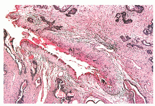
FIGURE 8-40 ▪ Post-TURP granuloma with central fibrinoid necrosis.

Reactive changes often are visualized in epithelium and stroma adjacent to postbiopsy TURP granulomas. Prostatic glands adjacent to the granulomas may appear compressed and reactive with slight to moderate nuclear atypia and small nucleoli, but macronucleoli are generally absent. Squamous metaplasia can be seen in about one-half of cases.191 Postbiopsy and post-TURP granulomas have been found in chips with both BPH and carcinoma. Localized vasculitis confined to the prostate has been reported in association with a few cases of post-TURP granulomas.153,189

Postbiopsy and post-TURP granulomas have been misdiagnosed as rheumatoid nodules and as tuberculous granulomatous prostatitis. A history of a previous procedure, in conjunction with the appearance of a palisading, fibrinoid granuloma, is most helpful in establishing the diagnosis of postbiopsy or post-TURP granulomatous prostatitis. In this setting it is not necessary to stain for organisms. Histochemical stains for organisms in these cases have been uniformly negative. Also, invariably negative are examinations for birefringent material with polarized light. Other types of granulomas are also in the differential diagnosis, such as allergic (eosinophilic) granulomatous prostatis and nonspecific granulomatous prostatitis.132 The former entity may overlap morphologically with early post-TURP granulomas in having numerous eosinophils, but allergic granulomatous prostatitis is seen in a different clinical context, with a clinical history of a hypersensitivity disorder (such as asthma) and a peripheral eosinophilia. Unlike nonspecific granulomatous prostatitis, postbiopsy and post-TURP granulomas are not centered on ducts and acini and also possess a striking fibrinoid central zone.


Malakoplakia

Malakoplakia uncommonly involves the prostate, with only about 30 cases reported to date.193, 194, 195, 196, 197, 198, 199, 200, 201, 202 In one review of malakoplakia occurring at all sites, the prostate was primarily involved in 10% of all cases.197 Although uncommon, malakoplakia, like other forms of granulomatous prostatitis, can be misdiagnosed as carcinoma by DRE, transrectal ultrasound (TRUS), and histopathologic examination.198, 199, 200 Patients with prostatic malakoplakia usually are in their 60s (range 49 to 85) and present with voiding symptoms (dysuria, frequency, urinary retention) and an enlarged, firm to hard prostate by DRE. There is almost always an associated urinary tract infection with E. coli. Immunosuppression has been associated with malakoplakia in general,197 but only two patients with prostatic malakoplakia were known to be immunosuppressed.201,202

Grossly, malakoplakia appears as soft, yellowish nodules in the prostate.195 Resected prostatic chips involved by malakoplakia are soft and yellow-brown to gray.200 Microscopically, there are sheets of macrophages, effacing normal prostatic architecture, with smaller numbers of lymphocytes, plasma cells, eosinophils, and neutrophils. The macrophages (also known as von Hansemann cells) contain round, often concentrically lamellated, basophilic intracytoplasmic inclusions that are 2 to 10 µm in diameter. They look targetoid, with a central basophilic body surrounded by a clear zone. These are the characteristic Michaelis-Gutmann bodies that represent calcified bacterial debris within phagolysosomes. These structures can also be extracellular.

The essential differential diagnostic distinction is the separation of malakoplakia from poorly differentiated carcinoma. There are three published cases in which an initial diagnosis of carcinoma was rendered.198, 199, 200 As in other types of granulomatous prostatitis, the diffuse growth of epithelioid histiocytes creates diagnostic difficulty. In one case misdiagnosed as clear cell carcinoma, the malakoplakia was diffuse and extensive, involving 60% of the TURP chips.200 The aforementioned immunohistochemical marker panel of pan-cytokeratin, PSA, PSAP, CD68, and lysozyme can also be diagnostically beneficial in selected cases. The presence of Michaelis-Gutmann bodies distinguishes malakoplakia from other types of granulomatous prostatitis.

Treatment is directed toward control of urinary tract infection.197 In one patient, failure of control resulted in a fatal prostate-rectal fistula.195 Overall, prognosis is related to extent of malakoplakia in the urinary tract and especially extent of renal involvement.197


Systemic Granulomatous Disease Affecting the Prostate

Several systemic granulomatous diseases can involve the prostate and even present primarily as a prostatic disorder (Table 8-4). All of these are uncommon to rare.

Allergic (eosinophilic) granulomatous prostatitis must be clearly defined and distinguished from other types of prostatitis that have eosinophils.132,148,149 The mere finding of eosinophils, even numerous eosinophils, does not equate with a diagnosis of allergic prostatitis. Rather, a histopathologic diagnosis of allergic (eosinophilic) granulomatous prostatitis should incorporate clinical findings of a hypersensitivity disorder (usually asthma or drug allergy), with or without a peripheral eosinophilia. Peripheral eosinophila without documented clinical hypersensitivity is not sufficient. Allergic (eosinophilic) granulomatous prostatitis is a rare condition, with an incidence of 0.06% in 3,600 TURP chip cases. Not a single case was diagnosed in 25,852 needle biopsies.148 It accounts for 1% of all cases of granulomatous prostatitis.149 Only a handful of cases have been well documented.132 Microscopically, the prototypical case has central fibrinoid necrosis with surrounding, palisaded epithelioid histiocytes and numerous eosinophils. No Charcot-Leyden crystals are seen. The differential diagnosis of allergic (eosinophilic) granulomatous prostatitis mainly includes other types of granulomatous prostatitis. Significant numbers of eosinophils can be seen in nonspecific granulomatous prostatitis (68% of cases), postbiopsy TURP cases, and less frequently in infectious granulomatous prostatitis (12% of cases).148


Sarcoidosis rarely involves the prostate, with only a few case reports.203, 204, 205 The noncaseating granulomas have been detected in needle biopsy,203,205 at radical prostatectomy,204 and at autopsy.

Wegener granulomatosis usually involves the upper respiratory tract, lungs, and kidneys, but in 2.7% to 7.4% of patients, prostatic involvement can occur.206, 207, 208, 209 Prostatic involvement at autopsy was seen in 7.4% of all Wegener cases,206 but only about 20 patients with Wegener granulomatosis have had symptoms referable to the prostate gland in the form of symptomatic prostatitis with urinary frequency, dysuria, hematuria, lower back pain, or acute urinary retention. Only a few patients (n = 10) had these prostatitis symptoms at initial clinical presentation.208,209 In a few patients localized induration on the DRE has caused clinical concern for carcinoma.209 Acute necrotizing granulomas have been detected in patients with prostatic Wegener granulomatosis in needle biopsy, in TURP chips, and at autopsy. An important microscopic finding is that of vasculitis, and this discovery, in conjunction with the necrotizing granulomas, should prompt a workup for Wegener granulomatosis in these rare patients who present with Wegener granulmatosis in the prostate. As in other anatomic sites, it is important to exclude infectious granulomatous disease before diagnosing Wegener granulomatosis, and so performance of histochemical stains for organisms is obligatory.


Foreign Body Granulomatous Prostatitis

A few examples of specific foreign body granulomatous reaction to Teflon (polytetrafluorethylene)210 and hair211, 212, 213 have been described. Teflon injection has been used to treat urinary incontinence, and this can cause prostatic nodularity by DRE and elevated serum PSA.210 Prostate needle biopsy demonstrates an exuberant foreign body giant cell reaction to the refractile, irregular fragments of Teflon. With polarization, the Teflon is highly birefringent. Detection of a Teflon granuloma does not rule out the coexistence of epithelial proliferative abnormalities. In one reported case, high-grade PIN was found, and in a second there was an associated Gleason score 7 adenocarcinoma.210 Hair granulomas with prostatic hair implantation due to long-term catheterization211 or perineal prostatic biopsy213 have also been seen.


Vasculitis

Inflammation of vessels in the prostate can represent a vasculitis confined to or isolated in the prostate or can represent a manifestation of systemic or nonprostatic confined vasculitis. Examples of localized vasculitis include vasculitis in post-TURP granulomatous prostatitis, giant cell arteritis (one case),214 and isolated nonfibrinoid arteritis in BPH (significance unknown). Systemic diseases that can produce vasculitis in the prostate include polyarteritis nodosa,215, 216, 217 Wegener granulomatosis, and hypersensitivity disorders causing allergic (eosinophilic) granulomatous prostatitis. Radiation therapy directed toward the prostate can also result in vasculitis in the prostate.


Infectious Prostatitis: Additional Types

Additional specific infections of the prostate have been reported for both immunocompetent and immunosuppressed patients. These include rare cases of prostatic actinomycosis,218 amebic prostatitis,219 Pseudomonas pseudomallei prostatitis,220 and prostatic Echinococcus221,222 in immunocompetent men. Microscopically, the prostatic response to these organisms is variable. For example, prostatic acute and chronic inflammation with fibrosis, granulation tissue, and abscess formation (with sulfur granules present) are evident in prostatic actinomycosis. A chronic, fibrosing prostatitis with eosinophils has been reported for echinococcal prostatic infection.221 It is uncertain how often sexually transmitted infections of the prostate by Neisseria gonorrhoeae and human papillomavirus occur and whether this is a clinical problem.223

In immunosuppressed patients, viral, bacterial, fungal, and parasitic prostatitis can occur. Most of these reports are of patients with acquired immunodeficiency syndrome (AIDS), although most cases of cytomegalovirus (CMV) prostatitis have been observed in patients undergoing immunosuppressive therapy following organ transplantation.224 In AIDS, patients can experience bacterial prostatitis, with progression toward abscess formation.225 Opportunistic infectious agents that have been reported in the prostates of patients with AIDS include CMV, Mycobacterium, Histoplasma, Candida, Pneumocystis, adenovirus, and Cryptococcus. Cryptococcal organisms may be sequestered in the prostate of patients with AIDS and thereby act as a reservoir for persistent infection.226 Microscopically, organisms can be identified in expressed prostatic secretions, prostate needle biopsy, or prostate glands from postmortem examination. The prostatic response to infection in patients with AIDS is variable and can be lacking,227 or there can be necrosis or abscess formation.228


Atrophy and Its Variants

Atrophy in the prostate is a common, age-related process that represents one of the benign lesions most often misdiagnosed as carcinoma.4, 5, 6 Prostatic atrophy begins at an early age and there is an increase in incidence and extent with age229,230; 67% of men aged 19 to 29 have atrophy and this increases to nearly 100% for men over 70.230,231 In needle biopsy, atrophy is an extremely common finding, detectable in 90% of peripheral zone biopsies.138 Atrophy in TURP chips usually represents cystic atrophy in nodular hyperplasia. In whole prostate glands, atrophy is principally localized to the peripheral zone.229

The cause of age-related atrophy is unknown. Factors that have been invoked include compression by hyperplastic nodules, inflammation, hormones, obstruction, nutritional deficiency, and systemic or local ischemia, such as that induced by arteriosclerosis.230, 231, 232, 233, 234 Atrophy in the prostate can also be caused by treatment, including radiation therapy and androgen deprivation therapy.


There are no clinical or radiologic features specific for atrophy although ultrasound examination may demonstrate cystic change that can be cystic atrophy.

Grossly, atrophy is visible only when there is cyst formation. Such cystic atrophy can occur in both the transition zone in BPH and the peripheral zone. However, atrophy is only definable at the light microscopic level, and it is not possible to tell macroscopically whether prostatic cysts are atrophic or not. These cysts have been termed retention cysts235 and impart a sponge-like appearance. Their size can range up to 2 cm in greatest diameter.235

Histologic classification of atrophy is useful for reasons of diagnostic awareness and differential diagnosis, but it is not absolutely necessary to subtype atrophy because there is currently no known clinical significance in doing so. For diagnostic recognition and research purposes atrophy may be classified as simple atrophy, simple atrophy with cyst formation, postatrophic hyperplasia (PAH), and partial atrophy.236 These patterns are often admixed. Sclerotic atrophy and proliferative atrophy (PA)/proliferative inflammatory atrophy (PIA) are also discussed here. The essential, unifying feature of all forms of atrophy is reduction of cytoplasmic volume of luminal epithelial cells (Box 8-3).


Simple Atrophy

Simple atrophy can be found in the peripheral zone with or without cyst formation. This is the most common type of atrophy, occurring in 89% of prostates with atrophy, sometimes alone and sometimes in combination with other types of atrophy.229 Simple atrophy is most often seen in combination with PAH (hyperplastic atrophy) and sclerotic atrophy.230 Microscopically, atrophy can be focal, involving just a few lobules, or it can involve virtually the entire peripheral zone. Simple acinar atrophy usually involves an entire lobule, with retention of a lobular architecture (Fig. 8-41). In noncystic atrophy there are aggregates of small dark acini with open lumens. The acini are rounded to angular in shape and are not crowded. Atrophic ducts ramifying into atrophic acini can be noted. Corpora amylacea are common in atrophic glands but crystalloids and intraluminal wispy blue mucin are decidedly uncommon. The stroma in the lobule is often altered with pale fibrosis and sometimes with a thickening of periacinar collagen (Fig. 8-42).232 Sclerosis around ducts can be marked (Fig. 8-43). Acute inflammation (of a moderate to severe grade) is observed in 15% of simple atrophy cases, whereas moderate to severe chronic inflammation is seen in 30% of cases (Fig. 8-44).126 The luminal epithelial cells in the atrophic foci are cuboidal (Fig. 8-45) to flattened (Fig. 8-46) and have a high nuclear/ cytoplasmic ratio. The nuclei are typically not enlarged but are often deeply staining, with condensed chromatin. However, nuclear atypia can be seen (Fig. 8-47), with prominent (>1 µm) nucleoli detectable in a minority of cases.126


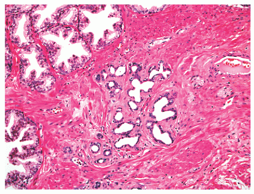
FIGURE 8-41 ▪ Simple atrophy, with a lobular configuration. Compare scant cytoplasm in atrophy versus adjacent benign nonatrophic glands where the luminal cells have a moderate amount of cytoplasm.

In needle biopsy, simple atrophy is eye-catching because it is often characterized by a haphazard distribution of small glands (Fig. 8-48). At higher magnifications, the small acini are seen to have distorted and angulated outer contours. This appearance, along with the stromal fibrosis of the atrophic lobule, and even chain-like (Fig. 8-47) and cord-like structures (Fig. 8-49) simulate an infiltrative process.


Simple Atrophy with Cystic Change

Cystic change in simple atrophy is a fairly common alteration, with gland size varying from mild dilatation to medium sized to larger cysts (Fig. 8-50), including the aforementioned 2-cm cysts. The epithelium and nuclei in these glands are markedly flattened. In the peripheral zone, a lobular arrangement of the cystically dilated, atrophic glands is sometimes evident, but with distortion the cystic, atrophic glands appear haphazardly arranged. Glands with cystic atrophy can masquerade as vessels. Simple cystic atrophy can also occur in areas of nodular hyperplasia in the transition zone (Fig. 8-51), where a flattened, atrophic-appearing epithelium can be found in nodules of usual epithelial and stromal hyperplasia.


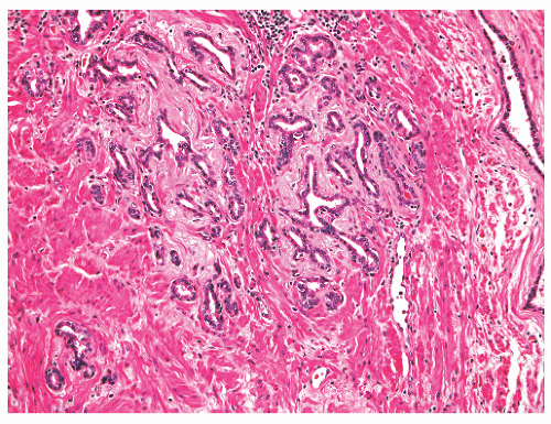
FIGURE 8-42 ▪ Atrophy with adjacent sclerotic stroma.


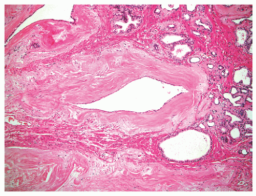
FIGURE 8-43 ▪ Atrophic prostatic duct with surrounding sclerosis.


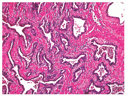
FIGURE 8-44 ▪ Atrophy with associated acute and chronic inflammation.


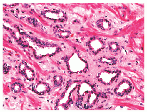
FIGURE 8-45 ▪ Atrophy with cytoplasmic volume loss.


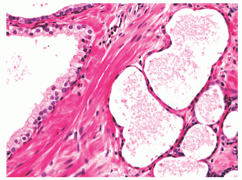
FIGURE 8-46 ▪ Atrophy with marked cytoplasmic volume loss and nuclear flattening.


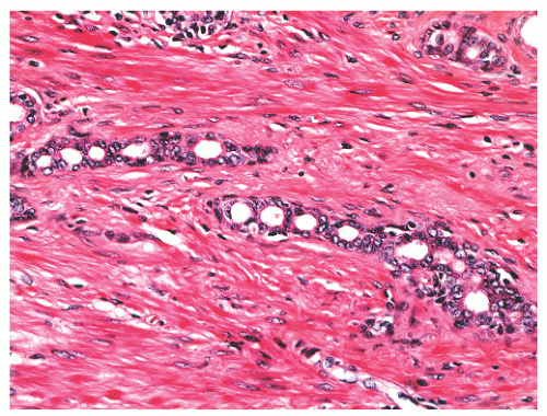
FIGURE 8-47 ▪ Atrophy with chain-like appearance and nuclear atypia with nuclear enlargement and nucleoli.


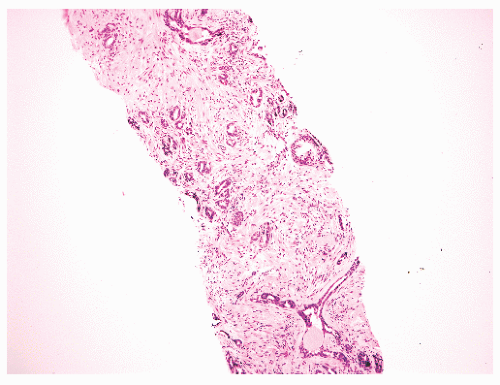
FIGURE 8-48 ▪ Atrophy with irregular distribution of small acini.


FIGURE 8-49 ▪ Atrophy with cord-like structures.


Sclerotic Atrophy

Sclerotic atrophy is a frequently detected type of peripheral zone atrophy, having been found in 74% of cases of atrophy in the whole gland.229 Like simple atrophy, it is usually admixed with other types of atrophy, and only 11% of atrophy cases consisted of pure sclerotic atrophy.230 This form of atrophy is remarkable for the lobular acinar disarray due to exuberant fibrosis. Accompanying chronic inflammation with often large collections of lymphocytes and histiocytes can be present. The acinar distortion and angulation in a background of stromal sclerosis can produce an infiltrative appearance (Fig. 8-52), especially in needle biopsy. Such an image may be highly alarming in needle biopsy, where lobular arrangements may be difficult or impossible to visualize. Yet, cytologically, the lining cells are small and bland; basal cells can be identified in some glands but not in all, owing to nuclear compression and crowding.


FIGURE 8-50 ▪ Cystic change in atrophy.


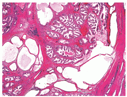
FIGURE 8-51 ▪ Cystic atrophy in BPH.


Partial Atrophy

Partial atrophy is a variant of atrophy in which the atrophic glands have relatively scant cytoplasm, but the glands appear as a crowded collection of pale glands rather than dark glands (Fig. 8-53).237,238 In many needle biopsies with partial atrophy, fully developed atrophy is also present.237,238 In about one-third of cases there is a disorganized rather than circumscribed growth pattern.238 Unlike other types of atrophy, stromal sclerosis is generally lacking and inflammation is rarely detected.238 The glandular outlines in partial atrophy vary from straight luminal borders to more undulating
luminal surfaces and papillary projections. A stellate shape is assumed by some glands (Fig. 8-54

Only gold members can continue reading. Log In or Register to continue

Stay updated, free articles. Join our Telegram channel

Jun 10, 2016 | Posted by in UROLOGY | Comments Off on Nonneoplastic Diseases of the Prostate and Seminal Vesicles

Full access? Get Clinical Tree

Get Clinical Tree app for offline access