Renal Artery Stenosis, Renovascular Hypertension, and Ischemic Nephropathy


Renal Artery Stenosis, Renovascular Hypertension, and Ischemic Nephropathy


Stephen C. Textor


The identification and management of renovascular disease presents a clinical challenge that is directly relevant to nephrologists, and also one that relates to many other medical specialties, including cardiovascular specialists, internal medicine physicians, vascular surgeons, and interventional clinicians. How best to manage renovascular disease remains controversial, in part because of the near simultaneous development of far more effective diagnostic tools, medical therapy, and revascularization techniques over the past decade that has ever been available before. It behooves clinicians caring for patients with renal disease to have a solid understanding of these concepts as part of their clinical responsibility to prevent an irreversible loss of kidney function and adverse effects of arterial hypertension.

Few conditions are more rewarding to treat than new onset severe hypertension and/or progressive renal insufficiency that reverses after the successful restoration of renal blood flow. Occlusive lesions of the main renal arteries can now be detected readily with any of a variety of imaging tools. Determining when to pursue renovascular lesions in clinical hypertension, renal insufficiency, or circulatory congestion; establishing their pathophysiologic role; and whether the hazards associated with revascularization are warranted are pressing concerns regularly faced by nephrologists. Although recent trial data fail to provide compelling evidence in favor of endovascular stenting for all patients with atherosclerotic disease, the validity of prospective clinical trials in this disorder has been fiercely challenged.1,2 Experienced clinicians understand that renal revascularization in these disorders sometimes should be undertaken both to improve hypertension and to salvage renal function.

Recognition that reduced renal perfusion activates pressor systems that raise systemic blood pressure remains one of the seminal observations and most widely studied mechanisms in cardiovascular physiology. Reversal of renovascular hypertension can provide major benefits to patients with accelerating hypertension (e.g., allowing effective blood pressure control and reduction of long-term drug therapy). Selecting patients and determining optimal timing for vascular intervention at a reasonable risk is rarely simple, however.

These issues are complicated further by the rapid expansion of endovascular procedures over the past two decades. Although restoring lumen patency in partially occluded vessels intuitively may seem beneficial, recent trials indicate that revascularization procedures carry both substantial costs and some risks, whereas the clinical benefits remain ambiguous.3,4 This is a remarkable turn of events, insofar as renovascular hypertension traditionally has been considered a prototype for “curable” secondary hypertension. Most diseases of the renal arteries are progressive, and the clinical manifestations develop gradually over time, either because the vascular compromise worsens or because adaptive mechanisms to offset hemodynamic effects become overwhelmed. Because advances in medical therapy have allowed more effective antihypertensive drug treatment than ever before, more patients are appearing clinically at later stages in their disorder with a manifest loss of kidney function and/or circulatory disorders.5 It may be argued that such patients face more severe consequences of renovascular compromise and may have less of a likelihood of benefit from restoring the renal circulation. Hence, it behooves nephrologists to recognize the importance of a close follow-up of vascular disease in the kidney, as with many other vascular conditions. Before moving forward with renal revascularization, both affected patients and physicians should consider carefully the potential benefits and risks. Understanding the pathophysiologic basis for the clinical syndromes associated with renal artery stenosis is an important first step in this process. This chapter will review the background and basis for much of the clinical information related to these disorders.

The basic clinical syndromes to be discussed are outlined in Figure 42.1. Many renal artery stenoses produce little hemodynamic effect and represent “incidental” disease. Such lesions sometimes may be found in asymptomatic, normotensive individuals. Because many renal arterial lesions are detected in patients with preexisting hypertension, the role of renovascular disease itself may be obscured. Renovascular hypertension denotes the syndrome of rising arterial pressures specifically caused by impaired renal perfusion that leads to the activation of pressure pathways. When the severity and duration of reduced blood flow threatens the viability of kidney tissue, many authors
have designated this condition as ischemic nephropathy, which some believe is a major cause for some patients reaching end-stage renal disease (ESRD).6,7 More recently, attention has been focused on the role of renovascular disease in impairing cardiac function, both by reducing the systemic excretion of sodium and volume and by producing abrupt rises in arterial afterload that magnify cardiac dysfunction. The primary task of the clinician is to elucidate the role of renal arterial stenosis in a given patient and to direct therapy accordingly.


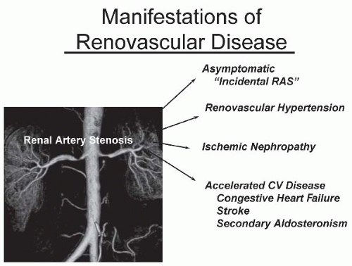
FIGURE 42.1 Clinical manifestations of renal artery stenosis range across a broad spectrum. Many, perhaps most, represent incidental lesions with minimal hemodynamic effects. Some reach levels wherein the activation of pressor mechanisms produces a rise in blood pressure, identified as renovascular hypertension, and at some point, threaten kidney function sufficiently to warrant the term ischemic nephropathy (see text). Particularly when the entire renal mass is affected, impaired kidney function and solute excretion from renovascular disease can accelerate cardiovascular morbidity, sometimes identified as one of the cardiorenal syndromes, with worsening congestive heart failure. The task facing the clinician is to identify where along this spectrum an individual patient lies. CV, cardiovascular; RAS, renal artery stenosis.


HISTORICAL PERSPECTIVE

Observations in the 1800s regarding blood pressure measurements revealed important connections between fluid volume, arterial pressure, and vascular resistance. How these observations ultimately led to the elucidation of the renin-angiotensin-aldosterone system has been reviewed.8 In 1898, Tigerstedt and Bergman established that extracts of the kidney had pressor effects in the whole animal, and these authors are credited with the identification of renin. The identification of each component of the renin-angiotensin system represents a remarkable series of research ventures spanning a half century and several investigators in many countries. Goldblatt and others provided seminal experiments with the development of an animal model in which reduced renal perfusion produced hypertension, published between 1932 and 1934. Numerous investigators thereafter identified the peptide nature of angiotensin, the role of renin substrate or angiotensinogen, the role of nephrectomy in sensitizing the animal to the pressor effects of angiotensin, and the sequential phases of renovascular hypertension. Hence, the renin-angiotensin system owes its initial discovery and nomenclature primarily to early studies related to the regulation of blood pressure by the kidney. Only recently have the multiple additional effects of angiotensin become evident regarding vascular remodeling, the modulation of inflammatory pathways, and interactions with fibrogenic mechanisms. Understanding that reduced renal blood flow produces sustained elevations in arterial pressure led to a broad study of the mechanisms underlying many forms of hypertension. Experimental models of two-kidney and one-kidney renal clips (two-kidney and one-kidney Goldblatt hypertension) represent some of the most extensively studies models of blood pressure and cardiovascular regulation.

Extension of these studies into clinical medicine followed soon thereafter. A time line highlighting these developments is illustrated in Figure 42.2.9 Some patients presented with malignant forms of hypertension during the late 1930s and 1940s, so designated due to remarkably poor survival if the patient’s blood pressure could not be lowered successfully. Few antihypertensive agents were known until the 1950s, and intervention consisted mainly of extremely low sodium intake diets and/or lumbar sympathectomy.

Recognition that some forms of severe hypertension were secondary to occlusive renovascular disease led surgeons to undertake unilateral nephrectomies for small kidneys in 1937.10 The fact that some of these were indeed pressor kidneys and blood pressure fell to normal levels provided proof of concept and led to more widespread use of nephrectomies. Unfortunately, achieving a cure of hypertension after the nephrectomy was rare, and Homer Smith reviewed the poor results overall in a 1956 paper discouraging this practice.10

The 1960s marked the introduction of methods of vascular surgery to restore renal blood flow. These procedures carried substantial morbidity associated with aortic surgery, but offered an opportunity to improve the renal circulation and to potentially reverse renovascular hypertension. One result of this development was a series of studies aiming to characterize the functional role of each vascular lesion in producing hypertension, thereby allowing a prediction of the outcomes of vascular surgery.10 A large, cooperative study of renovascular hypertension81 included major vascular centers and reported on the results of more than 500 surgical procedures. These results provided limited support for vascular repair,
but identified relatively high associated morbidities and mortalities, particularly in patients with atherosclerotic disease.


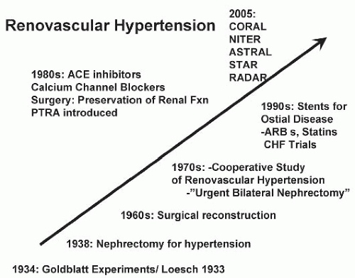
FIGURE 42.2 A time line with major events in the understanding of renovascular hypertension. Goldblatt and Loesch are identified as the original investigators in the 1930s who linked reduced kidney perfusion to the development of sustained hypertension. Surgical revascularization only became technically feasible in the 1960s, with the emergence of effective medical therapy and endovascular procedures in the 1980s and 1990s. The application of effective medical therapy, including statins, angiotensin-converting enzyme (ACE) inhibitors, and angiotensin-receptor blockers (ARBs), have led to clinical equipoise that is the basis for prospective, randomized trials comparing optimal medical treatments with or without revascularization, beginning around 2005. PTRA, percutaneous transluminal renal angioplasty; CORAL, cardiovascular outcomes for renal atherosclerotic lesions; NITER, nephrotherapy ischemic therapy; ASTRAL, angioplasty and stent for renal artery lesions; STAR, stent placement in patients with atherosclerotic renal artery stenosis and impaired renal function; RADAR, randomized, multicenter, prospective study comparing best medical treatment versus best medical trearment plus stenting in patients with hemodynamically relevant atherosclerotic renal artery stenosis; CHF, congestive heart failure.

In the 1980s and 1990s, further developments led to both improved medications and the introduction of endovascular procedures, including percutaneous angioplasty and stents. These both broadened the options for treating patients with vascular disease and raised new issues regarding timing and overall goals of intervention. Recent developments highlighted the need for intensive cardiovascular risk factor reduction and more stringent standards of blood pressure control. Antihypertensive medications have improved dramatically, both with regard to efficacy and tolerability. As emphasized in the following, the broad application of angiotensin-converting enzyme inhibitors and angiotensin-receptor antagonists for reasons other than hypertension alone changed the clinical presentation of disorders associated with renal artery stenosis. Uncontrollable hypertension is now less commonly the reason to intervene in renovascular disease. Often, the main objective is the long-term preservation of renal function. In recent years, endovascular techniques make possible renal revascularization with relatively low morbidity in many patients previously considered unacceptable surgical candidates. The challenge for clinicians is how and when to apply these tools most effectively in the management of individual patients.11


DEFINITIONS AND CLASSIFICATION

The syndrome of renovascular hypertension (RVH) refers to hypertension primarily mediated by the reduction of renal artery perfusion pressure. The prevalence of renovascular hypertension is not known with precision, but available data suggest that it occurs in 0.5% to 5.0% of the general hypertensive population.12 RVH can develop with a variety of arterial lesions, including arterial dissection, extrinsic compression, embolic infarction, or thrombosis (Table 42.1).
Atherosclerotic renal artery disease is the most common cause of renal artery stenosis, accounting for about 80% of renal arterial lesions. Fibrous renal artery diseases, as a group, account for less than 20% of renal arterial lesions. Reports of resistant hypertension suggest that when renovascular disease is a factor, more than 84% is atherosclerotic renal artery disease and 16% is fibromuscular renal artery disease.13 Fibrous renal artery disease has been reported in 2% to 6% of potential renal transplant donors (usually normotensive individuals).14,15 The vast majority of incidental renal artery lesions in angiographic series are atherosclerotic.


TABLE 42.1 Lesions Producing Renovascular Hypertension and Ischemic Nephropathy


Causes of Renal Artery Stenosis


Atherosclerotic renal artery disease


Fibromuscular dysplasias


Medial fibroplasia


Perimedial fibroplasia


Intimal fibroplasia


Medial hyperplasia


Endovascular aortic stent graft crossing the renal artery


Acute arterial embolism/thrombosis (e.g., antiphospholipid syndrome)


Arterial trauma


Aortic dissection


Neurofibromatosis


Arterial aneurysm


Arteriovenous malformation/fistulae


Cholesterol emboli


Systemic necrotizing vasculitis


Polyarteritis nodosa


In addition to atherosclerotic and fibrous renal artery disease, a number of less common clinical entities can produce renovascular hypertension. These include acute arterial thrombosis or embolism, cholesterol emboli, aortic dissection, renal arterial trauma, arterial aneurysm, arteriovenous malformation of the renal artery, neurofibromatosis, polyarteritis nodosa, and Takayasu arteritis. Recent expansion of the use of endovascular aortic stent grafts, sometimes with structural impingement of the main renal arteries, is a new addition to the list of iatrogenic causes of renovascular hypertension.4,16,17 Renal artery thrombosis occurring as a complication of umbilical artery catheterization has been recognized as causing renovascular hypertension in infants.18 Transplant renal artery atherosclerosis, intimal hyperplasia, or vascular kinking may contribute to renal transplant renovascular hypertension (see Chapter 82).

Table 42.2 presents a classification of atherosclerotic and fibrous renal artery diseases with a description of their morphology and histology. These types of renal artery occlusive diseases represent a heterogeneous group of diseases, occurring in different age groups and behaving differently with regard to their individual natural history. An appreciation of these differences may be important for therapeutic decision making in patients with renal artery occlusive disease.


TABLE 42.2 Histologic Classifications of Fibromuscular Dysplasia and Angiographic Appearance


Type


Frequency


Histology


Angiographic Appearance


Medial Medial fibroplasia


85%-100% most common


Alternating ridges of collagen/loss of elastic membrane


String of beads
Medial: bead diameter is larger than lumen diameter


Perimedial fibroplasia Medial hyperplasia


Rarer (10%-15%) Rarest


True smooth muscle hyperplasia: no fibrosis


Perimedial: bead diameter is smaller than lumen diameter Medial hyperplasia: smooth stenosis without beads


Intimal


< 10%


Circumferential deposition of collagen in intima: fragmented or duplicated internal elastic lamina


Concentric smooth stenosis: long, smooth vessel narrowing


Adventitial


<1%


Dense collagen replaces fibrous tissue in adventitia and surrounding tissue


Smooth stenosis or diffuse attenuation of vessel lumen


Fibromuscular Dysplasias

Fibromuscular dysplasia (FMD) lesions produce distortions of the luminal diameter of large- and medium-sized arteries due to nonatherosclerotic arteriopathies. The lesions usually involve the mid to distal vessel beyond the first 1 to 2 cm from the aorta (Fig. 42.3). The prevalence of clinically apparent renovascular FMD is estimated at 4 out of 1,000, with lower prevalence of cerebrovascular involvement (1 out of 1,000). Observations from screening angiographies in normotensive potential kidney donors indicate that some degree of FMDs can be observed in 3% to 6% of otherwise healthy, normotensive individuals.15 Clinically apparent FMDs are most common among young females between the ages of 15 and 50 years. It may be familial in 10% of cases and tends to involve both renal arteries. It has been associated with subclinical carotid fibromuscular disease in first-degree relatives, which, in some cases, is consistent with an autosomal dominant inheritance. Renal arteries are involved with FMDs in 65% to 70% of cases, whereas 25% to 30% involve cerebral vessels. Both sites are involved in approximately 15% of patients. FMDs may develop in association with hereditary disorders of the connective tissue, such as Ehlers-Danlos and Marfan syndromes.

The lesions of FMD develop as disruptions of vascular wall components with abnormal deposition of collagen
in bands, sometimes with disruption of the vascular elastic membrane. The molecular basis for these disruptions is unknown, although several candidate genes have been proposed. The primary subtypes of FMDs are designated as medial fibroplasia, intimal fibroplasia, and periadventitial fibroplasia, defined initially on the basis of histologic analysis of surgically resected vessels. Medial fibroplasia is the most common phenotype, and often manifests as a string-of-beads appearance with alternating stenoses with apparent luminal dilations, as summarized in Table 42.2. The other phenotypes appear as focal or elongated vascular occlusions. Medial fibroplasia affects the distal half of the main renal artery, frequently extends into the major branches, is often bilateral, and angiographically gives the appearance of multiple aneurysms wherein the diameters of the aneurysms are wider than the apparently unaffected portion of the main renal artery (Fig. 42.3A). Most cases of medial fibroplasia are diagnosed in women between the ages of 30 and 50 years. Although medial fibroplasia progresses to higher degrees of stenosis in about one-third of patients, complete arterial occlusion and/or ischemic atrophy of the kidney ipsilateral to the renal artery stenosis are rare. The stenotic lesions in medial fibroplasia are secondary to thickened fibromuscular ridges replacing the normal structure of the intima and the media of the artery. These thickened ridges alternate with thinned areas that may not have an internal elastic membrane, thereby becoming aneurysmal.


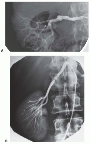
FIGURE 42.3 Examples of fibromuscular disease of the renal artery. A: An image of the string of beads appearance that is typical of medial fibroplasia with aneurysmal bulging beyond the artery. B: An image of a tight, focal lesion that is typical of intimal hyperplasia in a young male.

Perimedial fibroplasia (subadventitial fibroplasia) accounts for approximately 15% of fibrous renal artery lesions. This lesion also occurs predominantly in women, typically between the ages of 15 and 30 years. Angiographically, it is often characterized by a small string-of-beads appearance, with the beads being of similar or of smaller diameter compared to the diameter of the apparently unaffected portion of the renal artery. This lesion typically affects the distal half of the main renal artery, is frequently bilateral and highly stenotic, and may progress to total arterial occlusion. Collateral blood vessels and renal atrophy on the involved side are commonly observed.19,20

Medial hyperplasia and intimal fibroplasia account for only 5% to 10% of fibrous renal artery lesions. Intimal fibroplasia occurs primarily in children and teenagers and angiographically appears as a localized, highly stenotic, and smooth lesion with poststenotic dilation. It may occur in the proximal portion of the renal artery and when it does, it can resemble atheroma. Intimal fibroplasia is progressive and is occasionally associated with dissection or renal infarction and renal atrophy (Fig. 42.4C). Medial hyperplasia, also rare, is found predominantly in teenagers, and angiographically also appears as a smooth, linear stenosis, sometimes appearing as though a ligature were tied around the renal artery. There is considerable difficulty in distinguishing between intimal fibroplasia and medial hyperplasia by angiography alone, and these two types of fibrous artery disease are sometimes grouped together in the literature.21

Renal artery aneurysms can develop in up to 50% of these lesions, which sometimes produce local dissection and/or occlusion (Fig. 42.4C). Arteriovenous fistulae and thrombosis can also occur.

Rates of progression differ between histologic subtypes and are poorly understood. Many lesions remain below hemodynamic thresholds and have minimal clinical importance. Others progress to produce hypertension and occasional thrombosis, particularly in women, although this happens far more commonly with atherosclerotic lesions. Smoking appears to be the major risk factor for progressive disease in young women.

Takayasu arteritis is a variant form of systemic vasculitis that affects large arteries such as the aorta and its main branches, including the renal arteries. It mainly affects young women. It can cause discrete stenosis of the aorta
and its main branches, including the common carotid, and the subclavian and the renal arteries.22 Renal artery involvement is not uncommon in Asian and African patients and is a major cause of renovascular hypertension in these countries.23 Koide24 reported renovascular hypertension in 278 (18.8%) of 1,475 patients in a Japanese nationwide survey of Takayasu arteritis. Takayasu arteritis is a common cause of renovascular hypertension in Southeast Asia, including India and China, and accounts for between 20% and 60% of RVH cases.23,25


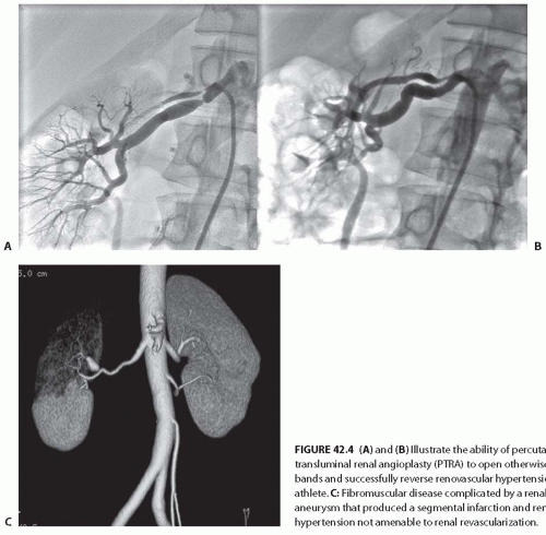
FIGURE 42.4 (A) and (B) Illustrate the ability of percutaneous transluminal renal angioplasty (PTRA) to open otherwise tight fibrous bands and successfully reverse renovascular hypertension in a young athlete. C: Fibromuscular disease complicated by a renal artery aneurysm that produced a segmental infarction and renovascular hypertension not amenable to renal revascularization.

A recent expansion in the use of endovascular procedures for aortic aneurysm repair (EVAR) has produced a new clinical source of renal artery occlusion (Fig. 42.5A). Many of these stent grafts are placed in close proximity to the origins of the renal arteries. Occlusion of the renal arteries is observed in up to 6% of these procedures, sometimes related to migration or inadvertent coverage of the vessels.4,26 Some grafts are designed for deliberate extension of uncovered struts above the renal arteries for anatomic reasons. Remarkably, the frequency of renal artery compromise is modest, and current endovascular procedures can restore luminal patency if promptly recognized.27 Identifying and treating vascular occlusion due to aortic stent grafts is important, because failure to achieve patency is associated with a clinically important loss of kidney function in up to 20% of subjects.28

Acute and chronic renal artery occlusion secondary to renal artery emboli, aortic or renal artery dissection, neurofibromatosis, arterial trauma, arteriovenous malformations, and polyarteritis nodosa are discussed elsewhere in this book.


Atherosclerotic Renal Artery Stenosis

Atherosclerotic renal artery stenosis (ARAS) most often develops in older patients (> 50 years of age) and is associated with systemic atherosclerosis. ARAS is the most common
cause of RVH and can contribute to a loss of renal function leading to ESRD (Fig. 42.5B,C). Atherosclerotic plaque often arises in the first 1 to 2 cm of the renal artery or may extend directly from the aorta into the renal ostium. Aortic and renal vascular calcification is often present. Some 75% to 80% of patients with renal artery atherosclerosis and renovascular hypertension have ostial atherosclerotic lesions, and 25% to 30% of lesions are in the nonostial location. ARAS is a manifestation of systemic atherosclerotic disease and is asso ciated with coronary, cerebrovascular, peripheral vascular, and aortic disease.29,30 The prevalence of ARAS appears to be increasing. This probably reflects the fact that more people are living long enough for atherosclerotic vascular disease in the visceral abdominal vessels to reach critical levels, thus aggravating hypertension when the kidney is affected. In a recent systematic analysis of patients undergoing an angiography of the peripheral or coronary circulations, ARAS was found in 11% to 42% of cases.30 Predictors of ARAS include a history of hypertension, the presence of renal functional impairment, coexisting vascular or coronary artery disease, the presence of abdominal bruits, and a history of smoking. Renal artery lesions are bilateral in 20% to 40% of such patients. Estimates of the prevalence of ARAS depend on the population screened. One population-based study of a cohort of 870 patients older than 65 years screened with renal artery duplex sonography found a 6.8% prevalence of ARAS, defined as greater than 60% stenosis. No differences in prevalence were detected between African Americans and Caucasians,31 although reported rates for surgically corrected renovascular hypertension are lower for African Americans.32 Men are affected more often than women, but this gender difference declines with advancing age. Recent series indicate a distinct shift toward women in series referred for revascularization.33,34 Several studies suggest that atherosclerotic renal vascular disease is associated with adverse coronary events33,35 and increased mortality. Autopsy series report an overall prevalence of 4% to 20%, with progressively higher rates for those older than 60 years (25% to 30%) and 75 years (40% to 60%). These studies suggest that ARAS leading to renal artery stenosis is the single most common cause of secondary hypertension in patients older than 50 years.36 It also commonly leads to systolic hypertension with wide pulse pressures. Furthermore, renal artery stenosis has been reported to contribute
to the decline in renal function in 15% to 22% of patients reaching ESRD.37,38


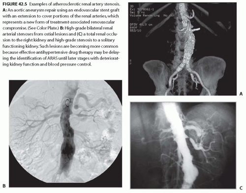
FIGURE 42.5 Examples of atherosclerotic renal artery stenosis. A: An aortic aneurysm repair using an endovascular stent graft with an extension to cover portions of the renal arteries, which represents a new form of treatment-associated renovascular compromise. (See Color Plate.) B: High-grade bilateral renal arterial stenoses from ostial lesions and (C) a total renal occlusion to the right kidney and high-grade stenosis to a solitary functioning kidney. Such lesions are becoming more common because effective antihypertensive drug therapy may be delaying the identification of ARAS until later stages with deteriorating kidney function and blood pressure control.


Adaptation and Progressive Vascular Occlusion in Atherosclerotic Renal Artery Stenosis

With improved antihypertensive drug therapy and the recognized potential for advanced ARAS to lead to reduced kidney function, clinicians need to consider the potential for progressive renovascular occlusion during medical therapy. This has been a controversial subject. Initial retrospective studies of serial angiograms suggested that progressive occlusion could develop in 40% to 50% of subjects37 with 15% progressing to total occlusion over periods between 3 to 5 years.39 A series of studies in the 1990s with ARAS followed prospectively with high-resolution Doppler ultrasound indicated that measurable hemodynamic progression occurred in nearly 50% of subjects over 5 years,40 although the rates of clinical progression defined by changes in kidney size (24%), loss of GFR, and/or progression to total occlusion were much lower.41 The 3-year cumulative incidence of renal artery disease progression for 295 renal arteries initially classified as normal, less than 60% stenosis, and greater than or equal to 60% stenosis, was 18%, 28%, and 49%, respectively. In this prospective series, the early progression to total occlusion occurred only in nine arteries (3%), all of which had a baseline reduction in lumen diameter greater than 60% (Fig. 42.6). The cumulative incidence of progression to total occlusion in patients with baseline stenosis of 60% or more was 4% at 1 year, 4% at 2 years, and 7% at 3 years. Factors associated with the risk of renal artery disease progression during the time of monitoring included systolic blood pressure (BP) greater than or equal to 160 mm Hg, diabetes mellitus, and high-grade stenosis (more than 60%) in either the ipsilateral or contralateral renal artery.37 One of the prospective treatment trials from Europe suggested that 16% of subjects treated without revascularization developed total occlusion, based on renography.39 Recent prospective treatment trials indicate far lower rates of disease progression. BP control and statin therapy in these trials have been more effective and more widely applied than before, with crossover rates from medical therapy to renal revascularization below 10% over reporting periods between 3 and 5 years.42 It should be emphasized that the epidemiologic and clinical hazard of progressive stenotic disease is closely related to the level of initial stenosis. For lesions that are more than 75% occluded, further progression is not only far more likely to occur, but the consequence regarding tissue ischemia and loss of functioning tissue is more severe (see Pathophysiology, which follows).


PATHOPHYSIOLOGY OF CLINICAL SYNDROMES WITH RENOVASCULAR DISEASE


Renovascular Hypertension

Renovascular occlusive disease from any cause that reaches a “critical” level can activate pressor mechanisms that tend to raise systemic arterial pressure and restore renal artery
perfusion pressures. Luminal occlusion of less than 60% (cross-sectional area) rarely produces any measurable gradient for either pressure or flow. Hence, a fall in renal perfusion pressure sufficient to initiate RVH occurs only when luminal occlusion is relatively severe, usually in the 70% to 80% cross-sectional occlusion range (Fig. 42.7). When critical stenosis develops and reduces renal perfusion pressure, multiple mechanisms are activated in the kidney to restore renal blood flow. Foremost among these pathways is the release of renin from the juxtaglomerular apparatus, leading to the activation of the renin-angiotensin-aldosterone system (RAAS). Release of plasma renin occurs only after poststenotic pressures fall by at least 10% to 20% compared with aortic pressures.43 This is mediated in part by the stimulation of neuronal nitric oxide synthase and cyclooxygenase 2 in the macula densa. Blockade of the RAAS at the time an experimental renal artery lesion is created prevents the development of hypertension. Animals genetically modified to lack the angiotensin (Ang 1) receptor fail to develop two-kidney one-clip hypertension.44 Experiments using kidney transplantation from AT1 receptor knockout mice indicate that both systemic and renal angiotensin receptors participate in additive fashion to blood pressure regulation.45


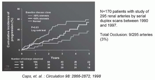
FIGURE 42.6 The progression of atherosclerotic renal artery stenosis (ARAS) of varying severity during a serial follow-up by Doppler ultrasound. There were 295 arteries that were followed sequentially at 6-month intervals for 5 years. Measurable hemodynamic progression (defined as an increase in peak systolic velocity of at least 100 cm per second) was identified in 31% by 3 years, and more than 50% of the group with more than 60% at baseline. Importantly, clinical progression was uncommon (defined by a change in serum creatinine, loss of kidney size, or total occlusion). Predictors of progression included systolic blood pressure, age, and diabetes. (Adapted from Caps MT, Perissinotto C, Zierler RE, et al. Prospective study of atherosclerotic disease progression in the renal artery. Circulation. 1998;98:2866-2872.) (See Color Plate.)

In the presence of an intact RAAS, systemic arterial pressures increase until renal perfusion is restored. Studies in both experimental models and humans indicate that additional mechanisms add to long-term elevation of blood pressure in the presence of renal artery stenosis, including activation of the sympathetic nervous system, impairment of nitric oxide generation, and release of endothelin as well as hypertensive microvascular injury to the nonstenotic kidney.

Although there are differences among species (rat, dog, rabbit) in experimental renovascular hypertension, two basic models of Goldblatt hypertension are recognized: the two-kidney, one-clip (2K-1C) model (in which one renal artery is constricted and the contralateral renal artery and kidney are left intact) and the one-kidney, one-clip (1K-1C) model (in which one renal artery is constricted and the contralateral kidney is removed). These two experimental models of renovascular hypertension are diagrammed in Fig. 42.8A,B. Mechanisms responsible for sustained RVH differ according to whether one or both kidneys are affected. Both of these models depend initially on impaired renal perfusion and activation of the RAAS with sodium retention. However, the presence of a normal contralateral kidney allows pressure natriuresis to occur, by which the elevated perfusion pressure produces sodium excretion in the nonstenotic kidney. Because the nonstenotic kidney eliminates excess sodium and volume, the level of perfusion pressure to the stenotic side remains reduced, leading to the ongoing activation of the renal artery underperfusion and RAAS stimulation. This sequence of events producing angiotensin II (Ang II)-dependent hypertension and secondary aldosterone excess with hypokalemia is summarized in Fig. 42.8A.

By contrast, 1K-1C hypertension represents a model in which the entire renal mass is exposed to reduced pressures
beyond a stenosis. There is no normal or nonstenotic kidney to counteract increased systemic pressures. As a result, sodium is retained and the blood volume is expanded, which eventually feeds back to inhibit the renin-angiotensin system (RAS) (Fig. 42.8B). Therefore, 1K-1C hypertension is typically not angiotensin dependent unless the removal of volume is achieved that reduces renal perfusion pressure and again activates the RAAS.


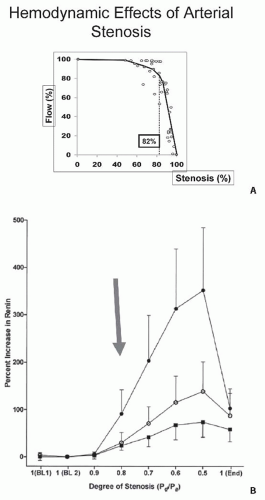
FIGURE 42.7 A: Arterial pressure depicted as a function of degree of luminal occlusion. These data highlight the fact that no change in postlesion pressure or flow can be identified until cross-sectional occlusion is severe, usually more than 70% to 80%. These experimental data in dogs are consistent with measurements in human subjects (B), wherein renal vein renin release was not detected during balloon occlusion until translesional gradients between the aorta and renal artery of at least 10% to 20% were produced. MAP, mean arterial pressure. (Data from May et al.229 and De Bruyne et al.43)


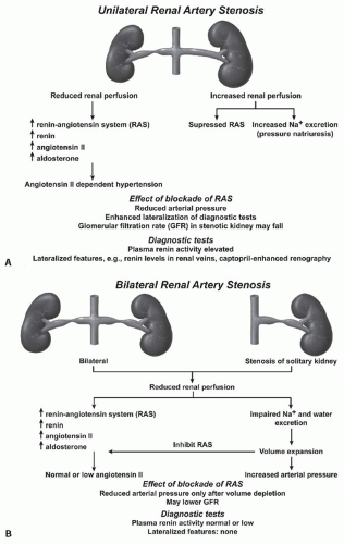
FIGURE 42.8 A: A schematic illustration of hormonal and hemodynamic effects of unilateral renal artery stenosis to produce renovascular hypertension. Unilateral disease is identified as 1-clip-2-kidney Goldblatt hypertension and manifests with the activation of the renin-angiotensin system so long as the contralateral kidney continues to undergo pressure natriuresis (see text). (Adapted from Safian and Textor91 .) Panel (B) illustrates the sequence of events with 1-clip-1-kidney Goldblatt hypertension, wherein no normal contralateral kidney is available to excrete sodium and volume in response to rising arterial pressures. In this model, the initial activation of the renin-angiotensin system is temporary due to volume expansion.

Most renovascular disease in humans is asymmetric and is thought most often to resemble 2K-1C renovascular models. The mechanistic differences between these forms of RVH have clinical implications regarding diagnosis and treatment. Many diagnostic studies classically used to evaluate the functional significance of renal artery lesions depend on comparisons of the different physiologic response of the two kidneys, which may not be evident if both kidneys are affected or if only one kid ney is present. Furthermore, diagnostic tests that depend on differences in responses to alterations in sodium status (such as the measurement of renal vein renin levels after sodium depletion or individual
kidney sodium reabsorption) may be problematic, because high levels of Ang II and aldosterone stimulate sodium reabsorption in both the stenotic and nonstenotic kidney.


The Phases of Renovascular Hypertension

Many of the primary physiologic drivers, such as activation of the RAAS, underlying renovascular hypertension manifest differently at various points in time. These changing manifestations complicate defining primary pathogenic mechanisms and establishing whether renovascular lesions are actually causal in hypertension. Rarely is it known exactly when critical levels of stenosis are attained. In experimental models, these observations have been summarized by separating two-kidney renovascular hypertension into sequential phases.46 In phase 1, renal ischemia and activation of the RAS are central and the BP elevation is renin dependent. The acute administration of Ang II antagonists or angiotensin-converting enzyme (ACE) inhibitors, removal of the renal artery stenosis (i.e., removal of the clip), or removal of the stenotic kidney normalizes the BP. In the absence of these maneuvers, a transition phase (phase 2) subsequently develops, bridging the acute (phase 1) and chronic (phase 3) phases of experimental renovascular hypertension. This transition phase variably lasts from a few days to several weeks depending on the experimental model and animal species. During this transition phase, plasma renin levels gradually fall, but the BP remains elevated. Salt and water retention are observed as a consequence of the effects of hypoperfusion of the stenotic kidney, augmented proximal renal tubular reabsorption of sodium and water, and secondary aldosteronism.47,48 In addition, the high levels of Ang II stimulate thirst, further contributing to an expansion of the extracellular fluid volume. The expanded extracellular fluid volume results in suppression of peripheral plasma renin activity (PRA). During this transition phase, the hypertension is still responsive to removal of the unilateral renal artery stenosis, to Ang II blockade, or to unilateral nephrectomy, although these maneuvers do not normalize the BP as promptly and consistently as in the acute phase. These changes may depend on the recruitment of additional mechanisms of Ang II action, including the generation of reactive oxygen species,49,50 the quenching of nitric oxide,51 and the generation of endothelium-derived substances such as endothelin52,53 and thromboxanes.54 Some of these mechanisms depend on slowly developing effects of Ang II at levels that do not reverse rapidly with direct Ang II blockade.55 Additional mechanisms recruited over time that sustain BP elevation and induce tissue injury are summarized in Table 42.3.

After several days or weeks, a chronic phase (phase 3) evolves, wherein the removal of the stenosis by unclipping the renal artery in the experimental animal or nephrectomy of the stenotic kidney fails to reduce the BP to baseline levels. The mechanism maintaining elevated arterial pressure—that is, the failure of “unclipping” to lower the BP in this chronic phase of 2K-1C hypertension—is multifactorial but is thought to reflect widespread arteriolar damage in the contralateral kidney consequent to elevated systemic pressure and the initial high levels of Ang II. The BP remains elevated even though the PRA has returned to a normal level. The pressure natriuresis of the contralateral kidney blunts the extracellular fluid volume expansion initially generated by the stenotic kidney (Fig. 42.8A), but because the contralateral kidney suffers vascular damage from prolonged exposure to the increased BP, its excretory function diminishes and
extracellular fluid volume expansion persists. In phase 3 of 2K-1C hypertension, acute blockade of the RAS fails to lower the BP. Sodium depletion may ameliorate the hypertension, but does not normalize it.


TABLE 42.3 Interactive Mechanisms Underlying Hypertension and Kidney Injury in Atherosclerotic Renal Artery Stenosis


Tissue Underperfusion


Recurrent Local Ischemia


Activation of the renin-angiotensin system Altered endothelial function: (endothelin, NO, prostaglandins) Sympathoadrenergic activation


▪ Increased reactive oxygen species


▪ Cytokine release/inflammation (NF-κ B, TNF, TGF- β , PAI-1, IL-1)


ATP depletion Tubulointerstitial injury Microvascular damage Immune activation Vascular remodeling Interstitial fibrosis Activation of the renin-angiotensin aldosterone system Sympatho-adrenergic activation Endothelin Disturbances of “oxidative stress” Oxidized-LDL


NO, nitric oxide; NF-κ B, nuclear factor kappa B; TNF, tumor necrosis factor; TGF-β, transforming growth factor-beta; PAI-1,plasminogen activating factor inhibitor; IL-1, interleukin 1; ATP, adenosine triphospate; LDL, low-density lipoprotein. From Textor SC. Atherosclerotic renal artery stenosis: overtreated, but underrated? J Am Soc Nephrol. 2008;19:656-659, with permission.


Exactly how and whether this sequence of events applies directly to humans is not known. All of these features obscure the diagnosis of true renovascular hypertension and limit predictability of the BP response to revascularization. Not surprisingly, a documented history of a brief duration of hypertension suggests a more favorable response to revascularization procedures.56

There are additional important clinical correlates of this process. First, many of the diagnostic studies that depend on the lateralization of effects have only modest predictive value when results are negative. As a general rule, these tests are most useful when results are positive, meaning that high-grade lateralization of renin release, differences in renal function, and changes in the glomerular filtration rate (GFR) after the administration of an ACE inhibitor most accurately predict improvement after revascularization when results are markedly positive. A negative test result, however, may also be associated with a beneficial outcome. Second, coexistent intrarenal disease, such as arteriolosclerosis with glomerulosclerosis, is usually associated with persistent hypertension despite the correction of renal artery stenosis, particularly for patients with ARAS.57 In such patients, a long duration of hypertension favors the development of arteriosclerotic lesions and renal injury in the contralateral kidney. Thus, older age and a long duration of hypertension for more than 3 to 5 years predict a poorer clinical BP outcome after renal revascularization. Most older patients with ARAS also have impaired renal function that itself predisposes one to longterm hypertension.11


The Pathogenesis of Ischemic Nephropathy

The activation of pressor mechanisms producing RVH often occurs with a minor loss of renal size or function, particularly with fibromuscular disease.58,59,60 Conversely, improved BP control after revascularization sometimes may be achieved without an appreciable improvement in kidney function. However, the more common clinical scenario with ARAS involves both increasing severity of hypertension and deteriorating renal function, often with a loss of renal size. Hence, the decision to consider renal revascularization commonly combines consideration of both the likelihood of salvage or the preservation of function, in addition to possible benefits regarding BP control.

Basal energy requirements for the kidney are met with less than 10% of blood flow, consistent with its filtration function. The term ischemic renal disease should be used with caution in this context.61 Under normal conditions, cortical blood flow provides vastly more oxygenated blood than is needed for tissue metabolism. Postglomerular vasa recta deliver a portion of kidney blood flow to the medulla. The major metabolic workload of the kidney takes place in medullary segments as a function of solute transport, leaving the medulla normally with substantial oxygen desaturation.62

The balance between regional oxygen saturation within the cortex and the medulla is carefully maintained over a variety of conditions.63 Remarkably, blood flow to the kidney can be reduced gradually to levels sufficient to reduce kidney volume, activate pressor mechanisms including the RAAS, and lower GFR without measurably disturbing cortical or medullary oxygenation.64 Lowered GFR is associated with a reduced filtered load of solutes and thereby reduced requirements for solute reabsorption.63 Venous oxygen levels from poststenotic kidneys therefore can be higher than those from normally functioning kidneys due to reduced metabolic oxygen consumption.64 Measurements of cortical and medullary deoxyhemoglobin by use of blood oxygen level-dependent magnetic resonance in human subjects confirm that both cortical and medullary oxygen saturation can be well preserved over a wide range of renovascular occlusion.65 These observations may explain the relative stability of kidney function observed in recent prospective treatment trials for ARAS for many years, despite an obvious reduction in renal blood flow beyond the stenotic lesion.

There are limits to the renal capacity for adaptation, however. When vascular occlusion is sufficiently severe, cortical blood flow and oxygenation falls.66 This in turn overwhelms the adaptive capacity of the medulla, with expanding hypoxia and activation of fibrogenic and inflammatory pathways.

Mechanisms underlying parenchymal renal damage differ from those responsible for generating hypertension. Remarkably, parenchymal fibrosis rarely develops in patients with fibromuscular disease with the exception of those experiencing renal infarction. This suggests that the activation of remodeling mechanisms in the poststenotic kidney partly is related to the atherosclerotic milieu itself, which produces microvascular proliferation and abnormal endothelial function within the kidneys.67 Recent experimental studies underscore the development of magnified renal microvascular changes distal to a stenosis in the renal artery in the context of atherosclerosis.68,69 An example of microvascular proliferation induced by cholesterol feeding (a surrogate for early atherosclerosis) and the subsequent rarefaction of renal small vessels beyond a main renal artery lesion is illustrated in Fig. 42.9. Numerous signaling pathways lead to the upregulation of cytokines and inflammatory mediators, including transforming growth factor (TGF)-β, within the poststenotic kidney.70,71 Over time, rarefaction of the distal arterioles develops and is associated with fibrogenesis and a loss of viable function.72,73 The sequence of events underlying the transition from a reversible loss of function beyond a vascular lesion to irreversible tissue fibrosis is not well understood. Atherosclerotic and inflammatory pathways can produce disturbances in endothelial function in small vessels that parallel tissue injury and accelerate cytokine signaling pathways.74,75 At some phase, microvascular rarefaction occurs that accompanies a fall in tissue oxygenation and activation of fibrogenesis.75 An irreversible loss of viable
microcirculation may explain some of the limitations observed after the restoration of large-vessel patency (e.g., with renal revascularization). Occasionally, renovascular disease is associated with nephrotic range proteinuria that can regress after renal revascularization.76 The mechanism of enhanced glomerular permeability in this context is unknown.


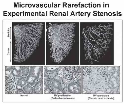
FIGURE 42.9 Reconstructions of the vascular structures using micro-computed tomography (CT) imaging in experimental renal artery stenosis. The atherosclerotic milieu produced by cholesterol feeding leads to microvascular (MV) proliferation and renders the animal especially susceptible to rarefication and obliteration of microvessels in the setting of high-grade renal artery stenosis. These changes eventually lead to interstitial fibrosis and a loss of kidney function. (From Lerman LO, Chade AR. Atherosclerotic process, renovascular disease and outcomes from bench to bedside. Curr Opin Nephrol Hyper. 2006;15:583-587, with permission.)


Circulatory Congestion and Flash Pulmonary Edema

Among other clinical manifestations, ARAS increasingly has been implicated in episodes of congestive heart failure and has been described as one of the cardiorenal syndromes.47 Several mechanisms contribute to this disturbance, including (1) Impaired sodium excretion due to reduced renal perfusion pressure and activation of the RAAS that affects both stenotic and contralateral kidneys and (2) sustained, and sometimes rapid, increases in systolic arterial pressure can abruptly add to pressure overload of the left ventricle (Fig. 42.10).77 Normally, the left ventricle compensates for changes in afterload with increased end-diastolic volume. This mechanism may be impaired, however, in patients with stiffened left ventricles due to left ventricular hypertrophy, precipitating in abrupt rises in end-diastolic pressure, left atrial, and pulmonary venous pressures. It is this abrupt rise in ventricular pressures that leads to sudden decompensation, often termed flash pulmonary edema, that was originally described by Pickering et al.78 In addition, the activation of sympathetic nervous pathways magnifies these effects and alters the transcapillary capacities for gas exchange. Reports from several case series suggest that renal revascularization can interrupt this cycle and reduce reoccurrences.79,80 In less dramatic cases, the presence of bilateral ARAS with reduced GFR may limit the effectiveness of diuretics and render congestive heart failure refractory to fluid removal short of external ultrafiltration.


Clinical Features of Renal Artery Stenosis


Renovascular Hyper tension

In the 1970s, a cooperative study of RVH compared clinical characteristics of patients with surgically proven RVH with those of patients with primary hypertension. In this study, the average age of onset for fibrous renal artery disease as the cause of renovascular hypertension was 33 years, and 16% of these patients were younger than 20 years. For atherosclerotic renal artery disease as the cause of renovascular hypertension, the average age at onset was 46 years, and 39% of these patients were older than 50 years.81 For these reasons, many argue that the clearly defined onset of hypertension below the age of 30 or above the age of 55 warrants the consideration of renovascular hypertension. Some features, such as the presence of an abdominal bruit, hypokalemia, and the absence of a family history of hypertension, were statistically more prevalent in RVH, but had little clinical predictive value. Recent studies suggest that for any level of office BP, patients with RVH may have higher nocturnal pressures and therefore higher overall pressure load as “nondippers.”82 As a result, target organ manifestations are more severe, including left ventricular hypertrophy (Fig. 42.11). A series of patients with treatment-resistant hypertension indicate that elevated cholesterol, impaired renal function, lower body mass index (BMI), and smoking provide positive clues. In practical terms, none of these features is sufficiently sensitive or specific to offer diagnostic precision. As noted previously, RVH rarely may be associated with nephrotic-range
proteinuria, which can regress with the correction of the vascular lesions.


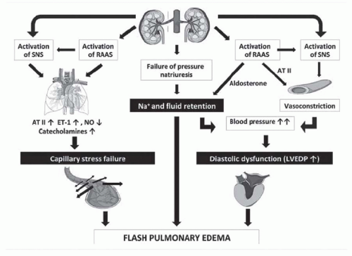
FIGURE 42.10 A schematic depicting the interplay of mechanisms leading to rapidly developing symptoms of left ventricular heart failure sometimes observed with renal artery stenosis. These include impaired volume excretion with bilateral atherosclerotic renal artery stenosis (ARAS), a rise in afterload and impaired left ventricular pump function, as well as disturbed capillary function within the pulmonary vasculature in part related to the activation of neurogenic and hormonal pathways. SNS, sympathetic nervous system; RAAS, renin-angiotensin-aldosterone system; AT II, angiotensin II; ET-1, endothelin 1; NO, nitric oxide; LVEDP, left ventricular end-diastolic pressure. (From Messerli FH, Bangalore S, Makani H, et al. Flash pulmonary oedema and bilateral renal artery stenosis: the Pickering syndrome. Eur Heart J. 2011;32(18):2231-2237, with permission.)


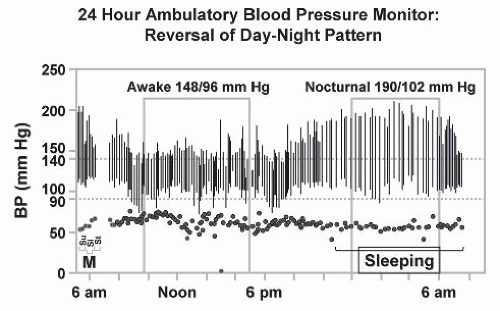
FIGURE 42.11 Ambulatory blood pressure (BP) monitoring in patients with renovascular hypertension commonly reveals a loss of nocturnal pressure fall, or in some cases, a complete reversal of the day-night pattern as shown here. This may be one of the features by which target-organ injury such as left ventricular hypertrophy is exaggerated as compared to patients with similar daytime blood pressure levels.232

BP elevations from RVH vary widely, often as a function of the rapidity of onset. Acute renal artery occlusion may only gradually produce an increase in pressure or it may produce a rapid increase in hypertension that may precipitate a hypertensive urgency or emergency. Before the current era of antihypertensive agents, 30% of Caucasian patients appearing in an emergency department with hypertensive urgency (defined as grade 3 or 4 hypertensive retinopathy) were ultimately found to have RVH. Hence, patients presenting with accelerated forms of hypertension should be considered candidates for renovascular hypertension (Table 42.4). Syndromes of polydipsia and accelerated hypertension with hyponatremia and hypokalemia, sometimes attributed to the dipsogenic actions of Ang II, also have been observed. Current antihypertensive medications have changed the clinical presentation of RVH, thus making them less severe.

Recent consensus documents regarding hypertension emphasize the need for effective populationwide BP control while limiting the number and expense of diagnostic studies.
When combined with the ambiguous results of prospective treatment trials in which medically treated individuals with ARAS fared as well as those treated with renal revascularization, some would argue that there is little evidence to support extensive diagnostic studies to identify all cases of renovascular hypertension. As a result, most patients with hypertension simply are treated and subjected to few laboratory investigations. For those who reach acceptable BP control without adverse effects, no further studies are performed. Hence, many if not most cases of true RVH are not detected (Fig. 42.12) unless hypertension becomes more difficult to treat or if renal dysfunction ensues. One important reason that RVH is less frequently detected is the availability of orally active antihypertensive agents that block the RAAS. Early studies beginning with captopril indicated that satisfactory BP control can be achieved in more than 86% of patients with RVH compared with less than 50% with previously available drugs. In recent years, the widespread application of ACE inhibitors and angiotensin receptor blockers (ARBs) for indications other than hypertension (e.g., congestive cardiac failure, diabetic nephropathy, and other proteinuric renal disease) increases the exposure of individuals with undetected renal artery stenosis to these drugs.


TABLE 42.4 Clinical Features Suggestive of Renovascular Disease


Clinical Clues


Age at onset of hypertension (< 30 or > 55 years)


Abrupt onset of hypertension


Acceleration of previously well-controlled hypertension


Hypertension refractory to an appropriate three-drug regimen


Accelerated retinopathy


Malignant hypertension/occasionally with hyponatremia


Systolic-diastolic abdominal bruit


Flash pulmonary edema


Evidence of generalized atherosclerosis obliterans


Acute renal failure with angiotensin-converting enzyme (ACE) inhibitor treatment


Laboratory Features of Renovascular Hypertension


Early activation of renin-angiotensin-aldosterone system (RAAS) Paroxysmal symptoms: sympathetic nervous system activation


Abnormal circadian rhythm: loss of nocturnal pressure fall


Accelerated target organ damage Left ventricular hypertrophy Microvascular disease Renal injury: fibrosis


Modified from Textor SC, Greco BA. Renovascular hypertension and ischemic renal disease. In: Floege J, Johnson RJ, eds. Comprehensive Clinical Nephrology . St. Louis, MO: Saunders/Elsevier; 2010: 451-468, with permission.


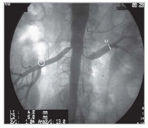
FIGURE 42.12 An aortogram revealing bilateral renal arterial disease performed during a coronary angiography. This individual was treated with antihypertensive drug therapy with excellent blood pressure (BP) control and normal kidney function and was identified only because imaging was included with coronary studies. Numerous series indicate that many such patients are otherwise never identified, but can be managed with medical therapy only. The number that progress to more advanced disease and/or loss of kidney function with current medical therapy may be less than 10% (see text).

One result of these changes has been the emergence of distinctive clinical syndromes that merit a further evaluation in patients at risk for ARAS. These are summarized in Figure 42.1. As a result, patients who typically undergo a diagnostic evaluation and renal revascularization are a subset of the population of patients with RVH.


Deterioration of Renal Function During Antihypertensive Drug Therapy

The availability of effective and tolerable drugs for hypertension has meant that many patients are primarily treated with medical management (see the following). Because some patients with renovascular disease are functioning near the lower end of autoregulation, further pressure reduction may curtail blood flow further. Some authors argue that a rise in serum creatinine more than 30% above pretreatment levels should prompt the exclusion of large vessel ARAS.83 The rapid deterioration of renal function following BP reduction with conventional antihypertensive agents or particularly following BP reduction with ACE inhibitors suggests the
presence of bilateral renal artery stenosis, or stenosis in a solitary functioning kidney.84,85,86,87,88 In one series, more than half of patients demonstrating an acute elevation in the plasma creatinine concentration that was either unexplained or occurred shortly after the institution of therapy with an ACE inhibitor had main renal artery disease, whereas the remainder presumably had a disease of the intrarenal vessels due to nephrosclerosis.88 Remarkably, most patients with renal artery stenosis tolerate ACE inhibition or ARB Rx with few adverse effects.89


Diagnostic Testing for Renovascular Hypertension and Ischemic Nephropathy


Goals of Evaluation

Given the array of diagnostic studies available and the need to focus the evaluation on patients most likely to benefit, it behooves clinicians to consider carefully the objectives of initiating expensive and sometimes ambiguous studies beforehand. As with all tests, the reliability and value of diagnostic studies depend heavily on the pretest probability of disease.90 Furthermore, it is helpful to consider from the outset exactly what objective is to be achieved. Is the major goal to exclude high-grade renal artery disease? Is it to exclude bilateral (as opposed to unilateral) disease? Is it to identify stenosis and estimate the potential for clinical benefit from renal revascularization? Is it to evaluate the role of renovascular disease in explaining deteriorating renal function? The specific diagnostic studies may differ depending on which of these is the predominant clinical objective (Table 42.5).

Noninvasive diagnostic tests for renovascular hypertension and ischemic nephropathy remain imperfect. For the purposes of this discussion, diagnostic tests fall into the following general categories (Table 42.6): (1) physiologic and functional studies to evaluate the role of stenotic lesions particularly related to activation of the RAS, (2) perfusion and imaging studies to identify the presence and degree of vascular stenosis, and (3) studies to predict the likelihood of benefit from invasive maneuvers, including renal revascularization.


Physiologic and Functional Studies of the Renin-Angiotensin System

Efforts have been made for many years to link the measurement of activation of the RAS as a marker of underlying renovascular hypertension. Although these studies are promising when studied in patients with known renovascular hypertension, they have lower performance results as diagnostic tests when applied to wider populations, as we and others have reviewed.91,92,93 PRA is modified by changes of sodium intake, volume status, renal function, and many medications. The sensitivity and specificity of measuring PRA are heavily dependent upon the a priori probability of renovascular hypertension. Although unilateral hypersecretion of renin is an expected finding in hemodynamically significant renal artery stenosis when renovascular hypertension is eventually proved, baseline PRA is elevated in only 50% of patients with renovascular hypertension.92 Tabulation of the number of patients whose hypertension was cured or improved by surgical renal revascularization indicates that probably no more than 50% of patients with renovascular hypertension have elevated PRA.94,95 In practice, the major utility of these studies often depends on their negative predictive value, specifically the certainty with which one can exclude significant renovascular disease if the test is negative. Because negative predictive value rarely exceeds 60% to 70%, these tests offer limited value in clinical decision making.


TABLE 42.5 Goals of Diagnostic and Therapeutic Intervention in Renovascular Hypertension and Ischemic Nephropathy


Goals of Diagnostic Evaluation


Establish presence of renal artery stenosis: location and type of lesion


Establish whether unilateral or bilateral stenosis (or stenosis to a solitary kidney) is present


Establish presence and function of stenotic and nonstenotic kidneys


Establish hemodynamic severity of renal arterial disease


Plan vascular intervention: degree and location of atherosclerotic disease


Goals of Therapy


I. Improved Blood Pressure Control


Prevent morbidity and mortality of high blood pressure


Improve blood pressure control and reduce medication requirement


II. Preservation of Renal Function


Reduce risk of renal adverse perfusion from use of antihypertensive agents


Reduce episodes of circulatory congestion (“flash” pulmonary edema)


Reduce risk of progressive vascular occlusion causing loss of renal function: “preservation of renal function”


Salvage renal function (i.e., recover glomerular filtration rate)


There are several reasons for the relatively low specificity and sensitivity of peripheral PRA in the diagnosis of renovascular hypertension. Most high-renin hypertension is not renovascular in origin; increased renin substrate can


be due to estrogen intake, pregnancy, cortisol excess, intrarenal microvascular ischemia (parenchymal disease), or accelerated or malignant hypertension, and the 15% to 20% of patients with primary (essential) hypertension and high renin levels constitute the majority of high-renin hypertensives. Renin secretion fluctuates widely and is influenced by sodium intake, posture and sympathetic tone,96 a variety of drugs, age, sex, and race.97 The utility of peripheral PRA is reportedly enhanced when measured in the morning with the patient in the seated position and when indexed against urinary sodium excretion; when measured under these exacting circumstances, a high peripheral PRA is found in 75% to 80% of patients with proven renovascular hypertension.98 Other investigators, measuring peripheral PRA under similar circumstances, failed to demonstrate significantly increased sensitivity of the peripheral PRA in predicting either the presence of ARAS and the response to therapy.92,99,100


TABLE 42.6 Noninvasive Assessment of Renal Artery Stenosis


Study


Rationale


Strengths


Limitations


Physiologic Studies to Assess the Renin-Angiotensin System


Measurement of peripheral plasma renin activity


Reflects the relationship between renin and sodium excretion


Measures the level of activation of the renin angiotensin system


Low predictive accuracy for renovascular hypertension; results influenced by medications and many other conditions


Measurement of captoprilstimulated renin activity


Produces a fall in pressure distal to the stenosis


Enhances the release of renin from the stenotic kidney


Low predictive accuracy for renovascular hypertension; results influenced by many other conditions


Measurement of renal vein renin activity


Compares renin release from the two kidneys


Lateralization predictive of improvement in blood pressure with revascularization


Nonlateralization often not predictive of the failure of blood pressure to improve after revascularization; results influenced by medications and many other conditions


Functional Studies to Assess Overall Renal Function


Measurement of serum creatinine


Measures overall renal function, estimated glomerular filtration rate


Readily available; inexpensive


Not sensitive to early changes in renal mass or single-kidney function


Urinalysis


Assesses urinary sediment and proteinuria


Readily available; inexpensive


Results are nonspecific and influenced by many other diseases


Nuclear clearance with [ 125I] iothalamate or chromium Cr 51-labeled pentetic acid (DTPA) to determine the glomerular filtration rate


Measures overall glomerular filtration rate


Useful for estimating glomerular filtration rate in patients with normal and abnormal renal function


Expensive; not widely available


Perfusion Studies to Assess Differential Renal Blood Flow


Captopril renography with technetium 99mTc mertiatide (99mTc MAG3)


Captopril-mediated fall in filtration pressure amplifies differences in renal perfusion


Normal study excludes renovascular hypertension in low-risk populations


Multiple limitations in patients with advanced atherosclerosis or creatinine ≥ 2.0 mg/dL (177 µmol/liter): false positives


Nuclear imaging with technetium mertiatide or technetiumlabeled pentetic acid (DTPA) to estimate fractional flow to each kidney


Estimates fractional flow to each kidney


Allows calculation of single-kidney glomerular filtration rate


Results may be influenced by the presence of obstructive uropathy/other unilateral pathology


Vascular Imaging Studies to Evaluate the Renal Arteries


Duplex ultrasonography


Shows the renal size and measures renal artery flow velocity as a means of assessing the severity of stenosis


Inexpensive; widely available, few false positives


Heavily dependent on operator’s experience; less useful than invasive angiography for the diagnosis of fibromuscular dysplasia and abnormalities in accessory renal arteries


Magnetic resonance angiography


Gadolinium contrast shows the renal arteries and perirenal aorta


No radiation; provides excellent images, newer functional studies (BOLD imaging)


Expensive; concerns about gadolinium now limiting use in renal failure


Computed tomographic angiography


Shows the renal arteries and perirenal aorta, provides functional nephrogram


Provides excellent images; stents do not cause artifacts


Expensive; the contrast is potentially nephrotoxic


BOLD, blood oxygen level dependent.


Modified from Safian RD, Textor SC. Medical progress: renal artery stenosis. N Engl J Med. 2001;344:431-442, with permission.

Only gold members can continue reading. Log In or Register to continue

Stay updated, free articles. Join our Telegram channel

May 29, 2016 | Posted by in NEPHROLOGY | Comments Off on Renal Artery Stenosis, Renovascular Hypertension, and Ischemic Nephropathy

Full access? Get Clinical Tree

Get Clinical Tree app for offline access