Pathology of the Paratesticular Region


Pathology of the Paratesticular Region


HELEN MICHAEL

JOHN R. SRIGLEY


INTRODUCTION

The paratesticular region is a relatively small anatomical compartment containing a disproportionately large number of anatomic structures. In addition to the testicular collecting system there are mesothelial and mesenchymal components representing extensions of the abdominal cavity and retroperitoneum. The epithelial, mesothelial, and connective tissue elements give rise to a wide variety of pathologic conditions including an interesting array of neoplasms and tumor-like lesions.


EMBRYOLOGY, ANATOMY, AND HISTOLOGY


Embryology

The embryology of the testis and paratesticular region is complex and has been the topic of various monographs.1, 2, 3, 4, 5 The gonads are first evident as paired mesenchymal ridges between the dorsal mesentery and the mesonephric ridge at about 4 weeks’ gestation (Fig. 13-1). The coelomic epithelium that covers these mesenchymal ridges extends into the underlying mesenchyme to form the primitive sex cords. Germ cells are first seen in the embryonic yolk sac. They then migrate along the wall of the hindgut and the dorsal mesenteric root into the developing gonads by the 6th week of gestation (Fig. 13-2). The sex cords proliferate under the influence of the germ cells. Testicular tissue with primitive seminiferous tubules is detectable by the 7th week of gestation. At about the same time, the primitive tunica albuginea forms as a layer of flattened cells around the gonad, and the primitive testis becomes separated from the overlying coelomic epithelium.

In the hilum of the developing testis, the sex cords form a network of cellular strands that admix with mesonephric cells from the degenerating mesonephric tubule (Fig. 13-3). Both sex cords and the mesonephros are thought to contribute to the formation of the rete testis. Hormones from the embryonic testis are thought to stimulate the mesonephric duct to develop into the male genital tract collecting system and, at the same time, to suppress development of the paramesonephric duct. By the end of the 4th month of gestation, the rete cords have merged with the epigenital tubules of the mesonephros, which form the efferent ductules and head of the epididymis (Fig. 13-4). The mesonephric duct develops into the epididymal body and tail and the vas deferens. The caudal portion of the vas deferens joins with the seminal vesicles, which are also derived from the mesonephric duct. The enlarging testis becomes separated from the degenerating mesonephros.

The descent of the testis into the scrotum through the inguinal canal is complex and not totally understood.2 At the same time that the mesonephros degenerates, a ligament called a gubernaculum descends from the lower pole of each gonad along each side of the abdomen, passing through the abdominal wall at the site of the future inguinal canal, and it attaches to the scrotal swelling. A peritoneal sac called the processus vaginalis develops bilaterally, ventral to the gubernaculum, and herniates through the abdominal wall along the pathway formed by the gubernaculum. The processus vaginalis carries with it extensions of the abdominal wall layers, thus forming the wall of the inguinal canal. The opening formed in the external oblique aponeurosis forms the superficial inguinal ring, and the defect in the transversalis fascia forms a deep inguinal ring. The external spermatic fascia is a thin membrane derived from the aponeurosis of the external oblique muscle at the outer abdominal ring. The cremasteric fascia originates from the lower border of the internal oblique muscle. The internal spermatic fascia is derived from the transversalis fascia.6 These layers become part of the spermatic cord and the wall of the scrotum.

By about 7 months’ gestation, the testes are located distal to the deep inguinal ring, a change in position that is attributed to growth of the trunk and pelvis of the embryo.2 The gubernaculum does not grow after it is formed, and the exact process of testicular descent into the scrotum is not well understood. The gubernaculum serves as an anchor that aids
in testicular descent, but it does not pull the testis caudally. Increasing abdominal pressure due to developing organs may have a role in the process. Androgenic and gonadotrophic hormones are also thought to be important in the descent of the testes into the scrotum.2 The testis is positioned posterior to the processus vaginalis, and it is located in the scrotum by about the 8th month of gestation. The layers of the inguinal canal then contract around the spermatic cord.


FIGURE 13-1 ▪ Embryology of the testis and paratestis. A: At 4 weeks, the genital ridges are apparent as mesenchymal condensations with a covering of coelomic epithelium that has proliferated. B: At 6 weeks, there is ingrowth of the coelomic epithelium with extension into the mesenchyme to form the primitive sex cords. (Reprinted from Langman J. Medical Embryology and Human DevelopmentNormal and Abnormal. 2nd ed. Baltimore, MD: Williams & Wilkins; 1972, Fig. 11-13, with permission.)

The vas deferens crosses anterior to the ureter as a result of the pathway of testicular descent into the scrotum.2,6 The blood vessels that supply the testis follow a pathway along the dorsal abdominal wall. The cranial part of the processus vaginalis is obliterated in the perinatal period. The tunica vaginalis is then an isolated sac lined by mesothelium.


FIGURE 13-2 ▪ Embryology of the testis and paratestis. A: At 3 weeks, the primordial germ cells form in the wall of the yolk sac. B: At 6 weeks, the primordial germ cells migrate to the wall of the hindgut, along the dorsal mesenteric root, and into the genital ridges. (Reprinted from Langman J. Medical Embryology and Human DevelopmentNormal and Abnormal. 2nd ed. Baltimore, MD: Williams & Wilkins; 1972, Fig. 11-14, with permission.)


Anatomy and Histology

The rete testis, efferent ductules, and epididymis represent a continuous conduit for seminiferous fluid to be transported from the seminiferous tubules to the vas deferens.


Rete Testis

The rete testis represents a group of anastomotic channels in the hilum of the testis (Fig. 13-5). These channels receive the contents of the seminiferous tubules. The rete also serves as a chamber for mixing the seminiferous tubule contents, a possible source of seminal fluid, a site of resorption of protein, and the site of a pressure gradient between the testis and the epididymis.7 There are intratesticular and
extratesticular portions of the rete testis. The tubulae rete are located in testicular interlobular septa, and they connect the two ends of each seminiferous tubule. They also connect to the mediastinal rete, which exit the testis as the extratesticular bullae retis. The latter structures anastomose to form the efferent ductules.


FIGURE 13-3 ▪ Embryology of the testis and paratestis. At 8 weeks of gestation, the tunical albuginea surrounds the developing testis, and the rete testis cords intermingle with mesonephric tubules at the hilum. (Reprinted from Langman J. Medical Embryology and Human Development—Normal and Abnormal. 2nd ed. Baltimore, MD: Williams & Wilkins; 1972, Fig. 11-15A, with permission.)

The rete testis is lined by low columnar, cuboidal, or simple squamous epithelium that rests on basal lamina surrounded by fibroblasts, myoid cells, collagen, and elastin. These connective tissue components constitute the wall of the rete testis. Microvilli are present on the luminal surfaces of the epithelial cells. Each cell also contains a single flagellum that can be seen by electron microscopy.8


FIGURE 13-4 ▪ Embryology of the testis and paratestis. A: By 4 months of gestation, the rete testis cords have merged with the epigenital tubules of the mesonephros. B: Diagram of the mature testis after descent showing the relationships of various structures. (Reprinted from Langman J. Medical Embryology and Human Development—Normal and Abnormal. 2nd ed. Baltimore, MD: Williams & Wilkins; 1972, Fig. 11-18, with permission.)


FIGURE 13-5 ▪ Complex anastomosing channels are present in the rete testis. The channels are lined by low columnar epithelium.


Efferent Ductules

Twelve to fifteen efferent ductules connect with the extratesticular rete testis. They make up most of the head of the epididymis and resorb seminal fluid components. The efferent ductule epithelium is composed of two layers, including columnar epithelial cells and flattened, cuboidal basal
cells. The luminal surface of the tubules is undulating. Sometimes, the epithelial cells may display nuclear atypia and cytoplasmic pigment reminiscent of that in the seminal vesicles.9 The epithelium is surrounded by basement membrane, smooth muscle, and some fibroblasts.8


Epididymis

Most of the head of the epididymis contains the efferent ductules. The body and tail contain a coiled, 4- to 5-m-long duct.7,10 The epididymis is involved with sperm storage, sperm transfer, sperm maturation, and sperm concentration.7 Sperm matures, develops motility, and is stored in the tail of the epididymis.11 The well-developed smooth muscle wall surrounding the epididymal tubules assists in transportation of sperm through the epididymis.

Unlike the wavy luminal surface of the efferent ductules, the epididymal ducts have a smooth luminal contour (Fig. 13-6). The epididymal epithelium contains tall columnar cells, dark columnar cells, clear cells, and basal cells. The columnar cells are ciliated, and the cilia decrease in length from the head to the tail of the organ. The duct lumen of the tail of the epididymis contains abundant spermatozoa, and clear cells are more prominent in the epithelium in this area. Periodic acid-Schiff-positive diastase-resistant intranuclear inclusions are often present in the columnar cells of the epididymal epithelium,12 most commonly in the distal portion. Electron-dense, membrane-enclosed bodies are identified by ultrastructural examination, but no viral features have been seen.12 Cytoplasmic lipochrome pigment may also be present. The epididymal epithelium is surrounded by both basement membrane and smooth muscle; the latter is important for sperm transport. The epithelium may have a cribriform pattern that should not be mistaken for hyperplasia.13, 14, 15


FIGURE 13-6 ▪ Normal epididymis. A: The head of epididymis is composed of efferent ductules. They contain pseudostratified epithelium with ciliated cells. The luminal border is undulating. B: Ductus epididymis. Cilia are prominent, and the pseudostratified lining cells have a smooth luminal border. Smooth muscle surrounds the duct.


Vas Deferens (Ductus Deferens)

The tail of the epididymis connects with the vas deferens, a 30- to 40-cm-long tubular structure that merges with the seminal vesicle to form the ejaculatory duct.7 The vas deferens epithelium contains pseudostratified columnar epithelial cells and cuboidal to flattened basal cells. Electron microscopic examination has shown principal cells, peg cells, mitochondria-rich cells, and basal cells in the epithelium.16 The luminal side of the epithelium is ciliated, and the cilia become shorter and less abundant as the seminal vesicle is approached. The epithelium is surrounded by a basement membrane. In adults, there is a layer of connective tissue containing elastic fibers between the basement membrane and the muscular wall of the vas deferens. The muscularis contains inner and outer longitudinal layers and a middle circular or oblique layer. Like the epididymis, the vas deferens epithelium may display cribriform architecture, intranuclear inclusions,12 and lipochrome pigment. The epithelium of the vas is thrown into folds, and some of these complex infoldings and outpouchings may reach into the muscularis layer.


Testicular Tunics

The tunica albuginea is internal to the tunica vaginalis. It is composed of thick fibrous tissue that contains smooth muscle cells and nerve fibers and surrounds the testis except at the testicular hilus. The myocytes may contract and lead to an increase in intratesticular pressure. Tumor-like lesions and rare neoplasms may arise from the tunica albuginea.

The tunica vaginalis is a layer of mesothelium and associated basement membrane that represents the inner lining of the intrascrotal structures. The visceral tunica vaginalis forms a serosal lining over the tunica albuginea, covering the testis and most of the epididymal head. It reflects
back on itself superiorly and posteriorly at the mediastinum testis to become the parietal tunica vaginalis. Urothelial metaplasia may occur in the tunica vaginalis,17 with formation of von Brunn nest-like structures.8 Squamous metaplasia may also occur, and after cystic transformation, it may account for some epidermoid cysts in the testis and paratestis.18,19

While the cranial part of the tunica vaginalis normally becomes obliterated in the perinatal period, sometimes residual mesothelium present in the spermatic cord may result in cysts or other lesions of mesothelial origin.


Spermatic Cord

During its descent into the scrotum, the testis brings with it the elements of the spermatic cord, including the vas deferens, blood vessels, and nerves.1,2,4,5 The cord is covered by the spermatic and cremasteric fascia that accompany the processus vaginalis through the abdominal wall into the inguinal canal.2,6 The cremasteric muscle is composed of skeletal muscle bundles present along the outer part of the spermatic cord and in the wall of the scrotal sac. The loose connective tissue matrix of the spermatic cord contains scattered bundles of smooth muscle.

The arteries present in the spermatic cord supply blood to the testis and paratesticular structures. They include the testicular (spermatic) artery, the artery of the vas deferens, and the cremasteric artery.6 The testicular artery originates from the aorta inferior to the renal artery and penetrates the tunica albuginea to provide the main blood supply to the testis and the epididymis. The anterior and posterior epididymal arteries arise from the testicular artery and supply the head of the epididymis and the epididymal body and tail, respectively. The artery of the vas deferens is a branch of the superior vesicle artery that accompanies the vas deferens and anastomoses with the main testicular artery or the posterior epididymal artery. The cremasteric artery is a branch of the deep epigastric artery. It supplies the cremasteric muscle and other coverings of the spermatic cord. It anastomoses with branches of the testicular artery.

The epididymal veins anastomose with the testicular veins6 to form the pampiniform plexus that invests the testicular artery.8 Further venous anastomoses eventually lead to the right and left testicular veins. The right testicular vein opens into the inferior vena cava at an acute angle, whereas the left testicular vein drains into the left renal vein at a right angle. Increased hydrostatic pressure from the perpendicular venous anastomosis on the left side is thought to account for the greater incidence of varicoceles on that side.

The lymphatic channels of the testis and epididymis arise from a superficial plexus beneath the tunica vaginalis and a deep plexus in the testis and epididymis.6 These vessels anastomose into four to eight larger channels that accompany the main testicular blood vessels through the spermatic cord to drain into the lateral paraaortic and preaortic lymph node groups.8

The spermatic cord nerves include the genital branch of the genitofemoral nerve, which innervates the cremasteric muscle and sends branches to the scrotal skin.6 The testicular plexus of sympathetic nerve fibers is derived from branches of renal and aortic nerve plexuses with contributions from the superior and inferior hypogastric plexuses. These nerves travel with the testicular artery as well as give off branches to the epididymis and the vas deferens. Nerves from the pelvic plexus may accompany the artery of the vas deferens.8


Congenital Abnormalities and Ectopia


Rete Testis

The term “dysgenesis of the rete testis” refers to the underdeveloped rete testis associated with cryptorchidism20 (Table 13-1).

Testicular abnormalities reported in cryptorchidism include Sertoli cell-only syndrome (23 cases), diffuse tubular hyalinization (8 cases), mixed tubular atrophy (3 cases), and maturation arrest of spermatogonia (4 cases).20

The rete has been described as diffusely hypoplastic in 37.5%, hypoplastic and cystic in 50%, and hyperplastic in 12.5% of 40 cryptorchid testes.20 The latter pattern is identical to the lesion described as (idiopathic) rete testis hyperplasia, characterized by tubular, papillary, and cribriform intratubular epithelial proliferations. Cystic change in the rete testis associated with cryptorchidism is manifested by dilated epithelial structures measuring up to 500 µm lined
by large cuboidal epithelial cells. The epithelium stains for both cytokeratin and vimentin in the same manner as the normal rete epithelium. Some cases have been associated with efferent duct atrophy, with small duct diameters and more space than usual between the ducts. Other associated findings included epididymal duct ectasia (21 cases), an underdeveloped muscular layer in the epididymis (11 cases), and fat and dilated veins in the mediastinum testis (3 cases each).


Table 13-1 ▪ CONGENITAL ABNORMALITIES AND ASSOCIATED CONDITIONS


Type


Associated Conditions


Rete testis


Dysgenesis


Cryptorchid testis, epididymal abnormalities


Cystic dysplasia


Renal agenesis or cystic dysplasia of kidney


Epididymis


Anomalies of attachment to testis


Cysts


Von Hippel-Lindau syndrome


In utero DES exposure


Agenesis


Absence of vas deferens and renal anomalies


Vas deferens


Congenital absence


Cystic fibrosis


Absent epididymis


Renal anomalies


Ectopic


Imperforate anus, hypospadias


Splenic-gonadal fusion (continuous form)


Peromelia, micrognathia, gastrointestinal abnormalities


Ectopic adrenal tissue


TTAGS


Nelson syndrome in some


Ectopic renal tissue


Undescended testis; rarely associated with extrarenal Wilms’ tumor


Cryptorchidism is also associated with anomalies of mesonephric structures, including luminal dilation of the spermatic duct system, immature muscle layers, and malformations of the epididymis. These multiple abnormalities associated with cryptorchidism suggest a primary developmental disorder of the mesonephros.21


Epididymis

Congenital anomalies of the epididymis include anomalies of the attachment to the testes, epididymal cysts, agenesis or accessory epididymis, ectopic epididymis, and duplication of the epididymis.

Anomalies of fusion, with detachment of the head of the epididymis from the testis, and anomalies of suspension have been reported in children who have had surgery for cryptorchidism.22, 23, 24 Fusion anomalies are more commonly associated with abdominal testes, and suspension anomalies are more often seen when the cryptorchid testis is located more distally. Some epididymal anomalies are associated with an absent testis. Anomalies of attachment of the epididymis to the testis include attachment of the caput and cauda with a detached corpus, attachment of the epididymal head only, attachment of the cauda only, and complete separation of the testes and epididymis.23 Epididymal anomalies may also be seen in about one-third of boys with hernias and hydroceles without cryptorchidism, especially if there is a patent processus vaginalis. Detachment of the head of the epididymis bilaterally results in infertility.

Epididymal cysts are usually asymptomatic lesions that are discovered incidentally on physical examination or ultrasound. Epididymal cysts may be acquired, but some authors believe that they have a congenital basis,25 perhaps related to maturation of the mesonephric ductal system. They are seen in patients with von Hippel-Lindau syndrome and in some patients exposed to diethylstilbestrol in utero26 (Table 13-1). The cysts may represent efferent ducts that did not fuse with the mesonephric duct during embryogenesis.

Epididymal agenesis is almost always associated with unilateral or bilateral absence of the vas deferens (Table 13-1). The epididymal head is usually present because it is composed of efferent ducts derived from the genital ridge, whereas the epididymal body and tail are of mesonephric derivation.25 A high frequency of renal anomalies is present in these patients.

Aberrant epididymal tissue is rare. It may be associated with an undescended testis. One case of ectopic epididymal tissue in the appendix testis has been reported.27 Ectopic epididymal tissue has also been associated with an inguinal hermia sac.28

Epididymal duplication is rare. It is characterized by a small accessory epididymis branching from the main epididymis. These patients are asymptomatic, and the lesions are discovered incidentally.


Vas Deferens

Congenital bilateral absence of the vas deferens is a well-studied abnormality of the wolffian ducts that represents a primary genital form of cystic fibrosis29 (Table 13-1). Mutations in the cystic fibrosis transmembrane conductance gene are associated with wolffian duct abnormalities, including unilateral and bilateral congenital absence of the vas deferens and idiopathic epididymal obstruction.29 The body and tail of the epididymis are also often absent, so the epididymal head may be prominent and distended with sperm. In some cases, there may be complete absence of the epididymis. Renal anomalies may be present due to the associated embryologic origins of these structures.25

Unilateral absence of the vas deferens occurs in <1% of healthy men. It represents the most frequent congenital anomaly of the vas deferens and involves the left side more often than the right side.25 It is often detected at the time of vasectomy. It is not a common cause of infertility if the contralateral vas is normal. The epididymis associated with the absent vas may be of variable length. Ipsilateral renal agenesis may be associated with this condition (Table 13-1).

Only a few cases of duplicated vas deferens have been reported.25 This term is restricted to cases where a second vas deferens is identified within the spermatic cord. The condition needs to be recognized at the time of vasectomy.

Ectopic vas deferens (persisting mesonephric duct) is a condition wherein the ureter enters the vas deferens.25 A triad of ectopic vas deferens, imperforate anus, and hypospadias has been described30 (Table 13-1); it has been present in about one-fourth of patients with ectopic vas deferens. The diagnosis of persisting mesonephric duct should be considered in male children with imperforate anus and recurring urinary tract infections.

One case of a large diverticulum of the vas deferens has been reported.31 A thickened scrotal vas deferens and azoospermia were present. Another rare congenital anomaly is crossed dystopia of the vas deferens, a condition in which the vas deferens crosses the midline and communicates with the contralateral seminal vesicles.31 Infertility is not associated with that lesion unless additional abnormalities are present. The diagnosis is made by vasography. Segmental aplasia of the vas deferens (skip vas) may be unilateral or bilateral; bilateral lesions result in infertility. Segments of vas deferens may also be hypoplastic. These conditions represent abnormal mesonephric duct development and may be associated with abnormalities of the epididymis and seminal vesicles.


Ectopic Splenic Tissue and Splenic-Gonadal Fusion

Splenic and gonadal tissue may fuse during embryogenesis.32 The left gonad is most frequently involved.33 There is a continuous form of this lesion in which a cord connects the spleen to the testis; many of those patients have marked defects of the extremities (peromelia), as well as micrognathia and gastrointestinal abnormalities (Table 13-1). Those patients may have a scrotal or inguinal mass that becomes apparent during an inguinal hernia operation or during surgery for an undescended testis. Small aggregates of splenic tissue may be found in the spermatic cord, or the cord may be composed entirely of splenic tissue.34

The discontinuous form of splenic-gonadal fusion is manifested by accessory splenic tissue in the paratestis region rather than continuity between the spleen and testis. Accessory splenic tissue has been reported in the epididymis, in the spermatic cord, and between the scrotal skin and the spermatic cord.33 The splenic tissue has the gross and microscopic characteristics of normal spleen.


Ectopic Adrenal Tissue

Ectopic adrenal cortical tissue is seen in the paratestis of 1.6% to 15% of male patients.35,36 It typically appears as yellow-orange nodules in the spermatic cord, epididymis, rete testis, tunica albuginea, and between the epididymis and the testis. It is most often seen in infants, but it may occur in adults. Nodules of ectopic adrenal tissue may measure 2 to 6 mm in diameter.37 They display three well-defined layers of adrenal cortex. The zona fasciculata is the predominant tissue. Adrenal medulla is not present in ectopic adrenal tissue. The characteristic zonation seen in the adrenal cortex is an important factor in distinguishing ectopic adrenal tissue from other steroid cell proliferations in this region.

Ectopic adrenal cortex may be the source for neoplasms such as the testicular tumor of the adrenal genital syndrome (TTAGS) that may be seen in the spermatic cord.38 Similar tumors are seen in patients with Nelson syndrome39 (Table 13-1). These lesions actually represent reversible hyperplasia of steroid-producing cells in response to high circulating levels of ACTH.40 Reduction of the ACTH levels causes regression of the lesion. TTAGS displays nests of cells with eosinophilic cytoplasm; the cell nests are separated by fibrous stoma. Intracytoplasmic lipofuscin may be present. Some authors have suggested that TTAGS arise from pluripotential cells in the testicular hilus rather than adrenal rests.38


Ectopic Renal Tissue

Ectopic renal tissue in the paratesticular region is rare and usually occurs in association with an undescended testis.41, 42, 43 One case occurred in a 36-year-old man with a painful inguinal swelling.41 An undescended testis was present with an adjacent ectopic kidney that measured 3 cm in diameter and contained renal cortex and medulla as well as some immature mesenchyme. An additional patient had heterotopic renal tissue that was discovered in association with an intrascrotal Wilms tumor44 (Table 13-1). The renal heterotopia consisted of renal tubules and immature glomeruli that may have arisen from caudal mesonephric elements present in the paratesticular area. Primary extrarenal nephrogenic rests occur rarely in the paratesticular region.45 They display blastema and immature glomeruli and tubules.


Extraparenchymal Leydig Cells

Clusters of Leydig cells are often present in the paratesticular region (Fig. 13-7). They have been reported in the tunica albuginea and rete testis,46,47 the adventitial tissue between the tunica albuginea and epididymis, the epididymis, the vas deferens, and the spermatic cord.36,48 A recent study found Leydig cells outside the testis in 90 of 97 orchiectomy specimens reviewed.49 The testicular tunics were involved in 50% of cases, and 14.4% of spermatic cords contained Leydig cells. These cells were associated with nerves in 25.5% of cases and with vascular spaces in 7.8% of orchiectomy specimens, but some aggregates of Leydig cells were not associated with either nerves or blood vessels.49 Leydig cells sometimes contain lipochrome pigment, Reinke crystals, and multiple nuclei.47 Large aggregates of extratesticular Leydig cells may be misinterpreted as a primary paratesticular Leydig cell neoplasm, spread from a testicular Leydig cell tumor or other rare neoplasms or metastatic disease. Extratesticular Leydig cells may form small nodules, but the location along nerves and vessels combined with the lack of an expansile paratesticular mass or testicular neoplasm distinguishes testicular adnexal Leydig cells from Leydig cell tumor of paratesticular or testis origin. In contrast to metastatic deposits, no atypia, mitosis, or stromal reaction is associated with extratesticular Leydig cell aggregate.


FIGURE 13-7 ▪ Perineural Leydig cells. Leydig cells have abundant eosinophilic cytoplasm and are often associated with nerve bundles in the paratesticular region.


Remnants of Wolffian and Müllerian Ducts

Remnants of the embryonic müllerian and wolffian ducts give rise to the appendix testis and the appendix epididymis, respectively. These structures may be subject to various pathologic processes, including cysts, infarcts, and tumors. Cysts usually occur in the retroperitoneum, mesentery, pelvis, or paratesticular region. However, one cyst thought to be derived from a wolffian duct remnant occurred in the liver of an infant boy. It was connected by a stalk to the head of the epididymis adjacent to his right abdominal undescended testis.50


Appendix Testis

The appendix testis is a remnant of the paramesonephric (müllerian) duct that is usually found on the anterosuperior aspect of the testis in the groove between the testis and the head of the epididymis.5,7 It is most commonly attached to the tunica vaginalis of the testis, but it may also be attached to the epididymis. The appendix testis can be identified in 80% to 90% of men, and it has been found bilaterally in 60% of patients.7 A thickened area of the tunica vaginalis or focal calcification may be the only evidence of this structure in some men. However, it is generally an ovoid structure that measures 0.5 to 2.5 cm in length.51 It is covered by cuboidal to columnar epithelium that may be stratified (Fig. 13-8). The connective tissue core contains blood vessels and, sometimes, smooth muscle cells. Stroma resembling ovarian stroma may also be present. Torsion and infarction of the appendix testis is usually seen in children or adolescents, and it is associated with scrotal pain52 that may be mistaken for processes involving other intrascrotal areas. Chronic torsion may cause detachment of the appendix testis.


Appendix Epididymis

The appendix epididymis is derived from the cranial aspect of the mesonephric (wolffian) duct. It is identifiable in only about 25% of specimens.53 It is a cystic structure that may contain eosinophilic fluid and is lined by cuboidal to columnar epithelium. The epithelium is surrounded by a basement membrane, with a small amount of connective tissue and mesothelial cells on the external aspect. The appendix epididymis may be pedunculated, or it may be adherent to the head of the epididymis. It may be involved by torsion and infarction causing scrotal pain, and sometimes it may become enlarged and suggest a mass lesion.


FIGURE 13-8 ▪ Appendix testis. The oblong structure has a fibrovascular core and is covered by columnar cells.


Aberrant Ductules and Paradidymis

The superior and inferior aberrant ductules (organ of Haller) and paradidymis (organ of Giraldes) are mesonephric duct (wolffian) remnants.8 They are near the head or body of the epididymis in the case of the superior aberrant ductule or the junction of the tail of the epididymis and the vas deferens in the case of the inferior aberrant ductule.7 Both are small tubules or cysts with low columnar epithelium surrounded by smooth muscle. They may be the source of some epididymal cysts, although epididymal cysts are also encountered in patients with the von Hippel-Lindau syndrome and in some patients who were exposed to diethyl-stilbestrol in utero.7,26,54

The paradidymis is usually located adjacent to the vas deferens in the area of the head of the epididymis. It is a small tubular structure similar to the aberrant ductules, and it may result in a spermatic cord cyst.28 It is important not to mistake the paradidymis for vas deferens in inguinal hernia specimens.55 The well-developed, three-layered muscular wall of the vas deferens aids in the distinction.


Hernia Sac Inclusions

Hernia sacs from young boys contain glandular or ductal structures in 1.5% to 6% of cases55, 56, 57 (Box 13-1). These embryonal rests must be distinguished from transected vas deferens and epididymal tissue because gland inclusions do not affect reproductive function.

The three types of embryonal rests seen in inguinal hernia sacs include vas deferens-like, epididymis-like and müllerian-like inclusions.56 The diameter of the remnants,
correlated with patient age, may aid in distinguishing them from normal vas deferens.56 Trichrome and immunohistochemical stains for smooth muscle actin are useful because they distinguish periglandular mesenchymal condensations in the inclusions from the subepithelial muscle layer seen in the vas deferens and epididymis. Vas deferens-like and müllerian-like inclusions do not display the luminal CD10 staining that is seen in normal vas deferens. Two types of epididymis-like inclusions have been reported.56 One group displays luminal decoration by CD10 and may represent aberrant wolffian ductules. The other group does not stain with this antibody and may represent müllerian remnants.


Congenital Hydrocele

Congenital hydroceles result from a persistent communication between the tunica vaginalis and the peritoneal cavity. This communication is normally obliterated after descent of the testis into the scrotum and usually before the age of 2 years.


Cystic Dysplasia

Cystic testicular dysplasia is rare, and it is an entity seen in infants and young children.58 It is usually a unilateral lesion that is associated with an ipsilateral urogenital lesion such as renal agenesis or cystic dysplasia of the kidney59 (Table 13-1). The lesion is usually manifested by painless testicular enlargement, although one recently reported patient complained of scrotal pain.60 Involvement of the rete may be segmental or diffuse. The rete testis in patients with this disorder displays cystically dilated channels (Fig. 13-9) that maintain the normal branching pattern of the rete and do not display epithelial proliferation.21 The rete spaces are lined by cuboidal to flattened epithelial cells that are surrounded by fibrous stroma. The cysts range in size from 1 to 5 mm in diameter and are visible on gross examination. Seminiferous tubules are normal. Cystic dysplasia of the rete testis does not appear to be caused by obstruction of the duct system, which causes extensive dilation of the rete channels as well as dilation of seminiferous tubules in young children. It is more likely related to an embryologic defect. During embryogenesis, the rete testis is formed from sex cord stromal tissue; the connection with epididymis (of wolffian duct origin) may be defective in this disorder, leading to dilated, blind-ended rete channels.21 Abnormal sodium metabolism has also been proposed as a cause for this disorder.26 Cystic dysplasia has also been reported in the epididymis.61


FIGURE 13-9 ▪ Cystic dysplasia. This gross photograph displays distorted rete testis architecture due to multilocular cystic dilation of the rete channels.


Inflammation and Infection

Inflammatory conditions of infectious or noninfectious origin may principally affect epididymis, tumor, or spermatic cord or may involve more than one of those structures. Furthermore, paratesticular inflammation may be secondary to orchitis. In this section the entities are discussed under headers of nonspecific and granulomatous inflammation, realizing that both may have active (acute) and chronic components. Diverse etiologic agents are associated with these conditions, and in some instances no identifiable agent is found.


Nonspecific Acute and Chronic Inflammations

Epididymitis represents the most common intrascrotal inflammation.62,63 Chronic infection and inflammation are associated with reactive changes and fibrosis that may simulate a neoplasm. Acute epididymitis is the most common cause of an acutely painful scrotum. Bacterial infection may be caused by a variety of organisms. It is often associated with anatomic abnormalities, and it is the most common type of epididymitis seen in older men.34 Sexually transmitted Chlamydia trachomatis or Neisseria gonorrhoeae epididymitis represent the most common cause of acute scrotal swelling in men under the age of 35 years.34 Sexually transmitted epididymitis is associated with underlying urologic abnormalities.64 Chlamydia trachomatis epididymitis is minimally destructive, with periductal and intraepithelial inflammation, epithelial proliferation, and, sometimes, squamous metaplasia.65 On the other hand, pyogenic infections such as N. gonorrhoeae are associated with destructive abscess formation and may create masses that mimic neoplasia. Low-risk and high-risk types of human papillomavirus have been detected in epididymal and ductus deferens tissue from some patients with epididymitis, although neither koilocytotic atypia nor dysplasia was identified.66 The male urogenital tract may therefore serve as a reservoir of HPV infection. Older men who develop bacterial epididymitis are more often infected with Escherichia coli.

Whereas epididymitis is rare in childhood, an increasing frequency has been reported in children admitted with the diagnosis of acute scrotum.67 In some cases, E. coli has been cultured from children with epididymo-orchitis who had no underlying urinary tract abnormalities.68

Chemical epididymitis occurs when sterile urine refluxes into the vas deferens, sometimes after heavy lifting or blunt abdominal trauma that increases the intra-abdominal pressure.69 The vas deferens and the tail of the epididymis become inflamed, but no organisms are identified.


Schistosomal funiculitis has been reported in a patient with chronic schistosomal infection of long duration.70 Dirofilaria conjunctivae infection has been seen in the spermatic cord, where it has simulated a neoplasm.71


Granulomatous Inflammation

The most common cause of granulomatous epididymitis is tuberculosis (Fig. 13-10). The incidence of this disease has increased because of human immunodeficiency virus infection and intravesical bacille Calmette-Guerin therapy for superficial bladder tumors.72 There is an association between renal and genital tuberculosis, with frequent involvement of the epididymis.73 Painful or painless scrotal enlargement is a common presenting symptom. Epididymal tuberculosis may present as a mass lesion in patients that have widespread disease. Secondary hydroceles may be associated with epididymal tuberculosis. Extensive infection can result in sinuses that communicate with the scrotum.72

Gross examination of tuberculous epididymitis reveals multiple small white to yellow nodules that typically contain caseous necrosis. Microscopic examination displays necrotizing granulomatous inflammation with palisading histiocytes and Langhans giant cells. Granulomas originate in the epididymal stroma and then enlarge, become confluent, and spread to and secondarily involve the tubules.74

Brucella75,76 and blastomycosis77 have also been implicated in granulomatous epididymitis clinically simulating neoplasia. Some cases of granulomatous epididymitis have not been associated with any detectable infectious agent.78 One such case occurred in a 41-year-old man who presented with a scrotal mass and had a 2.3-cm firm lesion in the tail of the epididymis.34 Epididymal tubules had necrotic walls, and there was a significant histiocytic infiltrate associated with squamous metaplasia.34 No necrosis or Langhans-type giant cells were seen, and no organisms could be identified on special stains.


FIGURE 13-10 ▪ Tuberculous epididymitis. This gross photograph shows partially necrotic tumor-like lesion involving epididymis and paratesticular soft tissue.

Granulomatous epididymitis may also be caused by fungal organisms. However, fungal epididymitis is very rare and usually associated with orchitis. Demonstration of the organisms with special histologic stains or culture is necessary if this diagnosis is a consideration.

Some cases of granulomatous epididymitis with no apparent infectious agent may have an ischemic etiology.79 In cases thought to be ischemic, granulomatous lesions display predominantly histiocytic infiltrates and typically involve tubular walls rather than the epididymal stroma.79 Areas of necrosis and squamous metaplasia may be present. Ischemic lesions are more frequently located in the head of the epididymis, where the blood supply is more easily compromised than in the body and tail.34 The vascular supply for the head of the epididymis consists of only the superior epididymal artery. The vascular supply for the body and tail of the epididymis includes numerous anastomoses between the inferior epididymal and vas deferens arteries, thereby protecting these areas against ischemia.

Xanthogranulomatous epididymitis is associated with Gram-negative bacteria and usually requires surgical excision.34,80 These lesions display prominent aggregates of foamy histiocytes in addition to plasma cells, lymphocytes, and neutrophils. One reported case80 was bilateral and so severe that it was difficult to distinguish the epididymis from the adjacent testis. Xanthogranulomatous funiculitis and epididymitis have been described in a quadriplegic patient with unsuccessful voiding.81 Other rare cases of xanthogranulomatous funiculitis have been reported.82 The lesion presents with spermatic cord enlargement, and the histologic features are identical to those of xanthogranulomatous epididymitis.


Sarcoidosis

Genitourinary involvement by sarcoidosis is very uncommon. The average age at onset of genital sarcoidosis is 33 years (range 2 to 67).83 Epididymal sarcoidosis has been reported mainly in black men, some of whom also had testicular sarcoidosis.84 The spermatic cord may also be involved. These lesions are often asymptomatic and unilateral, although they may be bilateral.85,86 Firm nodules may replace the entire epididymis. Nonnecrotizing granulomas may cause nodules that measure up to 2.5 cm in diameter and mimic a tumor.84,87,88 Special stains for acid-fast bacilli and fungi show no organisms. Diffuse involvement of the epididymis may also be present. Most patients have hilar adenopathy or reticulonodular lung infiltrates on chest radiographs. However, genitourinary sarcoidosis has been
reported in the absence of radiographically detectable lung or mediastinal disease.83


Sclerosing Lipogranuloma

Injection of lipids to increase the size of the genitalia has resulted in granulomatous lesions in the scrotum, the spermatic cord, or the epididymis.89,90 Some have presented as mass lesions requiring surgical excision.91,92 Most cases have been associated with a history of exogenous lipid injection or a history of trauma, but some may have been idiopathic.93 Most patients have presented with localized masses measuring from a few to several centimeters in size.5

Gross examination of resected sclerosing lipogranulomas reveals fragmented or intact specimens that are gray to yellow and solid or solid with small cysts on gross examination.5 Microscopic examination displays patchy fibrous tissue containing empty vacuoles of varying size without epithelial lining cells. Foreign body giant cells may be present. Areas of hyalinization may be seen, and inflammatory cells include histiocytes, lymphocytes, plasma cells, and eosinophils. This lesion is typically patchy in the involved area of the epididymis or spermatic cord. The differential diagnosis of sclerosing lipogranuloma in the epididymis includes adenomatoid tumor, sclerosing lipogranuloma, and signet ring carcinoma. None of those neoplasms contains the empty vacuoles devoid of lining cells that are characteristic of sclerosing lipogranuloma. Mesothelial-lined tubular structures typical of adenomatoid tumor are not seen in sclerosing lipogranulomas. Sclerosing liposarcoma displays atypical nuclei and rare lipoblasts in fibrous tissue alternating with “lipoma-like” areas. Neither atypical nuclei nor lipoblasts are seen in sclerosing lipogranuloma. Signet ring carcinomas contain mucinous cytoplasmic vacuoles that are not present in sclerosing lipogranuloma.


Other Rare Granulomas

Granulomas of paratesticular structures have been reported due to powder from surgical gloves.94 This was the etiology in the case of a 2-cm hard mass in the head of the epididymis and tunica albuginea in one patient.94 Microscopic examination of the mass showed fibrous tissue and epithelioid histiocytes intermingled with foreign body-type giant cells that contained crystalline material. Radiopaque contrast dye containing lipid has also been reported as a cause of granulomas of the vas deferens in a 35-year-old infertile man who underwent vesiculoepididymography.95 Partial resection of the vas deferens revealed a small nodule that showed acute, chronic, and granulomatous inflammation on microscopic examination. The granulomas contained refractile foreign material.

Cholesterol granulomas (also called cholesteatoma) of the tunica vaginalis simulating neoplasm have also been reported.96,97 One such lesion was described in a 52-year-old man who had a 7-cm mass that had been present since an episode of trauma 25 years earlier.97 Excision of the mass showed thickening of the tunica vaginalis. Microscopic examination showed granulomas and fibrosis with foreign body-type giant cells containing cytoplasmic cholesterol clefts.


Malakoplakia

About one-third of reported cases of testicular and paratesticular malakoplakia involve the epididymis. The testis is usually involved also,98,99 although sometimes only the epididymis is affected.100 Rare cases of epididymal malakoplakia present as masses in patients with remote histories of vasectomy or associated with a hydrocele, and they have occasionally required surgical excision.101 Other examples of this lesion have been detected incidentally in orchiectomy specimens from men with prostate cancer.100 Some patients have histories of urinary tract infections, especially with E. coli. On microscopic examination, malakoplakia displays aggregates of histiocytes and inflammatory cells with the characteristic targetoid Michaelis-Gutmann bodies. The inflammatory infiltrate may overshadow the population of histiocytes in some cases. Stains for iron (Prussian blue), calcium (von Kossa), and the periodic acid-Schiff stain may help identify these structures if they are not seen on routine H&E stains.


Meconium Periorchitis (Also Called Meconium Vaginalitis)

This lesion usually becomes evident in infants <1 month of age. Rare infants with this condition have had cystic fibrosis.102 However, several cases have been reported in black infants, a group that is rarely affected by cystic fibrosis. Meconium periorchitis is the result of perforation of the wall of the intestine in utero with subsequent extravasation of meconium into the tunica vaginalis. It is sometimes present as a firm, nontender scrotal mass that is separate from the testis and simulates a neoplasm.102 On gross examination, the tunica vaginalis and/or spermatic cord typically displays numerous nodules composed of yellow-green material. However, masses measuring up to 3 cm in diameter have been reported.102 The presence of numerous greenish-yellow small nodules on the tunica vaginalis or spermatic cord in young infants argues against a neoplasm. Microscopic examination shows myxoid tissue and spindle cells, macrophages, squamous cells with and without nuclei, rare lanugo hairs, and mesothelial hyperplasia.34 The lesion typically does not have much inflammatory infiltrate.


Sperm Granulomas

Sperm granulomas occur at the superior pole of the epididymis or in the vas deferens.103,104 They are more common in the vas deferens. They sometimes simulate neoplasms and have resulted in orchiectomies.62,103, 104, 105, 106, 107 One patient with an epididymal sperm granuloma had clinical features of an intratesticular tumor.107 The lesion was associated with testicular swelling and pain 2 years after trauma, and ultrasound studies revealed a solid, hypoechoic mass consistent with an intratesticular tumor. The possibility of a sperm granuloma should be considered when there is a firm, discrete, tender, persistent nodule in the epididymis or vas deferens, especially if the patient has a history of a vasectomy.104


More than 40% of sperm granulomas are related to a previous vasectomy,34 and 1% to 10% of men who have vasectomies develop sperm granulomas.104,108,109 Trauma, infection, obstruction, and previous surgery are also associated with this lesion. It may also be a complication of secondary oxalosis in patients with chronic renal failure.108 Most patients are younger than 40 years of age.103

The average size of sperm granulomas is 7 mm, although they may be as large as 4 cm, and they are sometimes multiple.104,106, 107, 108, 109, 110 They are firm, yellow-white lesions that contain sperm surrounded by neutrophils, histiocytes, and giant cells. Cystic spaces may be present due to obstruction and dilation of epididymal tubules, which may display squamous metaplasia.34 Dystrophic calcification may occur. The lesion eventually becomes replaced by fibrous tissue that may contain lipochrome pigment. Vasitis nodosa accompanies one-third of sperm granulomas present in the vas deferens.34


Vasitis Nodosa and Epididymitis Nodosa

Vasitis nodosa and epididymitis nodosa are usually postvasectomy changes. Vasitis nodosa is usually identified during a vasovasostomy from 1 to 15 years after vasectomy.111 The lesions occur in as many as half of men who have had vasectomies, and they may be bilateral. However, vasitis nodosa and epididymitis nodosa may also follow trauma, herniorrhaphy, and prostatectomy.112 They have been described in patients with primary infertility, chronic severe cystitis, and bladder diverticula.112 Most cases are asymptomatic, possibly because the lesions are small (<1 cm). Some patients have scrotal swelling, pain, and nodularity. Patients with vasitis nodosa usually have a firm nodule in the scrotal part of the vas. Both lesions display a proliferation of small ducts and gland-like structures in the walls of the vas deferens and epididymis in response to mechanical obstruction and increased intraluminal pressure. The glands may be located in a perineural location, resulting in confusion with adenocarcinoma.113, 114, 115 The gland proliferation seen in vasitis and epididymitis nodosa does not display mitotic figures or epithelial atypia, and the presence of sperm in gland lumina is evidence against a neoplasm. Extravasation of sperm leads to sperm granulomas and subsequent inflammation and fibrosis. Sperm granulomas coexist with vasitis and epididymitis nodosa in 70% of patients.34


Vascular Abnormalities and Ischemia


Arterial Venous Malformations

Arterial venous malformations are rare in the paratesticular region. A case of an intrascrotal, extratesticular lesion was detected after a bicycle accident resulted in a scrotal hematoma.116 A case has also been described in the spermatic cord.117 Some other arterial venous malformations have been reported in the “scrotum” without specification of exact locations.

There has been one reported case of a spermatic artery aneurysm.118 It presented as a round, firm, nonpulsating tender mass in the left spermatic cord of a 50-year-old man. It was attached to a segment of a muscular artery, and the cut surface showed aneurysmal dilation of the artery with blood clot. There was a transition from the wall of the artery to the aneurysm.118


Varicocele

Varicoceles represent abnormal venous dilation in the pampiniform vascular plexus. They occur in about 15% of men.119 They may be detected during investigation of infertility, or they may present with scrotal pain and swelling. They occur most commonly on the left side due to increased hydrostatic pressure from the left spermatic vein entering the left renal vein perpendicularly. The pampiniform plexus becomes dilated and tortuous. More unusual causes of varicoceles include compression of the renal vein, an aberrant renal vein, or an obstructed renal vein.119 Varicoceles that occur on the right side and those with recent onset in older men should prompt evaluation of a possible abdominal mass compressing veins downstream from the scrotum. Gross and microscopic examination of the rete testis in patients with varicoceles often reveals dilated rete testis veins that compress and obstruct the rete tubules, although some patients have dilated efferent ductule veins that result in dilated rete testis channels.120


Torsion

Testicular torsion has been diagnosed in one-third of patients under the age of 40 who present with acute scrotal pain and undergo emergency scrotal exploration.121 Torsion of the spermatic cord results in hemorrhagic infarction of the testis. The next most common entity in the patients with acute scrotal pain followed by emergency surgery is torsion of the appendix testis. Torsion of the appendix testis has been reported to be the most common cause of acute scrotum in children.122 Torsion of the epididymis has been described in a 11-year-old boy who presented with an acute scrotum and was found to have an abnormal attachment of the epididymis to the testis.123 Torsion of the testicular or epididymal appendages may simulate the clinical symptoms of testicular torsion. Gross and microscopic features of the structures affected by torsion are those of hemorrhagic infarction. Dark red, hemorrhagic tissue is present. Extravasated blood often overshadows any residual parenchymal architecture, and ischemic changes may obliterate normal histology.


Vasculitis

Polyarteritis nodosa is the most common vasculitis seen in the paratesticular region.34 Unilateral or bilateral epididymal enlargement and tenderness may be either the presenting symptom of the disease or a component of systemic disease.124, 125, 126 A review of autopsy tissue from patients with known polyarteritis nodosa showed epididymal vasculitis in two-thirds of cases.127 Isolated arteritis of the epididymis has also been reported, and it may be either an incidental finding
or associated with a mass.128 A case of limited Wegener granulomatosis involving the epididymis has been described in a 32-year-old man.129

Other types of vasculitis seen in the paratestis include Henoch-Schoenlein purpura,130 thromboangiitis obliterans, and granulomatous vasculitis. One 6-year-old boy with Henoch-Schoenlein purpura developed a swollen, painful right scrotum 24 hours after an appendectomy.130 The tunica albuginea, epididymis, spermatic cord, and testis displayed vascular necrosis, red blood cell extravasation, and vascular infiltration by neutrophils.130 There was hemorrhagic infarction of the testis and spermatic cord. Microscopic examination displayed necrotic vessel walls with areas of acute inflammation and extravasated red blood cells in the testis, tunica albuginea, epididymis, and spermatic cord.

Thromboangiitis obliterans has been reported in the epididymis and spermatic cord.34 The patients reported have been in the third or fourth decades of life, and they have presented with firm, tender scrotal masses.131, 132, 133 Gross examination of the lesions revealed enlarged, thickened vasa deferentia or epididymal nodules. Microscopic examination displayed arterial and venous thrombi and focal aggregates of mononuclear inflammatory cells and giant cells in the vessel walls.

Granulomatous vasculitis (Fig. 13-11) has been reported in the epididymis or spermatic cord in five patients, either with or without involvement of the adjacent testis.134, 135, 136 These lesions have been unilateral or bilateral and synchronous or metachronous. One 70-year-old patient had a painless paratesticular mass that was clinically suspicious for a neoplasm. He had no signs or symptoms of vasculitis before resection of the mass. Microscopic examination of the mass displayed a nonnecrotizing granulomatous vasculitis containing giant cells in arteries and veins of the spermatic cord.136

Two reported patients developed hemorrhagic infarction of the testis as a result of intimal fibroplasia of the spermatic artery.137 Both patients had orchiectomies because clinical features were suspicious for neoplasms. Microscopic examination of both lesions showed moderate or marked luminal obstruction of branches of the spermatic artery under the tunica albuginea and in the testis. The arterial wall intima contained a proliferation of loose connective tissue, but there were no abnormalities of the elastic lamina. These lesions resemble intimal fibroplasia of the renal artery seen in patients with arterial hypertension.34


FIGURE 13-11 ▪ Granulomatous vasculitis involving the spermatic cord. Small blood vessels are surrounded by giant cells.


Miscellaneous Cysts, Celes and Pseudotumorous Conditions


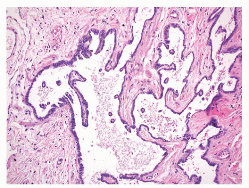
Cystic Transformation of the Rete Testis (Synonym: Also Called Giant Cystic Degeneration of the Rete Testis)

Nistal et al.138 studied 1,798 autopsy and 518 surgical specimens from the testis and epididymis, and identified cystic transformation of the rete testis in 20 autopsy and 18 surgical pathology specimens.138 The cystic transformation was thought to be due to several different etiologies. One cause of this lesion is mechanical compression of the extratesticular excretory ducts, resulting from neoplasia or other masses such as hematoceles. Cystic transformation may also follow postvasectomy inflammation or it may be related to infection such as epididymitis. An ischemic etiology has also been proposed in elderly men who have atherosclerosis in the epididymal branch of the testicular artery with associated atrophy of the head of the epididymis. Hormonal abnormalities may cause cystic transformation of the rete testis in patients with cirrhosis; these patients have increased peripheral conversion of androgen to estrogen and develop bilateral epididymal atrophy and columnar transformation of the rete testis epithelium. One case of giant cystic degeneration of the rete testis has been reported recently in an elderly adult man treated with LHRH for prostate adenocarcinoma.
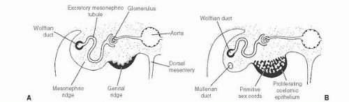
139 The patient age, the postulated effect to antiandrogen therapy, and the 10-cm size of the lesion are all unusual for this lesion. Malformations, such as cryptorchidism, that have dissociation between the rete testis (derived from the sex cords) and the epididymis (derived from the mesonephric duct) may also result in cystic transformation of the rete testis (Box 13-2).


Acquired Cysts of the Rete Testis

Acquired cysts of the rete testis have been reported in patients on hemodialysis, who may have oxalate crystals in the cyst lumens140 (Box 13-2). Benign rete testis cysts are lined by a single layer of flat or columnar epithelial cells.


Mesothelial Cysts

Mesothelial-lined cysts of the paratesticular area are rare and may involve the tunica albuginea, tunica vaginalis, epididymis, or spermatic cord.18,141, 142, 143, 144 They usually occur in men over 40 years of age. They may either be asymptomatic or patients may present with a painful mass simulating testicular tumors. These cysts may be single or multiple, but they are usually unilateral and located on the anterolateral surface of the testis.144 They measure from 0.3 to 0.4 cm in diameter and contain serous or blood-tinged fluid. The cysts are lined by single-layered, non-atypical, flattened, or cuboidal mesothelial cells. The epithelium is surrounded by hyalinized connective tissue. Squamous metaplasia may be present. The cysts are surrounded by fibrous tissue. They are benign lesions that should be treated conservatively.


Cysts of the Epididymis

Small benign cysts of the epididymis are common. They measure 1 to 2 cm in diameter. Pain and torsion may occur with larger cysts.145 Polycystic kidney disease has been associated with multiple epididymal cysts.146 Bilateral epididymal cysts have been described in a patient with von Hippel-Lindau disease and early-onset renal cell carcinoma.147 Epididymal cysts have also been reported in 10% of men exposed to DES in utero148 (Box 13-2).

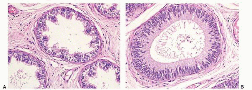
Epididymal cysts usually occur in the head of the epididymis. They are thought to arise from efferent ductules.
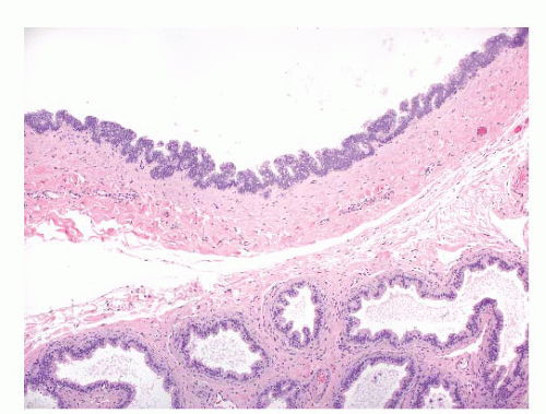
21 They may be unilocular or multilocular, and they are lined by a single layer of flat to cuboidal epithelial cells that may display variable numbers of cilia (Fig. 13-12). Cytologic atypia is not a feature of epididymal cysts. If spermatozoa are present in the cysts, the lesions are called spermatoceles. Small papillary structures containing connective tissue cores lined by a single layer of bland epithelium may protrude into the cysts.149


FIGURE 13-12 ▪ Cyst of epididymis. A dilated, cystic efferent duct is present adjacent to normal ducts.


Spermatocele

Spermatoceles result from cystic dilation of the efferent ductules, the tubules of the rete testis, or the aberrant ducts. Cysts may become large, with thin walls. Cloudy fluid present in spermatoceles is a result of sperm present in the cystic spaces. The fibromuscular walls are lined by cuboidal to columnar, sometimes ciliated, epithelium. The epithelial lining of long-standing spermatocele may be quite attenuated and may resemble a mesothelial or simple squamous layer.18 Sometimes they contain papillary proliferations lined by benign epithelial cells.149 Calcification may occur.150 Clusters of small blue cells have been reported in spermatocele and hydrocele specimens. They may represent sloughed rete testis epithelium and they may mimic the appearance of small cell carcinoma.151 Bland nuclei, lack of mitotic figures, and lack of staining with neuroendocrine markers are features indicative of benign epithelium.


Dermoid and Epidermoid Cysts

Epidermoid cysts are rare in the paratestis.152 They present either incidentally during hernia repair procedures or as painless enlarging masses located in the spermatic cord. They are lined by keratinizing squamous epithelium and contain intracystic keratinous debris. Dermoid cysts have also been reported in the spermatic cord.153,154 They contain skin appendage structures in addition to squamous epithelium.

Most epidermoid and dermoid cysts are considered benign neoplasms, in contrast to teratomas. Some epidermoid cysts may represent a metaplastic process derived from mesothelium. The possibility of extension or metastasis from a tumor in the testis should be excluded before making this diagnosis.


Hydrocele (Acquired)

A hydrocele is a fluid accumulation between the visceral and parietal layers of the tunica vaginalis. Congenital hydroceles have been discussed above (congenital abnormalities). Acquired hydroceles may be idiopathic or secondary to infections or neoplasms (Box 13-2). They represent the most common cause of painless scrotal swelling. They form over time, and neoplasm should be excluded in the case of a rapid formation of a hydrocele. Hydroceles usually transilluminate, except in cases where the tunica vaginalis is thickened. Ultrasound shows an anechoic fluid collection. The pathogenesis of acquired hydroceles reflects an imbalance between fluid secretion and resorption in the tunica vaginalis.155 Defective lymphatic drainage has been suggested as a cause of hydroceles.
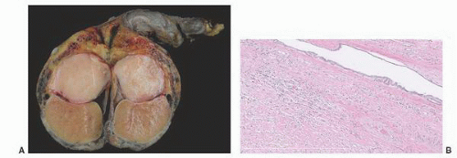
156


Chronic hydroceles may become inflamed, with a proliferation of fibrous tissue. The testis may become adherent to the parietal tunica vaginalis. These features may mimic neoplasms clinically.34,62,63 The resulting mass-like lesions may be more than twice the normal size of the normal testis, but they are diffuse and symmetrical.34 Patients in one series of nine patients ranged from 22 to 88 years of age.63 Most patients had hydroceles that were excised because of the clinical similarity to neoplasms.63 Microscopic examination showed features typical of a reactive process, including mesothelial hyperplasia with fibrosis and some chronic inflammation.63 Organizing hemorrhage in a hydrocele may also simulate a neoplasm until it is examined microscopically.


Hematocele

Hematoceles represent collections of blood in the tunica vaginalis. They may be acute or chronic, and they may have a mass effect. Causes include trauma, torsion, tumor, and surgery (Box 13-2). Varicoceles may be present, and minor trauma may cause rupture of one of the dilated blood vessels.69 Patients present with a mass and scrotal pain. Most hematoceles resolve spontaneously with conservative therapy, but some may become fibrotic and calcified.157


Endometriosis

Endometriosis has been reported in an elderly man who presented with a mass in the tail of the epididymis on a follow-up examination after taking diethylstilbestrol for 3 years for prostate carcinoma. On gross examination, a 5-cm mass was present between the vas deferens and the tail of the epididymis. Microscopic examination showed tubular glands lined by columnar to cuboidal epithelium without atypia. The glands were surrounded by stroma typical of that normally seen in the endometrium.


Prostatic-Type Glands

Prostatic-type glands have been found rarely in the epididymis.158 One example was found incidentally in a grossly normal epididymis in a 30-year-old man.158 The glands displayed the two cell layers normally seen in prostate glands. While these glands were different from the epididymal gland epithelium, there was a focal transition from epididymal ducts to the prostatic epithelium. The prostate glands stained positively for prostatic acid phosphatase (PAP) and prostatespecific antigen (PSA), but some epididymal duct tissue was also positive with these markers. The lesion probably represents prostatic metaplasia in the epididymis rather than true ectopic prostate glands.158,159


Metanephric Dysplastic Hamartoma

There is one report of metanephric dysplastic hamartoma presenting as an epididymal mass in an 18-month-old boy. Blastema associated with papillae, glomeruloid structures, and dysplastic tubules were seen on microscopic examination.5 This lesion must be distinguished from the rare Wilms tumors that have been reported in the paratesticular region.


Rosai-Dorfman Disease

A few cases of Rosai-Dorfman disease (sinus histiocytosis with massive lymphadenopathy) have been described in the epididymis.160 Patients may be either children or adults, and lymph nodes are also often involved.
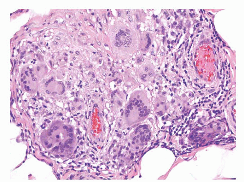
5 This process may result in nodules, or it may diffusely involve the epididymis. Microscopic examination shows sheets of histiocytes that contain oval nuclei and abundant pale cytoplasm with poorly defined cell borders. The histiocytes may contain phagocytosed lymphocytes, plasma cells, neutrophils, and erythrocytes.


Calcification, Bone, and Cartilage

An autopsy study of testes and associated rete testes showed a rare lesion called “nodular proliferation of calcifying connective tissue in the rete testis”161 in three men with histories of myocardial infarction. Polypoid projections in the rete spaces had connective tissue cores with calcification and were covered by benign, flattened epithelial cells.

Calcification may cause firm nodules in the epididymis, usually associated with sperm granuloma, filarial infection, or tuberculosis.162 Calcification of the epididymis may also be seen in patients on hemodialysis.163 Two cases of heterotopic bone trabeculae in the epididymis have been reported.18,164 Cartilage has also been seen in the epididymis of infants.165

Calcification of the seminal vesicles and vasa deferentia may indicate systemic disease.166 This lesion may be associated with diabetes, uremia with secondary hyperparathyroidism, prostatitis, infections (tuberculosis, schistosomiasis, gonorrhoea), and congenital abnormalities. Bilateral calcification of the vas deferens has been reported in a hemodialysis patient167 and, rarely, the condition is idiopathic.


Amyloidosis

One histochemical and ultrastructural study of six men with secondary amyloidosis showed amyloid deposits in the walls of the blood vessels of the epididymis and spermatic cord.168 Localized amyloidosis involving vasa deferentia has been described in two patients with prostate carcinoma.169


Inflammatory Myofibroblastic Tumor (Synonyms: Also Called Inflammatory Pseudotumor, Pseudosarcomatous Myofibroblastic Proliferation, and Proliferative Funiculitis)

Inflammatory myofibroblastic tumor and related lesions in the paratestis occur most often in the spermatic cord,62,170, 171, 172, 173, 174, 175, 176 but isolated cases have also been reported in the rete testis
and epididymis.171,172 They may present as mass lesions, or they may be incidental findings in inguinal hernia specimens. Some of these lesions in the epididymis may be a reaction to torsion and chronic ischemia.170,176


FIGURE 13-13 ▪ Inflammatory myofibroblastic tumor. A: Gross photograph showing circumscribed paratesticular tan tumor-like nodule; B: Note dense fibroblastic connective tissue and chronic inflammation adjacent to mesothelial lined space. Sclerosis is often present in long-standing cases.

These lesions are usually gray or tan, firm nodules that are <3 cm in size, although an occasional lesion has measured 7 cm.
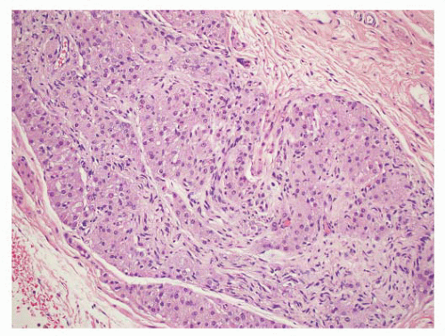
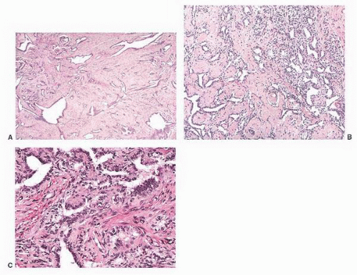
5 Some are well circumscribed (
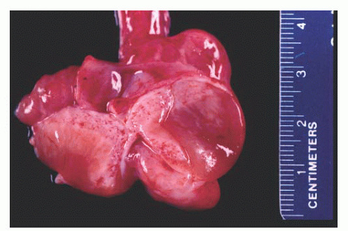
Fig. 13-13), but many are poorly demarcated. Areas of hemorrhage or cystic change may occur rarely. Inflammatory myofibroblastic tumors are composed of tapering spindle to stellate-shaped cells with vesicular nuclei and eosinophilic cytoplasm. The stroma is fibrous or myxoid. Cellularity varies in different parts of the lesions and may be greater in the central portion of the lesion. Inflammatory and giant cells may cause the lesion to resemble fasciitis seen elsewhere in the body.5 Hyalinized fibrous tissue characteristically surrounds blood vessels. The mitotic rate is usually low, although torsion has been associated with greater numbers of mitotic figures.176 Atypical mitotic figures are not present. It is possible that these lesions may progress to the densely collagenous and hyalinized “fibrous pseudotumors.”5 Immunohistochemical stains performed on inflammatory myofibroblastic tumors show cells will be of myofibroblastic phenotype; stains may be positive for actins, vimentin, desmin, and sometimes keratin,34 although these stains are not usually necessary for diagnosis of this lesion.

The differential diagnosis of inflammatory pseudotumors includes rhabdomyosarcoma, leiomyosarcoma, sclerosing liposarcoma, malignant fibrous histiocytoma, and spindle cell mesothelioma. The bland cytologic features, low mitotic rate, lack of atypical mitoses, and overall resemblance to fasciitis are features that are helpful in recognizing the reactive nature of this lesion and distinguishing it from malignant neoplasms.


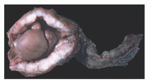
Fibrous Pseudotumor (Synonyms: Called Fibromatous Periorchitis, Nodular Periorchitis Additional Synonyms: Chronic Periorchitis, Proliferative Funiculitis, Fibrous Proliferation of Tunics, Fibroma, Nonspecific Testicular Fibrosis, Nodular Fibrous Periorchitis, Nodular Fibrous Pseudotumor, Inflammatory Pseudotumor, Reactive Periorchitis, Pseudofibromatous Periorchitis)

Paratesticular fibrous pseudotumors present as mass lesions. They are most often seen in the third decade of life,177,178 but one case has been reported in a 5-year-old boy.179 One patient had retroperitoneal fibrosis, one had Gorlin syndrome,180 and one lesion was associated with testicular infarction.
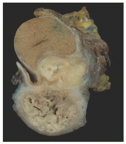
181 After adenomatoid tumors, these lesions represent the most common cause of masses in the paratestis. Patients have single or multiple nodules or plaques in the tunica vaginalis,182,183 epididymis,184,185 or spermatic cord, and the nodules range from 0.5 to
8 cm177 (
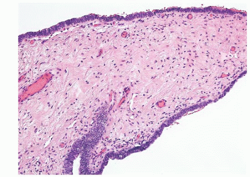
Fig. 13-14). In a diffuse form, dense fibrous tissue involves the tunica vaginalis.


FIGURE 13-14 ▪ Fibroma of the tunica vaginalis. Gross photograph showing extensive involvement of tunica vaginalis by white nodular fibrous tissue.

Microscopically, this lesion is characterized by dense, hyalinized, fibrous tissue often containing lymphocytes and plasma cells with occasional germinal centers.186 Calcification and ossification may occur.187 Three histologic types of fibrous pseudotumor have been recently described,188 although this subclassification is mainly of academic interest. Plaque-like lesions have dense fibrous stroma without significant inflammation, inflammatory sclerotic pseudotumors contain dense fibrous tissue with significant inflammation and myofibroblastic lesions have reactive appearing cells with numerous capillaries and sparse chronic inflammation.188 Paratesticular fibrous pseudotumor does not display significant nuclear atypia, mitotic activity, or necrosis. The differential diagnosis of paratesticular fibrous pseudotumor includes solitary fibrous tumor, leiomyoma, fibromatosis, spindle cell mesothelioma, and neurofibroma.188

Immunohistochemical stains performed on paratesticular fibrous pseudotumors display positive staining for smooth muscle actin in more than 80% of cases.188 Surprisingly, stains for cytokeratin, calretinin, and CD34 are positive in about half of the cases.188 All cases in a recent study were negative for B-catenin and ALK-1, and these lesions have a very low proliferation index as measured by Ki-67 staining.188

Potential cells of origin include myofibroblasts and submesothelial stromal cells, but paratesticular fibrous pseudotumors may be a nonspecific pattern resulting from a variety of pathogenetic processes.188 Past histories of trauma, surgery, infection, and inflamed hydroceles in some patients suggest a reactive etiology,63,189 although some patients have no such prior events. Some fibrous pseudotumors are thought to represent an advanced stage of inflammatory pseudotumors.149,177,190 Paratesticular fibrous pseudotumors are benign lesions, and local excision is curative.


“Tumor” of the Adrenogenital Syndrome and Related Lesions

This lesion develops in the paratesticular region of men with the adrenogenital syndrome who are not adequately treated, especially men with the salt-losing form of the syndrome. Nodules of steroid-type cells may occur in the epididymis, the spermatic cord, or the tunica albuginea.38,191, 192, 193 The paratesticular lesions may either extend from testicular nodules or be separate dark brown nodules measuring up to 1.5 cm in diameter.38 Sometimes, fibrous septa are present in the nodules. The microscopic appearance of these lesions is quite characteristic. Paratesticular nodules of steroid type cells are present. Bands of fibrous tissue and focal nuclear atypia are also seen.

Five cases of rete testis-associated nodular steroid cell nests associated with the rete testis have recently been reported.194 They are discrete conspicuous, unencapsulated nodules with vascular sinusoids between nests or cords of cells. They display strong melan A immunostaining, absent-to-weak inhibin staining and absent-to-moderate calretinin expression, in contrast to interstitial Leydig cells and testicular adnexal Leydig cells (TTAGS). The cells in rete testis-associated nodular steroid nests have been postulated to represent the precursor of testicular tumors of the adrenogenital syndrome.194

Patients with Nelson syndrome have ACTH-secreting pituitary adenomas following bilateral adrenalectomy for Cushing syndrome. These patients may also develop paratesticular steroid cell nodules.39,195,196 All reported cases have been bilateral, with nodules ranging up to 5.5 cm in diameter.195,196 They may produce cortisol and result in recurrence of Cushing syndrome. Gross and microscopic features are similar to those seen in patients with the adrenogenital syndrome. Bilateral small steroid cell nodules have also been described in children with Cushing syndrome and nodular hyperplasia of the adrenal cortex.38,197 They also have the same morphologic characteristics as the nodules associated with the adrenogenital syndrome.

Some masses of paratesticular steroid cells may also develop by stimulation of ectopic adrenocortical tissue found in about 10% of infants and some older patients.5,34,35,198,199


Hyperplasias


Hyperplasia of the Rete Testis (Synonym: Also Called Adenomatous Hyperplasia)

Hyperplasia of the rete testis includes both epithelial hyperplasia and smooth muscle hyperplasia. Epithelial hyperplasia may be real or apparent, so-called rete testis prominence.200

The cause of rete testis hyperplasia is not clear, but it may reflect hormonal factors including estrogen effect.201,202 Mice exposed in utero to DES have developed rete hyperplasia.203 Cryptorchidism, thought to be related to abnormalities of the hypothalamic-pituitary-testicular hormonal axis, is associated with both testicular atrophy and rete testis hyperplasia.204 One reported patient had been treated with DES for 15 months and androgen blockade for 19 months before diagnosis of rete testis hyperplasia.201 Increased numbers and stratification of rete epithelial cells has also been described after estrogen therapy in male-to-female transsexual patients.202 Columnar change of the rete epithelium has been associated with chronic liver failure,202 further supporting a hormonal basis for hyperplasia of the rete testis. Rete testis hyperplasia may also be associated with testicular atrophy and hypospermatogenesis.13,18,201,205,206 There is no clear dividing line between prominent rete testis associated with testicular atrophy and true rete hyperplasia. Rete hyperplasia may also be associated with epididymal cribriform change.13 It has also been described in a child with bilateral renal dysplasia207 and in patients with carcinoma of the prostate208 and breast.205

Some authors have proposed separating hyperplasia of the rete testis into two types: congenital and acquired.209 Lesions associated with cryptorchid testes, as well as some that are associated with testicular germ cell tumors, are included in the congenital group. Cases related to chemical
agents, some hormonal changes such as androgen blockage, and most lesions related to testicular germ cell tumors are considered acquired hyperplasias.


FIGURE 13-15 ▪ Adenomatous hyperplasia of rete testis. Complex, interconnecting proliferation of tubular channels with and without cystic dilation, low power (A, B). The lining cells are cuboidal to low columnar with innocuous cytology (C). (Reprinted from Amin MB. Selected other problematic testicular and paratesticular lesions: rete testis neoplasms and pseudotumors, mesothelial lesions and secondary tumors. Mod Pathol 2005;18:S131-S145, with permission.)

Nine patients in one study of rete testis hyperplasia201 ranged in age from 30 to 74 years of age (mean 59 years). Three patients presented with a clinically identifiable solid or cystic testicular hilar mass; however, the lesion is more likely to be an incidental finding. Rete testis hyperplasia may be multinodular and/or bilateral.

Two histologic types of rete testis hyperplasia have been described. Adenomatous hyperplasia (Fig. 13-15) displays a complex focal or diffuse interconnecting labyrinth of tubulopapillary channels that may be cystically dilated.200 Gland lumina may be empty or they may contain either sperm or eosinophilic secretions. A second type of rete testis hyperplasia represents an incidental finding in patients with testicular germ cell tumors. This lesion is characterized by a focal or diffuse epithelial proliferation within dilated rete testis spaces (Fig. 13-16). Hyaline globules within the hyperplastic rete epithelium may simulate the appearance of yolk sac tumor. Cells of rete hyperplasia are cuboidal, with pale, eosinophilic cytoplasm and round to oval, uniform nuclei. There is no cytologic atypia, necrosis, or mitotic activity. Epithelial cells stain positively for cytokeratin and epithelial membrane antigen, and the mesenchyme between the hyperplastic tubules displays variable staining for vimentin, muscle-specific actin, desmin, and S-100 protein.201 Ultrastructural examination of the epithelial cells shows intracellular junctions and complex interdigitation of cell membranes.201 Important indicators of the benign, reactive nature of this lesion are overall conformation to the normal branching tubular architecture of the rete testis and its continuity with nonhyperplastic rete testicular tubules.

Rete testis hyperplasia may resemble a primary or metastatic carcinoma.5,18,21,201,205,208 PSA and PAP stains may be useful in distinguishing rete hyperplasia from prostatic carcinoma.206

Only gold members can continue reading. Log In or Register to continue

Stay updated, free articles. Join our Telegram channel

Jun 10, 2016 | Posted by in UROLOGY | Comments Off on Pathology of the Paratesticular Region

Full access? Get Clinical Tree

Get Clinical Tree app for offline access