Fig. 10.1
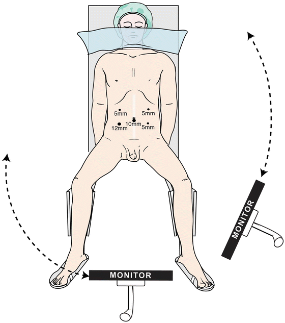
Patient positioning. With permission from Nakajima K, Milsom JW, Böhm B. Patient Preparation and Operating Room Setup. In: Milsom JW, Böhm B, Nakajima K, eds. Laparoscopic Colorectal Surgery, 2nd ed. Springer, New York 2006
Operative Technique (Video 10.1)
The author routinely utilizes the five following ports: one for the laparoscope, two for the operating surgeon, and two for the assistant. A 10-mm infraumbilical port is initially placed using an open technique. A balloon port—which creates an airtight seal—is preferred, as this port is mostly used for the camera. Five-millimeter ports are then placed in the right upper, left upper, and left lower quadrants, under direct laparoscopic vision. A 12-mm port is placed in the right lower quadrant and ultimately utilized for endoscopic stapling. A fascial suture (#0 Vicryl) is placed at the 12-mm stapling port site, using a suture passing technique for fascial closure at the end of the case. All ports are placed at least 8 cm apart on each side to prevent the instrument shafts from crossing each other. I generally use three monitors; however, two monitors are sufficient. One monitor is placed at the patient’s left leg and can swing to the left shoulder during splenic flexure mobilization; the other monitor is placed on the right and can be maneuvered between the patient’s legs for pelvic dissection (Fig. 10.2).


Fig. 10.2
One monitor is placed at the patient’s left leg and can swing to the left shoulder during splenic flexure mobilization; the other monitor is placed on the right and can be maneuvered between the patient’s legs for pelvic dissection. With permission from Memorial Sloan Kettering Cancer Center
Surgeon, Assistant, and Nurse Positioning
Depending on the operative step, all team members will stand in varying positions. It is important to adapt the position based on the patient’s body habitus and the particular step of the operation, to maximize ergonomics. For dissection of the mesocolon and mobilization of the splenic flexure, the surgeon and second assistant (camera person) stand on the patient’s right side, and the first assistant stands on the patient’s left. The nurse is positioned at the patient’s left leg. For mesocolon dissection, the surgeon views the monitor positioned near the patient’s leg. For splenic flexure mobilization, the second assistant moves between the patient’s legs; all team members look at the left-sided monitor, which is repositioned to the patient’s left shoulder. For pelvic dissection, the surgeon and second assistant stand on the patient’s right side, and the first assistant stands on the left side. All team members view the monitor between the patient’s legs.
Dissection of the Mesocolon and Vascular Pedicle
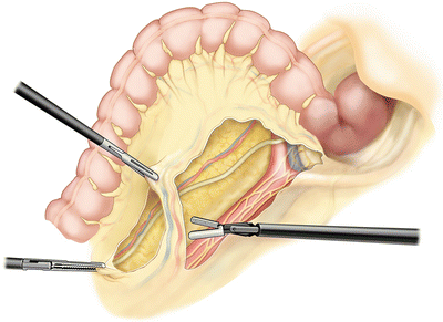
Following initial inspection of the peritoneal cavity, including liver, omentum, and pelvis, the patient is positioned in Trendelenburg for dissection of the mesocolon and vascular pedicle. The omentum is placed superiorly above the colon and onto the liver. The patient is tilted right-side down; this allows placement of the small intestine in the right upper quadrant, out of the area of dissection. Using 5-mm bowel graspers through the left-sided cannulas, the assistant holds the sigmoid ventrally under traction and to the left. In a medial to lateral fashion, the inferior mesenteric artery (IMA) is identified and the retroperitoneum is incised, starting at the sacral promontory to the right of (i.e., under) the vessel. Dissection is continued in a cephalad direction to the base of the IMA. Using gentle blunt dissection, the mesentery is dissected off the retroperitoneum. Dissection proceeds adjacent to and just below the IMA, which is swept ventrally, to ensure that the preaortic hypogastric sympathetic nerves are preserved and swept dorsally. Dissection beneath the mesentery is continued laterally, until the left ureter and gonadal vessels are identified and swept posteriorly. Once the origin of the IMA is identified, the peritoneum is incised anteriorly over the pedicle and away from the left colic artery. A peritoneal window is made just lateral to the inferior mesenteric vein (IMV). This permits ligation of the IMA and IMV pedicle, generally distal to the left colic artery (Fig. 10.3). We employ a bipolar device for vascular ligation, but occasionally use endoscopic staplers or clips. Care is taken to revisualize the left ureter before ligation and division of the IMA and IMV.


Fig. 10.3
A peritoneal window is made just lateral to the inferior mesenteric vein (IMV), permitting ligation of the IMA and IMV pedicle. With permission from Memorial Sloan Kettering Cancer Center
Splenic Flexure and Left Colon Mobilization
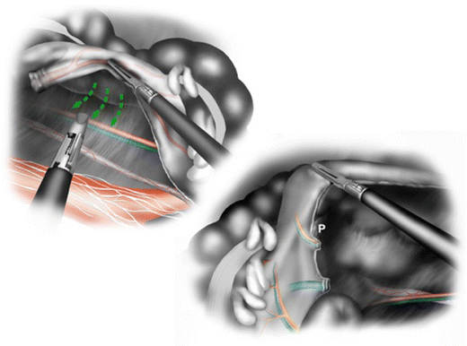
The second phase of the procedure is left colon mobilization. Through the peritoneal window, the left mesocolon is bluntly dissected from the underlying retroperitoneal structures, including the gonadal vessels, ureter, Gerota’s fascia, and pancreas (Fig. 10.4). If splenic flexure mobilization is necessary, the submesenteric dissection is continued until the spleen is visible and the lesser sac is entered. This can also be performed in a medial to lateral fashion by dissection just under the IMV adjacent to the ligament of Treitz and the pancreas. The IMV is divided adjacent to the pancreas, before it joins the splenic vein to form the portal vein. This enables full mobilization of the left colon and mesentery. The greater omentum is then freed from the transverse colon edge toward the midline, as far as necessary, to allow the descending colon to reach to the pelvis. The left colon is then mobilized by sharply dividing the lateral peritoneal attachments along the white line of Toldt (Fig. 10.5).


Fig. 10.4


Medial dissection. (Top): an avascular plane exits between Toldt’s fascia and the mesocolon, which is briefly dissected medial to lateral after IMA and IMV ligation. (Bottom): medial to lateral dissection beneath the left mesocolon provides excellent views of the distal pancreas (P), the base of the left transverse mesocolon and retroperitoneum. With permission from: Leroy J, Henri M, Rubino F, Marescaux J. Sigmoidectomy. In: Milsom JW, Böhm B, Nakajima K, eds. Laparoscopic Colorectal Surgery, 2nd ed. Springer, New York 2006
Stay updated, free articles. Join our Telegram channel

Full access? Get Clinical Tree


