Mechanisms of Immune Glomerular Injury


Mechanisms of Immune Glomerular Injury


William G. Couser


INTRODUCTION

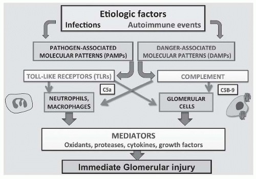
This chapter reviews the mechanisms of tissue injury that result in immune glomerular diseases, including glomerulonephritis and nephrotic syndrome (when presenting as a primary glomerular disease). The chapter is organized by individual diseases rather than by mechanisms to allow readers to appreciate more readily how the processes of tissue injury described here translate into the clinical disease entities encountered by clinicians and pathologists and covered in other chapters in this section. The mechanisms described derive from several decades of studies done at the molecular and cell culture levels in vitro as well as in an array of wellcharacterized animal models of glomerular diseases and in man. Cell cultures are not glomeruli or kidneys, and mice and rats are not humans, but years of experience have taught us that mechanisms defined in these experimental settings can be translated into an improved understanding of the very similar processes seen in human disease. For more information, the reader is referred to other reviews of the immune mechanisms that lead to glomerular disease.1,2 A schematic overview of the major pathogenic sequences currently believed to be operative in human glomerulonephritis (GN), and their interactions, is presented in Figure 45.1.


BASIC IMMUNE MECHANISMS: AN OVERVIEW


The Innate Immune Response (Figure 45.1)

For many decades, studies of the pathogenesis of human GN have focused on the adaptive immune system involving antibodies and T cells. However, more recently, increased attention has been given to the more ancient and well-conserved elements of the innate immune response, which occurs immediately after an initiating event without the latent period involved in the processing of antigen, antigen presentation, and specific immune responses.3 The innate immune system has two major arms: Toll-like receptors (TLRs) and complement, both of which may be involved in adaptive immunity as well.

TLRs are ancient and ubiquitous pattern recognition receptors present on all cell membranes and intracellularly between cytoplasm and endosomes.3 Over 10 TLR isoforms have been characterized that recognize conserved molecular patterns like peptidoglycans, lipopolysaccharides, and bacterial and viral nucleic acids (pathogen-associated molecular patterns [PAMPs]). TLRs also respond to certain endogenous cell-derived patterns (danger-associated molecular patterns [DAMPs]). Another related cytoplasmic group of receptors called Nod-like receptors (NLR) has recently been described as well.4 TLR ligation is central to activating the non-antigen-specific innate immune system in the immediate response to pathogens, but TLR activation is also required for adaptive, antigen-specific immune responses by facilitating conversion of dendritic cells to antigen-presenting cells.3,4 TLRs activate multiple intracellular signaling pathways, primarily via nuclear factor kappa B (NF-κB), that lead to the local release of a variety of cytokines, chemokines, and other inflammatory mediators by all cells, including inflammatory cells like neutrophils and macrophages, as well as all three resident glomerular cells (Fig. 45.1).3,4 Thus, TLRs and NLRs connect initiating pathogenic events like infection with a sequence of processes leading to immediate non-antigen-specific inflammation and tissue injury in GN that is associated with infections or autoimmunity or both (Fig. 45.1).


The Complement System (Figures 45.1 and 45.2)

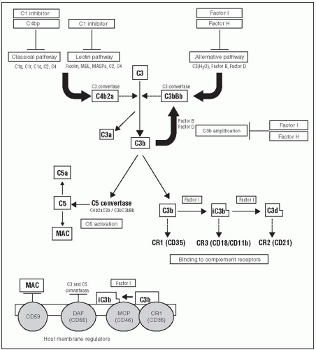
The complement (C) system and its regulatory proteins are also ancient components of the innate immune system with multiple roles in human GN (Fig. 45.2).5,6 C activation products are the principal mediators of antibody-induced GN. Nonimmunoglobulin zymogens, such as damaged cells and bacterial and viral proteins, can also activate C. C1q binding to immunoglobulins in the form of antigen-antibody complexes leads to classical pathway activation through C1, C4, and C2. Activation of the mannose-binding lectin (MBL) pathway is usually a consequence of microbial pathogens or galactosyl immunoglobulin G (IgG) binding to circulating MBL and proceeds through C4. The alternative C pathway (AP) is activated at low levels spontaneously, as well as by
foreign surfaces such as some microbial products and damaged cells. AP activation begins at C3 without involving C1, C4, or C2. In individual complement-mediated diseases, several of these pathways may be involved.5,6 Among the immunoglobulins, IgG subclasses IgG1 and IgG3 and IgM are classical C pathway activators, whereas IgG2 and IgG4 and normally glycosylated IgA activate C poorly.7 However, C activation and its sequelae need not involve immunoglobulin deposits and may occur by other mechanisms even in the presence of immunoglobulin deposits. All C activation pathways lead to cleavage of C5 and release of chemotactic factors such as C5a that attract inflammatory cells (neutrophils, macrophages, platelets) when activation occurs within, or adjacent to, the circulatory compartment. Cleavage of C5 by C5 convertases also leads to the release of C5b and the addition of C6, C7, C8, and multiple C9 molecules to form the lipophilic terminal membrane attack complex (MAC or C5b-9) (Fig. 45.1).8,9 Sublytic quantities of C5b-9 can insert into lipid bilayers of adjacent glomerular cell membranes and act in a fashion similar to receptor agonists. Sublytic C5b-9 initiates several signaling pathways and thus converts endothelial cells, mesangial cells, and podocytes to local inflammatory effector cells that can proliferate; release a variety of cytokines, growth factors, eicosanoids, oxidants, proteases, and other acute inflammatory mediators; as well as upregulate genes that encode matrix components and contribute to chronic overproduction of the extracellular matrix with scarring and sclerosis.9 Immunoglobulin-induced C activation products like C5a can also activate TLRs, thus linking the innate and adaptive immune systems (Fig. 45.2).10 C activation in vivo is tightly regulated by a number of circulating and cell-bound C regulatory proteins (CRPs) the functions of which, particularly those of CR1, factor H, membrane cofactor protein (MCP), and CD59, are also important in the development of several glomerular diseases (Fig. 45.2).5,6


FIGURE 45.1 The innate immune system in glomerular disease. Etiologic factors, usually infections or autoimmune events, are presented to the immune system as pathogen-associated molecular patterns (PAMPs) or danger-associated molecular patterns (DAMPs). These interact with the two principal arms of the innate immune system, Toll-like receptors, and the complement system. Toll-like receptors are present on both circulating inflammatory cells like neutrophils and macrophages and on resident glomerular cells including endothelial and mesangial cells and podocytes. Toll-like receptor activation induces inflammation through release of multiple mediators including cytokines, growth factors, oxidants, proteases, eicosanoids, and others. Activation of complement also leads to the attraction of inflammatory cells through the generation of chemotactic factors such as C5a and to the conversion of resident glomerular cells to inflammatory cells following sublytic C5b-9 insertion into cell membranes. The mediators released by both infiltrating neutrophils and macrophages, and resident glomerular cells cause glomerular injury that leads to morphologic and functional changes in diseased glomeruli.


The Adaptive Immune Response


Immunoglobulins

CD4 T-helper cells, activated by antigen-presenting dendritic cells and macrophages, stimulate B cells and plasma cells to make antibodies specific for nephritogenic antigens. Antigenic peptides capable of inducing GN may represent only a few amino acids of much larger proteins. Based on older studies of serum sickness in rabbits induced by single (acute) or repeated (chronic) injections of bovine serum albumin (BSA), glomerular immune deposits have long been attributed to the passive trapping of circulating, soluble antigen-IgG antibody complexes (ICs).11,12 In acute serum sickness, a single exposure to BSA is followed by a latent period of 5 to 7 days and then the production of the IgG antibody. As antigen forms immune complexes and disappears from the circulation,

there is an initial phase of antigen excess leading to the formation of small, soluble immune complexes followed by equivalence with midrange size complexes that are believed to be trapped in tissues, and finally, to antibody excess with large complexes that are cleared through the mononuclear phagocyte system with eventual elimination of the antigen. Glomerular deposits of antigens and antibodies in acute serum sickness form primarily in the mesangium and in a subendothelial distribution. In chronic serum sickness, repeated antigen administration is provided to maintain slight antigen excess and is associated with more subepithelial deposits, again attributed to the passive glomerular trapping of small, preformed immune complexes that crossed the capillary wall to localize beneath podocytes.12,13 Attribution of the tissue injury that occurs in serum sickness to the passive trapping of circulating complexes followed from observations that inflammation occurred only during the period that complexes could be detected in the circulation, corresponded with appearance of immune deposits in glomeruli and that both the antigen and antibody components of the immune complexes were constituents of the deposits.13 However, later studies using passively administered preformed immune complexes did induce some glomerular deposits, but generally failed to replicate the tissue injury seen in either acute or chronic serum sickness.14 Moreover, later measurements of circulating immune complex levels in human disease found little correlation between circulating complex levels, sizes, and disease activity. Other studies done in antiglomerular antibody (nephrotoxic nephritis [NTN]) models rather than in serum sickness demonstrated that antibody deposits activated C and other mediators that attracted circulating inflammatory cells—primarily neutrophils—which then caused tissue injury.15 Some of these discrepancies were resolved in 1978 when it was shown that the classic subepithelial immune complex deposits in the Heymann models of membranous nephropathy (MN) in rats formed in situ and were unrelated to circulating immune complexes. Instead, they resulted from antibody binding to endogenous glomerular components localized on the podocyte (see Membranous Nephropathy, which follows).14,16,17,18 Subsequent studies in both acute and chronic serum sickness using cationic BSA confirmed that deposits in these models involving exogenous antigens also formed locally and were unrelated to circulating immune complex levels or sizes.19,20 This new paradigm allowed the mediation of immune complex nephritis to be studied directly rather than by being extrapolated from findings in models of anti-GBM disease.


FIGURE 45.2 A schematic depiction of complement pathways and how they are activated as they relate to the pathogenesis of glomerulonephritis (GN). Activation via the classical, lectin, and alternative pathways leads to the formation of C3 convertases that cleave C3 to C3a and C3b. C3b can interact with complement receptors on cell surfaces such as CR1, CR2, and CR3 as well as contribute to the formation of C5 convertase. Cleavage of C5 results in the formation of the chemotactic factor C5a and C5b, which leads to the formation of the C5b-9 membrane attack complex (C5b-9) that is important in the mediation of several glomerular diseases. Factors H and I are circulating regulators of the alternative complement pathway, whereas CD59, decay accelerating factor (DAF), membrane cofactor protein (MCP), and complementary regulatory 1 (CR1) serve as membrane-bound regulators that protect cells from complement attack. MBL, mannose-binding lectin; MASP, MBL-associated serum protease; MAC, membrane attack complex (C5b-9) (Reproduced with permission from Pickering M, Cook HT. Complement and glomerular disease: new insights. Curr Opin Nephrol Hypertens. 2011;20(3):271-277.)

The variables that determine biopsy findings and clinical consequences in immune complex GN include: (1) where deposits form in the glomerulus (ICs of the same composition that form in a subendothelial distribution lead to exudative inflammatory cell infiltrates, in the mesangium to mesangial cell [MC] proliferation and matrix expansion, and in a subepithelial distribution to a noninflammatory lesion with podocyte injury, foot process effacement, and heavy proteinuria)18,21,22; (2) the biologic properties of the antibody (or antigen) itself, especially the capacity to activate complement, the Fc receptor affinity, the ability to form lattices that are necessary for complement activation to occur, or cryoprecipitability1,23; (3) the mechanism of deposit formation (when ICs form in situ the process usually induces tissue injury at the site, whereas passive trapping of ICs formed in the circulation has not been shown to be nephritogenic)1,14,18; and (4) the quantity of immune deposits formed (the more deposits that form, the more severe the disease).


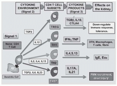
Although the existence of T cells sensitized to glomerular antigens was first demonstrated in glomerular basement membrane (GBM) disease over 40 years ago,24 experimental verification of the pathogenicity of T cells was delayed by several factors. The rapid expansion of immunopathology using fluoresceinated antibodies to visualize and characterize antibody deposits in human renal biopsies, the lack of good T-cell markers in both rodent models and in humans, and the conviction that all forms of GN were antibody mediated resulted in little research in this area for 2 decades.25 In 1984, the hypothesis that T cells could mediate GN independent of antibody was confirmed in ingenious experiments using bursectomized chickens that had no B cells, and later, in more conventional rodent models.26 In addition to providing help for B cells,28 antigen-specific CD4 T cells alone, sensitized to either self or nonself antigens that are localized in glomeruli, can induce antibody-independent tissue injury.27,28 All subsets of T cells are now implicated in GN, including dendritic antigen-presenting cells (DC) and CD4 helper cells of the Th1, Th2, and T regulatory cell (Treg) lineages. The best-established mechanism of T-cell mediated glomerular injury involves the recruitment of macrophages, which then act as the inflammatory effector cells. However, interleukin 17 (IL-17) producing Th17 cells have attracted the most attention recently and now seem likely to account for much of the T cell-induced inflammation that occurs in GN.29,30 Th17 cells are attracted by mechanisms involving chemokines and their receptors, and they release cytokines such as IL-9, IL-17, IL-21, IL-22, and tumor necrosis factor alpha (TNF-α),which induce other cells to produce additional proinflammatory chemokines that attract neutrophils and monocytes and also activate resident glomerular cells.29,30 Th17 cells have now been demonstrated in renal biopsies in several forms of human GN.31 The T-cell component of the adaptive immune response is regulated by CD4-derived Tregs (Fig. 45.3).26


DISEASES THAT USUALLY PRESENT AS GLOMERULONEPHRITIS


Postinfectious or Poststreptococcal Glomerulonephritis

The acute, diffuse exudative, and proliferative lesions of postinfectious or poststreptococcal glomerulonephritis (PSGN) were long regarded as the human equivalent of the acute
“one shot” serum sickness model in rabbits, leading to a prolonged search for the “nephritogenic” streptococcal antigen that has extended over several decades. Although many candidate proteins have been proposed, most have failed to meet strict criteria for causality such as being demonstrable in deposits, particularly subepithelial “humps,” inducing antibody that correlated with clinical disease, and inducing a similar disease in animal models.32 However, streptococcal pyogenic exotoxin B (SpeB) meets most of these criteria. SpeB is a small (28 kDa), cationic (pK 9.3) cysteine protease with C activating and plasmin-binding properties and represents 90% of the secreted extracellular protein made in vivo by nephritogenic strains of group A streptococci.32 Antibody to SpeB correlates with disease activity in PSGN and colocalizes with IgG and C3 in subepithelial “humps.”32,33 However, the intense exudative glomerular inflammatory response and subepithelial humps are not well explained by the analogy to acute serum sickness because intact circulating ICs do not form subepithelial IC deposits directly, and subepithelial IC deposits do not produce inflammation, presumably because complement activation products like C5a go directly into the urine and are not chemotactic for cells in the circulation.14,18 Moreover, IgG is sometimes absent, or only a minor constituent of the deposits, whereas C3 deposition has been reported to both precede and exceed detectable IgG.34,35 Several explanations for these apparent contradictions are plausible. They include observations that some subendothelial deposits are also present by electron microscopy (EM) in PSGN,34,35 perhaps because the antibody to SpeB exhibits molecular mimicry with endothelial cell antigens, and antiendothelial antibody deposits are generally rapidly cleared.36 In addition, SpeB alone is a zymogen that can activate C directly through the MBL pathway independent of IgG.32 SpeB also exhibits plasmin-binding properties that can facilitate C activation and might cause proteolysis of GBM and facilitate the transit of dissociated subendothelial ICs to form subepithelial humps.37 Finally, PSGN often exhibits autoimmune features including both IgM and IgG rheumatoid factors with cryoglobulin activity, antiendothelial antibodies, anti-DNA antibodies, and antineutrophil cytoplasmic antibodies (ANCA). Although the respective roles of these nonstreptococcal antibodies in mediating the disease, if any, remain undefined,
these findings suggest an autoimmune component to postinfectious GN that is consistent with current thinking about most other immune glomerular diseases (Table 45.1).38,39,40


FIGURE 45.3 The T-cell component of the adaptive immune system in glomerulonephritis (GN). Antigen is presented to naïve CD4 T cells by dendritic cells (Signal 1). Depending on the predominant cytokine environment, T cells differentiate into CD4 T-cell subsets that play different roles in the pathogenesis of glomerular disease. In the presence of transforming growth factor beta (TGF-β), T regulatory cells (Tregs) develop that make TGF-β, interleukin 10 (IL-10), and cytotoxic T-lymphocyte antigen 4 (CTLA-4) that downregulate and control the immune response. IL-12 stimulates differentiation into Th1 cells that make interferon gamma (IFNγ) and TNF and produce traditional T-cell/macrophage-mediated delayed-type hypersensitivity (DTH) reactions. IL-2, IL-4, and IL-13 favor the development of Th2 cells that make IL-4, IL-5, and IL-13, and lead to allergic-type hypersensitivity reactions involving immunoglobulin E (IgE) and eosinophils (Eos). The CD4 T cells most implicated in the pathogenesis of GN are Th17 cells that differentiate in the presence of TGFβ, IL-6, and especially IL-17, and those that produce IL-17a and IL-21 that facilitate recruitment of other inflammatory cells such as neutrophils (PMNs) and also cause tissue injury directly.


TABLE 45.1 The Most Common Complement Profiles and Autoimmune Features in Glomerulonephritis


Disease


Serum C Profile


Autoimmune Features


References


Poststreptococcal GN


AP, MBL; normal C1q, Low C3-C9


Anti-C1q, IgG AECA, anti-DNA, ANCA, PDI, cardiac myosin


37,42,45,46,47,48


IgA nephropathy


Normal, lectin pathway activation


Anti-glycan, mesangial cell


52,53


Anti-GBM nephritis


Normal, CP


Anti-GBM, ANCA (20%)


88,107,108


ANCA-positive GN


Normal, AP


Anti-MPO, PR3, cPR3, NET, DNA, endothelial cell, LAMP2


110,123,137,138,152


Lupus nephritis


CP, low C1q-C9


Anti-dsDNA, annexin, MPO, PR3, nucleosome, IgG, C1q, cardiolipin, MBL, NET


110,123,152


MPGN I


CP, low C1q-C9


Anti-C3 convertase (C3Nef),C4Nef, C1q IgM anti-IgG


MCD/FGS


Normal


None


Membranous nephropathy


Normal


Anti-PLA2R, DNA, NEP, aldose reductase, enolase SOD2


Dense deposit disease


AP, normal C1q, low C3-C9


C3Nef, C4Nef, anti-CFH, factor B, C1q


C3 nephropathy


AP, normal C1q, low C3-C9


C3Nef, anti-CFH


Most forms of GN exhibit major features of autoimmunity. GN, glomerulonephritis; AP, alternative pathway; MBL, mannose-binding lectin; IgG, immunoglobulin G; AECA, anti-endothelial cell antibodies; ANCA, antineutrophil cytoplasmic antibody; PDI, protein disulfide isomerase; anti-MPO, antimyeloperoxidase; PR3, proteinase 3; cPR3, complementary proteinase 3; NET, neutrophil extracellular trap; NEP, LAMP2, lysosomal membrane protein 2; C3Nef, C3 nephritic factor; CP, cofactor protein; MPGN, membranoproliferative glomerulonephritis; MCD/FGS, minimal change disease/focal glomerulosclerosis; anti-PLA2R, anti-phospholipase A2 receptor; SOD2, superoxide dismutase 2; anti-CFH, anti-complement factor H. NEP, neutral endopeptidase.


Other forms of postinfectious GN such as those associated with endocarditis, infected ventricular-atrial shunts, visceral abscesses, and Staphylococcus aureus infections with IgA deposits are clearly immunologically mediated, but the mechanisms involved in these diseases have been explored in much less detail.41


Immunoglobulin A Nephropathy

IgA nephropathy (IgAN) is the most common form of GN worldwide.42 IgAN is characterized by a focal proliferation of mesangial cells and mesangial matrix expansion accompanying diffuse mesangial aggregates of IgA, and often IgG, C3, and C5b-9.42,43,44 The disease is often associated with recurrent episodes of nephritis that immediately follow viral infections on mucosal surfaces of the upper respiratory or gastrointestinal tracts.42,43,44 Although usually assumed to be mediated by a mesangial trapping of circulating ICs, no exogenous antigens have been consistently identified. Animal models of IgAN that closely mimic both the pathologic, immunopathologic, and the clinical features of IgAN have been challenging to produce, in part because of substantial differences between the rodent and human IgA immune systems. IgA in mesangial deposits, and in IC form in the circulation, is polymeric (mucosal) IgA1 with a covalently linked secretory piece indicating a mucosal origin.45,46 In IgAN, a population of these IgA1 molecules exhibits deficient O-linked glycosylation at five sites in the hinge region of the molecule.42,43,44,45,46 The failure to normally glycosylate IgA1 can be inherited and is commonly seen in family members without renal disease,47 but the defect seems to occur epigenetically as well.48 Although underglycosylated pIgA1 is produced by mucosal B cells and is usually assumed to originate from mucosal surfaces where it should not enter
the bloodstream, it might also reach the circulation if abnormal trafficking of these cells to the bone marrow occurs.49 Underglycosylated IgA1 undergoes conformational change and exhibits altered biologic properties compared to normal IgA1, including increased tendencies to self-aggregate, to activate C, and to bind to other molecules like fibronectin, IgG, and collagen IV.45,46,50 In circulating macromolecular form, the underglycosylated IgA1 aggregates lack the glycosylated sites necessary to interact with asialoglycoprotein and the CD 89 receptors in the liver and spleen, thus evading normal clearing mechanisms and facilitating mesangial localization.45,46,51 It is not yet known if the “lanthanic” mesangial IgA deposits seen in 6% to 16% of normal donor kidneys without disease contain underglycosylated or normal IgA.42

Although IgG autoantibodies to MC antigens have been described in IgAN,52 Suzuki et al.53 were the first to report IgG antibodies directed to cryptic GalNac antigenic structures in the hinge region of aberrantly glycosylated IgA1 molecules (antiglycan antibodies). IgG antiglycan antibodies appear to correlate with disease activity in a way that has not been demonstrated with serum levels of IgA, IgA immune complexes, or IgA-fibronectin aggregates.53 Antiglycan antibodies form circulating ICs with underglycosylated IgA1 that can be passively trapped in the mesangium, although the mesangial trapping of ICs has not been demonstrated to be nephritogenic. Alternatively, IgG antiglycan antibodies could also lead to in situ IC formation with previously localized IgA aggregates. When IgG antibody does bind in situ to antigenic material in the mesangium54 or on the MC membrane (the antithymocyte serum [ATS] model in rats),56,57,58,59 the mesangial response to acute immune injury closely simulates the clinical and histopathologic features of human IgAN.55,60

In IgA nephropathy, MCs become activated through interactions between the IgA1 deposits and IgA Fca (CD89) receptors, TLRs (especially TLR4), and transferrin receptors (TfR, CD71).61,62 Innate immunity and TLR activation by IgA aggregates, perhaps containing or accompanied by PAMPs, may account for the recurrent episodes of acute injury with hematuria, particularly those that immediately follow infections.42,63 However, most experimental and clinical studies suggest a role for C in IgAN as well.5,6,63,64 C5b-9 generated from C activation induced by the interaction of IgA1 aggregates with MBL, or the in situ formation of ICs containing IgG antiglycan antibodies can induce MC transformation to a smooth muscle actin-expressing myofibroblastlike cells, upregulate genes for collagen I, and increase production of cytokines and growth factors such as IL-1, IL-6, TNF-α, platelet-derived growth factor (PDGF), transforming growth factor beta (TGF-β), epidermal growth factor (EGF), fibroblast growth factor (FGF), connective tissue growth factor (CTGF), and hepatocyte growth factor (HGF), all resulting in MC proliferation and matrix expansion.5,6,8,9 Of these, the best-established mediators of the glomerular response to immune injury are PDGF, which is the principal growth factor involved in the proliferation of MC that follows immune injury and the CTGF/TGF-β pathway that induces overproduction of mesangial matrix.65 The pattern of glomerular C deposition seen in active IgAN includes MBL, C4d, and C5b-9 (but not C1q) that colocalize with IgA1 and suggest the activation of both MBL and AP.66,67 Further evidence that C activation is important in IgAN includes observations that C deposits including MBL, C4d, and C5b-9 correlate with disease severity and prognosis.66,67,68


Rapidly Progressive, Crescentic Glomerulonephritis


Anti-Glomerular Basement Membrane Nephritis

Anti-GBM (aGBM) nephritis is characterized initially by an acute, focal necrotizing GN with crescents and linear deposition of IgG, usually with C3, on the GBM.67 With pulmonary alveolar hemorrhage it becomes Goodpasture syndrome (GPS). The role of aGBM antibody deposition inducing C activation, chemotactic factor release, and neutrophil-mediated injury was defined in NTN models in the 1960s,15 and the pathogenicity of human aGBM antibody was confirmed by the classic transfer studies of Lerner, Glassock, and Dixon69 in 1967. Studies in mice deficient in C3 and C4 primarily implicate the classical C pathway,56 which is activated by IgG1 and IgG3 aGBM that correlates with disease activity and recurrence in transplants.57,58,67 Circulating and deposited aGBM antibodies are of the same specificity, indicating that available antigens to bind antibodies is limited, and they are primarily of the IgG1 and IgG3 subclasses.58 Antibodies with apparently similar reactivity (but with lower titers, lower avidity, and primarily of the IgG2 and IgG4 subclasses) can be present in normal people.59

GBM antigens are also expressed in several extrarenal tissues, where they are sequestered by an endothelial cell layer impermeable to IgG.70,71 The unique fenestrated endothelium in glomeruli allows for free access of IgG to GBM. The GBM antigen itself consists of two normally sequestered (“cryptic”) epitopes, EA and EB, residing on the noncollagenous domain of both the a3 and a5 chains of the noncollagenous (NC)1 hexamer of type IV collagen and is synthesized in the glomerulus exclusively by podoctytes.70,71 Antibody directed to the a5 chain is associated with worse renal outcomes.71 Antibody deposition requires perturbation of the quaternary structure of the a3 45 NCI hexamer, possibly initiated by posttranslational modifications, proteolytic cleavage, or oxidant injury, which results in a conformational change in the a3 NC1 and a5 NCI subunits (“autoimmune conformeropathy”).70,71 In animal models, the nephritogenic GBM antigen has been mapped to as few as three amino acid sequences in a core residue,72 but both intermolecular and intramolecular epitope spreading occurs, suggesting immune reactivity may extend beyond the initial inducing autoantigen.72 Pulmonary toxins such as infections, smoke, and volatile hydrocarbons may damage the endothelium and expose the antigen in alveolar capillaries, thus accounting for the pulmonary manifestations in GPS.70,71 Whether such
extrarenal events have any role in autoimmunization is not known.

T-cell reactivity to GBM antigens was first demonstrated 4 decades ago, and a pathogenic role for GBM antigen-specific “sensitized cells “ was proposed,24 but the hypothesis was given little credence at the time.25 However, many subsequent studies have confirmed these original observations with newer technologies73 and documented that nephritogenic GBM antigens can induce a T-cell mediated GN with crescents, proteinuria, and decreased renal function in the absence of aGBM antibody.28,74,75 CD4 Th17 lymphocytes via the “IL-23/Th17 axis” have been shown to be central to the mediation of injury in aGBM models.31,76 Another unique feature of the T-cell response to GBM is the appearance of long-lived Tregs and inversion of the T-cell effector/regulatory cell ratio later in the disease. This may account for why recurrences of anti-GBM disease are so infrequent compared to other autoimmune GNs where Treg activity is often impaired.77

The aGBM immune response in humans is strongly linked to human leukocyte antigen (HLA) DRB1 alleles 1501, 0701, and 0101with DRB 1501 conferring a relative risk of over 8, whereas 0701 and 0101 are protective.78 This HLA linkage is the strongest yet identified in any autoimmune disease. Preceding infections or environmental toxins that might expose antigenic determinants in extrarenal tissue are possible triggering events. The disease can also be induced experimentally with a small nephritogenic T-cell epitope, pCol28,29

Only gold members can continue reading. Log In or Register to continue

Stay updated, free articles. Join our Telegram channel

May 29, 2016 | Posted by in NEPHROLOGY | Comments Off on Mechanisms of Immune Glomerular Injury

Full access? Get Clinical Tree

Get Clinical Tree app for offline access