Type of liver insult
Setting
Characteristic findings
Ischemic hepatic injury
Hypotension during surgery but may also occur with severe volume depletion and hepatic hypoperfusion at any time posttransplant
Marked but transient elevation of aminotransferases within 48–72 h, followed by mild transient cholestasis
DILI
One or multiple drugs with hepatotoxic potential or drug–drug interactions. Idiosyncratic or drug-dependent toxicity
Variable presentation, from asymptomatic elevation of liver enzymes to acute liver failure. Pattern of hepatic injury varies by drug (hepatocellular, cholestatic, mixed)
NAFLD
Metabolic syndrome, new-onset diabetes after transplantation
Mild elevation of aminotransferases, imaging studies or biopsy demonstrating fatty infiltration of the liver
De novo acute HBV or HCV infection
Traditional risk factors for infection with HBV and HCV: drug use, tattoos, unprotected sex, hemodialysis, transfusion of blood products
Marked elevation of aminotransferases with or without cholestasis. Positive HBc-IgM or HBV-DNA for acute HBV, HCV-RNA for acute HCV
Chronic HBV or HCV infection
HBV or HCV infection pre-transplant
Progression of liver disease with signs of hepatocellular dysfunction
CMV hepatitis
CMV-seropositive donor and CMV-seronegative recipient. Antirejection agents: antilymphocyte antibodies, muromonab, alemtuzumab
Wide spectrum of disease, from asymptomatic infection to acute and fulminant hepatitis. Positive PCR, CMV-pp65 antigenemia, immunohistochemistry
Approach to the Renal Transplant Recipient with Hepatic Dysfunction
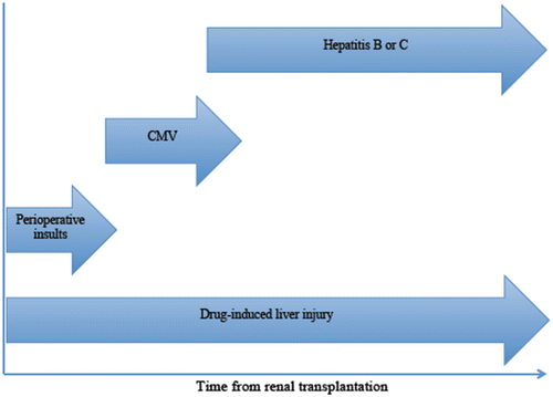
Evaluation of hepatic dysfunction in a renal transplant recipient is guided, in part, by its onset in relation to transplant, pattern, and duration (Fig. 23.1). In the immediate postoperative period, considerations include hepatic dysfunction due to intraoperative events. An abrupt rise in serum aminotransferases with a lesser elevation in bilirubin, alkaline phosphatase, and the international normalized ratio (INR) may reflect intraoperative hypotension and associated hepatic hypoperfusion, which can be confirmed by review of the operative records. If no additional insults contributed to hepatic dysfunction, prompt resolution can be anticipated. Currently favored anesthetic agents have a low rate of hepatotoxicity in contrast to halothane. Some anesthetics such as halothane undergo extensive hepatic metabolism, and in some susceptible individuals, immune-mediated hepatic injury may occur, which typically manifests with elevated serum aminotransferases within several days of surgery [4]. Although newer anesthetic agents are safer and less hepatotoxic than halothane, there have been case reports of significant hepatotoxicity associated with sevoflurane, desflurane, and isoflurane [5–7].


Fig. 23.1
Timeline of different liver diseases post-renal transplantation
The differential diagnosis for hepatic dysfunction shifts beyond the immediate postoperative period. Generally, immunosuppressive drugs are not particularly hepatotoxic, but freshly transplanted recipients may be prescribed other medications such as antibiotics that may cause DILI. Immunosuppressive regimens used in renal transplant recipients vary considerably by transplant center. The following agents are currently used in various combination regimens: glucocorticoids, azathioprine, mycophenolate mofetil, mycophenolic acid, cyclosporine, tacrolimus, everolimus, and sirolimus [8]. Most transplant centers favor combination of a calcineurin inhibitor (cyclosporine or tacrolimus), an antimetabolite (azathioprine, mycophenolate mofetil, or mycophenolic acid), and prednisone (for different lengths of time) [9]. Mild, and typically transient, elevations of serum aminotransferases are commonly seen in renal transplant recipients receiving calcineurin inhibitors such as cyclosporine or tacrolimus and often do not require further evaluation. However, a thorough workup including liver biopsy is indicated when persistent elevation occurs and no other plausible explanations for abnormal liver chemistries exist. If the postoperative course is complicated, for instance, by a wound infection, hepatic dysfunction may result due to the cholestatic effect of sepsis. In addition to surgical complications, therapeutic immunosuppression conveys a risk of opportunistic infections. The risk of CMV infection is greatest in CMV naïve recipients and those who receive additional immunosuppression for graft rejection. Hepatic involvement with hepatocellular dysfunction may be a feature of CMV disease [10]. Fibrosing cholestatic hepatitis, a severe and rapidly progressing form of liver injury typically resulting in devastating hepatic failure, described in individuals with hepatitis B or C, has also been reported in renal transplant recipients with CMV infection [11].
Elevations of serum aminotransferases and/or gamma-glutamyl transpeptidase may be due to ingestion of alcohol and/or hepatotoxic products including herbal remedies and health supplements; therefore, a detailed history is needed to elucidate consumption of these products in renal transplant recipients. The growing epidemic of obesity and the metabolic syndrome with associated detrimental effects on health and overall survival has not spared renal transplant candidates or recipients. NAFLD is the hepatic component of the metabolic syndrome with a disease spectrum ranging from simple hepatic steatosis to nonalcoholic steatohepatitis (NASH) and cirrhosis [12]. Accumulating evidence supports common comorbid risk factors for NAFLD and chronic kidney disease (CKD). In addition, epidemiologic studies show an increased incidence and prevalence of CKD in individuals with NAFLD, an association that appears to be independent of common risk factors such as obesity, diabetes mellitus, and hypertension [13, 14]. There are no data, however, about the incidence or prevalence of NAFLD in renal transplant recipients.
Widespread use of erythroid-stimulating agents such as erythropoietin and darbopoetin-α for treatment of chronic anemia has paralleled a lowered requirement for blood products with a decreased incidence and prevalence of iron overload syndromes in individuals undergoing dialysis and renal transplant recipients. Quantitative determination of iron in liver biopsy specimens and testing for genetic mutations responsible for hereditary hemochromatosis (C282Y and H63D) may be occasionally necessary to confidently distinguish primary hemochromatosis from secondary iron overload.
Hepatitis B in Renal Transplant Candidates and Recipients
The incidence and prevalence of hepatitis B virus (HBV) infection has markedly decreased over the past few decades in individuals with CKD as result of immunization, routine testing, reduced need for transfusion of blood products, awareness and enforcement of infection control precautions, and availability of effective antiviral agents [15]. There is, however, significant variation in prevalence of HBV infection in hemodialysis populations by country: 1 % in the United States, 5.9 % in Italy, 12 % in Brazil, and 1.3–14.6 % in Asian Pacific countries [15–18].
Liver biopsy has a valuable role in the evaluation of potential renal transplant candidates with HBV infection, as it is often difficult to estimate the severity of liver disease in this population based only on clinical data [19]. Assessment of fibrosis extent by noninvasive methods such as transient elastography (i.e., FibroScan®) may be another option in the future [20]. Elevations of serum aminotransferases are typically modest in HBV-infected individuals undergoing maintenance hemodialysis or with advanced CKD [21]. The histologic finding of cirrhosis on liver biopsy raises concern about hepatic decompensation after isolated renal transplantation. Combined liver–kidney transplantation should be considered if no clinical improvement is apparent after several months of treatment with suppressive antiviral therapy, especially in individuals with decompensated cirrhosis [22]. Milder liver disease on histology does not preclude isolated renal transplantation; however, accelerated progression may occur posttransplant if effective antiviral therapy is not used concomitantly with immunosuppression [19]. Endoscopy should be performed to detect gastroesophageal varices, which reflect more advanced liver disease. Determining the presence of markers of viral replication (hepatitis B e antigen [HBeAg] and HBV-deoxyribonucleic acid [DNA]) is key in potential renal transplant recipients; however, their absence does not preclude HBV reactivation posttransplant [19].
The effects of immunosuppressive therapy on HBV have been well characterized in several clinical settings including transplant recipients and individuals receiving systemic chemotherapy [23, 24]. The overwhelming majority of cases of HBV reactivation occur in hepatitis B surface antigen (HBsAg)-positive individuals; however, HBV reactivation has also been observed in renal transplant recipients with serological evidence of remote resolved infection manifest by absent HBsAg and positive total anti-hepatitis B core (HBc) pre-transplant (referred to as an “isolated core antibody”), and importantly is not a pattern induced by vaccination which results only in HBV surface antibody production [25]. Virologic and serologic markers, as well as the immunosuppressive regimen used, determine the risk of HBV reactivation in renal transplant recipients. The serologic and virologic statuses of the donor are important risk factors for de novo HBV infection in renal transplant recipients. HBV-naïve recipients of an allograft from an HBsAg-positive donor are predictably at highest risk. In contrast, the risk of de novo HBV infection from HBsAg-negative/anti-HBc-positive donors to HBsAg-negative/anti-HBc-negative recipients is very low [26]. HBV reactivation is accompanied by increased or reappearance of HBV-DNA in an individual with previously undetectable HBV-DNA. Increased HBV-DNA levels in renal transplant recipients with HBV reactivation typically precede elevations in alanine-aminotransferase (ALT). Reappearance of serum HBsAg in a recipient who only had total (“isolated”) anti-HBc, as evidence of remote resolved HBV infection, is well recognized and can be prevented by an oral antiviral HBV agent.
Renal transplant recipients with HBV infection have had poorer outcomes compared to noninfected controls. For instance, data from a meta-analysis demonstrate that HBsAg seropositivity is associated with 2.5-fold higher risk of death and 1.5-fold higher risk of renal allograft loss compared to HBsAg-negative controls [27]. The presence of HBsAg and either HBV-DNA or HBeAg is associated with increased posttransplant mortality from liver disease compared to renal transplant recipients with positive HBsAg but negative HBV-DNA and HBeAg [28]. A recent report from the United Network for Organ Sharing (UNOS) database suggests, however, that in recent years decreased survival is no longer occurring in HBV-infected renal transplant recipients, presumably reflecting the use of oral antiviral therapy, although liver failure is still more common than in uninfected controls [29].
The cornerstone of preventing HBV infection in renal transplant recipients is effective immunization of HBsAg-negative individuals with CKD, ideally during early stages of renal disease before immune function is compromised. All nonimmune individuals with CKD undergoing hemodialysis require hepatitis B vaccine. Following completion of the vaccination series, immunity should be verified with quantitative determination of hepatitis B surface antibody (anti-HBs) [30]. Importantly, individuals with CKD undergoing hemodialysis develop adequate immune response after HBV immunization at a much lower rate compared to the general population (40–50 % versus >95 %, respectively) [3]; therefore, in this population, it is recommended to administer four double doses (40 μg intramuscularly each dose) at 0, 1, 2, and 6 months [31]. Annual quantitative determination of anti-HBs is recommended in renal transplant candidates successfully immunized (anti-HBs titers above 10 mIU/mL) [19]. In addition, antiviral prophylaxis depending upon donor/recipient status is also important [32]. In the absence of antiviral prophylaxis against HBV, approximately 2–10 % of renal transplant recipients with HBV infection pre-transplant will develop reactivation [33]. Administration of antiviral agents to individuals at increased risk for HBV reactivation either prior to or immediately after renal transplantation is considered prophylaxis [34]. Several issues, however, remain unsettled about using prophylactic antiviral agents including the optimal length of therapy, efficacy in renal transplantation (as prophylactic approaches were derived from bone marrow transplant protocols), and the efficacy in HBeAg-negative or HBV-DNA-negative renal transplant candidates. In contrast to the prophylactic approach, a preemptive strategy implies routine testing of renal transplant recipients for HBV-DNA and initiation of antiviral therapy for individuals with newly detected HBV-DNA or marked increased HBV-DNA (i.e., ≥1 log) [35]. There are no data comparing one strategy over the other in renal transplant recipients, and decisions should be based on resource availability and local expertise. Importantly, either strategy results in better outcomes, including patient survival, compared to initiation of antiviral therapy once clinically significant HBV reactivation occurs [35].
Multiple agents are available for the treatment of HBV infection in the non-transplant setting: interferon alfa-2b, pegylated interferon alfa-2a, lamivudine, adefovir, entecavir, telbivudine, and tenofovir. The use of interferons (either standard or pegylated) has significantly decreased with the advent of potent oral antiviral agents with less frequent and more tolerable side effects, as well as low rates of resistance, resulting in effective viral suppression [36–38]. In addition, interferon is contraindicated in renal transplant recipients as it may precipitate severe and often irreversible graft dysfunction. All five oral anti-HBV agents are renally excreted and dose adjustments must be anticipated in individuals with CKD (Table 23.2). Experience with lamivudine for the treatment of HBV is the most extensive; however, mutations in the YMDD motif of the DNA polymerase commonly occur resulting in drug resistance that limits the use of this agent in clinical practice [39]. Entecavir and tenofovir are currently the mainstay therapies against HBV, primarily because of their high potency and low risk of resistance; however, data on the efficacy and safety of these agents in renal transplant recipients is scarce [40]. The standard dose of tenofovir is 300 mg once daily and reduced frequency instead of decreased dose is needed in individuals with impaired renal function (Table 23.2). Although the optimal frequency has not been established for individuals with creatinine clearance less than 10 mL/min, a weekly dose of 300 mg is recommended for individuals undergoing hemodialysis [40]. Entecavir is the agent of choice for individuals with impaired renal function; however, dose reductions should also be anticipated (Table 23.2). The dose of entecavir for individuals with normal renal function is 0.5 mg once daily [40]. All HBV-infected individuals are at increased risk of hepatocellular carcinoma, even in the absence of cirrhosis, and need to be screened with twice yearly ultrasound and alpha-fetoprotein.
Agent | CrCl | Recommended dose |
|---|---|---|
Entecavir (lamivudine naïve) | ≥50 | 0.5 mg QD |
30–49 | 0.25 mg QD or 0.5 mg Q48 h | |
10–29 | 0.15 mg QD or 0.5 mg Q72 h | |
<10 | 0.05 mg QD or 0.5 mg Q7 days | |
HD | 0.05 mg QD or 0.5 mg Q7 days | |
Entecavir (lamivudine resistant) | ≥50 | 1 mg QD |
30–49 | 0.5 mg QD or 1 mg Q48 h | |
10–29 | 0.3 mg QD or 1 mg Q72 h | |
<10 | 0.1 mg QD or 1 mg Q7 days | |
HD | 0.1 mg QD or 1 mg Q7 days | |
Tenofovir | ≥50 | 300 mg QD |
30–49 | 300 mg Q48 h | |
10–29 | 300 mg Q72–96 h | |
<10 | 300 mg Q7 days | |
HD | No recommendation available | |
Telbivudine | ≥50 | 600 mg QD |
30–49 | 600 mg Q48 h | |
<30 | 600 mg Q72 h | |
HD | 600 mg Q96 h (after HD) | |
Adefovir | ≥50 | 10 mg QD |
30–49 | 10 mg Q48 h | |
10–29 | 10 mg Q72 h | |
HD | 10 mg Q7 days (after HD) | |
Lamivudine | ≥50 | 100 mg QD |
30–49 | 100 mg × 1, then 50 mg QD | |
15–29 | 100 mg × 1, then 25 mg QD | |
5–14 | 35 mg × 1, then 15 mg QD | |
<5 | 35 mg × 1, then 10 mg QD |
Hepatitis C in Renal Transplant Candidates and Recipients
Hepatitis C virus (HCV) infection is a frequent problem in individuals with CKD [41]. Similar to HBV infection, the seroprevalence of anti-HCV antibodies in individuals undergoing maintenance hemodialysis varies by country: 2.7–3.9 % in the United Kingdom and Germany, 7.8 % in the United States, 16.4 % in Brazil, 22.2 % in Italy and Spain, and as high as 75–80 % in Morocco and Egypt [15, 42–44]. In the United States, the seroprevalence of HCV infection is approximately fivefold higher in individuals undergoing maintenance hemodialysis compared to the general population (7.8 % versus 1.6 %, respectively) [15, 45]. Nosocomial transmission within hemodialysis units is the most important risk factor and is proportional to the prevalence of HCV within individual hemodialysis units and time on hemodialysis [46].
HCV infection is associated with a 57 % higher risk of death in individuals with CKD undergoing hemodialysis compared to HCV-negative hemodialysis controls [47]. The increased mortality reflects not only a higher incidence of hepatic dysfunction but also faster progression to cirrhosis and frequent coinfection with other viruses that share common routes of transmission (i.e., HBV and human immunodeficiency virus [HIV]) as well as higher incidence and prevalence of cardiovascular diseases, anemia, hepatocellular carcinoma, and essential mixed (type II) cryoglobulinemia [48–50].
Routine testing for HCV is recommended in individuals with CKD regardless of the severity of kidney disease (Table 23.3). Screening with third-generation enzyme immunoassay (EIA) is accurate in individuals with CKD, and the polymerase chain reaction (PCR) assay for HCV-ribonucleic acid (RNA) is used to detect viremia in seropositive individuals and to exclude false negative EIA results if suspicion of HCV infection remains (i.e., individuals undergoing hemodialysis in units with high prevalence of HCV or renal transplant recipients with unexplained hepatic dysfunction). Individuals undergoing maintenance hemodialysis should have monthly screening for HCV with ALT. Importantly, ALT levels may remain within the so called “normal” range in HCV viremic individuals on hemodialysis; therefore, biannual or yearly testing with EIA or PCR is also recommended [42, 51].
Patient groups | Recommended tests |
|---|---|
Non-dialysis CKD patients | Test all patients, regardless of CKD stage, with third-generation EIA
Stay updated, free articles. Join our Telegram channel
Full access? Get Clinical Tree
 Get Clinical Tree app for offline access

|


