General Considerations
In addition to being a continuous daily home therapy, peritoneal dialysis (PD) offers a number of advantages for end-stage renal disease (ESRD) patients. Nevertheless, PD remains an underutilized form of renal replacement therapy. Recent data demonstrate that over 50% of ESRD patients in the United States prefer and request PD as the modality of choice for renal replacement therapy. However, only 12% of ESRD patients are initiated on this form of therapy. A variety of factors including timely insertion of PD access are likely responsible for the dramatic underutilization of PD. Recent attention has focused on increasing the use of this important modality of renal replacement therapy. To this end, interventional nephrologists have taken the initiative in performing PD access-related procedures, including catheter insertion, catheter removal, and repositioning of a migrated catheter. The safety and success of PD access-related procedures by nephrologists have been well documented.
Chronic Peritoneal Catheters
Chronic PD catheters are designed to be used for many months or years. They are constructed of soft materials ssuch as silicone rubber or polyurethane. The intraperitoneal portion usually contains 1-mm side holes, although one version has linear grooves or slots rather than side holes. All chronic PD catheters have one or two extraperitoneal Dacron cuffs that promote a local inflammatory response. This produces a fibrous plug that fixes the catheter in position, preventing fluid leaks and bacterial migration around the catheter. Chronic PD catheters are the most successful of all transcutaneous access devices, with longevity measured in years rather than days to months. Peritoneal access failure, however, is still a source of frustration for all continuous ambulatory peritoneal dialysis (CAPD) programs, and it is the reason why about 25% of patients drop out. Increasing the success of a CAPD program requires optimal use of peritoneal catheters. Currently, the method of catheter placement has more effect on outcome than catheter choice.
As shown in Figure 58–1, at first there appears to be a bewildering variety of chronic PDs. However, each portion of the catheter has only a few basic design options.

There are four designs of the intraperitoneal portion:
Straight Tenckhoff, with an 8-cm portion containing 1-mm side holes.
Curled Tenckhoff, with a coiled 16-cm portion containing 1-mm side holes.
Straight Tenckhoff, with perpendicular discs (Toronto-Western, rarely used).
T-fluted catheter (Ash Advantage) a T-shaped catheter with grooved limbs positioned against the parietal peritoneum.
There are three basic shapes of the subcutaneous portion between the muscle wall and the skin exit site:
Straight or gently curved.
A 150° bend or arc (Swan Neck).
A 90° bend, with another 90° bend at the peritoneal surface (Cruz “Pail Handle” catheter).
There are three positions and designs for Dacron cuffs:
A single cuff around the catheter, usually placed in the rectus muscle but sometimes on the outer surface of the rectus.
Dual cuffs around the catheter, one in the rectus muscle and the other in the subcutaneous tissue.
A disc-ball deep cuff with the parietal peritoneum sewn between the Dacron disc and silicone ball (Toronto–Western and Missouri catheters).
There are three internal diameters, each having an outer diameter of approximately 5 mm (Figure 58–2).
2.6 mm, the standard Tenckhoff catheter size.
3.1 mm, the Cruz catheter.
3.5 mm, the Flexneck catheter.
Figure 58–2.
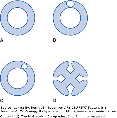
Comparison of cross-sectional dimensions of the intraperitoneal portion of several peritoneal catheters: A: Flexneck Tenckhoff catheter (silicone). B: Cruz Tenckhoff catheter (polyurethane). C: Standard Tenckhoff catheter (silicone). D: One intraperitoneal limb of the T-fluted catheter (Ash Advantage, silicone).
There are two materials of construction:
Silicone rubber (nearly all catheters).
Polyurethane (Cruz catheter).
The various intraperitoneal designs are all created to diminish outflow obstruction. The shape of the curled Tenckhoff catheter and the discs of the Toronto-Western catheter hold visceral peritoneal surfaces away from the side holes of the catheter. The grooves of the Advantage catheter distribute flow over the surface of the limbs that contact the parietal peritoneum, providing a much larger surface area for drainage than the side holes provide. An irritated omentum attaches firmly to the side holes of a catheter but only weakly to the grooves on a catheter (as demonstrated by the Blake surgical drain, with grooves on the catheter surface).
The subcutaneous catheter shapes all provide a lateral or downward direction of the exit site, which minimizes the risk of exit infection. An upward-directed exit site collects debris and fluid, increasing the risk of exit-site infection.
The optimal location for the standard deep cuff is within the rectus muscle. The subcutaneous cuff provides additional protection from bacterial contamination of the subcutaneous tunnel. The disc-ball deep cuff provides security of position of the catheter, since with the peritoneum sewn between the Dacron disc and intraperitoneal ball the catheter is fixed in position and cannot migrate outward. Similarly, the T shape of the Advantage catheter places the intraperitoneal limbs against the parietal peritoneum, preventing outward migration of the catheter.
The larger internal diameter of the Cruz and Flexneck catheters provides lower hydraulic resistance and more rapid dialysate flow during the early phase of outflow. In the latter part of outflow, the resistance to flow is determined mostly by the spaces formed by peritoneal surfaces as they approach the catheter, rather than the inside of the catheter. The Advantage catheter provides much larger entry ports for drainage of peritoneal fluid; limited clinical studies have demonstrated faster drainage of the peritoneum in the early and late phases of outflow and a decrease in residual peritoneal volume at the end of outflow.
The material from which peritoneal catheters are constructed has not affected the incidence of complications. There is no decrease in the incidence of peritonitis or omental attachment leading to outflow failure with polyurethane catheters, although they do have a weaker bond to the Dacron cuff, and loosening of this bond can create pericatheter leaks.
There is general agreement on the proper location of the components of chronic PD catheters (Figure 58–3):
The intraperitoneal portion should be between the parietal and visceral peritoneum and directed toward the pelvis to the right or left of the bladder.
The deep cuff should be within the medial or lateral border of the rectus sheath.
The subcutaneous cuff should be approximately 2 cm from the skin exit site.
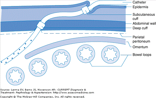
Placing the deep cuff within the abdominal musculature promotes tissue ingrowth and therefore avoids pericatheter hernias, leaks, catheter extrusion, and exit-site erosion. At the parietal peritoneal surface, the squamous epithelium reflects along the surface of the catheter to reach the deep cuff. If the deep cuff is outside the muscle wall, the peritoneal extension creates a potential hernia. At the skin surface, the stratified squamous epithelium follows the surface of the catheter until it reaches the superficial cuff. If the exit site is longer than 2 cm, the squamous epithelium disappears and granulation tissue is left, leading to an exit site with continued “weeping” of serous fluid; the potential for exit site infection is therefore increased.
Some peritoneal catheters have components that provide greater fixation of the deep cuff within the musculature. When the Missouri and Toronto-Western catheters are placed, the parietal peritoneum is closed between the ball (inside the peritoneum) and disc (outside the peritoneum). When the T-fluted (Ash Advantage) catheter is placed, the wings open in position adjacent to the parietal peritoneum and perpendicular to the penetrating tube. With these catheters, outward migration of the catheter is impossible.
When placing peritoneal catheters it is best to choose a deep cuff location that is free of major blood vessels (Figure 58–4).
Figure 58–4.
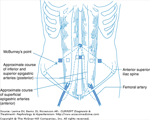
Major blood vessels and landmarks of the anterior abdominal wall. Open squares represent the preferred and safest points for the location of the deep cuff of a chronic peritoneal catheter within the medial or lateral border of the rectus muscle. Solid squares indicate the external landmarks used during blind insertion of a needle or cannula at the start of peritoneoscopic or blind catheter placement: One-half of the distance between the anterior superior iliac spine for the lateral border of the rectus and 2 cm below the umbilicus for the medial border of the rectus.
PD catheter insertion can be accomplished by any one of three techniques: the dissective or surgical, the blind or modified Seldinger, and the peritoneoscopic. The dissective technique utilized by most surgeons places the catheter by minilaparotomy, usually under general anesthesia. In the blind or modified Seldinger technique a needle is inserted into the abdomen, a guide wire is placed, a tract is dilated, and the catheter is inserted through a split sheath, all without visualization of the peritoneal cavity. Peritoneoscopic insertion uses a small (2.2-mm-diameter) optical peritoneoscope (Y-TEC Scope) for direct inspection of the peritoneal cavity and identification of a suitable site for the intraperitoneal portion of the catheter.
There are advantages and disadvantages of each technique of catheter placement, and the overall success of the catheters is as dependent upon the skill and experience of the physician performing the procedure as the method of placement. Each procedure has unique advantages and problems.
Dissective techniques securely place the deep cuff within the abdominal musculature. The techniques can be performed without any specialized equipment except for a stylet to straighten the catheter. Some types of catheters require surgical placement, such as the disc-and-ball Missouri or Toronto-Western catheters. The incision in the abdominal musculature requires surrounding tissues to first heal the wound and then grow into the deep cuff before the deep cuff is secure. Pericatheter leaks are frequent if the catheter is used immediately after placement. The dissective approach provides no visualization of adhesions and free spaces within the peritoneum. The catheter tip is advanced by “feel” and may be advanced to press against loops of bowel, or near adhesions, leading to early outflow failure of the catheter.
Blind placement procedures are convenient, can be performed anywhere in a hospital, and have the advantage of being low in cost. The needle, guidewire, dilators, and sheath are often packed in a kit with the peritoneal catheter. Bowel perforation is an occasional complication, usually not recognized until the catheter has been completely placed and is flushed. No visualization of the peritoneal space is provided to avoid impingement of the catheter tip on adhesions or visceral surfaces. The deep cuff is usually left just outside of the abdominal musculature, not within the rectus sheath.
Peritoneoscopic placement allows the best visualization of the peritoneal space. This avoids placing the catheter under bowel loops, under omentum, or against adhesions. The Quill expands to allow the deep cuff to advance into the musculature. The Y-TEC procedure can be performed in any room in the hospital. Specialized equipment must be purchased, however, and the physician must have some training in peritoneoscopic techniques. Of the three techniques, only the latter allows for direct visualization of the intraperitoneal structures. The use of this technique, most commonly employed by nephrologists, is rapidly expanding. Peritoneoscopic placement varies from laparoscopic techniques by using a much smaller scope and puncture size, only one peritoneal puncture site, a device to advance the cuff into the musculature, air in the peritoneum rather than CO2, and local rather than general anesthesia.
The preference of one technique over another must take into account the incidence of complications (pericatheter leakage, exit site and tunnel infection), the long-term catheter survival associated with each technique, the costs, ease, and timely insertion of the catheter, and factors contributing to risk of mortality (general anesthesia). To this end, peritoneoscopic placement of PD catheters by nephrologists has been rigorously compared to the surgical and the blind technique (Table 58–1). Both randomized and nonrandomized studies have documented the superiority of the peritoneoscopic technique in terms of a lower incidence of catheter complications (infection, outflow failure, pericatheter leak) and increased catheter survival. The avoidance of various complications by peritoneoscopic placement may relate to the decreased tissue dissection required with this technique. Extensive dissection (incising/splitting the rectus sheath/muscle as well as incising the parietal peritoneum) in the surgical technique may lead to loose attachment of the catheter to the abdominal wall, thereby increasing the incidence of pericatheter leaks, subsequent tunnel infection and peritonitis, and catheter loss.
Investigator (year)1 |
|---|


