Hysteroscopic Sterilization
Rafael F. Valle
Sterilization of women was greatly enhanced and simplified with the introduction of laparoscopy, which, since the late 1960s, has become one of the most common methods for tubal sterilization of women in the nonpuerperal state. Nonetheless, the method, although highly effective, in most cases is performed under general anesthesia. The risks associated with laparoscopy are also associated with tubal sterilization. For these reasons, new, simpler methods of tubal sterilization that are well tolerated by patients, effective, and perhaps eventually reversible are still being sought.
Modern hysteroscopy was introduced in the early 1970s as a method of visualizing the uterine cavity and uterotubal junctions, and the idea of occluding this area by hysteroscopy was revived with great interest. In 1878 Kocks attempted to occlude the uterotubal junctions blindly by a transcervically inserted electrode; despite numerous other attempts to improve this rudimentary technique by guiding an electrode with fluoroscopy, this method did not produce significant clinical success. It was associated with unacceptable failures and serious complications. Although the idea of using hysteroscopy as a guide to tubal occlusion goes back to the 1920s and indeed was explored in clinical trials in the 1950s, particularly by Japanese investigators, these preliminary attempts again did not produce significant acceptable results. It was not until the early 1970s that serious and well-designed clinical trials were initiated in several centers, using electrodes delivered directly under hysteroscopic guidance at the proximal segment of the intramural portion of the fallopian tubes. These clinical trials were performed particularly in West Germany by Lindemann, in Japan by Sugimoto, in Mexico by Quinones-Guerrero et al., and in the United States by Neuwirth. Once hysteroscopy was seen as a practical technique to deliver thin electrodes to the uterotubal junctions, other methods of tubal occlusion began to be explored.
The various hysteroscopic techniques to attempt occlusion of the intramural portion of the fallopian tubes are the following:
Electrocoagulation and cryocoagulation
Injection of chemicals for permanent closure, such as quinacrine; gelatin, resorcinol, and formaldehyde (GRF); and methyl cyanoacrylate (MCA)
Nondestructive occlusion by plastic formed-in-place plugs
Mechanical devices or tubal plugs that are placed at the proximal portion of the interstitial oviduct
Intratubal devices
Electrocoagulation and Cryocoagulation
Electrocoagulation
Electrocoagulation of the intramural portion of the fallopian tubes by hysteroscopy was the main hysteroscopic method of investigation in the early 1970s. Although the technique itself was fairly standard, the coagulation of this tubal area varied among investigators, particularly in the wattage and time of delivery; nonetheless, the preliminary results seemed comparable with 75% to 80% bilateral initial closure with one application and about 85% to 90% closure with a second application. However, the complications and specific failures were not uniform.
Because of reports of pregnancies and major complications associated with this technique, a hysteroscopic sterilization registry was established at Columbia University in 1975. Ten collaborators from the United States, Thailand, West Germany, India, and Singapore were enrolled in the study, and a total of 587 cases were analyzed with 333 (57%) found to have bilateral tubal closure at subsequent testing. There were 229 women (39%) who had one or both fallopian tubes open at subsequent testing or who had become pregnant despite tubal closure at patency test. Most distressing in the analysis of these 587 cases were the major complications, which included tubal and cornual ectopic pregnancies, uterine perforations, bowel damage, peritonitis, acute endometritis, excessive uterine bleeding, and one death as the result of bowel perforation and peritonitis. The overall rate of major complications was 4.3%. When this study was completed in 1977, hysteroscopic sterilization by electrocoagulation was just beginning to peak and expand to several centers. Nonetheless, in view of the failure rate and complications, most clinical trials were stopped altogether, and further investigation did not seem warranted.
Although multiple factors seemed to be related to the failures and complications, it was evident that the most important factor seemed to be lack of standardization and
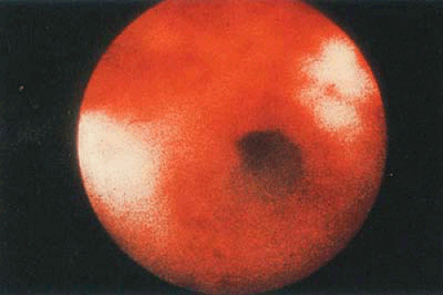
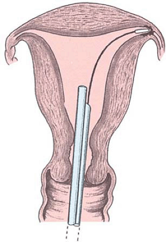
control of the current used and the time of its delivery. This was impossible to monitor, particularly thermal injury to this anatomic area, which is thinner than the uterus (Figs. 29.1, 29.2, 29.3, 29.4, 29.5).
control of the current used and the time of its delivery. This was impossible to monitor, particularly thermal injury to this anatomic area, which is thinner than the uterus (Figs. 29.1, 29.2, 29.3, 29.4, 29.5).
Cryocoagulation
Cryocoagulation of the endometrium and uterotubal junctions has also been investigated as a method to occlude the fallopian tubes for permanent sterilization. The tissue effects of cryosurgery are coagulation necrosis, a result of biochemical and biophysical changes, and cellular and nuclear damage produced by the subfreezing temperatures. The final result is avascular necrosis of tissues. Because of the rapid regeneration of the endometrium when cryosurgery has been applied, collagen plugs 2 mm in diameter and 8 mm long have been placed at the cornual junctions following freeze–thaw cycles and abrasion. The initial experiments have been performed in animals and in selected human volunteers prior to hysterectomy; nonetheless, although no extended clinical trials have been performed to determine the permanent damage to the tissue and the results of tubal occlusion and subsequent recanalizations, the initial results appear promising.
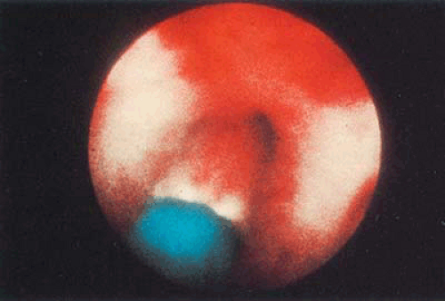 FIGURE 29.3 Electrode (from Fig. 29.2) at the proximal portion of the intramural oviduct. |
Hysteroscopic Injection of Chemicals
Using hysteroscopy as a platform to deliver chemicals into the fallopian tube to produce tubal occlusion has constituted another attempt at tubal sterilization, either permanent or temporary. Of the various sclerosing substances used to produce permanent occlusion of the fallopian tubes, the most relevant have been quinacrine, GRF, and MCA (Fig. 29.6).
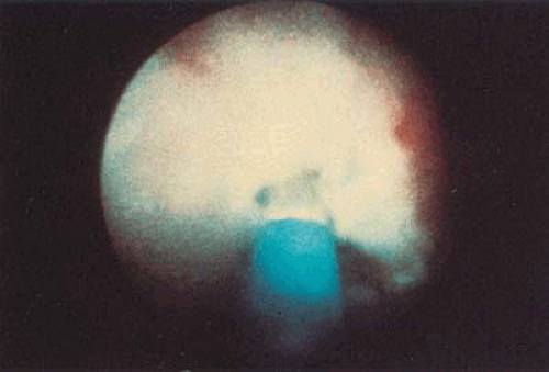 FIGURE 29.4 Blanching of tissue produced by transmission of the electric current as electrode is activated. |
Quinacrine
Zipper et al. studied the effects of quinacrine in the fallopian tubes by transcervical injection in 1970. A 50% bilateral occlusion was obtained with one injection of quinacrine in 1,000 patients; when the instillation rate was increased to
three at 1-month intervals, the closure rate rose to 80%. In 1974, Alvarado et al. described the preliminary results with the hysteroscopic instillation of quinacrine to attain tubal occlusion in 30 patients, and only six out of 16 cases under hysterosalpingographic control showed bilateral obstruction. After 3 months of application, of 16 of 30 patients, bilateral obstruction was achieved in six cases, unilateral obstruction in six, and bilateral patency in four. No further clinical trials were performed, and quinacrine delivered under hysteroscopy was not deemed feasible for tubal closure.
three at 1-month intervals, the closure rate rose to 80%. In 1974, Alvarado et al. described the preliminary results with the hysteroscopic instillation of quinacrine to attain tubal occlusion in 30 patients, and only six out of 16 cases under hysterosalpingographic control showed bilateral obstruction. After 3 months of application, of 16 of 30 patients, bilateral obstruction was achieved in six cases, unilateral obstruction in six, and bilateral patency in four. No further clinical trials were performed, and quinacrine delivered under hysteroscopy was not deemed feasible for tubal closure.
Gelatin, Resorcinol, and Formaldehyde
Of the tissue adhesives studied in an attempt to occlude the fallopian tubes, a combination of gelatin, resorcinol, and formaldehyde was investigated in 1964 by the Battelle Laboratories. This adhesive substance permitted ingrowth and, being biodegradable, could be used safely in these investigations. Although GRF has never been used in human clinical trials, some preliminary studies in animals show its potential for tubal blockage. Nonetheless, although theoretically the substance could be delivered through hysteroscopy, perhaps difficulty in confining the substance to the fallopian tubes and/or avoiding reflux have halted any further attempts to study this approach.
Methyl Cyanoacrylate
Another tissue adhesive evaluated for occlusion of the fallopian tubes is MCA (Crazy Glue), which—injected as a fluid—polymerizes rapidly on hydration. One of the problems in using MCA for surgical application has been its localized effect on surrounding tissues, which developed necrosis, inflammation, and fibrosis. Although the application of MCA to the fallopian tubes has been tried by means of a balloon catheter to prevent reflux, hysteroscopy was also attempted by Lindemann with fair success.
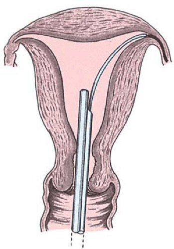 FIGURE 29.6 Various sclerosing materials have been injected into the oviduct by a catheter delivered through the hysteroscope. |
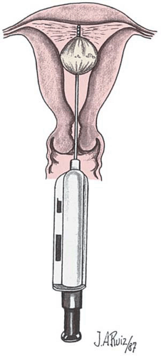
The use of MCA has been facilitated by a device called FEMCEPT for its delivery. The FEMCEPT system requires transcervical introduction of a cannula with a distal balloon. Insufflation places the tip of the device with lateral openings, facilitating delivery of substances directly into the fallopian tubes without reflux. The device has been designed so as to deliver a volume of 0.06 mg (0.2 in tubes) of MCA per instillation (Fig. 29.7). Bilateral tubal closure rate has been achieved in these preliminary trials in 72% of tubes with a single application, and in 96% with two applications 1 month apart (Table 29.1).
Hysteroscopic Tubal Occlusion with Silicone Rubber Plugs
In 1975, Erb proposed the use of silicone rubber plugs directly formed in place to block the oviducts as a method of tubal occlusion, using a cornual salpingo guide guided directly under fluoroscopy.
Early Experimental Trials
Silicone was believed to be an ideal material with which to form plugs because during other uses in the body it appeared to be non–tissue damaging and noninvasive. In studies conducted by Erb on rabbits and monkeys (Fig. 29.8), pathologic examination of the removed organs after being instilled with silicone for up to 284 days showed no invasiveness of the material into the tissues and no tissue damage other than some flattening of the tubal cilia. Standard pathologic investigation techniques were used in these studies, as well as electron microscopy evaluation of these organs (Fig. 29.9).
Erb got his ideas from Corfman and Taylor and also from Hefnawi et al., who in animal studies found silicone to be 100% effective when the material was retained in the isthmus of the tube. The first application to humans was made by Rakshit, who had a very simple technique. He used a low-viscosity solution of silicone, which he injected through the cervical canal by means of a large, cone-shaped syringe to fill the uterine cavity and the fallopian tubes. He had a 50% success rate with this technique and reported no pregnancies in a period of 11 to 24 months in 22 patients who had bilateral occlusion. The excess rubbery solution from the uterine cavity was simply wiped out.
Erb reasoned that if there could be a direct application of the silicone to the tubal ostia at the cornual end, a higher success rate of filling the tubal lumen could be obtained. He also believed that if the material were made more viscous, it would not flow into the abdominal cavity or the uterine cavity as it did so readily with Rakshit’s technique. He therefore added a catalyst (stannous octoate) to the mix, which made it cure more rapidly and become more viscous in its liquid form.
Erb et al. worked originally with rabbits. Through hysterotomy, they applied the material with a catheter directly into the cornual opening of the fallopian tubes. Afterward, the does were exposed to bucks for up to 280 days, and none of them became pregnant. Following this, Erb et al.
attempted to apply the procedure in women fluoroscopically with a cannula to the cornual opening of the tube; however, since they could not find a proper fitting, they abandoned this approach and began looking at hysteroscopy as a means of visualizing and cannulating the tube.
attempted to apply the procedure in women fluoroscopically with a cannula to the cornual opening of the tube; however, since they could not find a proper fitting, they abandoned this approach and began looking at hysteroscopy as a means of visualizing and cannulating the tube.
TABLE 29.1 Electrocoagulation, Cryosurgery, Sclerosing Chemicals, and Tissue Adhesives | ||||||||||||||||||||||||||||||||||||||||||||||||||
|---|---|---|---|---|---|---|---|---|---|---|---|---|---|---|---|---|---|---|---|---|---|---|---|---|---|---|---|---|---|---|---|---|---|---|---|---|---|---|---|---|---|---|---|---|---|---|---|---|---|---|
| ||||||||||||||||||||||||||||||||||||||||||||||||||
Plug and Instrument Design
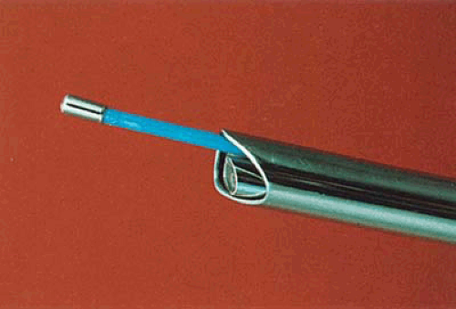
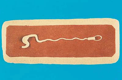
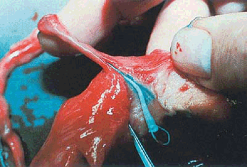
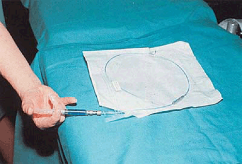
To make this a successful procedure, Erb then developed the guide assembly (Figs. 29.10 and 29.11), the obturator tip (Fig. 29.12), a mixer-dispenser (Fig. 29.13), and the fluid flow actuator (Fig. 29.14). These instruments were developed for the hysteroscopic application of the silicone to the fallopian tubes. The obturator tip (see Fig. 29.12) is a hollow, premolded silicone rubber tip that measures in its original form 235 mm. This is placed on the end of the guide assembly, which is 75 cm long and 1 mm in diameter (Fig. 29.15A). It is made of polysulfone tubing that has over approximately two-thirds of its length a second polysulfone tubing measuring 2 mm in diameter, which butts up against the base of the obturator tip (Figs. 29.15B and 29.16A). The silicone flows through the inner guide, then through the hollow tip; the flowing silicone cross-links to this tip, making the tip a part of the final formed-in-place plug (Figs. 29.16B and 29.17A). With proper viscosity and timing, the viscous liquid silicone
will flow through the obturator tip into the fallopian tube to the ampullar section (Figs. 29.17B, 29.18, 29.19). When it cures, which takes about 5 minutes, the plug has a dumbbell shape: It is larger on either end than in the middle of the isthmus portion. This is what is believed to be the basic success of the silicone plug over other types of intraluminal devices. The tip, because of the design of the guide assembly, can be separated from the attached cured silicone in the remainder of the guide assembly, thus leaving the cured plug (Fig. 29.20) with its tip attached in the tube from the cornu through the isthmus into the ampullar section (Figs. 29.21 and 29.22).
will flow through the obturator tip into the fallopian tube to the ampullar section (Figs. 29.17B, 29.18, 29.19). When it cures, which takes about 5 minutes, the plug has a dumbbell shape: It is larger on either end than in the middle of the isthmus portion. This is what is believed to be the basic success of the silicone plug over other types of intraluminal devices. The tip, because of the design of the guide assembly, can be separated from the attached cured silicone in the remainder of the guide assembly, thus leaving the cured plug (Fig. 29.20) with its tip attached in the tube from the cornu through the isthmus into the ampullar section (Figs. 29.21 and 29.22).
Stay updated, free articles. Join our Telegram channel

Full access? Get Clinical Tree


